Haemoproteus spp. and Leucocytozoon californicus Coinfection in a Merlin (Falco colombarius) - MDPI
←
→
Page content transcription
If your browser does not render page correctly, please read the page content below
pathogens
Article
Haemoproteus spp. and Leucocytozoon californicus
Coinfection in a Merlin (Falco colombarius)
Simona Nardoni 1 , Francesca Parisi 1, *, Guido Rocchigiani 1 , Renato Ceccherelli 2 ,
Francesca Mancianti 1 and Alessandro Poli 1
 1   Dipartimento di Scienze Veterinarie- Università di Pisa, Viale delle Piagge n. 2, 56124 Pisa, Italy;
     simona.nardoni@unipi.it (S.N.); guido.rocchigiani.g@gmail.com (G.R.); francesca.mancianti@unipi.it (F.M.);
     alessandro.poli@unipi.it (A.P.)
 2   Centro Recupero Uccelli Marini e Acquatici-CRUMA, via delle Sorgenti 430, 57121 Livorno, Italy;
     apusvet.cruma@libero.it
 *   Correspondence: francesca.parisi@vet.unipi.it; Tel.: +39-0502216982

 Received: 14 March 2020; Accepted: 2 April 2020; Published: 4 April 2020

 Abstract: The Leucocytozoon genus comprises numerous widely distributed parasites which have
 been less investigated than other avian hemoprotozoa. Their occurrence is common, with very
 variable prevalence values and pathogenicity degrees. Leucocytozoon species are characterized by a
 great taxonomic diversity, and infections are usually restricted to birds of the same family. In the
 present paper, a mixed hemosporidia infection by Leucocytozoon californicus and Haemoproteus sp. in
 an adult male merlin (Falco columbarius) which died during hospitalisation is reported, indicating,
 for the first time, a newly described avian host species. A molecular investigation was carried out
 through cytochrome b gene analysis, revealing a 100% match with L. californicus and Haemoproteus spp.
 A blood smear examination allowed us to detect Leucocytozoon fusiform mature gametocytes and
 different degrees of maturity of Haemoproteus gametocytes. Histopathology revealed foci of necrosis,
 hemorrhagic areas and extramedullary hematopoiesis in the liver, the presence of microthrombi in
 the heart and lung and scattered hemorrhages in the lung.

 Keywords:   Leucocytozoon californicus;               Haemoproteus sp.;       Falco columbarius;        PCR;
 anatomopathological findings

1. Introduction
     Avian hemoprotozoa encompass different genera of blood parasites, including Leucocytozoon,
Haemoproteus and Plasmodium. These three parasite genera are commonly reported as being
pathogenic [1,2]. Almost all parasite species present gametogony and sporogony in blood suckling
dipteran vectors, infected through the blood meal. Exoerytrhrocytic schizonts develop in different
tissues, while gametocytes occur into the blood cells of infected birds. Nevertheless, developmental
stages in the avian host remain only partially studied [3]. Although several parasite species have
been encountered from a variety of different countries, information about complete life-cycles of most
Haemoproteus is lacking, especially for exoerythrocytic stages. In this genus, the sporozoites, inoculated
into birds by Ceratopogonidae and Hippoboscidae Diptera, undergo schizogony in the endothelial cells
and probably in fixed macrophages, or myofibroblasts, depending on the parasite species. Gametocytes
develop in mature erythrocytes. Simuliidae are the cyclic vectors of Leucocytozoon parasites that
inoculate the sporozoite stages. The exoerythrocytic stages in birds firstly occur in the hepatocytes, in
macrophages and other reticuloendothelial cells, then further schizogony stages are observed in the
lungs, and less often in the liver, spleen, kidneys, heart, skeletal musculature and other organs. The
gametocytes colonize erythroblasts, erythrocytes and mononuclear leukocytes [3].

Pathogens 2020, 9, 263; doi:10.3390/pathogens9040263                           www.mdpi.com/journal/pathogens
Pathogens 2020, 9, 263                                                                                2 of 8

     Among hemoprotozoa, Plasmodium spp have the widest host range [4]. Although the majority
of Haemoproteus and Leucocytozoon species are characterised by great taxonomic diversity, in birds,
they are relatively species-specific, with infections usually restricted to subjects within the same
taxonomic family.
     Haemoproteus spp are the most common varieties. There are up to 200 species within the genus and
they have been reported in 1700 species of birds. Haemoproteus parasites have low pathogenicity and
infections are usually subclinical; sometimes they cause mild clinical signs and are rarely fatal [1,2].
     Leucocytozoon appears to be the least investigated among avian haemoprotozoa genera. It
is reported especially in Northern temperate areas, where both Leucocytozoon and black flies are
common [5]. It generally occurs in Anseriformes, turkeys, raptors, wild birds and Columbiformes [6].
Leucocytozoon spp are usually considered nonpathogenic in adult raptors, even if they are reported as
a possible cause of mortality during reproduction [7] and in nestlings, and as a consequence, need
to switch to a new avian host. Pathogenicity would also be correlated with parasite species and
parasitemia level [8].
     Recent studies suggest that some species among avian hemoprotozoa could sometimes be lethal
for birds, much more frequently than previously reported [1,2,9]. From this perspective, studies on
the distribution and the diversity of avian haemoprotozoa are necessary. These studies would be
extremely useful to understand wildlife diseases, particularly the virulence and the mortality rates in
avian species due to these parasites.
     The present case report describes, for the first time, a coinfection due to Leucocytozoon californicus
and Haemoproteus sp. in a merlin (Falco columbarius) from Italy.

2. Results

2.1. Blood Smears
      An examination of blood smears revealed the presence of different intracellular stages of parasites
consistent with Haemoproteus sp. and Leucocytozoon sp. (Figure 1A). Haemoproteus sp gametocytes
were found in RBCs, while it was difficult to establish the host cells for Leucocytozoon, since the
parasite dramatically changes the morphology of infected cells and no young stages compatible
with Leucocytozoon have been successfully identified in blood cells. Leucocytozoon sp.-infected cells
per 1000 blood cells were 10 ± 4, while Haemoproteus sp.-infected RBCs per 1000 cells were 66 ± 6.
Mature gametocytes were the only clearly visible parasite stage. They filled up the whole cellular
space, replacing cell cytoplasm, which sometimes formed elongated “horns” (Figure 1A,B). The
measurements of mature gametocytes are presented in Table 1. Microgametocytes stained more lightly
than the macrogametocytes. The cytoplasm was extremely pale blue and the nucleus was pale pink.
Macrogametocytes could be distinguished from microgametocytes because of their darker appearance,
i.e., cytoplasm stained dark blue and nucleus light red, and small vacuoles and magenta volutin
cytoplasmatic granules could be frequently seen (Figure 1B). The nucleus of macrogametocytes was
also smaller than the that of microgametocytes.
      Immature and mature gametocytes of Haemoproteus sp were visible in RBCs. Young gametocytes
(Figure 1C) were roundish to oval with an even outline, a peripheral dark nucleus and a large white
vacuole. They grew toward the host cell nucleus and had no contact with the host cell membrane
or nucleus. Very small, scattered pigment granules of hemozoin could be seen at low magnification.
Ring gametocytes developed in length and width and evolved in medium grown gametocytes of
elongated shape with a wavy outline. At this stage, they did not fill the erythrocytes up to their
poles; they were appressed to the nucleus, but rarely in contact with the envelope of the host cells,
and irregular to roundish small pigment granules could be seen (Figure 1A). Mature gametocytes
could be easily differentiated both in macro- and micro- gametocytes. Macrogametocytes (Figure 1E)
were sausage-shaped, with an angular outline. The mature stages filled the erythrocytes up to their
poles, although not completely encircling the nucleus, they were closely appressed to it and rarely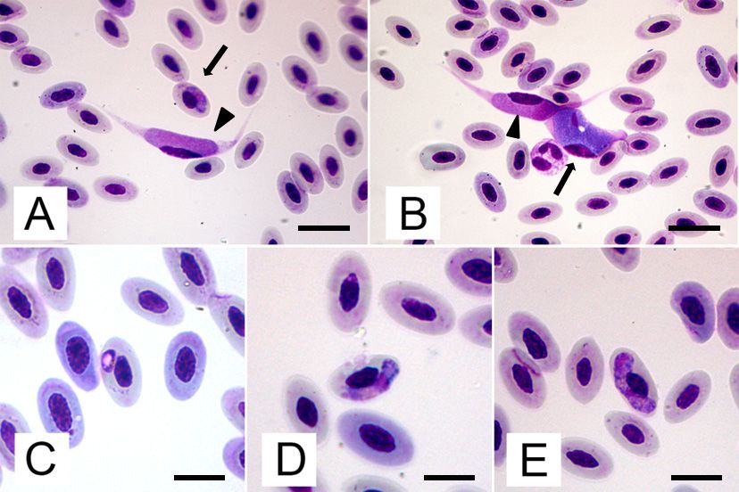
Pathogens 2020, 9, 263                                                                                                                 3 of 8

appeared to be in contact with the envelope of the host cells, with some variations. The cytoplasm was
moderately coarse and stained pink, with few small, scattered malaria-like pigment granules. Granules
of volutin could also occur. Vacuoles regressed, but sometimes a small vacuolar residue could be
observed. The nucleus was terminal or subterminal, far from the host cell nucleus, and stained bright
pink (average length 11.5 ± 1.5 and width 2.6 ± 0.4).
     The general development and configuration of microgametocytes are similar to the
macrogametocytes,
            Pathogens 2020,albeit   withREVIEW
                           9, x FOR PEER   a slightly defined outline. The cytoplasm was finely3 of            granular
                                                                                                                 8      and
stained pink, with medium size malaria and valutin pigment granules accumulated on the membrane
            moderately coarse and stained pink, with few small, scattered malaria-like pigment granules.
of the polarGranules
              regions.  of The
                           volutinvacuole    residue
                                     could also        could beregressed,
                                                occur. Vacuoles   seen. Nucleus     was median
                                                                          but sometimes            or submedian,
                                                                                         a small vacuolar residue   close to
the host cellcould
               nucleus,     with some
                    be observed.           variations,
                                    The nucleus          and stained
                                                 was terminal           brightfar
                                                               or subterminal,  pink.
                                                                                  from the host cell nucleus, and
              stained bright pink (average length 11.5 ± 1.5 and width 2.6 ± 0.4).

                  Coinfection
      Figure 1. Figure             of Haemoproteus
                        1. Coinfection of Haemoproteus sp.sp.    and Leucocytozoon
                                                            and Leucocytozoon                 californicus
                                                                               californicus from              from
                                                                                                   a blood smear  of aaFalco
                                                                                                                         blood smear
                 colombarius: A)  Early  stage of  gametocytes   of  Haemoproteus     sp.  in  RBC
      of a Falco colombarius: A) Early stage of gametocytes of Haemoproteus sp. in RBC (arrow) and    (arrow)  and  mature
                 microgametocyte of L. californicus (arrowhead); B) Mature macrogrametocyte (arrow) and
      mature microgametocyte of L. californicus (arrowhead); B) Mature macrogrametocyte (arrow) and
                 microgametocyte (arrowhead) of L. californicus; C) Ring-shaped young gametocyte of Haemoproteus
      microgametocyte     (arrowhead)
                 sp in RBC;                 of L. californicus;
                             D) Mature microgametocyte            C) Ring-shaped
                                                           of Haemoproteus  sp. in RBC; young
                                                                                           E) Maturegametocyte       of Haemoproteus
                                                                                                       macrogametocyte    of
      sp in RBC; Haemoproteus
                 D) Maturesp.   microgametocyte        of Haemoproteus
                                  in RBC. (Modified Wright’s                  sp. Scale
                                                               solution staining;   in RBC;     andMature
                                                                                          bar A E)   B 20 µm, macrogametocyte
                                                                                                               C–E 15 µm).        of
      Haemoproteus sp. in RBC. (Modified Wright’s solution staining; Scale bar A and B 20 µm, C–E 15 µm).
                   The general development and configuration of microgametocytes are similar to the
      Table 1.macrogametocytes,
               Measurements of   albeit with gametocytes
                                    mature   a slightly defined
                                                            andoutline. The cytoplasm
                                                                cell-parasite complexwas  finely granular californicus
                                                                                      of Leucocytozoon     and
             stained pink, with medium size malaria and valutin pigment granules accumulated on the membrane
      from blood smears of the examined Falco columbarius.
              of the polar regions. The vacuole residue could be seen. Nucleus was median or submedian, close to
              the host cell nucleus, with some variations,  and stained bright
                                                         Macrogametocytes          pink.
                                                                                (n=10)          Microgametocytes (n=10)
                            Figure .
                                                        Min – Max (Mean ± SD) µm              Min – Max (Mean ± SD) µm
              2.2. Molecular Analysis
                                          Lenght            19.0 – 21.6 (20.5 ± 0.7)              18.0 – 20.9 (19.4 ± 1.2)
                    Nested-PCR was positive
                   Parasite                    for Leucocytozoon
                                           Width             8.2 –and
                                                                   10.2Haemoproteus/Plasmodium
                                                                         (9.1 ± 0.9)               DNA.    The
                                                                                                    5.8 – 7.2   nucleotide
                                                                                                              (6.5  ± 0.6)
              sequence of the isolates (493Area
                                             bp and 417 bp, respectively)
                                                          87.0 – 147.8 (123.9showed
                                                                               ± 24.4)a 100% identity
                                                                                                  69.1 with
                                                                                                       – 98.0Leucocytozoon
                                                                                                              (83.7 ± 11.4)
              californicus (Figure 2) [GenBank
                                          Lenghtaccession number:    KR422359.1]
                                                              3.1 – 4.8 (4.1 ± 0.7) and with Haemoproteus     sp. H-FATI1
                                                                                                    6.9 – 8.9 (7.7  ± 0.8)
              [GenBank:    EF607289.1], isolated
              Parasite nucleus             Widthfrom a Falco tinnunculus
                                                              2.0 – 2.9 (2.5by
                                                                             ± Krone
                                                                               0.4) et al. [10], who3.3
                                                                                                      provided
                                                                                                        – 5.4 (4.2a ±
                                                                                                                    detailed
                                                                                                                      0.9)
              phylogenetic tree.           Area              6.7 – 11.7 (8.8 ± 2.0)               20.8 – 32.0 (27.2 ± 4.4)
           Cell-parasite complex           Area            128.7 – 230.0 (189.6 ± 40.7)         130.0 – 197.50 (163.2 ±25.6)

2.2. Molecular Analysis
      Nested-PCR was positive for Leucocytozoon and Haemoproteus/Plasmodium DNA. The nucleotide
sequence of the isolates (493 bp and 417 bp, respectively) showed a 100% identity with Leucocytozoon
californicus (Figure 2) [GenBank accession number: KR422359.1] and with Haemoproteus sp. H-FATI1
Pathogens 2020, 9, 263 4 of 8
[GenBank: EF607289.1], isolated from a Falco tinnunculus by Krone et al. [10], who provided a detailed
Pathogens 2020, 9, x FOR PEER REVIEW 4 of 8
phylogenetic tree.
Pathogens 2020, 9, x FOR PEER REVIEW 4 of 8
Figure 2. Phylogenetic tree showing the Leucocytozoon sequencing results.
Figure 2. Phylogenetic tree showing the Leucocytozoon sequencing results.
2.3. Pathological Investigations
Figure 2. Phylogenetic tree showing the Leucocytozoon sequencing results.
2.3. Pathological Investigations
Necropsy revealed poor body conditions with a reduction of muscle mass and fat deposits. The
2.3. Pathological
Necropsy Investigations
revealed poorthebody
proventriculus was empty and onlyconditions with a reduction
gross abnormality of muscleduring
findings observed mass and fat deposits.
the necropsy The
were
proventriculus was
Necropsy empty
revealed and
poor the
body only gross
conditions abnormality
with a
severe hepatomegaly and scattered hemorrhages in the liver and the lung.reduction of findings
muscle observed
mass and fat during
deposits. the
The necropsy
were proventriculus
severe was empty
hepatomegaly and and the only gross abnormalityinfindings observed during the necropsy
Histologically, tissue stages ofscattered hemorrhages
Leucocytozoon, the liver
such as macromeronts andandthe lung.
micromeronts, were not
were severe hepatomegaly and scattered hemorrhages in the liver and the lung.
Histologically,
detected. However, tissue stages
histopathology of Leucocytozoon, such as macromeronts and micromeronts, were not
Histologically, tissue stages ofinvestigations
Leucocytozoon, suchallowed us to observe
as macromeronts tissue changes,
and micromeronts, were not probably
detected. However,
related to detected.
the colonization histopathology
of host tissues
However, histopathology investigations allowed us to observe tissue
(Figure 3).allowed us to observe tissue changes, probably
investigations changes, probably
related related
to the tocolonization
the colonization of of
host
hosttissues (Figure
tissues (Figure 3). 3).
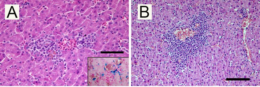
Hematoxylin
Figure 3. Figure and eosin-stained
3. Hematoxylin and eosin-stainedtissue
tissue from Falco
from aaFalco colombarius:
colombarius: (A)ofFoci
(A) Foci of necrosis
necrosis and and
hemorrhages in the liver; insert: Perl’s stain, macrophages laden with hemosiderin;
hemorrhages in the liver; insert: Perl’s stain, macrophages laden with hemosiderin; (B) Myeloid (B) Myeloid
precursorsprecursors in extramedullary hematopoiesis in the liver; (C) Thrombus in a cardiac capillary with
in extramedullary hematopoiesis in the liver; (C) Thrombus in a cardiac capillary with
necrosis of the surrounding tissue; (D) Thrombi in pulmonary vessels. (H–E; Scale bar 100 µm).
necrosis of the surrounding tissue; (D) Thrombi in pulmonary vessels. (H–E; Scale bar 100 µm).
Figure 3. Hematoxylin and eosin-stained tissue from a Falco colombarius: (A) Foci of necrosis and
hemorrhages in the liver; insert: Perl’s stain, macrophages laden with hemosiderin; (B) Myeloid
precursors in extramedullary hematopoiesis in the liver; (C) Thrombus in a cardiac capillary with
necrosis of the surrounding tissue; (D) Thrombi in pulmonary vessels. (H–E; Scale bar 100 µm).Pathogens 2020, 9, 263 5 of 8
In the liver, small necrotic foci were surrounded by scattered Perl’s-positive hemosiderin-laden
macrophages, which are interpreted as posthemorrhagic lesions (Figure 3A). Multifocal aggregates of
myeloid precursors cells, interpreted as foci of extramedullary hematopoiesis, were observed with a
perivascular arrangement (Figure 3B). Hepatic sinusoids were dilated due to blood congestion. Mature
stages of the parasite were evident within the thrombi in medium and small vessels of examined
organs. Necrosis of miocardiocytes and microthrombi in small vessels (Figure 3C) were detected in
the heart. In the lung, the gametocytes formed clusters together with red blood cells and oxyphilic
filaments of fibrin, obliterating the lumina of alveolar capillaries vessels (Figure 3D).
3. Discussion
The present report describes, for the first time, a parasite coinfection of Haemoproteus sp. and L.
californicus in F. colombarius, a widely distributed diurnal raptor. Based on our findings, this avian
species could be an additional host for L. californicus.
Gametocytes of Haemoproteus sp. developed in RBCs, while it was not easy to identify the host
cell for L. californicus, since the parasite dramatically modifies the morphology of infected cells. In fact,
even if the parasite is reported to infect blood cells, it seems that Leucocytozoon spp. would usually
infect cells with a single nucleus. Therefore these protozoa could infect RBCs (since avian RBCs are
nucleated), single nucleus WBCs, or single nucleated thrombocytes (another characteristic feature of
avian blood [11]. Moreover, we could not identify any possible immature stage of L. californicus, since
the only immature stage we found was characterised by the presence of pigment granules, which are
known to be absent in Leucocytozoon spp. [3]. On the other hand, mature gametocytes in the blood smear
were easily be ascribable to L. californicus or Haemoproteus sp., since the L. californicus host cell–parasite
complex was characterised by parasitic forms developing in a fusiform structure, as reported in other
cases of infection of diurnal raptors [12,13]. This was very different to the sausage-shaped mature
gametocytes of Haemoproteus spp. Leucocytozoon gametocytes, whose size was comparable to that
described in kestrel by Sacchi and Prigioni [14]. However, parasite width and area agreed with the
description reported by Walther et al. [15].
Numerous gametocytes were microscopically detectable in blood smears, suggesting a massive
presence in the host’s body. This was probably due to the high individual susceptibility of the host,
suffering the consequences of both physical injury and hospitalization. Such events could have played
a role in impairing the immune system, leading to death, probably caused by hemosporidia infection,
as supported by both clinical and histological evidence.
Pathological findings suggest that the hemosporidian parasite infection could have been fatal
for the subject. Hematological changes, probably related to the high parasitaemia, could have led
to the formation of thrombi exiting in capillary obstructions. Moreover, the death resulted from
cardio-respiratory failure, caused by clots due to subsequent disseminated intravascular coagulation.
Parasitic tissue stages were not detected in samples from organs, while indirect signs of parasite
tissue colonization, such as necrosis and hemorrhages, were observed, in agreement with Valkiūnas [3]
and Donovan et al. [2]. Moreover, extramedullary hematopoiesis represents a typical finding in avian
malarial disease [16,17], probably due to a compensation mechanism of severe anemia, the most
common clinical finding in erythrocytic stages of the parasites.
The apicomplexan cytochrome b gene is genetically informative, and it has been proven to be
suitable to investigate the evolution and divergence of avian malarial parasites in different host groups,
to analyze the areas of parasite transmission [18], and to carry out phylogenetic studies for Leucocytozoon
species [19].Pathogens 2020, 9, 263 6 of 8
4. Materials and Methods
4.1. Case History
A male adult F. colombarius was hospitalized as a consequence of an injury to the flight feathers
due to a gunshot wound, at the Centro Recupero Uccelli Marini ed Acquatici-CRUMA, a wild bird
care center in Livorno (Italy). Despite a satisfactory clinical response and wound remission, a sudden
clinical worsening with respiratory distress arose, leading to the quick death of the bird.
4.2. Blood Smears
Blood in ethylenediaminetetraacetic acid (EDTA) was collected before death. Smears were
prepared on a glass slide, fixed in methanol and stained with modified Wright’s solution. Each smear
was examined through 100–150 fields at low magnification (400X) and then at least 100 fields at
high magnification (1000X). The measurements of parasite and infected host cells were made based
on morphometric parameters of gametocytes as previously proposed, i.e., the length, width, and
area of macrogametocytes and microgametocytes, parasite nucleus, and area of host cell parasite
complex [20]. The parameters were determined using a semiautomated morphometric system (LAS 4.1,
Leica Microsystems, Wetzlar, Germany) and statistically analysed using IBM SPSS Statistics 21 system.
4.3. Molecular Analysis
Part of blood was stored at 4 ◦ C for molecular analysis. DNA was extracted using the
blood/cultured cell genomic DNA extraction mini kit (Fisher Molecular Biology, Roma, Italy),
following the manufacturer recommendations. Hemoprotozoa DNA was detected with a nested
PCR protocol targeting the cytochrome b mitochondrial gene, according to Hellgreen et al. [19] Primers
for first step amplification were HaemNFI (5’-CATATATTAAGAGAAITATGGAG- 3’) and HaemNR3
(5’-ATAGAAAGATAAGAAATACCATTC-3’). Two second step PCR were then performed, both using
the product of the first step as a template. HaemF (5’-ATGGTGTTTTAGATACTTACATT-3’) and HaemR2
(5’-GCATTATCTGGATGTGATAATGGT-3’) internal primers were used for detection of Haemoproteus
and Plasmodium [21] amplifying a 478 bp sequence. Leucocytozoon lineages were amplified by HaemFL
(5’-ATGGTGTTTTAGATACTTACATT-3’) and HaemR2L (5’-CATTATCTGGATGAGATAATGGIGC-3)
amplifying a 480 bp sequence. All PCRs were carried out in a total volume of 40 µL, using 6 and 3 µL
of template DNA for the first and the second reaction, respectively, 8 µL of reaction buffer, 1 mM of
each primer, 1.25 U of wonder Taq DNA polymerase (Euroclone, Milan, Italy), and 24 and 27 µL of
nuclease-free water for the first and the second reaction. Thermal cycling was performed in a C1000
thermal cycler (Bio-Rad, Hercules, CA, USA) using the following conditions: 30 s at 94 ◦ C, 30 s at 50
◦ C and 45 s at 72 ◦ C for 20 cycles for the first reaction and for 35 cycles for the second. Positive and
negative samples were added for quality control. Amplified DNA products, as well as molecular weight
markers (Sharp Mass TM 100 Plus Ladder, Euroclone, Milano, Italy), were subsequently submitted to
electrophoresis in 2% agarose gel at 100 V for 30 min. The gel was prestained with EuroSafe -Fluorescent
Nucleic Acid (Euroclone, Milano, Italy).
The PCR products were then sequenced by a commercial laboratory (BMR Genomics, Padova,
Italy). The sequences were compared to those already deposited in the NCBI database using the BLAST
search engine.
4.4. Pathological Investigations
Necropsy was carried out and representative tissue samples were collected from heart, lung,
liver, spleen, kidney, proventriculus, intestine, and central nervous system. Samples were fixed in
10% buffered formalin (pH 7.4), routinely processed and embedded in paraffin. Four-micron thick
sections were stained with hematoxylin and eosin for general vision and specific stain (Periodic Acid
Schiff—PAS) for fungi, acid-fast bacteria (Ziehl Neelsen stain) and iron-loaded hemosiderin pigment
(Perl’s stain).Pathogens 2020, 9, 263 7 of 8
5. Conclusions
The present study reported, for the first time, the occurrence of L. californicus in coinfection with
Haemoproteus sp. in F. colombarius. The concomitance between the detection of RBC stages of parasites
and the lack of identification of tissue forms in the sampled organs, together with pathological signs
attributable to pre-erythrocytic stages and extramedullary hematopoiesis, highlights the fatal outcome.
Author Contributions: Conceptualization, S.N. and F.P.; formal analysis and investigation, G.R., F.P. and R.C.;
data curation, S.N., F.P, A.P. and F.M.; writing—original draft preparation, F.P., S.N.; writing—review and editing,
S.N., F.P.; supervision, A.P. and F.M. All authors have read and agreed to the published version of the manuscript.
Funding: This research received no external funding.
Conflicts of Interest: The authors declare no conflict of interest.
References
1. Ferrell, S.T.; Snowden, K.; Marlar, A.B.; Garner, M.; Lung, N.P. Fatal hemoprotozoal infections in multiple
avian species in a zoological park. J. Zoo Wildl. Med. 2007, 38, 309–316. [CrossRef]
2. Donovan, T.A.; Schrenzel, M.; Tucker, T.A.; Pessier, A.P.; Stalis, I.H. Hepatic hemorrhage, hemocoelom, and
sudden death due to Haemoproteus infection in passerine birds: Eleven cases. J. Veter. Diagn. Investig. 2008,
20, 304–313. [CrossRef] [PubMed]
3. Valkiūnas, G. Avian Malaria Parasites and Other Haemosporidia; Informa UK Limited: Colchester, UK, 2004;
pp. 36–45.
4. Greiner, E.C.; Ritchie, B. Parasites. In Avian Medicine: Principles and Application; Ritchie, B., Harrison, G.,
Harrison, L., Eds.; Wingers: Lake Worth, FL, USA, 1994; pp. 1007–1029.
5. Ojanen, U.; Rätti, O.; Adler, P.; Kuusela, K.; Malmqvist, B.; Helle, P. Blood feeding by black flies (Diptera:
Simuliidae) on black grouse (Tetrao tetrix) in Finland. Èntomol. Fenn. 2002, 13, 153–158. [CrossRef]
6. Özmen, Ö.; Haligür, M.; Yukari, B.A. A study on the presence of leucocytozoonosis in wild birds of Burdur
district. Turk. J. Vet. Anim. Sci. 2005, 9, 1273–1278.
7. Norris, K.; Anwar, M.; Read, F. Reproductive effort influence the prevalence of haemoprotozoan parasites in
Great Tits. J. Anim. Ecol. 1994, 63, 601–610. [CrossRef]
8. Redig, P.T.; Cruz-Martinez, L. Raptors. In Handbook of Avian Medicine; Elsevier BV: Amsterdam, The
Netherlands, 2009; pp. 209–242.
9. Olias, P.; Wegelin, M.; Zenker, W.; Freter, S.; Gruber, A.D.; Klopfleisch, R. Avian Malaria Deaths in Parrots,
Europe. Emerg. Infect. Dis. 2011, 17, 950–952. [CrossRef] [PubMed]
10. Krone, O.; Waldenstrom, J.; Valkiunas, G.; Lessow, O.; Muller, K.; Iezhova, T.A.; Fickel, J.; Bensch, S.
Haemosporidian blood parasites in European birds of prey and owls. J. Parasitol. 2008, 94, 709–715.
[CrossRef] [PubMed]
11. Zhao, W.; Cai, B.; Qi, Y.; Liu, S.; Hong, L.; Lu, M.; Chen, X.; Qiu, C.; Peng, W.; Li, J.; et al. Multi-Strain
Infections and ‘Relapse’ of Leucocytozoon sabrazesi Gametocytes in Domestic Chickens in Southern China.
PLoS ONE 2014, 9, e94877. [CrossRef] [PubMed]
12. Greiner, E.C.; Kocan, A.A. Leucocytozoon (Haemosporida; Leucocytozoidae) of the Falconiformes. Can. J.
Zool. 1977, 55, 761–770. [CrossRef] [PubMed]
13. Valkiūnas, G.; Sehgal, R.N.; Iezhova, T.A.; Hull, A.C. Identification of Leucocytozoon toddi Group
(Haemosporida: Leucocytozoidae), with Remarks on the Species Taxonomy of Leucocytozoids. J. Parasitol.
2010, 96, 170–177. [CrossRef] [PubMed]
14. Sacchi, L.; Prigioni, C. Occurrence of Leucocytozoon and Haemoproteus (Apicomplexa, Haemosporina)
in Falconiformes and Strigiformes of Italy. Annal. Parasitol. Hum. Compar. 1984, 59, 219–226. [CrossRef]
[PubMed]
15. Walther, E.; Valkiūnas, G.; Wommack, E.A.; Bowie, R.C.K.; Iezhova, T.A.; Sehgal, R.N.M. Description
and molecular characterization of a new Leucocytozoon parasite (Haemosporidae: Leucocytozoidae)
Leucocytozoon californicus sp. nov., found in American kestrel (Falco sparverius sparverius). Parasitol. Res.
2016, 115, 1853–1862. [CrossRef] [PubMed]Pathogens 2020, 9, 263 8 of 8
16. Silveira, P.; Belo, N.O.; Lacorte, G.A.; Kolesnikovas, C.K.; Vanstreels, R.E.; Steindel, M.; Catao-Dias, J.;
Valkiūnas, G.; Braga, É.M. Parasitological and new molecular-phylogenetic characterization of the malaria
parasite Plasmodium tejerai in South American penguins. Parasitol. Int. 2013, 62, 165–171. [CrossRef]
[PubMed]
17. Vanstreels, R.E.T.; Da Silva-Filho, R.P.; Kolesnikovas, C.K.M.; Bhering, R.C.C.; Ruoppolo, V.; Epiphanio, S.;
Amaku, M.; Junior, F.F.; Braga, É.M.; Catao-Dias, J. Epidemiology and pathology of avian malaria in penguins
undergoing rehabilitation in Brazil. Veter. Res. 2015, 46, 30. [CrossRef] [PubMed]
18. Waldenström, J.; Bensch, S.; Hasselquist, D.; Östman, Ö. A New Nested Polymerase Chain Reaction Method
Very Efficient in Detecting Plasmodium and Haemoproteus Infections from Avian Blood. J. Parasitol. 2004,
90, 191–194. [CrossRef] [PubMed]
19. Hellgren, O.; Waldenström, J.; Bensch, S. A new Pcr assay for simultaneous studies of Leucocytozoon,
Plasmodium, and Haemoproteus from Avian blood. J. Parasitol. 2004, 90, 797–802. [CrossRef] [PubMed]
20. Valkiūnas, G. Bird Haemosporida; Institute of Ecology: Vilnius, Lithuania, 1997; p. 608.
21. Bensch, S.; Stjernman, M.; Hasselquist, D.; Hansson, B.; Westerdahl, H.; Pinheiro, R.T. Host specificity in
avian blood parasites: A study of Plasmodium and Haemoproteus mitochondrial DNA amplified from birds.
Proc. R. Soc. B Biol. Sci. 2000, 267, 1583–1589. [CrossRef] [PubMed]
© 2020 by the authors. Licensee MDPI, Basel, Switzerland. This article is an open access
article distributed under the terms and conditions of the Creative Commons Attribution
(CC BY) license (http://creativecommons.org/licenses/by/4.0/).You can also read


