Integrated clinical treatment of external cervical resorption: Case report
←
→
Page content transcription
If your browser does not render page correctly, please read the page content below
Research, Society and Development, v. 10, n. 5, e54410515340, 2021
            (CC BY 4.0) | ISSN 2525-3409 | DOI: http://dx.doi.org/10.33448/rsd-v10i5.15340

Integrated clinical treatment of external cervical resorption: Case report
Tratamento clínico integrado de reabsorção cervical: Relato de caso
Tratamiento clínico integrado de la reabsorción cervical: Reporte de caso

                                                   Received: 04/22/2021 | Reviewed: 04/30/2021 | Accept: 05/03/2021 | Published: 05/16/2021

                                                                                            Cássio Messias Beija Flor Figueiredo
                                                                                            ORCID: https://orcid.org/0000-0002-5872-5480
                                                                                                         São Paulo State University, Brazil
                                                                                                           E-mail: cbcassio04@gmail.com
                                                                                                       Leonardo Raniel Figueiredo
                                                                                            ORCID: https://orcid.org/0000-0002-9001-2085
                                                                                                         São Paulo State University, Brazil
                                                                                                   E-mail: leonardo_raniel@hotmail.com
                                                                                                                 Luy de Abreu Costa
                                                                                            ORCID: https://orcid.org/0000-0003-3661-8219
                                                                                                         São Paulo State University, Brazil
                                                                                                       E-mail: luyabreucosta@gmail.com
                                                                                                           Paulo Koji Hara Sonoda
                                                                                            ORCID: https://orcid.org/0000-0003-1269-812X
                                                                                             Federal University of Triângulo Mineiro, Brazil
                                                                                                        E-mail: paulosonoda@hotmail.com
                                                                                                   Julliana Cariry Palhano Freire
                                                                                            ORCID: https://orcid.org/0000-0001-7652-102X
                                                                                                         State University of Paraíba, Brazil
                                                                                                    E-mail: jullianapalhano@hotmail.com
                                                                                                              Eduardo Dias-Ribeiro
                                                                                            ORCID: https://orcid.org/0000-0002-6321-4159
                                                                                                      Federal University of Paraíba, Brazil
                                                                                                      E-mail: eduardo_ufpb@hotmail.com
                                                                                                                 Celso Koogi Sonoda
                                                                                            ORCID: https://orcid.org/0000-0001-8639-1767
                                                                                                         São Paulo State University, Brazil
                                                                                                         E-mail: celso.k.sonoda@unesp.br

      Abstract
      External cervical resorption (ECR) has an inflammatory nature and the proximity to the gingival sulcus favors
      contamination and progression of the lesion. Change in crown color, inflammation of the marginal gingiva or even the
      presence of secretion in the gingival sulcus are the main clinical signs. Being an asymptomatic lesion, it can be
      neglected and its progression can jeopardize the tooth involved. This report describes the treatment of a patient who
      presented two teeth with ECR. On clinical examination, the crown of tooth 17 showed a pinkish translucency on the
      occlusal surface. On tooth 12, this spot was dark and located in the cervical third of the labial surface of the crown.
      Both the teeth were asymptomatic, and the radiographic examination showed an image comparable with root
      resorption in the cervical third of the crown. On tooth 17, the middle and cervical third of the crown was
      compromised and the pulp vitality test was negative. The treatment for the case was extraction. A tomographic
      examination of tooth 12 demonstrated pulpal involvement and biologic width violation. The vitality test was positive.
      After endodontic treatment, the tooth was extruded by 4 mm, the resorbed area was exposed and restored with
      composite resin. A 39-month clinical and radiographic control showed integrity of the root surface and the
      periodontium. It was found that early diagnosis influences the prognosis of treatment considering the speed of
      progression of resorption. It emphasizes the importance of clinical and radiographic control of the clinical conditions
      that predispose to ECR.
      Keywords: Root resorption; Orthodontic extrusion; Endodontics.

      Resumo
      A reabsorção cervical externa (RCE) possui natureza inflamatória e a proximidade com o sulco gengival favorece a
      contaminação e progressão da lesão. Alteração de cor da coroa, inflamação da gengiva marginal ou mesmo a presença
      de secreção no sulco gengival constituem os principais sinais clínicos. Por se tratar de lesão assintomática pode ser
      negligenciada e sua progressão pode condenar o dente envolvido. Este relato descreve o tratamento do caso de um
      paciente que apresentou dois dentes com RCE. No exame clínico a coroa do 17 apresentava por transparência
      coloração rósea na face oclusal. No 12 essa mancha era escura e se localizava no terço cervical da face vestibular da
      coroa. Ambos os dentes estavam assintomáticos e ao exame radiográfico apresentavam imagem compatível com
                                                             1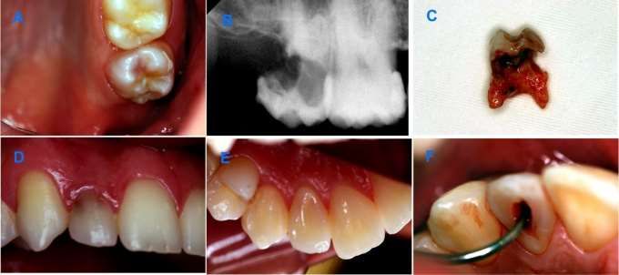
Research, Society and Development, v. 10, n. 5, e54410515340, 2021
                (CC BY 4.0) | ISSN 2525-3409 | DOI: http://dx.doi.org/10.33448/rsd-v10i5.15340

         reabsorção radicular no terço cervical da coroa. No 17, o terço médio e cervical da coroa estava comprometida e o
         teste de vitalidade pulpar foi negativo. O tratamento para o caso foi a exodontia. No 12 foi feito exame tomográfico
         que demonstrou o envolvimento da polpa e comprometimento da distância biológica. O teste de vitalidade foi
         positivo. Após o tratamento endodôntico, o dente foi extruído em 4 mm, a área reabsorvida foi exposta e restaurada
         com resina composta. O controle clínico e radiográfico de 39 meses demonstrou integridade da superfície radicular e
         do periodonto. Constatou-se que o diagnóstico precoce influi no prognóstico do tratamento considerando a velocidade
         de progressão da reabsorção. Torna importante o controle clinico e radiográfico das condições clínicas que predispõe
         a RCE.
         Palavras-chave: Reabsorção da raiz; Extrusão ortodôntica; Endodontia.

         Resumen
         La reabsorción cervical externa (CER) tiene un carácter inflamatorio y la proximidad al surco gingival favorece la
         contaminación y progresión de la lesión. El cambio de color de la corona, la inflamación de la encía marginal o
         incluso la presencia de secreción en el surco gingival son los principales signos clínicos. Al tratarse de una lesión
         asintomática, puede descuidarse y su progresión puede condenar el diente afectado. Este informe describe el
         tratamiento del caso de dos dientes con CER. En el examen clínico, la corona de 17 mostró una transparencia rosada
         en la superficie oclusal. Em el 12, esta mancha era oscura y estaba ubicada en el tercio cervical de la superficie
         vestibular. Ambos dientes estaban asintomáticos y el examen radiográfico mostró una imagen compatible con
         reabsorción radicular en el tercio cervical de la corona. El tercio medio y cervical de la corona del 17, estaba
         comprometido y la vitalidad pulpar fue negativa. El tratamiento fue la extracción. Examen tomográfico del diente 12
         demostró afectación pulpar y compromiso de la distancia biológica. La vitalidad pulpar fue positiva. Después del
         tratamiento de endodoncia, se extruyó el diente en 4 mm, y la área reabsorbida ha sido restaurada. El control clínico y
         radiográfico de 39 meses demostró integridad de la superficie radicular y del periodonto. Se encontró que el
         diagnóstico precoz influye en el pronóstico del tratamiento considerando la tasa de progresión de la reabsorción. Hace
         que sea importante el control clínico y radiográfico de las condiciones clínicas que predisponen al CER.
         Palabras clave: Resorción radicular; Extrusión ortodóncica; Endodoncia.

1. Introduction
         Root resorption corresponds to the loss of dental tissue due to the action of clastic cells on cementum and dentin
(Patel, et al., 2009). In permanent teeth, it is considered a pathological process and can be classified as internal or external
according to its location. A specific type of external root resorption located in the cervical region is termed by some authors as
External Cervical Resorption. Other authors prefer the term “Invasive Cervical Resorption”, due to its aggressive and
irreversible character (Heithersay, 1999). It affects mostly the teeth present in the upper arch, especially the central incisors,
irrespective of the gender (Mavridou, et al., 2017).
         Its etiology can be related to previous orthodontic treatment, dental trauma, tooth whitening procedures,
parafunctional habits, poor hygiene, and malocclusion (Heithersay, 1999; Mavridou, et al., 2017; Perlea, et al., 2017). These
causes could act individually or in combination (Mavridou, et al., 2017). It is usually diagnosed on radiographic examinations
performed for other reasons. Clinically, it can be characterized as a small defect in the gingival margin of the crown or even by
the pinkish color of the tooth enamel, resulting from tissue loss beneath it (Heithersay, 1999). In the absence of these signs and
being asymptomatic, the case may be neglected leading to premature loss of the dental elements involved (Patel, et al., 2018).
Diagnostic methods based on radiography (Heithersay, 1999) and tomography (Patel, et al., 2018) have been proposed to favor
the treatment of lesions. Currently, cone beam computed tomography (CBCT) is the best diagnostic technique due to its three-
dimensional properties (Patel, et al., 2018). In this case aspects such as height, circumference, and extent of its spread at the
root could be assessed. The proximity of resorption to the root canal would also be better measured.
         Such a resource would significantly favor the correct diagnosis of the injury, avoiding its confusion with other
pathologies common to this location, such as cervical caries, abrasion, abfraction, and erosion, which despite presenting with
different clinical signs, could confuse the professional (Consolaro, et al., 2014; Trope, 1998; Patel, et al., 2018). A correct
diagnosis of ECR is important to define the treatment strategy since inadequate treatments could be ineffective. When properly

                                                                2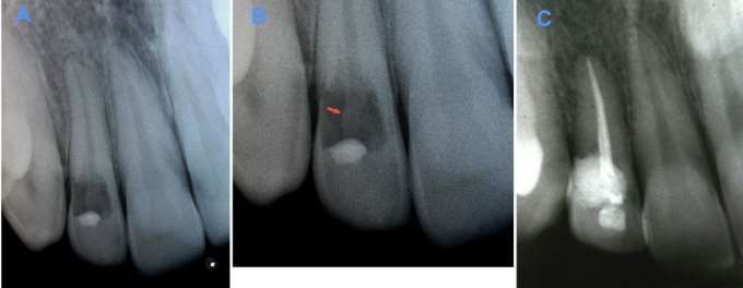
Research, Society and Development, v. 10, n. 5, e54410515340, 2021
                (CC BY 4.0) | ISSN 2525-3409 | DOI: http://dx.doi.org/10.33448/rsd-v10i5.15340

conducted, treatment has proven effective for these cases, with good prognosis (Lo Giudice, et al., 2016). The present case
report demonstrates the treatment of two teeth compromised by ECR, in the same patient, which presented different outcomes.
2. Methodology
        The purpose of this paper was to report, in a descriptive way, a case study (Pereira, et al., 2018) involving ECR. The
importance of this dental condition in oral health, as well as the techniques available for its treatment, its advantages and
disadvantages were based on scientific articles obtained in the Pubmed database. Likewise, the results obtained were discussed,
observing the advantages and disadvantages in their indication. For its realization, the diagnosis, the treatment plan, the
expected results as well as the risks and benefits were informed to the patient. In the same way, the destination of the personal
information collected was exposed to the patient with easy-to-understand language that he consented to through the Informed
Consent Form.

3. Case Report
        A 27-year-old male patient, sought the Dental Trauma Clinic for treatment of tooth 12 that had a darkened area on the
crown and secretions in the gingival sulcus. The patient had completed orthodontic treatment, started two years back, and had
no history of trauma or significant medical history. In the panoramic radiography, a radiolucent image could be seen in the
cervical region of teeth 12 and 17. Clinically, tooth 17 was asymptomatic and the crown had an area of pinkish color that
showed under the enamel of the occlusal surface (Figure 1A). Agenesis of tooth 18 was reported. The pulp vitality test was
negative and the periapical radiography showed a radiolucent area that started on the distal surface and involved the middle
and cervical third of the crown (Figure 1B). On probing, there was profuse bleeding of the periodontium, furcation
involvement and rigidity of the cavity walls. After clarification and consent by the patient, the treatment plan was extraction
(Figure 1C). Clinically, tooth 12 had secretion in the gingival sulcus, was asymptomatic and the vitality test was positive. The
labial and lingual surfaces of the crown were healthy (Figure 1D, E). There was a small layer of composite resin on the
cervical third of the labial surface of the crown. The underlying dentin was darkened (Figure 1D).

Figure 1. A- Pinkish aspect of the occlusal surface of tooth 17. B- Radiographic image of the resorbed area in the cervical and
middle third of the crown of tooth 17. C- Aspect of the molar after extraction showing short roots and extent of resorption. D-
Clinical condition of tooth 12 with darkened area of the cervical region of the crown. E- Integrity of the lingual surface of the
crown of tooth 12. F: Coronary access for canal biomechanics.

                                                                3
Research, Society and Development, v. 10, n. 5, e54410515340, 2021
(CC BY 4.0) | ISSN 2525-3409 | DOI: http://dx.doi.org/10.33448/rsd-v10i5.15340
Source: Authors.
On probing the labial surface, there was profuse bleeding and a 5 mm gingival sulcus depth (Figure 1F). Periapical
radiography showed integrity of the periodontal ligament and a radiolucent area that involved the cervical and middle third of
the crown (Figure 2A). It was also possible to notice an image suggestive of the outline of the root canal in the center of the
lesion (Figure 2B).
Figure 2. A- Radiographic aspect of tooth 12 with radiolucent area in the cervical region of the crown. B-: Details of the
periapical radiography with an image suggestive of the root canal in the center of the crown (arrow). C- 39-month radiographic
control showing the integrity of the periodontal ligament.
Source: Authors.
In the CT scan (Scanora 3D CBCT - Soredex Palodex Group Oy. – Finland), the coronal section revealed that the
cervical limit of the lesion in the distal surface of the crown was approximately 1 mm from the alveolar bone crest (Figure 3A)
with involvement of the pulp cavity. In the sagittal section, it was observed that the cervical limit of resorption was located
approximately 3 mm from the alveolar bone crest. In this image (Figure 3B), it was possible to notice the presence of remains
of the dentinal wall of the root canal. In the axial section, the lesion involved the entire labial surface of the crown (Figure 3C).
4Research, Society and Development, v. 10, n. 5, e54410515340, 2021
(CC BY 4.0) | ISSN 2525-3409 | DOI: http://dx.doi.org/10.33448/rsd-v10i5.15340
Figure 3. Cone beam computed tomography images. A- Coronal section showing impairment of periodontal biological width
on the distal surface of the crown and pulp involvement by resorption. B-: Sagittal section showing the location and extent of
resorption on the labial surface of the crown. The remaining dentinal wall of the root canal is also noted. C-: Axial section
showing extent of the lesion involving the labial surface of the crown.
Source: Authors.
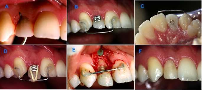
The treatment plan was explained to the patient and it consisted of endodontic treatment followed by orthodontic
extrusion, and restoration of the resorbed area with composite resin. During the coronary opening for endodontic treatment
(Figure 1F), it was found on probing that the communication of the resorbed area with the external environment was located 2
mm below the marginal gingiva (Figure 4A) and that the dentinal walls were rigid. The root canal length was 24 mm. Calcium
hydroxide dressing was used in the canal for 14 days and the final obturation was performed with gutta-percha cones and
calcium hydroxide cement (Sealapex - Sybron Kerr). For orthodontic extrusion, a 0.7-inch orthodontic wire was bonded with
an acid etching system and composite resin on the labial surfaces of teeth 11 and 13 to obtain anchorage. A bracket (Figure 4B)
was bonded to the labial surface of tooth 12 and a 0.25-inch orthodontic wire loop (Figure 4C) was bonded to the lingual
surface, using an acid etching system and composite resin. Device activation was achieved by using 1/8-inch orthodontic
elastic (Morelli Ortodontia) (Figure 4D).
After thirty days 4 mm of extrusion were obtained and the excess of the incisal edge was removed with a diamond
bur. Apically positioned flap expose the cavity (Figure 4E). After two weeks, the area was restored with an acid etching system
and composite resin. A 39-month clinical control demonstrated absence of mobility and a gingival sulcus depth of 2 mm
(Figure 4F). On radiographic examination, the integrity of the root surface and the periodontium was observed (Figure 2C).
Figure 4. A- Probing demonstrating the location of resorption and communication with the pulp chamber. B and C- Device
installed for orthodontic extrusion. D- Activation of the appliance with orthodontic elastics. E- Flap surgery to expose the
resorbed area. F- Clinical control after 39 months.
5Research, Society and Development, v. 10, n. 5, e54410515340, 2021
(CC BY 4.0) | ISSN 2525-3409 | DOI: http://dx.doi.org/10.33448/rsd-v10i5.15340
Source: Authors.
4. Discussion
Histologically, ECR occurs due to the loss of protection that cementoblasts and the cemental layer exert on the root
surface (Patel, et al., 2009; Patel, et al., 2018). The resorbed dentin and cementum are replaced by fibrovascular tissue
originating from the periodontal ligament (Gartner, et al., 1976). The progression of the lesion is a consequence of its
contamination by bacteria from the gingival sulcus, which penetrate the dentinal tubules directing resorption to the center of
the root. Upon reaching the supragingival region, the well-vascularized granulation tissue can be seen through the enamel as a
pink stain. The resorptive process presents difficulties in affecting tooth enamel as it is more mineralized and devoid of
pathways that promote communication between pulpal and periodontal tissues (Makkes, et al., 1975).
According to estudies (Patel, et al., 2018) the lesions in this case can be classified as 2Bp, that is, it involves the
coronary third of the root, extending apically to the alveolar bone crest, with a 90° to 180° contour of the root and with pulp
involvement. These lesions could be mistaken for dental caries because they have similar radiographic characteristics. In this
case, clinical aspects such as hard remaining dentin and profuse bleeding after periodontal probing contributed to the diagnosis
of ECR (Trope, 1998; Patel, et al., 2018). Due to furcation involvement through resorption and the short length of the roots, the
treatment plan for tooth 17 was extraction. In the case of tooth 12, a healthy remaining root favored an alternate treatment
option.
The difference between diagnosis of external and internal resorption can interfere with the treatment plan. Unlike
internal resorption, the pulp tissue may not be involved in the etiology of the ECR. If the lesion does not reach the pulp and it
presents with vitality, there is no need for endodontic treatment (Frank, et al., 1987; Bergmans, et al., 2002; Gonzales, et al.,
2007). Radiographically, internal resorption can be described as a symmetrical, eccentric lesion, with clear, defined, soft, and
smooth margins. The density of the radiolucency is uniform and the contour of the pulp chamber and the root canal cannot be
followed through the lesion (Cvek, 1973). In contrast, the external root resorption can be asymmetrical, with undefined edges
and variations in the density of the radiolucency in the lesion body (Gulabivala, et al., 1995). The canal wall can be traced in
the middle of the lesion, since it can be overlapping the root canal (Cvek, 1973). One of the resources for the diagnosis of
internal and external resorption is the use of the parallax principle (Clark's technique) in radiographic imaging (Gulabivala, et
al., 1995). However, radiographs are deficient in providing details of the remaining structures because they are based on a two-
6Research, Society and Development, v. 10, n. 5, e54410515340, 2021
(CC BY 4.0) | ISSN 2525-3409 | DOI: http://dx.doi.org/10.33448/rsd-v10i5.15340
dimensional image. Thus, CBCT was used because it offers a three-dimensional image with greater precision in the location
and dimension of the lesion (Patel, et al., 2018). Sagittal section demonstrated enhanced details of the existence of remains of
the vestibular wall of the root canal, a characteristic of external resorption. This could be due to the presence of the pre-dentin
layer that forms the root canal wall, which being a less mineralized structure, is less prone to resorption (Patel, et al., 2018;
Bergmans, et al., 2002). With the coronal and axial sections, it was possible to detect the biologic width violation and the
involvement of the pulp tissue.
The strategy to stop the resorptive process in cases of ECR, is to remove the connective tissue and blood supply that
provide favorable conditions for maintaining the clasts next to the dentin. The resorbed area must be debrided and restored to
avoid revascularization and subsequent action of the clasts (Heithersay, 1999). Among the techniques described for the
treatment of ECR, the use of materials such as mineral trioxide aggregate (MTA), intentional replantation, surgery to increase
the clinical crown and orthodontic extrusion are mentioned Krug, et al., 2019). In this case, the use of MTA would result in
compromising aesthetics due to its dark color.16 Intentional replantation could lead to trauma to the root surface with a risk of
resorption. Clinical crown augmentation surgery would result in elongation of the crown and involve the periodontium of
adjacent teeth with exposure of the cementum (Batenhorst, et al. 1974; Krug, et al. 2019). Thus, orthodontic extrusion was
used to expose the resorbed area and recover the biological width in a less traumatic way, allowing restoration with an
aesthetic material (Heithersay, 1973; Batenhorst, et al., 1974).
In endodontic treatment, calcium hydroxide was used as a temporary dressing because of its bactericidal capacity. The
release of hydroxyl ions by this material would provide action against bacteria and their toxins, which could eventually be
present in the root canal and dentinal tubules (Safavi, et al., 1993). This property is also very important to prevent root
resorption, especially when associated with contamination (Trope, et al., 1995). According to the literature, a sudden traumatic
injury would be the most likely cause for ECR (Patel, et al., 2018). In the case of tooth 17, it could occur due to the traumatic
action of the impacted third molar (Schriber, et al., 2020). However, given the history of agenesis of the third molar and the
absence of some other type of trauma, reinforced orthodontic treatment seems a potential cause. Several predisposing factors,
isolated or combined, could contribute to its onset and progress (Mavridou, et al., 2017). Thus, the type of orthodontic
appliance used, the type of tooth movement, the force applied and the duration of treatment could be considered (Krishnan,
2005; Dudic, et al., 2017). In molars, the use of bands could expose dentin in the cervical region (Krishnan, 2005). The 39-
month clinical control showed normal mobility. Radiographically integrity of the root surface and the periodontium were
observed indicating the effectiveness of the treatment plan.
5. Conclusion
It can be seen from the case that early diagnosis is important for the prognosis, considering the speed of progression of
resorption. It emphasizes the importance of clinical and radiographic control of clinical conditions that could predispose to
ECR. Considering orthodontic treatment as a potential etiological factor, it would be interesting to develop a study to assess the
relationship between different types of tooth movement, the intensity and frequency of the force used and the occurrence of
ECR.
References
Bergmans, L., Van Cleynenbreugel, J., Verbeken, E., Wevers, M., Van Meerbeek, B., & Lambrechts, P. (2002). Cervical external root resorption in vital teeth.
Journal of clinical periodontology, 29(6), 580–585. https://doi.org/10.1034/j.1600-051x.2002.290615.x
Batenhorst, K. F., Bowers, G. M., & Williams Jr, J. E. (1974). Tissues changes resulting from facial tipping and extrusion of incisors in monkeys. Journal of
periodontology, 45(9), 660–668. https://doi.org/10.1902/jop.1974.45.9.660
7Research, Society and Development, v. 10, n. 5, e54410515340, 2021
(CC BY 4.0) | ISSN 2525-3409 | DOI: http://dx.doi.org/10.33448/rsd-v10i5.15340
Consolaro, A., Cardoso, M. A., Almeida, C. D. C. M., Souza, I. A. O., & Capelloza Filho, L. (2014). The clinical meaning of external cervical resorption in
maxillary canine: transoperative dental trauma. Dental Press Journal of Orthodontics, 19(6), 19-25. https://doi.org/10.1590/2176-9451.19.6.019-025.oin
Cvek, M. (1973). Treatment of non-vital permanent incisors with calcium hydroxide. II. Effect on external root resorption in luxated teeth compared with
effect of root filling with guttapucha. A follow-up. Odontologisk revy, 24(4), 343–354.
Dudic, A., Giannopoulou, C., Meda, P., Montet, X., & Kiliaridis, S. (2017). Orthodontically induced cervical root resorption in humans is associated with the
amount of tooth movement. European Journal of Orthodontics, Volume 39, Issue 5, Pages 534–540, https://doi.org/10.1093/ejo/cjw087.
Frank, A. L., & Bakland, L. K. (1987). Nonendodontic therapy for supraosseous extracanal invasive resorption. Journal of endodontics, 13(7), 348–355.
https://doi.org/10.1016/S0099-2399(87)80117-6
Gartner, A. H., Mack, T., Somerlott, R. G., & Walsh, L. C. (1976). Differential diagnosis of internal and external root resorption. Journal of endodontics,
2(11), 329–334. https://doi.org/10.1016/S0099-2399(76)80071-4
Gonzales, J. R., & Rodekirchen, H. (2007). Endodontic and periodontal treatment of an external cervical resorption. Oral surgery, oral medicine, oral
pathology, oral radiology, and endodontics, 104(1), e70–e77. https://doi.org/10.1016/j.tripleo.2007.01.023
Gulabivala, K., & Searson, L.J. (1995). Clinical diagnosis of internal resorption: an exception to the rule. International endodontic journal, 28(5), 255–260.
https://doi.org/10.1111/j.1365-2591.1995.tb00310.x
Heithersay, G. S. (1999). Clinical, radiologic, and histopathologic features of invasive cervical resorption. Quintessence international (Berlin, Germany:
1985), 30(1), 27–37.
Heithersay, G. S. (1973). Combined endodontic-orthdontic treatment of transverse root fractures in the region of the alveolar crest. Oral surgery, oral
medicine, and oral pathology, 36(3), 404–415. https://doi.org/10.1016/0030-4220(73)90220-x
Heithersay, G. S. (1999). Invasive cervical resorption following trauma. Australian endodontic journal: the journal of the Australian Society of Endodontology
Inc, 25(2), 79–85. https://doi.org/10.1111/j.1747-4477.1999.tb00094.x
Krishnan, V. (2005). Critical issues concerning root resorption: a contemporary review. World journal of orthodontics, 6(1), 30–40.
Krug, R., Soliman, S., & Krastl, G. (2019). Intentional replantation with an atraumatic extraction system in teeth with extensive cervical resorption. Journal of
endodontics, 45(11), 1390–1396. https://doi.org/10.1016/j.joen.2019.07.012
Lo Giudice, G., Matarese, G., Lizio, A., Giudice, R. L., Tumedei, M., Zizzari, V. L., & Tetè, S. (2016). Invasive cervical resorption: a case series with 3-year
follow-up. The International journal of periodontics & restorative dentistry, 36(1), 103–109. https://doi.org/10.11607/prd.2066
Makkes, P. C., & Thoden van Velzen, S. K. (1975). Cervical external root resorption. Journal of dentistry, 3(5), 217–222. https://doi.org/10.1016/0300-
5712(75)90126-8
Mavridou, A. M., Bergmans, L., Barendregt, D., & Lambrechts, P. (2017). Descriptive analysis of factors associated with external cervical resorption. Journal
of endodontics, 43(10), 1602–1610. https://doi.org/10.1016/j.joen.2017.05.026
Patel, S., Dawood, A., Wilson, R., Horner, K., & Mannocci, F. (2009). The detection and management of root resorption lesions using intraoral radiography
and cone beam computed tomography - an in vivo investigation. International endodontic journal, 42(9), 831–838. https://doi.org/10.1111/j.1365-
2591.2009.01592.x
Patel, S., Foschi, F., Condon, R., Pimentel, T., & Bhuva, B. (2018). External cervical resorption: part 2 - management. International Endodontic Journal
Nov;51(11):1224-38.
Patel, S., Foschi, F., Mannocci, F., & Patel, K. (2018). External cervical resorption: a three-dimensional classification. International endodontic journal,
51(11), 1224–1238. https://doi.org/10.1111/iej.12946
Patel, S., Kanagasingam, S., & Pitt Ford, T. (2009). External cervical resorption: a review. Journal of endodontics, 35(5), 616–625.
https://doi.org/10.1016/j.joen.2009.01.015
Patel, S., Mavridou, A. M., Lambrechts, P., & Saberi, N. (2018). External cervical resorption-part 1: histopathology, distribution and presentation.
International endodontic journal, 51(11), 1205–1223. https://doi.org/10.1111/iej.12942
Pereira, A. S., Shitsuka, D. M., Parreira, F. J., & Shitsuka, R. (2018). Metodologia da pesquisa científica [recurso eletrônico] 1. ed. – Santa Maria, RS: UFSM,
NTE. 1 e-book.
Perlea, P., Imre, M., Nistor, C. C., Iliescu, M. G., Gheorghiu, I. M., Abramovitz, I., & Iliescu, A. A. (2017). Occurrence of invasive cervical resorption after
the completion of orthodontic treatment. Romanian journal of morphology and embryology = Revue roumaine de morphologie et embryologie, 58(4), 1561–
1567.
Safavi, K. E., & Nichols, F. C. (1993). Effect of calcium hydroxide on bacterial lipopolysaccharide. Journal of endodontics, 19(2), 76–78.
https://doi.org/10.1016/S0099-2399(06)81199-4
Schriber, M., Rivola, M., Leung, Y. Y., Bornstein, M. M., & Suter, V. (2020). Risk factors for external root resorption of maxillary second molars due to
impacted third molars as evaluated using cone beam computed tomography. International journal of oral and maxillofacial surgery, 49(5), 666–672.
https://doi.org/10.1016/j.ijom.2019.09.016
Trope, M. (1998). Root resorption of dental and traumatic origin: classification based on etiology. Practical periodontics and aesthetic dentistry : PPAD,
10(4), 515–522.
8Research, Society and Development, v. 10, n. 5, e54410515340, 2021
(CC BY 4.0) | ISSN 2525-3409 | DOI: http://dx.doi.org/10.33448/rsd-v10i5.15340
Trope, M., Moshonov, J., Nissan, R., Buxt, P., & Yesilsoy, C. (1995). Short vs. long-term calcium hydroxide treatment of estabilished inflammatory root
resorption in replanted dog teeth. Endodontics & dental traumatology, 11(3), 124–128. https://doi.org/10.1111/j.1600-9657.1995.tb00473.x
9You can also read