Unexpected Reason for Non healing Oral Ulcers: Syphilis - Lester DR Thompson, MD
←
→
Page content transcription
If your browser does not render page correctly, please read the page content below
Head and Neck Pathology
https://doi.org/10.1007/s12105-021-01348-y

    CASE REPORTS

Unexpected Reason for Non‑healing Oral Ulcers: Syphilis
Frank Deng1 · Lester D. R. Thompson2 · Jinping Lai3

Received: 5 May 2021 / Accepted: 11 June 2021
© The Author(s), under exclusive licence to Springer Science+Business Media, LLC, part of Springer Nature 2021

Abstract
Syphilis is a sexually transmitted infectious disease caused by Treponema pallidum and characterized by a complex and
variable clinical presentation. Cases of unexpected oral syphilis presenting as non-healing ulcers are uncommonly reported.
We report 3 cases (one female and two males, aged 35, 35, and 56 years, respectively) in which patients presented with
non-healing oral ulcers. Biopsies revealed surface ulceration and a significant neutrophilic infiltrate rather than the more
conventional plasma cell infiltrate seen with most reported syphilis infections, potentially leading to an inaccurate diagno-
sis. Treponema pallidum immunohistochemistry highlighted spirochetes within the epithelium, with additional diagnostic
confirmation by serum T. pallidum particle agglutination assay. Sexual history documentation by the clinician with non-
specific oral ulcers is paramount to aiding diagnosis and leading to proper management. Further, it is important to perform
immunohistochemistry for T. pallidum in oral biopsies from non-healing ulcers, especially when clinical history raises the
differential diagnosis or when other clinical manifestations may support this consideration.

Keywords Oral ulcer · Syphilis · Treponemal pallidum · Mouth diseases · Sexual behavior · Spirochaetales ·
Immunohistochemistry · Differential diagnosis

Introduction                                                                 of oral syphilis can be challenging [3]. However, the inci-
                                                                             dence of oral syphilis has been increasing, and thus oral
Syphilis is an infectious disease of the spirochete Treponema                lesions may represent an important diagnostic indicator of
pallidum, and is transmitted by sexual contact in most cases                 infection [4].
(congenital cases are not considered herein). Infection is                       Mucosal ulcers are a common patient complaint, with
divided into four stages: primary, secondary, latent, and ter-               syphilitic infection only one of many different etiologies
tiary [1]. The first sign of primary syphilitic infection is the             to be considered [5]. The objectives of this study were to
appearance of a small, painless papule that develops into a                  report three cases of syphilis with primarily oral manifes-
hard chancre with nontender lymphadenopathy 1–3 weeks                        tations, highlighting the importance of an accurate sexual
after exposure. Chancres are usually identified on the exter-                history and maintaining a high index of suspicion for syphi-
nal genitalia, but oral sex may lead to syphilis transmission,               lis. Histopathological studies of oral syphilis commonly
with the lips most commonly affected [2]. There is a wide                    report a significant subepithelial plasma cell infiltrate [6,
diversity of etiologies for oral ulcers, and thus recognition                7]. However, cases of oral syphilis with a predominant neu-
                                                                             trophilic infiltrate are rarely reported. This study discusses
                                                                             this uncommon histology which can lead to misdiagnosis of
* Jinping Lai                                                                primary or secondary oral syphilis.
  jinping.x.lai@kp.org
1
     Warren Alpert Medical School of Brown University,
     Providence, RI, USA
                                                                             Case Representation
2
     Department of Pathology, Woodland Hills Medical
     Center, Southern California Permanente Medical Group,
                                                                             Case 1
     Woodland Hills, CA, USA
3
     Department of Pathology, Kaiser Permanente Sacramento
                                                                             A 35-year-old female presented with a 2-month long non-
     Medical Center, 2025 Morse Ave, 95825 Sacramento, CA,                   healing ulcer on the right lateral tongue with an associated
     USA

                                                                                                                                13
                                                                                                                           Vol.:(0123456789)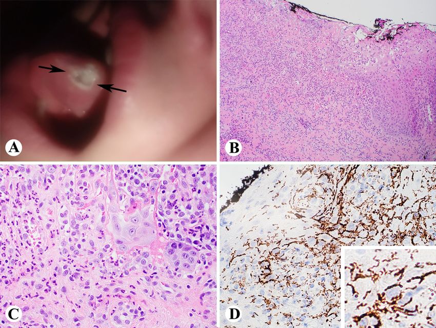
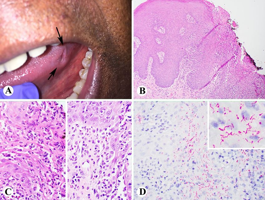
Head and Neck Pathology

burning sensation. She noted changing her diet to avoid               Case 2
spicy and hot foods due to pain. The patient had done noth-
ing to attempt to alleviate her symptoms. Significant past              A 35-year-old male presented with a three month-long his-
medical history included diabetes mellitus, chlamydia infec-          tory of left-sided tongue discomfort. He reported oral sex
tion, and gastroesophageal reflux disease (GERD). She was             with females only. Past medical history was significant for
a non-smoker. Clinical examination revealed a 1 cm right              alcoholic cirrhosis. Physical exam revealed white plaques
tongue ulcer (Fig. 1a). An unrelated right torus palatini was         of varying sizes on the hard palate, bilateral tonsillar fossa,
noted.                                                                and on the left lateral tongue and lower lip mucosa (Fig. 2a).
   An excisional biopsy was performed. Sections revealed              Upon further examination, similar lesions were identified
ulcerated squamous epithelium with associated granulation             on his penis.
tissue (Fig. 1b, c). Acute inflammation was seen extend-                 An excisional biopsy was performed on both the tongue
ing into the subjacent stroma. Based on these findings, a             and lower lip. Histologic sections showed ulcerated squa-
Gomori Methamine silver stain and herpes simplex virus                mous mucosa with adjacent pseudoepitheliomatous hyper-
(HSV) and T. pallidum spirochete immunohistochemistry                 plasia and a rich acute inflammatory infiltrate (Fig. 2b, c).
were performed. Innumerable T. pallidum organisms were                Special studies included a negative Grocott’s methenamine
highlighted via immunohistochemistry (Fig. 1d). No fun-               silver stain and acid-fast bacillus (AFB) stains for fungal
gal organisms were identified. Serologic testing confirmed            organisms and mycobacteria, respectively, while numerous
a diagnosis of syphilis (positive T. Pallidum IgG and IgM             organisms were identified by immunohistochemistry stain
by T. Pallidum particle agglutination assay (TP-PA) with a            of T. pallidum (Fig. 2d). Human immunodeficiency virus
Rapid Plasma Reagin (RPR) titer 1:128). The patient was               (HIV) and HSV serology screens were negative. Serologic
treated with penicillin-G (2.4 million units) and has not             testing confirmed a diagnosis of syphilis (positive T. Pal-
returned for additional evaluation.                                   lidum IgG and IgM by TP-PA with a RPR titer 1:32). The

Fig. 1  Case 1 showing a 1 cm right tongue ulcer (a); biopsy showing ulceration, granulation tissue and active inflammation (b and c, H&E
stain); and T. Pallidum immunostain showing innumerable T. Pallidum spirochetes (d and inset) (b, 100x; c and d, 400x)

13
Head and Neck Pathology
Fig. 2 Case 2 showing a 1.4 cm lip white plaque (a); biopsy show- stain); and immunostain of T. Pallidum showing numerous T. Palli-
ing ulcerated squamous mucosa with adjacent pseudoepitheliomatous dum spirochetes (d and inset) (b, 100x; c and d, 400x)
hyperplasia and a rich acute inflammatory infiltrate (b and c, H&E
patient was treated with penicillin-G (2.4 million units) and Discussion
all lesions resolved by the time he was seen one month later.
Syphilis has experienced an increased incidence in the 21st
century [1, 4], compared to levels in the previous century
Case 3 [8]. Yet, recognition of the disease through its many vari-
ous presentations challenges even the most experienced of
A 56-year-old male presented with an 8-week-long indenta- clinicians [7–10].
tion on the left lateral tongue. He reported it was of a sud- Primary syphilis often presents as an isolated papule at
den onset and associated with soreness. He denied trauma. the site of inoculation, that rapidly erodes and becomes an
He smoked cigars occasionally. Pertinent past medical his- indurated chancre, with surrounding erythema. A classic
tory included anorectal pain. Previous serology screens for chancre is usually not painful but can become so if suprain-
sexually acquired infections were negative within the past 2 fected with bacteria. The vast majority of chancres present as
years. Physical exam revealed a 5 mm ulcer on the left lateral anogenital lesions, but chancres may present extragenitally
tongue (Fig. 3a), without additional findings. usually after orogenital contact, most often in the oral cav-
The biopsy showed surface ulceration with both acute ity or on the lips [11]. Secondary syphilis presents in the
and chronic inflammation and adjacent squamous epithe- oral cavity in many forms, including multiple white mucous
lial hyperplasia (Fig. 3b, c). Immunohistochemistry for T. patches, condyloma lata, and split papules [12]. Oral ulcers
pallidum spirochetes (Fig. 3d) was positive. Serologic test- are a relatively common clinical presentation, due to a wide
ing confirmed a diagnosis of syphilis (positive T. Pallidum variety of causes, which makes recognition of oral syphilis
IgG and IgM by TP-PA with a RPR titer 1:64). The patient challenging. The differential diagnoses include infections
was treated with doxycycline due to a penicillin allergy. No (bacterial, viral fungal), trauma, neoplasm, autoimmune dis-
lesion remained at 3-month follow-up. eases, and allergies, etc. Factors such as duration, pattern
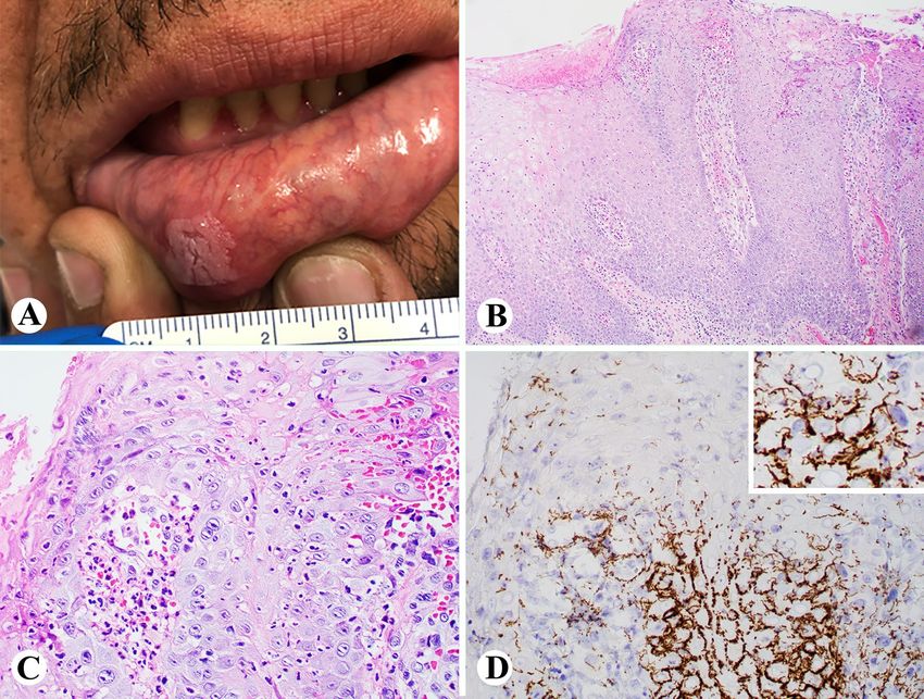
13Head and Neck Pathology Fig. 3 Case 3 showing a 5 mm lateral tongue ulcer (a); biopsy show- immunostain of T. Pallidum showing many T. Pallidum spirochetes ing surface ulceration with both acute and chronic inflammation and (d and inset) (b, 100x; c and d, 400x) adjacent squamous epithelial hyperplasia (b and c, H&E stain); and of recurrence, clinical appearance, mucosal location, and carcinoma [16]. However, syphilitic oral chancres develop presence or absence of systemic symptoms are useful clues at inoculation sites and often spontaneously resolve within to determining an ulcer’s cause [13]. The clinical manifes- 3–8 weeks, unlike oral tuberculosis, which often includes tations of ulcers are nonspecific, requiring detailed clinical constitutional symptoms for a significant time frame [15]. history, including social and sexual history, along with his- Most syphilis cases are diagnosed based on a combina- tory of trauma, dental appliance use, food or additive aller- tion of clinical, microscopic, immunohistochemical, and gies, aphthous history, among other causes, with supporting serologic findings. Histology of a subepithelial plasma laboratory investigations to reach a definitive diagnosis. cell-predominant infiltrate is the most common finding Acute oral ulcers develop due to a wide variety of etiolo- that prompts T. pallidum immunohistochemistry studies gies. The most common causes include trauma but may also [7]. The immunohistochemistry study is specific for T. pal- be the oral manifestations of autoimmune disorders, con- lidum, helping to exclude the spirochete commensals nor- nective tissue disease, gastrointestinal tract disease (Crohn mally identified in the oral cavity, and potentially difficult disease; ulcerative colitis), and the nonspecific aphthous to separate on Warthin-Starry stains, or other non-specific stomatitis, along with contact allergies or reactions (amal- spirochete histochemistry studies. Further serologic test- gam, toothpaste sensitivity, cinnamon allergy, among oth- ing is generally advocated to document disease. In our ers) [14]. Past reports have highlighted the many protean reported cases, all patients had chancres affecting the oral manifestations of syphilis, with presentations that mimic mucosa (tongue and lip). Microscopic exam highlighted oral tuberculosis, HIV-associated Kaposi sarcoma, recur- acute inflammation rather than a plasmacytic cell popula- rent aphthous ulcer, and oral squamous cell carcinoma [15]. tion, making the diagnosis more challenging. Syphilis was However, when the history is of painless oral ulceration, confirmed with the immunohistochemistry reaction for T. the differential diagnosis is narrowed to syphilis, lupus ery- pallidum and serology tests of TP-PA and RPR. Thus, thematosus, traumatic ulceration from neuropathy, and/or in nonspecific oral ulcers, careful consideration must be 13
Head and Neck Pathology
given to infectious etiologies, using a panel approach of Declarations
fungal stains along with selected immunohistochemistry
studies (HSV, T. pallidum, cytomegalovirus) directed Conflict of interest All authors declare that they have no conflict of
by possible additional histologic features. This targeted interest regarding this study.
approach is especially important in potentially at-risk Ethical approval This clinical investigation was conducted in accord-
patients (immunocompromised, past sexually transmitted ance and compliance with all statutes, directives, and guidelines of
infection history, multiple sexual partners, males who have an Internal Review Board authorization involving a retrospective
sex with males, and sex worker contact). data analysis involving human subjects in research with appropriate
informed consent.
A relatively low 21% of patients with syphilis are diag-
nosed at an early stage, and the high proportion of patients Informed consent Patient granted consent for publication of their
with early latent syphilis, suggests that primary and second- data. The consent form has been filed and can be produced upon
request.
ary syphilis frequently goes undiagnosed or misdiagnosed
[8, 17]. Diagnosis of syphilis is frequently based on clinical
and serological findings. However, biopsies may provide an
important histologic basis to further direct specific serologic
testing. In these reported cases, syphilis was not suspected References
clinically until microscopic evaluation and additional special
studies were performed to identify the causative T. pallidum 1. Peeling R, Mabey D, Kamb M, Chen X, Radolf J, Benzaken A
(2017) Syphilis. Nat Rev Dis Primers 3: 7073 https://doi.org/10.
agent. It is most helpful to have a complete clinical history
1038/nrdp.2017.73.
that may provide some presumptive evidence to aid in rec- 2. Kelner N, Rabelo G, da Cruz Perez D, Assuncao J Jr, Witzel A,
ognizing and confirming the diagnosis of syphilis and thus Migliari D, Alves F. Analysis of nonspecific oral mucosal and der-
improve health outcomes. mal lesions suggestive of syphilis: a report of 6 cases. Oral Surg
Oral Med Oral Pathol Oral Radiol. 2014;117(1):1–7. doi:https://
Treatment of syphilis is determined by disease stage, but
doi.org/10.1016/j.oooo.2012.04.028.
penicillin-G remains the drug of first choice in allergy-free 3. Fitzpatrick S, Cohen D, Clark A. Ulcerated Lesions of the
patients for all stages [11]. Alternative therapies are used for Oral Mucosa: Clinical and Histologic Review. Head Neck
penicillin-allergic patients. Pathol. 2019;13(1):91–102. doi:https:// d oi. o rg/ 1 0. 1 007/
s12105-018-0981-8.
4. Matias M, Jesus A, Resende R, Caldeira P, Aguiar M. Diagnos-
ing acquired syphilis through oral lesions: the 12 year experi-
ence of an Oral Medicine Center. Braz J Otorhinolaryngol.
Conclusion 2020;86(3):358–63. doi:https://doi.org/10.1016/j.bjorl.2018.12.
010.
5. Fourie J, Boy S. Oral mucosal ulceration - a clinician’s guide to
Oral ulcers represent a wide diversity of etiologies. For any diagnosis and treatment. S Afr Dent J. 2016;71:500–8.
nonhealing oral ulcer, a broad differential diagnosis must 6. Barrett A, Villarroel Dorrego M, Hodgson T, Porter S, Hopper
include infectious etiologies, including fungal, mycobacte- C, Argiriadou A, Speight P. The histopathology of syphilis of the
oral mucosa. J Oral Pathol Med. 2004;33(5):286–91. doi:https://
rial, viral and spriochetal infections. There are important
doi.org/10.1111/j.0904-2512.2004.00099.x.
public health related concerns when managing sexually 7. Ficarra G, Carlos R. Syphilis: the renaissance of an old disease
transmitted infections, and so a broader awareness of syphi- with oral implications. Head Neck Pathol. 2009;3(3):195–206.
lis as it presents in the oral cavity may aid it better detection doi:https://doi.org/10.1007/s12105-009-0127-0.
8. Ghanem K, Ram S, Rice P. The Modern Epidemic of Syphilis.
and management [1, 18–20]. Our presentation highlights the
N Engl J Med. 2020;382(9):845–54. doi:https://doi.org/10.1056/
broad spectrum of histologic features that can be seen with NEJMra1901593.
oral syphilis, which underscores the need for a high index of 9. Carbone P, Capra G, Nelson B. Oral Secondary Syphilis. Head
suspicion for syphilis when presented with oral ulceration, Neck Pathol. 2016;10(2):206–8. doi:https://doi.org/10.1007/
s12105-015-0623-3.
whether showing classical plasmacytic infiltration or acute
10. Smith MH, Vargo RJ, Bilodeau EA, Anderson KM, Trzcinska
inflammation. A, Canterbury CR, Fantasia JE, Rawal YB. Oral Manifesta-
tions of Syphilis: a Review of the Clinical and Histopathologic
Characteristics of a Reemerging Entity with Report of 19 New
Authors’ Contributions Each author contributed important intellectual Cases. Head Neck Pathol. 2021. doi:https://doi.org/10.1007/
content during manuscript drafting or revision and accepts account- s12105-020-01283-4.
ability for the overall work by ensuring that questions pertaining to 11. Siu A, Landon K, Ramos D. Differential diagnosis and manage-
the accuracy or integrity of any portion of the work are appropriately ment of oral ulcers. Semin Cutan Med Surg. 2015;34(4):171–7.
investigated and resolved. doi:https://doi.org/10.12788/j.sder.2015.0170.
12. Little J. Syphilis: an update. Oral Surg Oral Med Oral Pathol. Oral
Radiol Endod. 2005;100(1):3–9. doi:https://d oi.o rg/1 0.1 016/j.t ripl
Funding No external funding was obtained for this study.
eo.2005.03.006.
13Head and Neck Pathology
13. Bruce AJ, Dabade TS, Burkemper NM. Diagnosing oral ulcers. 18. Hertel M, Matter D, Schmidt-Westhausen A, Bornstein M.
JAAPA. 2015;28(2):1–10. doi:https://doi.org/10.1097/01.JAA. Oral syphilis: a series of 5 cases. J Oral Maxillofac Surg.
0000459826.63026.67. 2014;72(2):338–45. doi:https://doi.org/10.1016/j.joms.2013.07.
14. Scully C, Shotts R. ABC of oral health. Mouth ulcers and 015.
other causes of orofacial soreness and pain. Br Med J. 19. Streight K, Paranal R, Musher D. The oral manifestations of syph-
2000;321(7254):162–5. doi:https://d oi.o rg/1 0.1 136/b mj.3 21.7 254. ilitic disease: a case report. J Med Case Rep. 2019;13(1):227.
162. doi:https://doi.org/10.1186/s13256-019-2171-z.
15. Huang S, Lu R, Yang J, Zhou G. A nonspecific ulcer on upper lip 20. Strieder L, Leon J, Carvalho Y, Kaminagakura E. Oral syphilis:
presented as the first and sole sign of syphilis. Journal of infection report of three cases and characterization of the inflammatory
chemotherapy: official journal of the Japan Society of Chemo- cells. Ann Diagn Pathol. 2015;19(2):76–80. doi:https://doi.org/
therapy. 2020;26(12):1309–12. doi:https://doi.org/10.1016/j.jiac. 10.1016/j.anndiagpath.2015.01.003.
2020.07.010.
16. Lehman J, Rogers R. 3rd: Acute oral ulcers. Clin Dermatol. Publisher’s Note Springer Nature remains neutral with regard to
2016;34(4):470–4. doi:https://doi.org/10.1016/j.clindermatol. jurisdictional claims in published maps and institutional affiliations.
2016.02.019.
17. Wong N, Huang S, Zheng H, Chen L, Zhao P, Tucker J, Yang L,
Goh B, Yang B. Stages of syphilis in South China – a multilevel
analysis of early diagnosis. BMC Public Health. 2017;17(1):135.
doi:https://doi.org/10.1186/s12889-016-4004-y.
13You can also read