Monilinia species of fruit decay: a comparison between biological and epidemiological data - Unibo
←
→
Page content transcription
If your browser does not render page correctly, please read the page content below
A. Di Francesco, M. Mari                                     Italian Journal of Mycology vol. 47 (2018) ISSN 2531-7342
DOI: https://doi.org/10.6092/issn.2531-7342/7817

 Monilinia species of fruit decay: a comparison
 between biological and epidemiological data
 ___________________________________________
                                Alessandra Di Francesco, Marta Mari

 CRIOF - Department of Agricultural and Food Science, Alma Mater Studiorum University of Bologna
 Via Gandolfi, 19, 40057 Cadriano, Bologna, Italy

 Correspondig Author e-mail: alessand.difrancesc3@unibo.it

 Abstract

    The fungal genus Monilinia Honey includes parasitic species of Rosaceae and Ericaceae. The Monilinia genus
    shows a great heterogeneity, it is divided in two sections: Junctoriae and Disjunctoriae. These sections were
    defined by Batra (1991) on the basis of morphology, infection biology, and host specialization. Junctoriae spp.
    produce mitospore chains without disjunctors, they are parasites of Rosaceae spp.; M. laxa, M. fructicola, M.
    fructigena and recently M. polystroma, represent the principal species of the section, and they are responsible
    of economically important diseases of Rosaceae, new species such as M. yunnanensis, and M. mumecola could
    afflict European fruits in the future in absence of a strict phytosanitary control. The Disjunctoriae section
    includes species that produce mitospores intercalated by appendages (disjunctors); they parasitize Rosaceae,
    Ericaceae, and Empetraceae. The principal Disjunctoriae species are M. vaccinii-corymbosi, M. urnula, M.
    baccarum, M. oxycocci, and M. linhartiana. This study has the aim to underline the importance of Monilinia
    spp., and to describe their features.

    Keywords: Monilinia spp; Junctoriae; Disjunctoriae; Prunus; Vaccinium

 Riassunto

    Il genere Monilinia Honey include diverse specie che attaccano in particolar modo le piante delle famiglie
    Rosaceae ed Ericaceae. Monilinia è un genere molto eterogeneo, infatti è suddiviso in due sezioni: Junctoriae
    e Disjunctoriae. Le due sezioni sono state definite da Batra (1991) sulla base della morfologia, della capacità
    infettiva e dell’ospite attaccato. La sezione Junctoriae è caratterizzata dalla produzione di catene di mitospore
    prive di setti di separazione fra una spora e l’altra. Essa comprende specie parassite di piante appartenenti alle
    Rosaceae. M. laxa, M. fructicola, M. fructigena e recentemente M. polystroma, appartengono a questa sezione e
    rappresentano la principale problematica fitosanitaria per drupacee e pomacee. Non vanno sottovalutate anche
    altre specie della sezione Junctoriae parassite del pesco e dell’albicocco, che al momento risultano essere
    presenti solo in Oriente: M. yunnanensis e M. mumecola. E’ comunque necessario attuare severi controlli
    fitosanitari per evitarne la diffusione. La sezione Disjunctoriae include specie che producono mitospore
    intercalate da disgiuntori e che sono parassite di Ericaceae, Rosaceae ed Empetraceae. Le specie di Monilinia
    appartenenti a questa sezione sono M. vaccinii-corymbosi, M. urnula, M. baccarum, M. oxycocci e M.
    linhartiana. Questo lavoro ha lo scopo di sottolineare l’importanza delle specie di Monilinia e di descriverne le
    caratteristiche.

    Parole chiave: Monilinia spp; Junctoriae; Disjunctoriae; Prunus; Vaccinium

                                                                                                                   13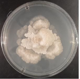
A. Di Francesco, M. Mari                               Italian Journal of Mycology vol. 47 (2018) ISSN 2531-7342
DOI: https://doi.org/10.6092/issn.2531-7342/7817

 Introduction

 The genus Monilinia Honey includes several aggressive and economically important pathogens of the
 Rosaceae, Ericaceae and Empetraceae. They are present worldwide and affect in particular stone and pome
 fruits, and also berry fruits. These pathogens are often referred as “brown rot” agents. Monilinia spp. belong
 to the Junctoriae or Disjunctoriae section. These sections were defined on the basis of conidial morphology,
 fungal life cycle and host specialization (Batra, 1991).
 M. fructicola, M. laxa, M. fructigena, M. polystroma, M. mumecola and M. yunnanensis constitute the
 Junctoriae section. They do not have disjunctors between the mature mitospores within the conidial chains.
 Other Monilinia species such as M. oxycocci M. baccarum, M. urnula, M. vaccinii-corymbosi and M.
 linhartiana, have disjunctors, narrow barren separating cells. The endostromata occur in the young fruits,
 and the infection develops in the ovary chamber (Batra, 1991).
 M. fructicola, M. laxa, M. fructigena and M. polystroma are well-known pathogens affecting rosaceous fruit
 production all over the world (Petroczy et al., 2012). The other species such as M. oxycocci, M. baccarum,
 M. urnula, M. vaccinii-corymbosi inhabit Vaccinium hosts specially in North America. M. fructicola
 originates in America, Australia and New Zeland (EPPO/CABI, 2010). It was detected for the first time in
 French peach orchards (EPPO, 2002) and then in Spain (De Cal et al., 2009), Switzerland (Bosshard et al.,
 2006), Hungary (Petroczy and Palkovics, 2009), and Italy (Pellegrino et al., 2009; Montuschi et al., 2011;
 Marinelli et al., 2013) on peaches. This wide diffusion by M. fructicola could be related to sexual exchange
 for its ability to produce ascospores from mummified fruits (Byrde and Willets, 1977).
 M. laxa is the most prevalent (85-90%) (Larena et al., 2005) in Europe, but since M. fructicola was detected,
 in peach orchards both species easily can co-existed ( Boehm et al., 2001; Villarino et al., 2013). These
 species can be distinguished for different growth characteristics. Nevertheless M. laxa remains a quarantine
 pathogen in China and in some areas of North America (Martini and Mari, 2014).
 M. fructigena is prevalent on pome fruits, only 10% of isolates derived from stone fruits in Europe (Martini
 and Mari, 2014). M. polystroma, was found for the first time in Japan (Cotè et al., 2004) and later in
 Hungary (Petroczy and Palkovics, 2009), Czech Republic (EPPO, 2011), Italy (Martini et al., 2014), and
 Croatia (Di Francesco et al., 2015). It shows similar morphological characteristics to M. fructigena. M.
 mumecola represents a new apricot pathogen that causes white rot; it resembles to M. laxa, but it differs from
 this and to the other brown rot fungi for conidia dimensions and colony characteristics (Harada et al., 2004).
 Generally, Monilinia spp. hyphae differentiate melanised aggregates, the stromata (Holst-Jensen et al., 1997)
 which in early spring on mummified fruits could produce apothecia, the teleomorphic stage. The anamorphic
 stage is produced on infected leaves, blossoms, flowers and fruits.
 Although the identification of Monilinia spp. has been based on morphological analysis, from ‘90s it was
 also realized by molecular diagnostics.
 The objective of this review is to provide a deep analysis of Monilinia species, with respect to host
 specialization.
 Taxonomy and morphology
 Since 1928 the genus Monilinia belongs to the Sclerotiniaceae family, Ascomycota. These fungi are
 characterised by the apothecia originated from pseudosclerotia formed in mummified fruits or in debris. The
 apothecia are cup or disk-shaped with asci containing each eight ascospores. Apothecia are recorded more
 frequently in M. fructicola than M. laxa. (De Cal et al., 2014).
 Monilinia spp. can also produced microconidia (spermatia), pyrose or globose no-germinative cells, may be
 involved in the fertilization process (Martini and Mari, 2014). The hyphae are septate, hyaline or variously
 pigmented: grey, tan, olive green or black. Monilinia species are difficult to distinguish because present
 14
A. Di Francesco, M. Mari                                   Italian Journal of Mycology vol. 47 (2018) ISSN 2531-7342
DOI: https://doi.org/10.6092/issn.2531-7342/7817

 similar life cycles, symptoms, and host range (Batra, 1991). They could be identified by morphological, and
 cultural characteristics, such as growth rate, growth pattern, colour, conidia and germ tube dimension (De
 Cal and Melgarejo, 1999).
 As previously specified, Batra (1991) distinguished two section: Disjunctoriae and Junctoriae. The first
 includes most of Monilinia species with the characteristic mitospores intercalated by disjunctors (Holst-
 Jensen et al., 1997), and able of teleo and anamorphic reproduction. Disjunctoriae species could affect more
 than one host species, by vectors such as wind, rain, and insects (Batra, 1991).
 The second section includes species that produce mitospores without disjunctors and no teleomorphic
 reproduction. These species affect principally Rosaceae hosts. Spores are dispersed by atmosphere factors
 and insects and produce the infection in pre or post ripening.
 Monilinia taxonomic classification is complicated because this genus shares features with the Sclerotinia
 (Gjaerum, 1969; Penrose et al., 1976) and also Ciboria genera (Honey, 1936; Batra, 1991).
 Disease cycle
 The life cycle of brown rot diseases comprises the following three phases (Byrde and Willetts, 1977): (i)
 blossom blight and twig canker in early spring, (ii) brown rot in late spring and summer, and (iii) mummified
 fruits on trees and soil (Rungjindamai et al., 2014).
 The pathogen overwinters in mummified fruits and twig cankers, and conidia from mummied fruits or
 ascospore from apothecia are blown on floral parts by wind, rain or insects.
 The infected tissues turn dark and new masses of conidia are produced. The infection advances rapidly into
 blossoms and fruit spurs. Rot of fruits develops in clustered fruits, in fruit contact spots, and in insect or wind
 damaged fruits, under moist environmental conditions. The infection can remain latent until the fruit
 ripening. The pathogen quiescence capability and the brown rot incidence were often related to the fruit
 development stage (Lee and Bostock, 2007).
 Latent infections present a typical pattern with the subcuticular infection of unripe fruit followed by the stop
 of the pathogen growth. As the fruit ripens, the fungal growth resumes resulting in post- harvest rot
 (Rungjindamai et al., 2014).

 Monilinia spp. features

 Monilinia laxa Aderhold & Ruhland
 The disease caused by this fungus is usually known as European brown rot because it has been reported in
 almost all European countries. The disease is also present in Asia, America, Africa and Australia. In China
 and North America M. laxa is considered a quarantine pathogen.
 Peach, apricot, plume, sweet cherry and sometimes apple are the principals M. laxa hosts. M. laxa shows on
 potato dextrose agar (PDA) at 22°C a lower growth (2-11 mm/24h) than M. fructicola (EPPO, 2002). The
 colony margin is lobed, and the sporulation is sparse, black rings and arcs associated with the formed rosette
 are visible in the bottom of the Petri dish. The stromata are greyish or hazel in colour (EPPO, 2002).
 Conidial size is 11.5-17 x 8-11 μm, and germ tubes are short and branching near spore (Martini and Mari,
 2014). Symptoms are visible since the blossom stage. M. laxa could cause on different parts of plant different
 disease known as: brown blossom bright, brown canker rot, twig blight, brown fruit rot. On blossoms the
 pathogen causes bud formation or petals fall; tissues become brown and necrotic. Blossoms seems to be the
 most susceptible in the phase of full bloom (Holb, 2008). Symptoms on fruits are small, circular, brown spots
 that under high temperatures and humidity completely invade tissues in a few days (Fig. 1.).

                                                                                                                 15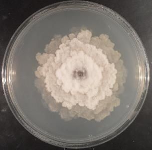
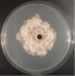
A. Di Francesco, M. Mari                                    Italian Journal of Mycology vol. 47 (2018) ISSN 2531-7342
DOI: https://doi.org/10.6092/issn.2531-7342/7817

 Fig. 1. Monilinia laxa strain (Criof Collection) grow on Potato Dextrose Agar (PDA).
 Fig. 1. Ceppo di Monilinia laxa (Collezione Criof) allevato su substrato agarizzato PDA.

 Monilinia fructicola Winter
 M. fructicola is usually known as a North America fungus. It was also found in Asia and Oceania. In Europe
 is considered a quarantine pathogen by EPPO until 2001.
 The species Chaenomeles Lindl., Crataegus L., Cydonia Mill., Eriobotrya Lindl., Malus Mill., Prunus L.
 and Pyrus L. constitute only a part of the potential host plants of M. fructicola (EFSA, 2011). M. fructicola
 has a greater growth rate (9-20 mm/24h) respect to M. laxa on PDA at 22°C. The colony margin is entire and
 the sporulation is abundant, especially the production of microconidia. The stromata are greyish or hazelin in
 colour, with irregular crusts that may appear in old colonies (Fig.2.). The conidial and germ tube size are
 greater than those of M. laxa (14.5-16 x 9.5-11 μm) (Angeli et al., 2017).

 Fig. 2. Monilinia fructicola strain (Criof Collection) grow on Potato Dextrose Agar (PDA).
 Fig. 2. Ceppo di Monilinia fructicola (Collezione Criof) allevato su substrato agarizzato PDA.

 Symptoms are visible on blossoms, buds, branches, twigs and fruits. M. fructicola affects mainly fruits where
 the infection could remain latent if the environmental conditions are not favourable to the pathogen. The
 infection occurs through natural openings or during the ripening phase (Ogawa et al., 1995). Also young and
 immature fruits, especially peaches and plums, are very susceptible to this pathogen. Infected fruits that
 remain attached to the branches or fall to the soil, are the stroma substrate (mummies) where subsequently
 apothecia are formed.
 Monilinia fructigena Aderhold & Ruhland
 M. fructigena causes brown rot and blossom blight of stone and pome fruit trees worldwide. It has a more
 restricted distribution than the other species, it occurs in Europe and Asia, but not in North America. It is a
 quarantine pathogen in Canada, United States, Australia and New Zealand (USDA, 2010). Apple represents
 the principal M. fructigena host, but it was found also in peach, pear, sour cherry, plum and apricot
 (https://www.cabi.org/isc/datasheet/34747).

 16
A. Di Francesco, M. Mari Italian Journal of Mycology vol. 47 (2018) ISSN 2531-7342
DOI: https://doi.org/10.6092/issn.2531-7342/7817
M. fructigena colonies show lower growth rate respect to the species mentioned above (Fig. 3.). Conidia are
very large (17-21 x 10-13 µm) and form often two germs tube per conidia (van Leeuwen and van Kersten,
1998). Colonies are cream and yellow in colour, and could produce concentric rings with entire margins. The
sporulation is less abundant respect to M. fructicola. Symptoms are rarely found on blossoms and twigs, they
are more frequently found on fruits if the ripening phase is late.
The principal symptoms are the presence of circular and concentric brown spots on fruits. The pathogen is
also known to be the cause of black rot, especially on fruits stored in the dark (Hrustic et al., 2012).
Fig. 3. Monilinia fructigena strain (Criof Collection) grow on Potato Dextrose Agar (PDA).
Fig. 3. Ceppo di Monilinia fructigena (Collezione Criof) allevato su substrato agarizzato PDA.
Monilinia polystroma van Leeuwen
M. polystroma has a more restricted distribution respect to M. laxa or M. fructicola; the pathogen was
detected in Asia (China, Japan), and recently in Europe (Hungary, Serbia, Italy, Croatia, and Switzerland)
(Petróczy and Palkovics, 2009; EPPO 2011; Vasić et al., 2013, Martini et al. 2014, Di Francesco et al.,
2015). It causes a fruit rot on Malus, Prunus, Pyrus spp. and survives on mummified fruits.
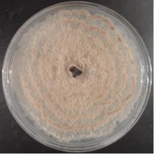
M. polystroma colonies reach after 6 days on PDA at 20°C, 50-60 mm of diameter (Fig. 4.). Colony margins
are regular, buff/pale luteous in colour (van Leeuwen et al., 2002). The black stromata are firstly discrete but
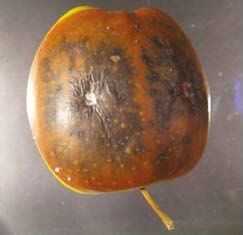
in time coalesce. Conidia are globose, ovoid or lemon shaped (12-21 x 8-12μm). The principal symptom is
the presence of yellowish exogenous stromata on peaches, pears and apples 15 days after the ripening. The
mantle of stromata formed by the host cuticle protects them against abiotic and biotic factors (van Leeuwen
et al., 2002). Also under the field conditions, M. polystroma seems to colonize the fruits faster than M.
fructigena (van Leeuwen et al., 2002) (Fif. 5.).
Fig. 4. Monilinia polystroma strain (Criof Collection) Fig. 5. Monilinia polystroma symptoms on Cripps
grow on Potato Dextrose Agar (PDA). Pink apple.
Fig. 4. Ceppo di Monilinia polystroma (Collezione Fig. 5. Sintomi di Monilinia polystroma su mela
Criof) allevato su substrato agarizzato PDA. Cripps Pink.
17A. Di Francesco, M. Mari Italian Journal of Mycology vol. 47 (2018) ISSN 2531-7342 DOI: https://doi.org/10.6092/issn.2531-7342/7817 Monilinia yunnanensis Hu & Luo M. yunnanensis is one of the Monilinia spp. detected in China on peaches. It is closely related to M. fructigena (Zhu et al., 2016). Colonies show regular margins with a daily growth of 8.5 mm, and are grey- green in coulor. After more than 10 days of incubation at 20°C, M. yunnanensis mycelium begins to develop stromata. Conidia are large and similar to those of M. fructigena (10–21×7–12 μm). M. yunnanensis produced indistinguishable symptoms from M. fructigena on peaches, and the lesions diameter on stone fruits reached 25.5 mm. Monilinia mumecola Harada M. mumecola is present only in Japan on Mume trees (Prunus mume Sieb. & Zucc.), a typical Japanese apricot. M. mumecola reaches its maximum mycelial growth (83 mm) at 20°C in 13 days (Harada et al., 2004). Colony is olive brown in colour, margins are slightly lobed, and the sporulation is scarce. The stromata on plates are greyish brown. Conidia are globose, sub-globose to broadly ellipsoid, hyaline (15-23 x 15-20 μm). The symptoms on infected fruits are a light brown rot, properly called “white rot” without sporodochia production (Harada et al., 2004). Monilinia vaccinii-corymbosi Honey M. vaccinii-corymbosi causes a serious disease of blueberry (Vaccinium corymbosum) in North America regions. In Europe it was firstly found in Austria (Gosch, 2003). The infection of open blueberry flowers, is followed by internal colonization and subsequent mummification of developing fruits. Symptoms consist of leaf and shoot blight. The highest incidence of infected shoots occurs on the lower part of canopy, more close to the soil. The blueberry fruits initially appear healthy, however they turn in dark bluish-purple colour. From late summer until early spring, the fungus overwinters as pseudosclerotia or “mummies” that produce apothecia which discharge the meiospores. Pseudosclerotia are dark brown, robust, hollow, distinctively ribbed, flattened and opened at both poles. Apothecia are reddish brown to umber, cup shaped with recurved margin when young. Ascospores are hyaline, ellipsoid measuring 15–19.2 μm x 8–10.7 μm. Macroconidia are observed in spring. They appear as a dense greyish covering on the convex side of bent current year twigs, on petioles, and along midribs of blighted leaves. Macroconidia are lemon shaped, hyaline, measuring 21.9–30.1 μm x 11.7 –15.3 μm (Munda, 2011). The colonies show a slow growth reaching 7-8 cm in diameter after 21 days at 20°C. The mycelium is white to beige in colour and compact, the reverse is brown, with yellow or honey pigmentation in some cultures. The production of macroconidia is scarce (Gosch, 2003). Monilinia urnula Whetzel This pathogen causes a disease of Vaccinum vitis-idaea called Mummy berry. It is widespread especially in Europe, mainly in Scandinavia, Austria and United Kingdom (Woronin, 1888; Dennis, 1968; Gjaerum, 1969;). It was reported also in Japan (Kobayashi, 2007). As M. vaccinii-corymbosi has a complicated life cycle. From harvesting time the mummies, compact masses of fungal tissue formed in infected berries, are found on the soil (Goheen, 1953). Pseudosclerotia are reddish brown, hollow, open above and below. Apothecia could occurs in late spring depending on weather conditions, although were rare to find. They arose from mummified fruits (pseudosclerotia) that pass the winter in the field. Ascospores are hyaline, ellipsoid measuring 11.4 -15 μm x 5 –6.5 μm. Macroconidia are lemon shaped, hyaline measuring 25.6 –41.4 μm x 15.5 –27.5 μm. Mature macroconidia are separated by spindle like structures (disjunctors), firstly described by Woronin (1888) in M. urnula, and later found to be present in all species belonging to the Disjunctoriae section. The colonies on PDA are slow growing; at 25°C they reach 90 mm diameter in 21 days. Mycelium is white and beige on 18
A. Di Francesco, M. Mari                                  Italian Journal of Mycology vol. 47 (2018) ISSN 2531-7342
DOI: https://doi.org/10.6092/issn.2531-7342/7817

 reverse, black stromata, buried in the agar medium, are observed in old colonies. Only microconidia are
 present especially in mature colonies. They are globose, hyaline, 2 -3 μm wide.
 Monilinia baccarum Whetzel
 M. baccarum is a pathogen of Vaccinium myrtillus L., typical of Scandinavia, Austria, Belgium, Germany
 and United Kingdom (Rehm, 1885; Woronin, 1888; Dennis, 1968; Gjaerum, 1969; Palmer, 1988; Batra,
 1991). It causes the blight of newly emerging shoots; the infected berries turn pale, dry, shrivel, mummify
 and fall to the ground. They are called “white berries” due to the fine whitish layer of host cells that covers
 the berries (Batra, 1991).
 Pseudosclerotia are light grey, sometimes greyish pink, apothecia are present from the end of May are dark
 brown. Ascospores are hyaline, ellipsoid measuring 14.5 –20.8 μm x 5.9 –7.3 μm. Macroconidia are lemon
 shaped, hyaline measuring 19–28 μm x 14 -21 μm, they are separated by disjunctors. The colonies are slow
 growing and reach the diameter of 50 mm in 21 days. The mycelium is white, greyish brown on reverse,
 black superficial stromata develop in old colonies.
 Monilinia oxycocci Honey
 M. oxycocci causes a disease of Vaccinium macrocarpon Ait. known as cranberry cotton ball . It is typical of
 Canada and North - East of United States.
 The disease cycle includes both primary and secondary infection stages. In the spring as cranberry shoot
 growth resumes, pseudosclerotia germinate and produce apothecia. Ascospores infect young, succulent
 cranberry shoots. Conidia are carried by wind, or insects, to floral stigmata where secondary infection
 occurs. Conidia germinate on the stigma, germ tubes grow through the stylar canal, and white cotton-like
 mycelia fill the locules of developing fruit. The greatest numbers of conidia were produced at 16°C, but at
 20°C conidia grow up better.The entire pericarp is colonized and develops into a pseudosclerotium in which
 the pathogen overwinters (Sanderson and Jeffers, 2001).
 The colonies on PDA medium reach the diameter of 28 mm after 16 days at 20°C (Sanderson and Jeffers,
 2001), the conidia production is generally low, less than 1000 conidia/ml, resulting similar to those of M.
 fructigena.
 Monilinia linhartiana Dennis
 M. linhartiana represents the most important pathogen of quince fruits (Cydonia oblonga Mill.) in South of
 Spain, where it often destroys entire crops (Moral et al., 2011). The life cycle of this pathogen is very similar
 to that of M. vaccinii-corymbosi on blueberry.
 The M. linhartiana mycelium produces on fruits sporodochia with conidiophores and conidia arranged often
 in concentric zones. The conidia are one-celled, hyaline, lemon shaped. They have smooth walls and
 disjunctors, and are arranged in chains of up to 30 conidia (Pârvu and Pârvu, 2014).

 Concluding remarks

 Monilinia spp. are economically important pathogens, very difficult to control. The most studied Monilinia
 spp. are those responsible of the brown rot disease of stone fruit crops such as peach, apricot, apple, sweet
 and sour cherry (Tab. 1.). Brown rot disease is principally caused by M. laxa, M. fructicola and M.
 fructigena belonging to Junctoriae section.
 This work focuses also on other Monilinia spp. less studied, because rare or restricted in their distribution
 (Munda, 2011), and responsible of less significant damage; the majority of these species belong to

                                                                                                               19
A. Di Francesco, M. Mari Italian Journal of Mycology vol. 47 (2018) ISSN 2531-7342
DOI: https://doi.org/10.6092/issn.2531-7342/7817
Disjunctoriae section. They need further studies to better understand their adaptive radiation in fungal
speciation.
Between the Disjunctoriae spp., M. urnula, M. baccarum, M. oxycocci and M. vaccinii-corymbosi infect
North America Vaccinium spp. causing a great economical impact on blueberry fruit production (Tab. 1.).
Although morphological differences and host affinity, M. oxycocci and M. vaccinii-corymbosi are similar to
M. fructigena; whereas M. mumecola is more closely related to M laxa on the base of symptoms on flowers
and twigs of Prunus spp., cultural characteristics and sequence data in the ITS region of ribosomal DNA
(Harada et al., 2004). Based upon comparison of sequence data for the ITS, glyceraldehyde-3-phosphate
dehydrogenase (G3PDH) and β-tubulin genes and cultural characters (conidia size and stromatic tissue
production), M. yunnanensis seems to be closely related to M. fructigena (Hu et al., 2011). Also M. oxycocci,
M. vaccinii-corymbosi and M. baccarum that parasitize plants in the Vaccinium genus are similar to M.
fructigena regarding endostromata structure.
Phylogenetic studies showed how these microorganisms could be genetically similar, and it represent a risk
of high variability through their ability to adapt at different environmental conditions or sexual
recombination (Holst –Jensen et al., 1997; Moral et al., 2011).
Tab. 1. Host affinity and morphological characteristics of Monilinia spp colonies.
Tab. 1. Pianta ospite e differenze morfologiche tra le specie di Monilinia.
Colony characteristicson nutrient
Species Section Host Conidial dimension
medium
Colour grey/hazel
M. fructicola Junctoriae Apple, peach pear 14.5-16 x 9.5-11 μm
Entire margin and abundant sporulation
Colour creamy/yellow
M. fructigena Junctoriae Pome and stone fruit 17-21 x 10-13 μm Concentric rings with entire margin and
not abundant sporulation
M. Peach, pear, Colour buff/pale
Junctoriae 12-21 x 8-12 μm
polystroma apple Regular margin and black stromata
Colour grey/hazel
Peach, apricot, plume,
M. laxa Junctoriae 11.5-17 x 8-11 μm Rosette with lobed margin and abundant
sweet cherry
sporulation
Mume Colour olive/brown.
M. mumecola Junctoriae 15-23 x 15-20 μm
(Japanese apricot) Lobed margin, slightly sporulation
Colour grey/green
M. Regular margin and not abundant
Junctoriae Peach 10–21 x 7–12 μm
yunnanensis sporulation
Colour white/grey
M.
Disjunctoriae Quince fruit Concentric zones with entire margin and
linhartiana 10.5-17 x 8.5-11 μm
not abundant sporulation
Colour white/beige
M. vaccinii-
Disjunctoriae Blueberry 15–19.2 x 8–10.7 μm with brown stromata, slight lobed margin
corymbosi
and not abundant sporulation
Coulor white/greyish
M. baccarum Disjunctoriae Black raspberry 19–28 x 14-21 μm with black stromata, slight lobed margin
and not abundant sporulation
Colour white/beige
25.6–41.4 x 15.5–27.5 with buried black stromata, slight lobed
M. urnula Disjunctoriae Cranberry
μm margin.
Only microconidia
Colour greyish,
M. oxycocci Disjunctoriae Cranberry 17-21 x 10-13 μm slight lobed margin.
Not abundant sporulation
20A. Di Francesco, M. Mari Italian Journal of Mycology vol. 47 (2018) ISSN 2531-7342
DOI: https://doi.org/10.6092/issn.2531-7342/7817
References
Angeli S.S., Amorim L. (2017). Comparative analysis of Monilinia fructicola and M. laxa isolates from
Brazil: monocyclic components of peach brown rot. Ciencia rural, 47.
Batra L.R. (1991). World species of Monilinia (Fungi): their ecology, biosystematics and control. Mycologia
Memoir, 16: 246.
Byrde R.J.W., Willets H.J. (1977). The brown rot fungi of fruit. Pergamon press, New York, 171.
Boehm E.W.A., Ma Z., Michailides T.J. (2001). Species-specific detection of Monilinia fructicola from
California stone fruits and flowers. Phytopathology, 91: 428-439.
Bosshard E., Hilber-Bodmer M., Schärer H.J., Bünter M., Duffy B. (2006). First report of the quarantine
brown rot pathogen Monilinia fructicola on imported stone fruits in Switzerland. Plant Disease, 90:
1554.
Côté M.J., Tardif M.C., Meldrum A.J. (2004). Identification of Monilinia fuctigena, M. fructicola, M. laxa,
and M. polystroma on inoculated and naturally infected fruit using muliplex PCR. Plant Disease 88:
1219-1225.
Di Francesco A., Fruk M., Martini C., Jemric T., Mari M. (2015). First report of asiatic brown rot (Monilinia
polystroma) on apple in Croatia. Plant Disease, 99: 1181.
De Cal A., Euguen B., Melgarejo P. (2014). Vegetative compatibility groups and sexual
reproduction among Sspanish Monilinia fructicola isolates obtained from peach and nectarine
orchards, but not Monilinia laxa. Fungal Biology, 118: 484-494.
De Cal A., Gell I., Usall J., Viñas I., Melgarejo P. (2009). First report of brown rot caused by
Monilinia fructicola in peach orchards in Ebro Valley, Spain. Plant Disease, 93: 763.
De Cal A., Melgarejo P. (1999). Effects of long-wave UV light on Monilinia Growth and Identification of
Species. Plant Disease, 83: 62-65.
Dennis R.W.G. (1968). British Ascomycetes. Lehre, Cramer, 445.
European Food Safety Authority (2011). Pest risk assessment of Monilinia fructicola for the EU territory and
identification and evaluation of risk management options. European Food Safety Authority Journal, 9:
2119.
European and Mediterranean Plant Protection Organization (2002). First report of Monilinia fructicola in
France. European and Mediterranean Plant Protection Organization Reporting Service 2002/003.
European and Mediterranean Plant Protection Organization (2011). https://gd.eppo.int/taxon/ MONIPO/
distribution/PL.
European and Mediterranean Plant Protection Organization/ European and Mediterranean Plant Protection
Organization (2010). Monilinia fructicola. Distribution Maps of Plant Diseases, Map No. 50, Edition 8.
CAB International, Wallingford, UK.
Gjaerum H.B. (1969). Some fruit inhabiting Sclerotinias in Norway. Friesia, 9: 18-28.
Goheen A.C. (1953). The cultivated highbush blueberry. U.S. Dept. Agr. Yearbook of Agriculture, 1953:
784-789. HALL.
Gosch C. (2003). Monilinia vaccinii-corymbosi on high bush blueberries (Vaccinium corymbosum L.): also
in Europe. European Journal of Horticultural Science, 68: 238–241.
Harada Y., Nakao S., Sasaki M., Sasaki Y., Ichihashi Y.,Sano T. (2004). Monilia mumecola, a new brown
rot fungus on Prunus mume in Japan. Journal of General Plant Pathology, 70: 297–307.
Holb I.J. (2008) Monitoring conidial density of Monilinia fructigena in the air in relation to brown rot
development in integrated and organic apple orchards. European Journal of Plant Pathology, 120: 397–
408
Holst-Jensen A., Kohn L.M., Schumacher T. (1997). Nuclear rDNA Phylogeny of the Sclerotiniaceae.
Mycologia, 89: 885-899.
Honey E.E. (1936). North American species of Monilinia. I. Occurrence, grouping, and life-histories.
American Journal of Botany, 23: 100-106.
21A. Di Francesco, M. Mari Italian Journal of Mycology vol. 47 (2018) ISSN 2531-7342
DOI: https://doi.org/10.6092/issn.2531-7342/7817
Hrustic J., Mihajlović M., Tanović B., Delibašić G., Stanković I., Krstić B., Bulajić A. (2012). First
Report of brown rot caused by Monilinia fructicola on nectarine in Serbia. Plant Disease, 97:
147.
Hu M.J., Cox K.D., Schnabel G., Luo C.X. (2011). Monilinia species causing brown rot of peach in China.
Plos one, https://doi.org/10.1371/journal.pone.0024990.
Kobayashi T. (2007). Index of fungi inhabiting woody plants in Japan. Host, Distribution and Literature.
Kyoiku, Kyokai Publishing Co., 1227.
Larena I., Torres R., De Cal A., Liñan M., Melgarejo P., Domenichini P. (2005). Biological control of
postharvest brown rot (Monilinia spp.) of peaches by field applications of Epicoccum nigrum.
Biological Control, 32: 305-310.
Lee M.H., Bostock R.M. (2007). Fruit exocarp phenols in relation to quiescence and development
of Monilinia fructicola infections in Prunus spp.: A Role for Cellular Redox? Phytopathology, 97: 269-
277.
Marinelli E., Vitale S., Valente M.T., Riccioni L. (2013). Segnalate in Lazio infezioni di Monilinia fructicola
su drupacee. Informatore agrario, 2: 55-57.
Martini C., Lantos A., Di Francesco A., Guidarelli M., D’Aquino S., Baraldi E. (2014). First Report
of Asiatic Brown Rot Caused by Monilinia polystroma on Peach in Italy. Plant Disease, 98:
1585.
Martini C., Mari M. (2014). Monilinia fructicola, Monilinia laxa (Monilinia rot, Brown rot). Postharvest
decay-control strategies, Academic Press, 233-265.
Montuschi C., Ceredi G., Mari M. (2011). Monilia fructicola è arrivata anche in Emilia-Romagna.
Agricoltura, 90-92.
Moral J., Muñoz-Díez C., Cabello D., Arquero O., Lovera M., Benítez M.J., Trapero A. (2011).
Characterization of Monilia disease caused by Monilinia linhartiana on quince in southern Spain. Plant
Pathology, 60: 1128–1139.
Munda A. (2011). Monilinia pathogens of cultivated Vaccinium species in Slovenia. Acta agricolturae
Slovenica, 97: 99-104.
Ogawa J.M., Zehr E.I., Bird G.W., Ritchie D.F., Uriu K., Uyemoto J.K. (1995). Compendium of stone fruit
diseases, APS, St. Paul, MN, 98.
Palmer J.T. (1988). Investigations into the Sclerotiniaceae. Lejunia , 127: 1–40.
Pârvu M., Pârvu A.E. (2014). Parasitic fungi Sclerotiniaceae: morphology and ultrastructure. Microscopy:
advances in scientific research and education (A. Méndez-Vilas, Ed.), 530-537.
Pellegrino C., Gullino M.L., Garibaldi A., Spadaro D. (2009). First Report of brown rot of stone
fruit caused by Monilinia fructicola in Italy. Plant Disease, 93: 668.
Penrose L.J., Tarran J., Wong A.L. (1976). First record of Sclerotinia laxa Aderh. & Ruhl. in New South
Wales: differentiation from S. fructicola (Wint.) Rehm. by cultural characteristics and electrophoresis.
Journal of Agriculturtal Research, 27: 547–55.
Petróczy M. and Palkovics L. (2009). First report of Monilia polystroma on apple in Hungary. European
Journal of plant Pathology, 125: 343-347.
Petroczy M., Szigethy A., Palkovic L. (2012). Monilinia species in Hungary: morphology, culture
characteristics, and molecular analysis. Trees, 26: 153–164.
Rehm H. (1885). Ascomyceten: Sclerotinia baccarum. Fasc. XVI. Hedwigia, 24: 7-17.
Rungjindamai N., Jeffries P., Xu X.M. (2014). Epidemiology and management of brown rot on stone fruit
caused by Monilinia laxa. European Journal of Plant Pathology, 140: 1-17.
Sanderson P.G., Jeffers S.N. (2001). Vegetative growth and conidium production by Monilinia
oxycocci in vitro. Mycologia, 93: 9-16.
United States department of agriculture (2010). Asian/European brown rot of Rosaceae - Monilinia
fructigena. https://nt.ars-grin.gov/taxadescriptions/factsheets/index.cfm?thisapp=Moniliniafructigena
van Leeuwen G.C.M., Baayen R.P., Holb I.J., Jeger M.J. (2002). Distinction of the Asiatic brown rot fungus
Monilia polystroma sp. nov. from Monilia fructigena. Mycological Research, 106: 444–451.
22A. Di Francesco, M. Mari Italian Journal of Mycology vol. 47 (2018) ISSN 2531-7342
DOI: https://doi.org/10.6092/issn.2531-7342/7817
van Leeuwen G.C.M., van Kersten H.A. (1998). Delineation of the three brown rot fungi of fruit crops
(Monilinia spp.) on the basis of quantitative characteristics. Canadian Journal of Botany, 76: 2042–
2050.
Vasić M., Duduk N., Ivanović M.S. (2013). First Report of Brown Rot Caused by Monilia
polystroma on Apple in Serbia. Plant Disease, 97: 145.
Villarino M., Egüen B., Lamarca N., Segarra J., Usall J., Melgarejo P., De Cal A. (2013). Occurrence of
Monilinia laxa and M. fructigena after introduction of M. fructicola in peach orchards in Spain.
European Journal of Plant Pathology, 137: 835-845.
Woronin M. (1888). Uber die Sclerotienkrankheit der Vaccinieen-beeren. Mem. Acad. Imp. Sci. St.
Petersbourg VII, 36: 1-49.
Zhu X.Q., Niu C.W., Chen X.Y., Guo L.Y. (2016). Monilinia species associated with brown rot of cultivated
apple and pear fruit in China. Plant Disease, 100: 2240-2250.
23You can also read