CAT DISSECTION A LABORATORY GUIDE

Page created by Mike Walker
 
CONTINUE READING
CAT DISSECTION A LABORATORY GUIDE
8546d_fm_i-iv   6/26/02   3:51 PM   Page 1 mac62 mac62:1253_GE:

          CAT DISSECTION
          A LABORATORY GUIDE
CAT DISSECTION A LABORATORY GUIDE
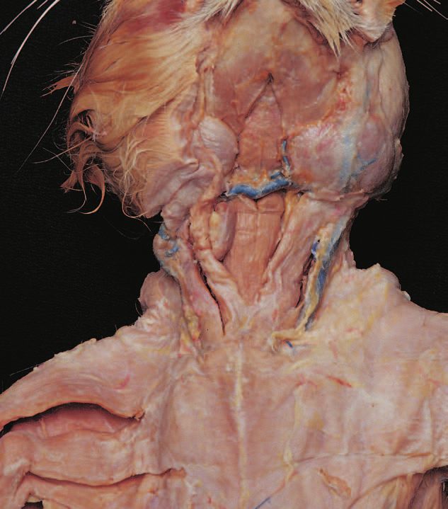
8546d_fm_i-iv   6/26/02   3:51 PM   Page 2 mac62 mac62:1253_GE:
CAT DISSECTION A LABORATORY GUIDE
8546d_fm_i-iv   6/26/02   3:51 PM   Page 3 mac62 mac62:1253_GE:

           CAT DISSECTION
           A LABORATORY GUIDE

                                                                    CONNIE ALLEN
                                                                  VALERIE HARPER
                                                                   Edison Community College

          John Wiley & Sons, Inc.
CAT DISSECTION A LABORATORY GUIDE
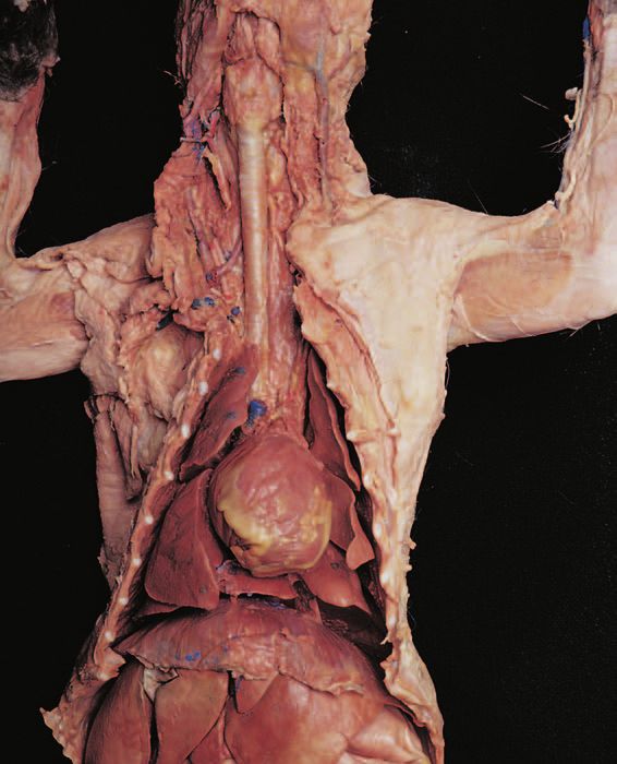
8546d_fm_i-iv   6/26/02       12:17 PM       Page 4 mac62 mac62:1253_GE:

          Senior Editor                          Bonnie Roesch
          Associate Editor                       Mary O’Sullivan
          Production Editor                      Sandra Russell
          Senior Marketing Manager               Clay Stone
          Senior Designer                        Kevin Murphy
          Book Designer                          Nancy Field
          Photo Manager                          Hilary Newman
          Illustration Editor                    Anna Melhorn
          Production Management Services         TechBooks

          This book was typeset in 10/12 Times Roman by TechBooks, Inc. and printed and bound
          by Von Hoffmann Press. The cover was printed by Von Hoffmann Corporation.

          The paper in this book was manufactured by a mill whose forest management programs
          include sustained yield harvesting of its timberlands. Sustained yield harvesting principles
          ensure that the number of trees cut each year does not exceed the amount of new growth.

          This book is printed on acid-free paper.     ∞

          Copyright © 2003 by John Wiley & Sons, Inc. All rights reserved.

          No part of this publication may be reproduced, stored in a retrieval system, or transmitted in any
          form or by any means, electronic, mechanical, photocopying recording, scanning or otherwise,
          except as permitted under Section 107 or 108 of the 1976 United States Copyright Act, without
          either the prior written permission of the Publisher or authorization through payment of the
          appropriate per-copy fee to the Copyright Clearance Center, 222 Rosewood Drive, Danvers,
          MA 01923, (508) 750-8400, fax (508) 750-4470. Requests to the Publisher for permission should
          be addressed to the Permissions Department, John Wiley & Sons, Inc., 605 Third Avenue, New
          York, NY 10158-0012, (212) 850-6008, E-mail: PERMREQ@WILEY.COM. To order books or for
          customer service, call 1-800-CALL-WILEY(225-5945).

          Allen and Harper
          Cat Dissection
          A Laboratory Guide

          ISBN 0-471-26457-1

          Printed in the United States of America.

          10 9 8 7 6 5 4 3 2 1
CAT DISSECTION A LABORATORY GUIDE
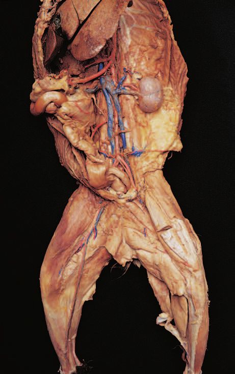
8546d_c01_1-42   6/25/02    4:32 PM   Page 1 mac48 Mac 48: 420_kec:

           CAT DISSECTION
           A LABORATORY GUIDE

           OUTLINE
            Preface, p. 2
            A. Preparing the Cat, p. 2                                Dissection 3: Endocrine Organs, p. 24
            B. Removing the Skin, p. 3                                Dissection 4: Blood Vessels, p. 26
            C. Opening Ventral Body Cavities, p. 3                    A. Arteries, p. 26
            Dissection 1: Skeletal Muscles, p. 4                      B. Veins, p. 29
            A.   Dissecting Skeletal Muscles, p. 4                    Dissection 5: Lymphatic System, p. 30
            B.   Muscles of the Head and Neck, p. 4
            C.   Muscles of the Chest, p. 6                           Dissection 6: Respiratory System, p. 32
            D.   Muscles of the Abdomen, p. 8                         Dissection 7: Digestive System, p. 34
            E.   Muscles of the Back and Shoulder, p. 10              A. Mouth, Oropharynx, and Salivary Glands, p. 34
            F.   Muscles of the Arm and Forearm, p. 12                B. Esophagus and Abdominal Organs, p. 35
            G.   Muscles of the Thigh, p. 15
            H.   Muscles of the Leg, p. 18                            Dissection 8: Urinary and Reproductive
                                                                      Systems, p. 38
            Dissection 2: Brachial and Lumbosacral                    A. Urinary System, p. 38
            Plexuses and Major Nerves, p. 20                          B. Male Reproductive System, p. 40
            A. Brachial Plexus, p. 20                                 C. Female Reproductive System, p. 41
            B. Lumbosacral Plexus, p. 22
                                                                                                                         1
CAT DISSECTION A LABORATORY GUIDE
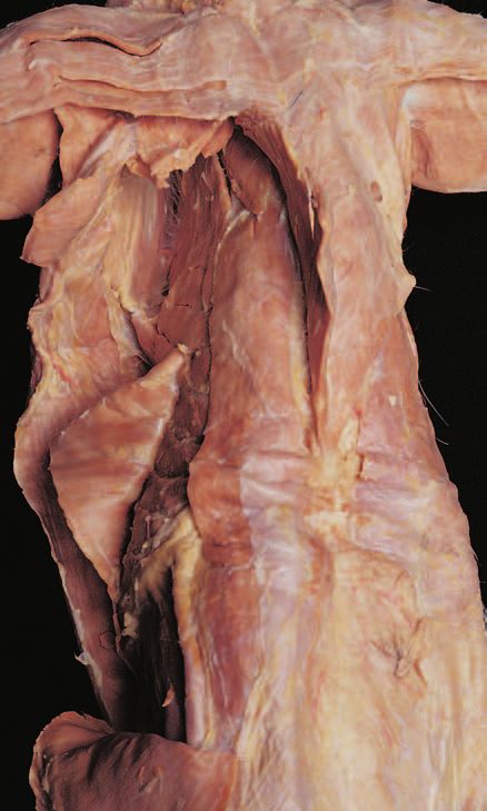
8546d_c01_1-42     6/21/02      1:34 PM    Page 2 mac62 mac62:1253_GE:

          2       Cat Dissection

          PR E FAC E

          A. Preparing the Cat
          1. With gloves on, remove the cat from its bag and lay
             the cat on a dissecting tray. Keep any liquid preserv-
             ing solution that remains in the bag.
          2. Review the directional terms for the cat in Figure
             CP.1. Note the differences between four-legged
             animals and humans.
             • Anterior is toward the cephalic (head) end of the
               cat
             • Posterior is toward the caudal (tail) end of the cat
             • Superior is toward the dorsal (back) surface
             • Inferior is toward the ventral (belly) surface

                                       Superior
                                       (dorsal)

          Posterior                                            Anterior                                (a) Incisions for skinning
          (caudal)                                             (cephalad)

                                       Inferior
                                       (ventral)

          F I G U R E C P. 1   Directional terminology for the cat.

          3. Place your cat ventral surface up on the dissecting
             tray.                                                                                                                  Diaphragm
          4. Identify the gender of your cat. Males have a scrotum
             and a prepuce, a small mound anterior to the scrotum
             in which the penis is located. Females have a urogeni-
             tal aperture, an opening located anterior to the anus
             that is a common passageway for the urinary and
             reproductive systems. Four or five teats (nipples) are
             present on both male and female cats. Be able to
             identify both sexes externally.
          5. Prepare a label for your cat with the names of your
             group members and the gender of your cat.
          6. Follow the instructions for skinning the cat if you are
             dissecting skeletal muscles, or the instructions for                                     (b) Opening ventral body cavities
             opening the ventral body cavities if you are dissecting                             Incision line
             an organ system.
                                                                            F I G U R E C P. 2     Cat incisions.
CAT DISSECTION A LABORATORY GUIDE
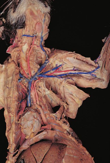
8546d_c01_1-42   6/26/02   12:26 PM    Page 3 mac62 mac62:1253_GE:

                                                                                                              Cat Dissection        3

          B. Removing the Skin                                             prevent the growth of bacteria and mold. Dispose of
                                                                           fascia and fat as indicated by your instructor. Do not
          1. Referring to Figure CP.2a, pinch the skin on the ven-         forget to attach the label identifying your cat before
             tral surface of the neck. Using scissors, carefully           storing it.
             make a small, longitudinal incision at the midline
             through the skin only. Use care not to cut into the
             underlying muscle layer.
                                                                        C. Opening Ventral
          2. Continue cutting longitudinally along the midline
             toward the lower lip and then posteriorly, stopping an-
                                                                        Body Cavities
             terior to the genital area.
                                                                        1. At the midline, just above the pubic bone, carefully
          3. Cut the skin around the neck.                                 make a longitudinal incision through the abdominal
                                                                           muscles. Refer to Figure CP.2b. Continue the incision
          4. Make a horizontal cut across the chest and continue
                                                                           to the ribs.
             cutting down the midline of the extremities as indi-
             cated in Figure CP.2a. Make diagonal cuts in the           2. Cut either to the right or left of the sternum, cutting
             groin and continue midline down the extremities. Cut          through the costal cartilages. Continue cutting midline
             the skin around all paws.                                     through the neck.
          5. Use your fingers to carefully peel the skin from the       3. Cut horizontal incisions at the top and at the base of
             underlying muscles. Cutaneous muscles, such as the            the neck.
             platysma, are attached to the undersurface of the skin
                                                                        4. Cut horizontal incisions anterior and posterior to the
             and will be removed as you peel away the skin.
                                                                           diaphragm as indicated in Figure CP.2b, and cut the
          6. Continue peeling the skin until it is only attached at        diaphragm away from the ventral body wall. Open the
             the face and the tail. Cut around the base of the tail,       flaps to expose the thoracic and abdominal cavities,
             leaving the skin on the tail. Cut the skin around the         leaving the diaphragm intact.
             face of the cat, leaving the skin on the face, ears, and
                                                                        5. Use a scalpel to make a longitudinal cut down each
             forehead. Peel the skin from the head and save it.
                                                                           inner wall of the rib cage. Bend the walls outward to
          7. Carefully remove as much fat and superficial fascia as        break the ribs, allowing the flaps of the thoracic wall
             possible with your fingers or forceps.                        to stay open.
          8. Wrap the skin around the cat and follow your instruc-      6. Dispose of fascia and fat as indicated by your instruc-
             tors directions for storing your cat in the plastic bag.      tor. Do not forget to attach the label identifying your
             The skin will prevent the tissues from drying out and         cat before storing it.
CAT DISSECTION A LABORATORY GUIDE
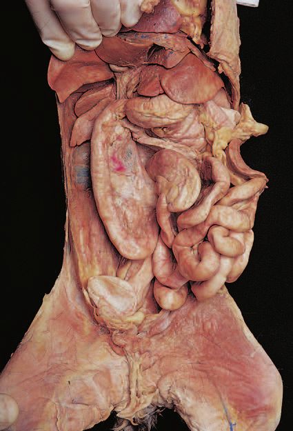
8546d_c01_1-42   6/21/02    1:34 PM    Page 4 mac62 mac62:1253_GE:

          4      Cat Dissection

                                                                          may result in cutting muscles or other structures. Blunt dis-
          D I S S E C TI O N 1: S KE L E TA L                             section is a technique that uses blunt probes and forceps
          MUSCLES                                                         to remove fascia and separate muscles. To observe a deep
                                                                          muscle, you will have to cut the superficial muscle at the
          Many skeletal muscles of the cat are similar to human mus-      midline and reflect (pull back) the edges toward the origin
          cles. This dissection will reinforce your knowledge of hu-      and insertion.
          man skeletal muscles and allow you to observe the fascia
          that surrounds, protects, and compartmentalizes these mus-
          cles. Assemble your dissection equipment and safety
          glasses, put on your gloves, and obtain your cat. Position
                                                                          B. Muscles of the
          your cat within the dissection tray, including the tail. Keep   Head and Neck
          any remaining preserving fluid in the bag to keep your cat
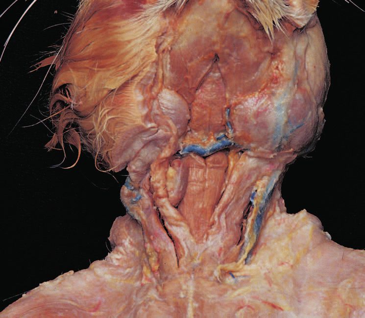
          moist and inhibit bacterial and mold growth.                    1. Refer to Figure C1.1 to locate the following superfi-
                                                                             cial muscles on the cat. Cats have a platysma, but this
                                                                             muscle was most probably removed during the skin-
                                                                             ning process.
          Procedure                                                          • Masseter
                                                                             • Digastric
          A. Dissecting Skeletal Muscles                                     • Mylohyoid
                                                                             • Sternohyoid
          It is important to carefully remove the fascia to observe          • Sternothyroid
          the individual muscles. However, using scissors or scalpels        • Sternomastoid (sternocleidomastoid in humans)
CAT DISSECTION A LABORATORY GUIDE
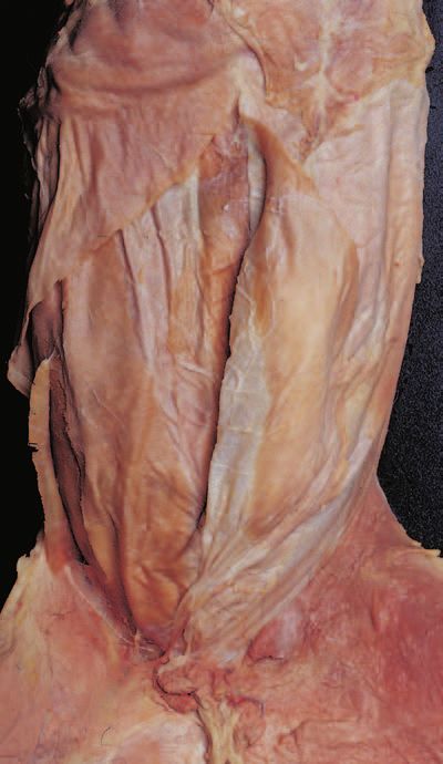
8546d_c01_1-42    6/25/02      4:32 PM   Page 5 mac48 Mac 48: 420_kec:

                                                                                           Cat Dissection   5

                                                                           Masseter
                                                                           Mylohyoid
                                                                           Digastric

                                                                           Sternohyoid

                                                                           Sternomastoid

                   Digastric

                                                                                            Masseter

                                                                                            Mylohyoid

                                                                                           Sternothyroid

                                                                                           Sternomastoid

                   Sternohyoid

          F I G U R E C 1. 1   Superficial muscles of the head and neck.
CAT DISSECTION A LABORATORY GUIDE
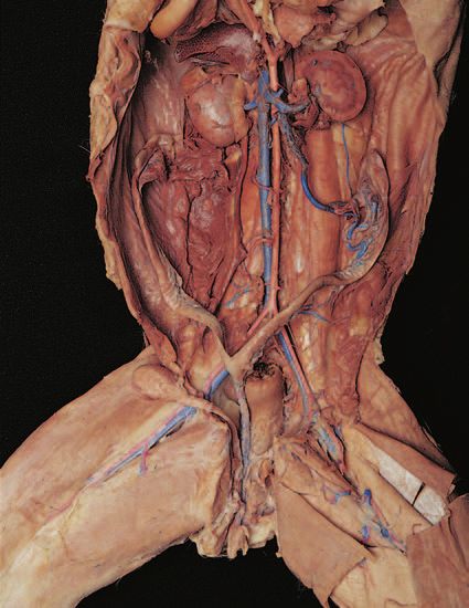
8546d_c01_1-42     6/21/02      1:34 PM        Page 6 mac62 mac62:1253_GE:

          6        Cat Dissection

          C. Muscles of the Chest
          1. Refer to Figure C1.2a to locate the following superfi-
             cial muscles on the chest of the cat:
             • Pectoantebrachialis (not in humans)
             • Pectoralis major
             • Pectoralis minor
             • Xiphihumeralis (not in humans)

                                                                                                  Pectoantebrachialis

                                                                                                  Pectoralis major

                                                                                        Pectoralis minor

                                                                                        Xiphihumeralis

                                                                                                           Pectoantebrachialis

                                  Pectoralis
                                  major

                                                                                                           Pectoralis minor

                                                                                                           Xiphihumeralis

                                                              (a) Superficial muscles

          F I G U R E C 1. 2   Muscles of the chest.
8546d_c01_1-42    6/21/02      1:34 PM     Page 7 mac62 mac62:1253_GE:

                                                                                                                      Cat Dissection     7

          2. Cut and reflect the pectoralis major, pectoralis minor,      4. If advised by your instructor, cut and reflect these mus-
             and the xiphihumeralis.                                         cles to observe the internal intercostal muscles that run
                                                                             obliquely to the external intercostals.
          3. Refer to Figure C1.2b to locate the following deep
             muscles on the ventral thorax of the cat:
             • External intercostals
             • Serratus ventralis (serratus anterior in humans)

                                                                                                       Pectoralis
                                                                                                       major (cut)

                                     Pectoralis
                                     minor (cut)

                                                                                                       External
                                     Serratus                                                          intercostals
                                     ventralis

                                                                  (b) Deep muscles

          F I G U R E C 1. 2   Muscles of the chest, continued.
8769d_c01_1-42 11/15/02 1:11 PM Page 8 ymac6 Yes Mac 6:1st shift:101_lkt:8769d_PD3:

          8     Cat Dissection

          D. Muscles of the Abdomen                                  3. Cut and reflect the very thin internal oblique to
                                                                        observe the underlying:
          1. Refer to Figure C1.3 to locate the following superfi-      • Transverse abdominis; often, the transverse abdo-
             cial muscles on the abdomen of the cat:                      minis is attached to the underside of the internal
             • Rectus abdominis                                           oblique.
             • External oblique
          2. Cut and reflect the very thin external oblique to
             observe the underlying:
             • Internal oblique

                                     Latissimus dorsi

                                     Rectus abdominis

                                     External oblique

                                     Internal oblique

                                     Transverse abdominis
8546d_c01_1-42   6/25/02        4:32 PM   Page 9 mac48 Mac 48: 420_kec:

                                                                                             Cat Dissection   9

                                                                          Rectus abdominis

                                                                          Latissimus dorsi

                                                                          External oblique (cut)

                                                                          Internal oblique (cut)

                                                                          Linea alba

                                                                          Transverse abdominis

           F I G U R E C 1. 3   Muscles of the abdomen.
8546d_c01_1-42     6/25/02      4:32 PM     Page 10 mac48 Mac 48: 420_kec:

          10         Cat Dissection

          E. Muscles of the                                                  2. Cut and reflect the trapezius muscles and the
                                                                                latissimus dorsi.
          Back and Shoulder
                                                                             3. Refer to Figure C1.4b to locate the following deep
          1. Refer to Figure C1.4a to locate the following superfi-             muscles:
             cial muscles:                                                      • Splenius
             • Trapezius muscles—The cat has three separate mus-                • Levator scapulae ventralis (levator scapulae in
               cles, compared with a single human trapezius.                      humans)
               —Clavotrapezius                                                  • Rhomboideus capitis (not in humans)
               —Acromiotrapezius                                                • Rhomboideus (rhomboideus major and minor in
               —Spinotrapezius                                                    humans)
             • Deltoid muscles—The cat has three separate deltoid               • Supraspinatus
               muscles, compared with one in humans.                            • Infraspinatus
               —Clavobrachialis (clavodeltoid)                                  • Teres major
               —Acromiodeltoid
               —Spinodeltoid
             • Latissimus dorsi

                        Clavotrapezius                                                                      Acromiotrapezius

                                                                                                            Spinotrapezius

                        Levator scapulae
                                                                                                            Latissimus dorsi
                        ventralis

                        Spinodeltoid

                        Acromiodeltoid
                                                                                                            External oblique

                        Clavobrachialis
                        (clavodeltoid)

                        Triceps brachii

                                           Clavotrapezius

                                           Clavobrachialis                                                 Acromiodeltoid
                                           (clavodeltoid)
                                                                                                           Levator scapulae ventralis

                                           Triceps brachii                                                 Acromiotrapezius
                                                                                                           Spinodeltoid

                                                                                                           Spinotrapezius

                                                                                                           Latissimus dorsi

                                           External oblique

                                                                      (a) Superficial muscles

          F I G U R E C 1. 4   Muscles of the shoulder.
8546d_c01_1-42    6/25/02      4:32 PM      Page 11 mac48 Mac 48: 420_kec:

                                                                                                              Cat Dissection   11

                                                                                        Levator scapulae ventralis

                  Clavotrapezius                                                        Rhomboideus capitis
                  Clavobrachialis
                  (clavodeltoid)

                  Acromiodeltoid

                  Triceps brachii

                                                                                        Rhomboideus
                  Levator scapulae ventralis
                                                                                        Supraspinatus
                  Spinodeltoid
                                                                                        Teres major
                  Acromiotrapezius
                                                                                        Infraspinatus
                  Spinotrapezius                                                        Spinalis dorsi
                                                                                        Longissimus
                  Latissimus dorsi
                                                                                        Iliocostalis

                  External oblique

                                     Superficial                                        Deep

                                                                                                  Splenius

                                 Levator scapulae
                                 ventralis

                                 Supraspinatus

                                                                                                  Rhomboideus

                                 Teres major                                                      Infraspinatus
                                 Triceps brachii
                                                                                                  Latissimus dorsi (cut)

                                                                     (b) Deep muscles

          F I G U R E C 1. 4   Muscles of the shoulder, continued.
8546d_c01_1-42   6/25/02    4:32 PM    Page 12 mac48 Mac 48: 420_kec:

          12      Cat Dissection

          F. Muscles of the                                             4. Lift the extensor carpi radialis longus to observe the
                                                                           underlying muscle (see Figure C1.5b):
          Arm and Forearm                                                  • Extensor carpi radialis brevis

          1. Using Figure C1.5a, locate the following muscles on        5. Using Figure C1.5b, locate the following muscles on
             the lateral arm:                                              the medial arm:
             • Brachialis                                                  • Biceps brachii—Cut and reflect the pectoante-
             • Triceps brachii lateral head                                  brachialis muscle to better observe the biceps brachii
             • Triceps brachii long head                                   • Epitrochlearis (not in humans)

          2. Cut and reflect the lateral head of the triceps brachii    6. Using Figure C1.5b, locate the following muscles on
             muscle and identify the:                                      the medial forearm:
             • Triceps brachii medial head                                 • Flexor carpi radialis
                                                                           • Palmaris longus
          3. Using Figure C1.5a, locate the following muscles on           • Flexor carpi ulnaris
             the lateral forearm. These muscles are listed from            • Pronator teres
             anterior to posterior:
             • Brachioradialis
             • Extensor carpi radialis longus
             • Extensor digitorum communis
             • Extensor digitorum lateralis
             • Extensor carpi ulnaris
8769d_c01_1-42 11/15/02 1:11 PM Page 13 ymac6 Yes Mac 6:1st shift:101_lkt:8769d_PD3:

                                                                                                      Cat Dissection   13

                        Brachioradialis
                        Clavodeltoid
                                                                                Extensor carpi ulnaris
                        Acromiodeltoid
                                                                                Extensor digitorum lateralis
                        Brachialis
                                                                                Extensor digitorium communis
                         Triceps brachii
                         (medial head)                                          Extensor carpi radialis longus

                        Spinodeltoid

                                                                                Triceps brachii
                                                                                (lateral head) cut

                                                                                Triceps (long head)

                          Brachioradialis

                          Clavodeltoid                                         Extensor carpi ulnaris
                          Acromiodeltoid                                       Extensor digitorum lateralis
                          Brachialis                                           Extensor digitoris communis
                          Triceps brachii                                      Extensor carpi radialis longus
                          (medial head)

                                                                               Triceps brachii (lateral head) cut

                                                                               Triceps brachii (long head)

                                                           (a) Lateral view

          F I G U R E C 1. 5   Muscles of the arm and forearm.
8546d_c01_1-42     6/25/02        5:42 PM       Page 14 mac48 Mac 48: 420_kec:

          14         Cat Dissection

                                 Brachioradialis

                                 Extensor carpi
                                 radialis longus

                                 Extensor carpi
                                 radialis brevis
                                                                                        Clavobrachialis
                                 Flexor carpi
                                                                                        Biceps brachii
                                 radialis
                                                                                        Pectoantebrachialis
                                 Palmaris longus
                                                                                        Pectoralis major
                                 Flexor carpi
                                 ulnaris
                                                                                        Epitrochlearis

                                 Pronator teres

                               Brachioradialis                                          Clavobrachialis
                               Extensor carpi
                               radialis longus                                          Biceps brachii
                               Extensor carpi
                                                                                        Pectoantobrachialis
                               radialis brevis
                               Flexor carpi radialis
                                                                                        Pectoralis major
                               Palmaris longus                                          Epitrochlearis

                               Flexor carpi ulnaris

                               Pronator teres

                                                                      (b) Medial view

          F I G U R E C 1. 5    Muscles of the arm and forearm, continued.
8546d_c01_1-42    6/25/02      4:32 PM    Page 15 mac48 Mac 48: 420_kec:

                                                                                                                 Cat Dissection          15

          G. Muscles of the Thigh                                            2. Using Figure C1.6b, locate the following superficial
                                                                                muscles on the medial thigh:
          1. Thighs of four-legged animals have broad lateral and               • Sartorius
             medial surfaces. Note how the quadriceps and                       • Adductors
             hamstring muscles are distributed on the lateral and               • Gracilis
             medial surfaces of the cat, and compare this with the           3. Cut and reflect the sartorius and the gracilis muscles.
             distribution in humans. Using Figure C1.6a, locate the
             following superficial muscles on the lateral thigh:             4. Using Figure C1.6c, locate the following deep
             • Sartorius                                                        muscles on the medial thigh:
             • Tensor fasciae latae                                             • Iliopsoas
             • Gluteus medius                                                   • Pectineus
             • Gluteus maximus                                                  • Adductor longus
             • Caudofemoralis (not in humans)                                   • Adductor femoris (adductor magnus in humans)
             • Vastus lateralis                                                 • Vastus lateralis
             • Biceps femoris                                                   • Rectus femoris
             • Semitendinosus                                                   • Vastus medialis
                                                                                • Semimembranosous

                                                                                                                      Gluteus medius
                   Gluteus maximus                                                                                    Tensor fasciae latae
                   Caudofemoralis
                                                                                                                      Fascia latae

                   Biceps femoris                                                                                     Sartorius

                   Semitendinosus
                                                                                                                      Vastus lateralis
                   Gastrocnemius

                                                          (a) Superficial muscles, lateral view

          F I G U R E C 1. 6   Muscles of the thigh.
8546d_c01_1-42     6/21/02      1:34 PM    Page 16 mac62 mac62:1253_GE:

          16         Cat Dissection

                                    Femoral artery                                                 Femoral vein
                                                                                                   Adductors

                                    Sartorius

                                                                                                   Gracilis

                                                                                                    Femoral vein
                                   Femoral artery                                                   Adductors

                                   Sartorius

                                                                                                    Gracilis

                                                            (b) Superficial muscles, medial view

          F I G U R E C 1. 6   Muscles of the thigh, continued.
8546d_c01_1-42    6/26/02      7:10 AM     Page 17 mac48 Mac 48: 420_kec:

                                                                                                                              Cat Dissection     17

                                                            Sartorius
                                                            (cut)
                                                            Iliopsoas
                                                                                                       Gracilis
                                                            Rectus femoris                             (cut)

                                                            Pectineus
                                                            Adductor longus
                                                            Vastus lateralis
                                                                                                           Adductor femoris
                                                            Vastus medialis
                                                                                                       Semimembranosus
                                                            Sartorius
                                                            (cut)
                                                                                                       Semitendinosus
                                                            Gracilis
                                                            (cut)

                                         Sartorius (cut)                                                                      Pectineus
                                         Iliopsoas
                                                                                                                              Adductor longus
                                                                                                                              Adductor femoris
                                         Vastus lateralis

                                         Rectus femoris                                                                       Gracilis (cut)
                                         (under fascia)
                                                                                                                              Vastus medialis

                                         Sartorius                                                                            Semimembranosus
                                         (cut)

                                                                           (c) Deep muscles, medial view

          F I G U R E C 1. 6   Muscles of the thigh, continued.
8546d_c01_1-42     6/26/02      12:26 PM    Page 18 mac62 mac62:1253_GE:

          18         Cat Dissection

          H. Muscles of the Leg                                            3. Identify the calcaneal tendon (Achilles tendon) that
                                                                              attaches the gastrocnemius to the calcaneal bone.
          1. Using Figure C1.7a, locate the following muscles on           4. Place the skin back over your cat and follow your in-
             the lateral leg:                                                 structor’s directions to prepare the cat for storage in
             • Gastrocnemius                                                  the plastic bag. Be sure to attach your group’s identi-
             • Soleus                                                         fication tag.
             • Peroneus
             • Extensor digitorum longus                                   5. Clean your tabletop with disinfectant.

          2. Using Figure C1.7b, locate the following muscles on           6. Wash your dissection tools, dissection tray, and hands
             the medial leg:                                                  before leaving the lab.
             • Tibialis anterior
             • Flexor digitorum
             • Gastrocnemius

                                                                                                     Biceps femoris

                                                Semitendinosus
                                                                                                     Posterior tibial nerve
                                                                                                     Gastrocnemius
                                                                                                     Peroneus
                                                                                                     Soleus
                                                                                                     Extensor digitorum
                                                                                                     longus

                                                                                                     Biceps femoris

                                                Semitendinosus

                                                                                                     Gastrocnemius
                                                                                                     Peroneus
                                                                                                     Soleus
                                                                                                     Extensor digitorum longus

                                                                       (a) Lateral view

          F I G U R E C 1. 7   Muscles of the leg.
8546d_c01_1-42    6/25/02      4:32 PM        Page 19 mac48 Mac 48: 420_kec:

                                                                                                      Cat Dissection   19

                                      Sartorius
                                                                                     Gracilis (cut)

                                                                                     Semimembranosus

                                                                                     Semitendinosus
                                      Tibialis
                                      anaterior
                                                                                     Gastrocnemius
                                      Tibia
                                                                                     Flexor digitorum

                                                                                     Achilles tendon

                                                                                     Gracilis (cut)

                                                                                     Semimembranosus

                                     Sartorius                                       Semitendenosus

                               Tibialis anterior                                     Gastrocnemius
                                                                                     Flexor digitorum
                                          Tibia

                                                                                     Achilles tendon

                                                                   (b) Medial view

          F I G U R E C 1. 7   Muscles of the leg, continued.
8546d_c01_1-42   6/21/02    1:34 PM    Page 20 mac62 mac62:1253_GE:

          20      Cat Dissection

                                                                         2. Reflect these muscles to expose the nerves of the
          DISSECTION 2: BRACHIAL                                            brachial plexus. Using a blunt probe, dissect out these
          AND LUMBOSACRAL                                                   nerves.
          PLEXUSES AND MAJOR                                             3. Using Figure C2.1, identify the following four nerves
                                                                            of the brachial plexus: the musculocutaneous, radial,
          NERVES                                                            median, and ulnar. Start with the most superior (ante-
                                                                            rior) nerve in this plexus and work inferiorly (posteri-
          This dissection illustrates the structure of a plexus. You
                                                                            orly).
          will observe the network of spinal nerves forming each
          plexus. The major nerves arising from the brachial and         4. The musculocutaneous nerve, the most superior
          lumbosacral plexuses are the same as the human.                   nerve of the brachial plexus, separates into two divi-
             Assemble your dissection equipment and safety glasses,         sions. The superior division courses under and inner-
          put on your gloves, and obtain your cat. Position your cat        vates the coracobrachialis muscle, and the inferior di-
          within the dissection tray, including the tail. Keep any re-      vision runs beneath and innervates the biceps brachii
          maining preserving fluid in the bag to keep your cat moist        muscle.
          and inhibit bacterial and mold growth.
                                                                         5. The radial nerve is the largest brachial plexus nerve
                                                                            and is located inferior to the musculocutaneous nerve.
                                                                            This nerve innervates the three heads of the triceps
          Procedure                                                         muscles, as well as muscles of the forearm.
                                                                         6. The median nerve is inferior to the radial nerve and
          A. Brachial Plexus                                                also follows a similar track as the brachial artery and
                                                                            vein. This nerve continues to innervate muscles of the
          1. After placing your cat dorsal side down on the dis-            forearm.
             secting tray, carefully transect (cut through the middle
                                                                         7. The ulnar nerve is inferior to the median nerve and
             of) the pectoralis major and minor muscles, if this
                                                                            continues to innervate muscles of the forearm and
             was not done in the muscle dissection lab.
                                                                            front paws.
8546d_c01_1-42   6/21/02   1:34 PM    Page 21 mac62 mac62:1253_GE:

                                                                                   Cat Dissection   21

                                                                     Pectoralis muscles (cut)

                                                                     Subscapular nerve

                                                                     Biceps brachii muscle

                                                                     Radial nerve
                     Musculocutaneus nerve

                     Axillary nerve                                  Median nerve

                                                                     Ulnar nerve

                                                                     Triceps brachii muscle

          FIGURE C2.1      Brachial plexus.
8546d_c01_1-42   6/21/02    1:34 PM   Page 22 mac62 mac62:1253_GE:

          22      Cat Dissection

          B. Lumbosacral Plexus                                           cle to expose the wide sciatic nerve (Figure C2.3).
                                                                          Follow the course of this nerve as it travels down the
          1. Using Figure C2.2 and Figure C2.3, identify the four         posterior thigh and divides into the medial tibial
             major nerves of the lumbosacral plexus: the femoral,         nerve and lateral common fibular (peroneal) nerve.
             sciatic, tibial, and common peroneal.                     4. Place the skin back over your cat and follow your
          2. With your cat dorsal side down, note the femoral             instructor’s direction to prepare the cat for storage in
             nerve in the lumbar area emerging from the psoas             the plastic bag. Be sure to attach your group’s identi-
             major muscle. This nerve travels with the femoral            fication tag.
             artery and vein through the femoral triangle and onto     5. Clean your tabletop with disinfectant.
             the surface of the thigh (Figure C2.2).
                                                                       6. Wash your dissection tools, dissection tray, and hands
          3. Turn your cat over with the dorsal side up and tran-         before leaving the lab.
             sect the biceps femoris muscle, if not done previously
             in the muscle dissection. Reflect the ends of this mus-

                                   Femoral nerve

                                                                       Ventral view

          FIGURE C2.2      Lumbar plexus.
8546d_c01_1-42   6/21/02   1:34 PM     Page 23 mac62 mac62:1253_GE:

                                                                                           Cat Dissection   23

                                                                           Spinal cord

                                                                           Spinal nerves

                                                                           Biceps femoris (cut)
                           Vastus lateralis

                                                                           Common fibular
                           Sciatic nerve
                                                                           (peroneal) nerve

                                                                           Tibial nerve

                                                             Dorsal view

          FIGURE C2.3      Sacral plexus.
8546d_c01_1-42   6/21/02    1:34 PM    Page 24 mac62 mac62:1253_GE:
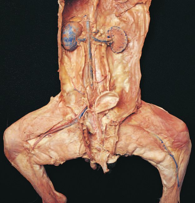
          24      Cat Dissection

                                                                         4. The bean-shaped adrenal glands are located superior
          DISSECTION 3:                                                     and medial to the kidneys. Both the kidneys and adre-
          ENDOCRINE ORGANS                                                  nal glands are retroperitoneal, or located behind the
                                                                            peritoneum.
          The major endocrine organs of the cat have similar loca-
                                                                         5. The female gonads are called ovaries and are very
          tions and structure compared with humans. Assemble your
                                                                            small, oval organs located inferior to the kidneys.
          dissection equipment and safety glasses, put on your
          gloves, and obtain your cat. Position your cat within the      6. The male gonads, the testes, are located outside of
          dissection tray, including the tail. Keep any remaining pre-      the abdominopelvic cavity in the scrotum. Before
          serving fluid in the bag to keep your cat moist and to in-        opening the scrotum, your instructor will tell you
          hibit bacterial and mold growth.                                  whether or not to proceed to view the testes at this
                                                                            time, or to wait for the reproductive system.
                                                                         7. Place the skin back over your cat and follow your in-
          Procedure                                                         structors directions to prepare the cat for storage in
                                                                            the plastic bag. Be sure to attach your group’s identi-
          1. Place the cat on its back with the ventral side up. Use        fication tag.
             Figure C3.1 to help you identify the endocrine organs.
                                                                         8. Clean your tabletop with disinfectant.
             If you have not opened the ventral body cavities, refer
             to the instructions in the preface.                         9. Wash your dissection tools, dissection tray, and hands
                                                                            before leaving the lab.
          2. There are two main endocrine organs in the thoracic
             cavity: the thyroid and the thymus. The thyroid
             gland has two dark lobes similar to the human that
             are on either side of the trachea inferior to the larynx.
             The thymus is lighter colored with small lobules, and
             is located inferior to the thyroid gland on the trachea,
             partially covering the heart.
          3. There are three main endocrine organs in the abdomi-
             nal cavity: the pancreas, adrenal glands, and gonads.
             Locate the diaphragm that separates the thoracic and
             abdominopelvic cavities. Reflect the stomach and
             look beneath it for the light, glandular-looking
             pancreas. It is close to the curve in the first part of
             the small intestine (the duodenum) and extends to the
             left toward the spleen.
8546d_c01_1-42   6/21/02   1:34 PM       Page 25 mac62 mac62:1253_GE:

                                                                                    Cat Dissection   25

                              Trachea                                   Thyroid gland

                              Thymus

                              Pancreas
                                                                        Adrenal gland
                                                                        Kidney

                                                                        Ovary ( )

                              Testis (   )

          FIGURE C3.1      Endocrine organs, ventral view.
8546d_c01_1-42   6/21/02    1:34 PM     Page 26 mac62 mac62:1253_GE:

          26      Cat Dissection

                                                                            9. The first major branch off each subclavian artery is
          DISSECTION 4: BLOOD                                                  the vertebral artery. Follow the right and left sub-
          VESSELS                                                              clavian arteries to the first rib. As each subclavian
                                                                               artery crosses the first rib, it becomes the axillary
          Double-injected cats are usually used to identify blood              artery. Follow the axillary artery into the arm,
          vessels. Arteries are injected with red latex, and veins are         where it becomes the brachial artery. Distal to the
          injected with blue latex. Blood vessels differ slightly in lo-       elbow, the brachial artery divides to form the radial
          cation from cat to cat. It is important to understand that           and ulnar arteries.
          these slight differences in location are normal and also oc-
                                                                           10. Lift up the heart and follow the aortic arch as it de-
          cur in humans. Observe the fascia that protects and secures
                                                                               scends and forms the thoracic aorta. Follow the
          blood vessels. Carefully remove the fascia with blunt in-
                                                                               thoracic aorta and observe where it passes through
          struments to separate blood vessels from other structures.
                                                                               the diaphragm with the esophagus and inferior vena
             Position your cat within the dissection tray, including
                                                                               cava, and becomes the abdominal aorta. The ab-
          the tail. Keep any remaining preserving fluid in the bag to
                                                                               dominal aorta is retroperitoneal. You must move
          keep your cat moist and inhibit bacterial and mold growth.
                                                                               aside the visceral organs and remove the parietal
                                                                               peritoneum lining the dorsal body wall to observe
                                                                               the aorta.
          Procedure                                                        11. Refer to Figure C4.2 to identify the following arter-
                                                                               ies that are located below the diaphragm.
          A. Arteries                                                      12. Locate the celiac trunk, the first branch off the
                                                                               abdominal aorta. The celiac trunk branches into the
           1. Place your cat in a dissecting tray with the ventral
                                                                               hepatic artery, the left gastric artery, and the splenic
              surface facing upward. If you have not opened the
                                                                               artery.
              ventral body cavities, refer to the instructions in the
              preface.                                                     13. Posterior (caudal) to the celiac trunk is the superior
                                                                               mesenteric artery, whose branches can be observed
           2. Identify the following major organs: heart, trachea,
                                                                               traveling through the mesentery of the small
              lungs, diaphragm, stomach, spleen, pancreas, liver,
                                                                               intestine.
              small intestine, and large intestine.
                                                                           14. Follow the abdominal aorta to the level of the
           3. Using your scissors, cut open the pericardial sac sur-
                                                                               kidneys and observe the paired renal arteries
              rounding the heart to expose the heart.
                                                                               branching off and traveling to the kidneys.
           4. Refer to Figure C4.1 to identify the following arter-
                                                                           15. The gonadal arteries, testicular arteries in males
              ies that are located above the diaphragm.
                                                                               and ovarian arteries in females, are the next major
           5. Identify the pulmonary trunk exiting from the right              branches off the abdominal aorta. Follow these
              ventricle. Locate its branches, the right pulmonary              arteries to the gonads (testes in males and ovaries in
              artery and the left pulmonary artery, and follow                 females).
              them to the lungs.
                                                                           16. The inferior mesenteric artery branches off the ab-
           6. Identify the ascending aorta as it exits the left                dominal aorta posterior (caudal) to the gonadal arter-
              ventricle.                                                       ies. Branches of the inferior mesenteric artery travel
                                                                               through the mesentery of the large intestine.
           7. Identify the aortic arch. In cats, there are only two
              branches off the aortic arch, the brachiocephalic            17. Iliolumbar arteries are large branches off the
              artery (first branch) and the left subclavian artery.            abdominal aorta posterior to the inferior mesenteric
              Identify these branches. Compare this branching                  arteries.
              with the human.
                                                                           18. The abdominal aorta ends when it divides into the
           8. The brachiocephalic artery divides into the right sub-           right and left external iliac arteries, and the
              clavian artery, the right common carotid, and the left           internal iliac artery. There is no common iliac ar-
              common carotid. Locate the subclavian artery as it               tery in the cat.
              turns laterally and travels toward the upper extrem-
                                                                           19. Follow one external iliac artery into a thigh, where it
              ity. Locate the right and left common carotid ar-
                                                                               becomes the femoral artery.
              teries as they travel along the trachea. At the level
              of the larynx, the common carotid arteries divide to         20. The femoral artery travels down the thigh and be-
              form the external and internal carotid arteries.                 comes the popliteal artery in the popliteal area.
8546d_c01_1-42   6/25/02     4:32 PM        Page 27 mac48 Mac 48: 420_kec:

                                                                                                                       Cat Dissection   27

                              Right common                                         External carotid a.
                              carotid a.                                           Internal carotid a.
                              External jugular v.
                                                                                   Left common carotid a.
                              Internal jugular v.                                  Vertebral a.
                                                                                       Subclavian a.
                              Brachiocephalic veins
                                                                                              Axillary a.
                              Subclavian v.

                              Axillary v.                                                                      Radial a.

                           Azygos v.

                                                                                                                       Ulnar a.

                                                                                                                   Brachial a.

                                                                                                     Aortic arch
                        Brachiocephalic a.

                        Superior vena cava                                                        Thoracic aorta

                                                                                                         External jugular v.
                     Left brachiocephalic v.

                     Right common carotid a.
                                                                                                         Left common carotid a.

                     Right subclavian a.                                                                 Left brachial a.
                                                                                                         Brachial v.
                                                                                                         Axillary a.
                     Brachiocephalic a.                                                                  Left subclavian v.

                     Azygous v.                                                                          Left subclavian a.
                     Aortic arch
                                                                                                         Superior vena cava

                     Heart                                                                               Thoracic aorta

                     Diaphragm

                                                                    Ventral view

          FIGURE C4.1        Blood vessels above the diaphragm.
8546d_c01_1-42   6/25/02    4:32 PM      Page 28 mac48 Mac 48: 420_kec:

          28      Cat Dissection

                                         Inferior vena cava                       Thoracic aorta
                                         Hepatic veins
                                         Hepatic a.                               Celiac trunk
                                         Gastric a.                               Splenic a.
                                                                                  Superior mesenteric a.

                                                                                  Renal a.
                                                                                  Renal v.
                                                                                  Gonadal v.
                                                                                  Gonadal a.

                                         Inferior mesenteric a.                   Iliolumbar a.
                                                                                  Iliolumbar v.
                                                                                  Internal iliac a.
                                         Common iliac v.
                                                                                  External iliac a.
                                         External iliac v.
                                         Internal iliac v.
                                                                                  Femoral a.
                                         Femoral v.

          FIGURE C4.2      Blood vessels below the diaphragm.

                        Thoracic aorta                                                 Adrenal gland
                        Celiac trunk
                        Superior mesenteric a.                                         Kidney

                        Left renal a.                                                  Left renal v.
                                                                                       Inferior vena cava
                                                                                       Abdominal aorta
                        Inferior mesenteric a.

                                                                                       External iliac a.

                        Femoral a. and v.

                        Great saphenous a. and v.

                                                                   Ventral view

          FIGURE C4.2      Blood vessels below the diaphragm, continued.
8546d_c01_1-42   6/21/02   1:34 PM    Page 29 mac62 mac62:1253_GE:

                                                                                                             Cat Dissection      29

          B. Veins                                                       9. Refer to Figure C4.1 to identify veins cephalic to the
                                                                            diaphragm.
           1. Blood leaving tissues travels through veins back to       10. Locate the radial and ulnar veins in the forearm.
              the heart. Remember that some veins are superficial           These veins are adjacent to their corresponding
              (close to the surface), whereas others are deep.              arteries. The radial and ulnar veins merge to form
              Many of the deep veins are adjacent to arteries with          the brachial vein.
              the same name.
                                                                        11. The brachial vein becomes the axillary vein that is
           2. Refer to Figure C4.2 to identify veins located caudal         adjacent to the axillary artery in the axillary regions.
              to the diaphragm.
                                                                        12. In the shoulder area, the axillary vein becomes the
           3. Observe the large superficial vein traveling along the        subclavian vein.
              medial surface of the leg ascending into the thigh.
              This is the great saphenous vein, and it joins the        13. Each subclavian vein unites with an external jugular
              femoral vein, a deep vein, traveling through the              vein to form either the right or left brachiocephalic
              thigh adjacent to the femoral artery.                         vein. The brachiocephalic veins merge to form the
                                                                            superior vena cava (precava). Follow the superior
           4. The femoral vein becomes the external iliac vein in           vena cava unit it enters the right atrium.
              the groin region. The internal iliac vein joins the ex-
              ternal iliac vein to form the common iliac vein.          14. Blood draining from the face and skull enters the ex-
                                                                            ternal jugular vein. The internal jugular vein drains
           5. The right and left common iliac veins unite to form           the brain. Identify the large external jugular vein
              the inferior vena cava (postcava in cat).                     traveling along the lateral surface of the neck until it
           6. The renal veins and gonadal veins carry blood from            joins with the subclavian vein to form the brachio-
              the kidneys and gonads to the inferior vena cava.             cephalic vein.

           7. The hepatic portal vein probably does not contain         15. Place the skin back over your cat and follow your
              blue latex and may appear brown from the presence             instructor’s directions to prepare the cat for storage
              of coagulated blood. The hepatic portal vein receives         in the plastic bag. Be sure to attach your group’s
              blood from the digestive organs and carries this              identification tag.
              blood to the liver. The hepatic portal vein is formed     16. Clean your tabletop with disinfectant.
              from the gastrosplenic vein and the superior mesen-
              teric vein.                                               17. Wash your dissection tools, dissection tray, and
                                                                            hands before leaving the lab.
           8. Follow the inferior vena cava through the
              diaphragm, into the thoracic cavity, and into the
              right atrium.
8546d_c01_1-42   6/21/02    1:34 PM    Page 30 mac62 mac62:1253_GE:

          30      Cat Dissection

                                                                         4. The spleen is located in the upper left quadrant poste-
          DISSECTION 5:                                                     rior and lateral to the stomach, and may be a dark
          LY M P H AT I C S Y S T E M                                       brownish-red color.
                                                                         5. The thoracic duct (left lymphatic duct) can some-
          The lymphatic system of the cat is complementary to the
                                                                            times be found in the dorsal part of the thoracic cav-
          human with the organs being similar in location and struc-
                                                                            ity, especially if your cat has been triple injected with
          ture compared with the human. Assemble your dissection
                                                                            latex. Move the lungs and heart aside and look just to
          equipment and safety glasses, put on your gloves, and ob-
                                                                            the left of the midline next to the descending
          tain your cat. Position your cat within the dissection tray,
                                                                            (thoracic) aorta. The thoracic duct will be very thin
          including the tail. Keep any remaining preserving fluid in
                                                                            (116 inch) and may be reddish-brown, with a seg-
          the bag to keep your cat moist and inhibit bacterial and
                                                                            mented look that is caused by the presence of valves.
          mold growth.
                                                                            You may be able to trace it to where it enters the
                                                                            junction of the left subclavian and external jugular
                                                                            veins. The right lymphatic duct is smaller and is not
          Procedure                                                         as easy to find.
                                                                         6. Place the skin back over your cat and follow your
          1. You may have already looked at the lymphatic organs
                                                                            instructor’s directions to prepare your cat for storage
             in your previous dissections. If your cat is triple
                                                                            in the plastic bag. Be sure to attach your group’s
             injected with yellow or green latex for the lymphatic
                                                                            identification tag to the cat or bag.
             system, it will be easier to find the lymphatic organs
             and very thin vessels.                                      7. Clean your laboratory tabletop with disinfectant.
          2. As you dissected the blood vessels, you may have            8. Wash your dissection tools, dissection tray, and hands
             noted small, bean-shaped lymph nodes in the cervi-             before leaving the lab.
             cal, axillary, and inguinal areas. Because these nodes
             are small, they are easy to miss if you do not know
             their structure or location.
          3. The noncapsulated thymus is over the anterior sur-
             face of the heart and sometimes is also a little supe-
             rior to the heart. This gland may have been identified
             in the endocrine system.
8546d_c01_1-42   6/21/02    1:34 PM   Page 31 mac62 mac62:1253_GE:

                                                                                   Cat Dissection   31

                                                                              Thymus

          FIGURE 5.1       Thymus.

                                                                     Spleen

          FIGURE 5.2       Spleen.
8546d_c01_1-42   6/21/02   1:34 PM    Page 32 mac62 mac62:1253_GE:

          32      Cat Dissection

                                                                          6. Cut the trachea in cross section and pull the cut por-
          DISSECTION 6:                                                      tion toward you. Carefully separate the connective
          R E S P I R ATO RY SYSTE M                                         tissue between the esophagus and the trachea with a
                                                                             blunt probe. Observe the dorsal side of the trachea
          The respiratory system of the cat is complementary to the          and identify the trachealis muscle that connects the
          human. The structure of the larynx, trachea, lungs, and di-        free edges of the tracheal cartilages.
          aphragm are similar to the human. Assemble your dissec-
                                                                          7. If you have already studied the cardiovascular sys-
          tion equipment and safety glasses, put on your gloves, and
                                                                             tem, ask your instructor for permission to remove
          obtain your cat. Make sure all parts of the cat are inside
                                                                             the heart and great vessels from the cat.
          the dissection tray, including the tail. Keep any remaining
          preserving fluid in the bag to keep your cat moist and in-      8. With the heart removed, you can easily identify the
          hibit bacterial and mold growth.                                   end of the trachea in the thoracic cavity at its bifur-
                                                                             cation into the right and left primary bronchi.
                                                                          9. Dissect away lung tissue on the left side to follow
          Procedure                                                          the left primary bronchus as it branches into the
                                                                             secondary bronchi. If you keep dissecting, you may
           1. Use Figure C6.1 to help you identify the bolded                want to use a dissecting microscope to observe
              structures listed below in the cat.                            smaller tertiary bronchi.
           2. Observe the external nares (choanae), nasal cavity,        10. On the right side, you should find the anterior, me-
              and oral pharynx.                                              dial, posterior, and mediastinal lobes of the lung.
                                                                             The latter lobe will be more midline.
           3. Locate the larynx, the prominent thyroid cartilage
              in the anterior neck region, and the cricoid cartilage     11. On the left side, you should find the anterior,
              inferior to the thyroid cartilage. Use the blunt probe         medial, and posterior lobes.
              to separate the larynx from the muscles and connec-
                                                                         12. Identify the hilus of the lung on its medial border,
              tive tissue.
                                                                             along with the primary bronchus, pulmonary
           4. Your instructor may divide the lab groups in half to           artery, and pulmonary veins.
              observe two different views of the larynx as listed
                                                                         13. Look deep into the thoracic cavity and identify the
              below:
                                                                             shiny parietal pleura that covers the ribs and inter-
              • Half of the lab groups will cut the complete larynx
                                                                             costal muscles. The visceral pleura also glistens and
                away from the laryngopharynx at the hyoid bone.
                                                                             covers the lungs themselves.
                Pull the larynx toward you, look into the top of
                the larynx, and identify: the epiglottis (elastic car-   14. Observe the muscular diaphragm that forms the
                tilage), glottis, false vocal cords (anteriorly), and        thoracic cavity floor and its relationship to the lungs
                true vocal cords (posteriorly).                              and the pleura of the lungs.
              • The other half of the lab groups will make a longi-
                                                                         15. Place the skin back over your cat and follow your
                tudinal cut through the thyroid cartilage, the lar-
                                                                             instructor’s directions to prepare your cat for storage
                ynx, and through the superior part of the trachea.
                                                                             in the plastic bag. Be sure to attach your group’s
                Observe the following structures: the epiglottis,
                                                                             identification tag to the cat or bag.
                glottis, false vocal cords, and true vocal cords.
                                                                         16. Clean your tabletop with disinfectant.
           5. Examine the trachea, following it into the thoracic
              cavity. Feel the C-shaped tracheal cartilages.             17. Wash your dissection tools, dissection tray, and
              Check to see if the thyroid gland is still present, or         hands before leaving the lab.
              if it was removed in a previous dissection.
8546d_c01_1-42   6/21/02       1:34 PM   Page 33 mac62 mac62:1253_GE:

                                                                                 Cat Dissection        33

                                                                                Epiglottis of larynx
                                                                                Thyroid cartilage
                                                                                of larynx

                                                                                Thyroid gland

                                                                                Trachea

                 Heart
                                                                                Thymus

                 Right lung:                                                    Left lung:
                                                                                  Anterior lobe
                   Anterior lobe

                   Medial lobe                                                    Medial lobe

                   Mediastinal lobe                                               Posterior lobe
                   Posterior lobe

                                                                                Diaphragm

                                                                               Thyroid
                                                                               cartilage

                                                                               Thyroid gland
                                                                               Trachea

                      Heart

                      Right lung:                                              Left lung:
                                                                                Anterior lobe
                         Anterior
                         lobe
                         Medial
                         lobe
                                                                                Medial
                         Mediastinal                                            lobe
                         lobe
                                                                                Posterior
                         Posterior                                              lobe
                         lobe
                                                                               Diaphragm

                                                                Ventral view

          FIGURE C6.1       Respiratory system, ventral view.
8546d_c01_1-42     6/21/02      1:34 PM    Page 34 mac62 mac62:1253_GE:

          34         Cat Dissection

                                                                          2. Identify the vestibule, hard palate, soft palate,
          DISSECTION 7: DIGESTIVE                                            canine teeth, tongue, lingual frenulum, and
          SYSTE M                                                            oropharynx. Compare the teeth with human teeth.
                                                                          3. Using a hand lens or magnifying glass, observe the
          The cat digestive system and organs are quite similar to
                                                                             papillae on the tongue.
          that of the human in location and structure. This dissec-
          tion also clearly demonstrates the location and structure of    4. To expose the salivary glands, remove the skin on
          the mesentery and parts of the peritoneum that are not re-         one side of the head inferior to the ear (see Figure
          alistically portrayed in models. Assemble your dissection          C.7.1), trim away the connective tissue in the area
          equipment and safety glasses, put on your gloves, and ob-          between this and the masseter muscle. Look for tiny,
          tain your cat. Position your cat within the dissection tray,       dark lymph nodes (bean-shaped) in this area. The
          including the tail. Keep any remaining preserving fluid in         parotid gland is a light-colored gland on the cheek
          the bag, to keep your cat moist and to inhibit bacterial and       area inferior to the ear. You may be able to identify
          mold growth.                                                       the parotid duct traversing the masseter to enter the
                                                                             oral cavity. The smaller submandibular gland is
                                                                             inferior and a little posterior to the parotid gland. The
                                                                             sublingual gland, just anterior to the submandibular
          Procedure                                                          gland, is the smallest salivary gland is more difficult
                                                                             to find.
          A. Mouth, Oropharynx, and
          Salivary Glands
          1. To observe the oral cavity structures, you may need to
             use a bone cutter to cut through the mandible and
             separate it from the maxilla.

                                                                                                                          Parotid gland

          Submandibular
          gland

          F I G U R E C 7. 1   Salivary glands.
8546d_c01_1-42   6/21/02   1:34 PM    Page 35 mac62 mac62:1253_GE:

                                                                                                            Cat Dissection      35

          B. Esophagus and                                                  index finger; cut open this area to observe the con-
                                                                            striction caused by the sphincter. To the left of and
          Abdominal Organs                                                  posterior to the stomach is the long, narrow, dark-
                                                                            colored spleen that hugs the left abdominal wall (not
           1. If you have dissected the respiratory system, you             a digestive organ).
              have previously observed the laryngopharynx,
              epiglottis, larynx, and trachea. The laryngopharynx        7. Lift the stomach, and reflect it back to reveal the
              also leads to the esophagus that is posterior to the          granular, usually brownish-gray pancreas. The head
              trachea. Follow the esophagus through the thoracic            of the pancreas is in the C-shape of the first section
              cavity to the diaphragm, locating the esophageal              of the small intestine, the duodenum, and the tail of
              hiatus where the esophagus penetrates through the             the pancreas is near the spleen. Find the common
              diaphragm to the abdominal cavity.                            bile duct entering the duodenum and follow it to-
                                                                            ward the liver until you find the junction of the
           2. Use Figure C.7.2a and Fig C.7.2b as a reference, to           common hepatic duct with the cystic duct.
              identify the bolded structures.
                                                                         8. The small intestine of the cat has three divisions, as
           3. Observe the yellowish, fat-filled “apron” that covers         does the human: the duodenum, jejunum, and
              the abdominopelvic viscera. This is the double-               ileum. Note the mesentery that attaches the small
              layered serous membrane, the greater omentum,                 intestine to the posterior body wall. Spread the
              that can be deflected back or totally removed                 mesentery to observe the branches of the superior
              according to your instructor’s directions.                    mesenteric artery and vein. Follow the small intes-
           4. Observe the peritoneum that lines the abdominal               tine through its entire length. The ileum ends in the
              cavity and also covers the exterior of the abdominal          inferior right quadrant, where it joins with the large
              organs. The peritoneal cavity is the large cavity that        intestine at the ileocecal junction or sphincter.
              is filled with the abdominopelvic organs.                     Make an incision in this area to observe the sphinc-
                                                                            ter. Note that the small intestine has a smaller diam-
           5. The next obvious structure in the abdomen is the              eter, a greater length, and is very coiled compared
              large, brown or reddish-brown liver on the right side         with the large intestine.
              inferior to the diaphragm. Look for a small, greenish
              sac, the gallbladder, on the inferior surface of the       9. The large intestine, or colon, is composed of the
              liver, and the cystic duct. The falciform ligament            cecum, a short ascending colon, transverse colon,
              separates the right and left lobes of the liver and at-       descending colon, and rectum. Just inferior to the
              taches the liver superiorly to the abdominal wall.            ileocecal junction is the cecum, or blind pouch.
                                                                            Identify the ascending, transverse, and descending
           6. To the left of and partially posterior to the liver is        parts of the colon plus the mesocolon that affixes
              the stomach. Identify the lesser omentum, the                 the colon to the posterior body wall. Now identify
              serous membrane that attaches the liver to the lesser         the rectum and the anus.
              curvature of the stomach. Note the constricted junc-
              tion of the esophagus and the stomach, the                10. Place the skin back over your cat and follow your
              esophageal sphincter. Cut open the stomach along              instructor’s directions to prepare your cat for storage
              its greater curvature to reveal the rugae, if present.        in the plastic bag. Be sure to attach your group’s
              If the cat’s stomach is stretched, rugae are absent; if       identification tag to the cat or bag.
              the stomach is contracted, rugae will be present.         11. Clean your laboratory tabletop with disinfectant.
              Identify the parts of the stomach: the cardia, fun-
              dus, body, pylorus, and the pyloric sphincter. Roll       12. Wash your dissection tools, dissection tray, and
              the firm sphincter area between your thumb and                hands before leaving the lab.
8546d_c01_1-42     6/25/02        4:32 PM      Page 36 mac48 Mac 48: 420_kec:

          36         Cat Dissection

                                                                                       Diaphragm

                                 Gallbladder

                                                                                       Lobes of liver

                                                                                       Stomach

                                                                                       Spleen

                                 Greater omentum

                                                                                       Diaphragm
                                      Gallbladder

                                                                                       Lobes of liver

                                                                                       Stomach

                                                                                       Spleen

                                      Greater
                                      omentum

                                                                    (a) Ventral view

          F I G U R E C 7. 2 a    Digestive organs, superficial.
8546d_c01_1-42    6/21/02        1:34 PM       Page 37 mac62 mac62:1253_GE:

                                                                                                        Cat Dissection   37

                                                                                       Diaphragm

                                                                                       Lobes of liver
                                   Gallbladder
                                                                                       Stomach
                                   Pyloric valve                                       Lesser omentum

                                                                                       Mesentery
                                   Duodenum of
                                   small intestine                                     Jejunum
                                   Pancreas                                            Ileocecal junction
                                                                                       Cecum

                                                                                       Ileum

                                   Urinary bladder

                                                                                         Diaphragm
                                                                                         Lobes of liver
                                 Gallbladder

                                 Pyloric valve                                           Stomach
                                                                                         Lesser omentum

                                 Duodenum                                                Mesentery
                                                                                         Jejunum
                                 Pancreas                                                Ileocecal junction
                                                                                         Cecum

                                                                                         Ileum

                                 Urinary bladder

                                 Urethra

                                                                    (b) Ventral view

          F I G U R E C 7. 2 b   Digestive organs, deep.
8546d_c01_1-42   6/25/02    4:32 PM    Page 38 mac48 Mac 48: 420_kec:

          38      Cat Dissection

                                                                         2. Reflect the abdominal viscera that were observed in
          DISSECTION 8: URINARY                                             the digestive system dissection.
          AND REPRODUCTIVE                                               3. Remove the peritoneum from the kidneys if not re-
          SYSTE M S                                                         moved in a prior dissection and carefully remove the
                                                                            adipose capsule surrounding the kidneys. Locate the
          Typically, the urinary and reproductive systems are studied       adrenal glands that are not attached to the kidneys,
          together, because of their close association of structures        but are superior and medial to them.
          and their embryologic derivations. The urinary and repro-
                                                                         4. Locate the renal hilus on the medial surface of each
          ductive systems of the male cat are similar to the human.
                                                                            kidney and identify the renal artery, renal vein, and
          The female cat has more differences compared to the hu-
                                                                            the ureter passing through the renal hilus.
          man, because she has litters rather than one offspring dur-
          ing one pregnancy. Assemble your dissection equipment          5. Follow the renal artery to where it branches off the
          and safety glasses, put on your gloves, and obtain your cat.      abdominal aorta and the renal vein to where it enters
          Position your cat within the dissection tray, including the       the inferior vena cava.
          tail.
                                                                         6. Follow the ureters to the urinary bladder, a
                                                                            retroperitoneal, muscular sac. If you have a female
                                                                            cat, be careful not to mistake the uterine horns for the
                                                                            ureters. Observe the entrance of the ureters into the
          Procedure                                                         posterior wall of the urinary bladder, and the peri-
                                                                            toneal folds that secure the urinary bladder to the
          A. Urinary System                                                 abdominal wall.
          1. Refer to Figure C8.1a if you have a male cat, or Fig-       7. The urethra will not be dissected out at this time be-
             ure C8.1b if you have a female. Identify the bolded            cause of its location, but will be located in the repro-
             urinary structures described.                                  ductive system dissection that follows.
8546d_c01_1-42     6/25/02        4:32 PM   Page 39 mac48 Mac 48: 420_kec:

                                                                                     Cat Dissection         39

                                                                                 Left kidney
                                                                                 (sectioned)
                  Inferior
                  vena cava
                                                                                 Left ureter
                  Abdominal
                  aorta

                                                                                 Urinary
                                                                                 bladder
                  Vas deferens

                  Spermatic
                  cord                                                           Urethra

                                                                                 Epididymis
                  Scrotum
                  with testis                                                    Penis

                                                                                  Left kidney (sectioned)

                                                                                  Left ureter

                 Vas deferens                                                     Urinary bladder

                 Inguinal canal
                                                                                  Urethra

                 Spermatic
                 cord

                                                                                  Penis
                 Scrotum with
                 testis

                                                                  Ventral view

          FIGURE C8.1a            Male urinary and reproductive organs.
8546d_c01_1-42   6/25/02   4:32 PM    Page 40 mac48 Mac 48: 420_kec:

          40      Cat Dissection

          B. Male Reproductive System                                   9. To properly observe the accessory sex glands and
                                                                           the urethra, you need to cut the cat’s pelvis. Using a
           1. Using Figure C8.1a for reference, identify the               sharp scalpel, make a midline incision to cut through
              bolded male reproductive structures listed below.            the muscles covering the symphysis pubis and then
                                                                           carefully cut through the center of the pubic symph-
           2. Because a male cat has a retractable penis, you may          ysis cartilage.
              need to check for the external urethral orifice first
              to find the penis and the sheath-like prepuce cover-     10. Spread the thighs apart and bend the pelvic bones
              ing it. To observe the glans penis, make an incision         back to expose the prostate gland, paired bul-
              in the prepuce.                                              bourethral glands, urethra, and penis. The prostate
                                                                           can be palpated as a small, hard mass surrounding
           3. Identify the scrotum or scrotal sac covering the             the urethra. The cat anatomy is similar to, but not
              paired testes, which may not be very obvious if you          identical to, the human. There are no seminal vesi-
              have a young male.                                           cles in the cat.
           4. Carefully, make a lateral incision in one side of the    11. The bulbourethral glands are located posterior to
              scrotum and remove the loose fascia and inner fi-            the prostate gland dorsal to the penis. Carefully cut
              brous connective tissue to expose one testis. Is the         the proximal end of the penis to expose the white
              scrotal sac open to both testes?                             swellings of the bulbourethral glands.
           5. Note the epididymis on the medial and posterior          12. Make a longitudinal incision in the penis and iden-
              surfaces of the testis, and inspect its tiny, coiled         tify the two columns of corpora cavernosa, one
              tubules. You may want to use a hand lens for this.           column of corpus spongiosum, and the spongy ure-
           6. Identify the ductus (vas) deferens that begins at the        thra. You may wish to cut a cross section of the
              tail of the epididymis and travels toward the body in        penis to identify all three columns of tissue and the
              the spermatic cord.                                          urethra.

           7. Observe the spermatic cord and cut away the con-         13. Observe the dissection of a female cat from another
              nective tissue to identify the ductus (vas) deferens,        lab group.
              testicular artery, testicular vein, and autonomic        14. Read steps 9–11 of the female cat reproductive
              nerves within it. Follow the ductus (vas) deferens           system dissection for clean-up directions.
              through the inguinal canal into the pelvic cavity.
           8. Trace the path of the ductus (vas) deferens in the ab-
              dominopelvic cavity as it arches around the ureter,
              and continues posterior to the bladder to join the
              small prostate gland at the urethra. Inside the pelvic
              cavity, the testicular blood vessels and autonomic
              nerves travel near the ureters, taking a different
              route from the ductus (vas) deferens.
8546d_c01_1-42   6/21/02   1:34 PM   Page 41 mac62 mac62:1253_GE:

                                                                                                          Cat Dissection      41

          C. Female Reproductive System                                6. The urinary bladder and urethra will be ventral to
                                                                          the body of the uterus and the vagina. Using a blunt
           1. Using Figure C8.1b as a reference, identify the             probe, separate the connective tissue that holds the
              bolded structures below.                                    urethra to the vagina and move the urethra to the
                                                                          side. Locate the posterior union of the urethra with
           2. The cat’s uterus is quite different from a human. The       the vagina.
              uterus in a cat is Y-shaped and is called a bipartate
              uterus. The base of the Y is the body of the uterus      7. Just caudal to the union of the urethra and the
              and the upper two branches are the uterine horns,           vagina is the urogenital sinus that opens to the exte-
              where multiple fetuses may be located if your cat is        rior in the urogenital oriface. The female cat has
              pregnant.                                                   the urogenital orifice as one opening for both the
                                                                          urinary and reproductive systems similar to the male
           3. In the pelvic cavity, locate the small, oval ovaries        cat and human male.
              caudal and lateral to the kidneys and the small uter-
              ine tubes that have tiny fimbriae curved over the        8. Observe the dissection of a male cat from another
              ovaries. Note the thin mesentery that attaches these        lab group.
              structures to the body wall.                             9. Place the skin back over your cat and follow your
           4. To follow the uterus to the vagina, you will need to        instructor’s directions to prepare your cat for storage
              cut the cat’s pelvis. With a sharp scalpel, make a          in the plastic bag. Be sure to attach your group’s
              midline incision through the muscles covering the           identification tag to the cat or bag.
              pubic symphysis and then cut through the center of      10. Clean your tabletop with disinfectant.
              the cartilage of the pubic symphysis.
                                                                      11. Wash your dissection tools, dissection tray, and
           5. Spread the thighs and bend the pelvic bones back to         hands before leaving the lab.
              expose the urethra (anterior) and vagina (posterior).
8546d_c01_1-42   6/25/02    4:32 PM       Page 42 mac48 Mac 48: 420_kec:

          42      Cat Dissection

                                                                               Left kidney

                             Ovary
                                                                               Ovarian
                                                                               ligament

                             Right horn                                        Left horn
                             of uterus                                         of uterus

                             Right
                             ureter                                            Body of
                             Urinary                                           uterus
                             bladder
                                                                              Vagina
                             Urethra

                             Urogenital
                             sinus

                                                                             Left kidney (sectioned)

                         Ovary                                               Ovarian ligament

                                                                             Ovarian vein

                         Right
                         uterine horn

                                                                             Left horn of uterus
                         Right ureter
                                                                             Body of uterus
                         Urinary bladder
                         Urethra                                             Vagina

                         Urogenital sinus

                                                              Ventral view

          FIGURE C8.1b     Female urinary and reproductive organs.
You can also read