Alopecia areata is driven by cytotoxic T lymphocytes and is reversed by JAK inhibition
←
→
Page content transcription
If your browser does not render page correctly, please read the page content below
letters

Alopecia areata is driven by cytotoxic T lymphocytes and
is reversed by JAK inhibition
Luzhou Xing1,7, Zhenpeng Dai2,7, Ali Jabbari2,7, Jane E Cerise2,3, Claire A Higgins2, Weijuan Gong2,
Annemieke de Jong2, Sivan Harel2, Gina M DeStefano2,4, Lisa Rothman2, Pallavi Singh2, Lynn Petukhova2,
Julian Mackay-Wiggan2, Angela M Christiano2,5,8 & Raphael Clynes1,2,6,8

Alopecia areata (AA) is a common autoimmune disease                              with considerable similarity to human AA. Broad-acting intralesional
resulting from damage of the hair follicle by T cells. The                       steroids are the most commonly used therapy for AA, with varying
immune pathways required for autoreactive T cell activation                      success. Progress in developing effective, rationally targeted therapies
in AA are not defined limiting clinical development of                           has been limited by our lack of mechanistic understanding of the
rational targeted therapies1. Genome-wide association studies                    underlying key T cell inflammatory pathways in AA.
(GWAS)2 implicated ligands for the NKG2D receptor                                   We2 and others5 have previously identified a cytotoxic subset of
(product of the KLRK1 gene) in disease pathogenesis.                             CD8+NKG2D+ T cells within the infiltrate surrounding human AA
Here, we show that cytotoxic CD8+NKG2D+ T cells are both                         hair follicles, as well as concomitant upregulation in the follicle itself
necessary and sufficient for the induction of AA in mouse                        of the ‘danger signals’ ULBP3 (ref. 2) and MICA5, two NKG2D ligands
models of disease. Global transcriptional profiling of mouse                     (NKG2DLs) whose importance in disease pathogenesis has also been
and human AA skin revealed gene expression signatures                            suggested by genome-wide association studies2.
indicative of cytotoxic T cell infiltration, an interferon-g                        To determine the contribution of CD8+NKG2D+ T cells to AA
(IFN-g) response and upregulation of several g-chain (gc)                        pathogenesis, we used the C3H/HeJ mouse model6, which sponta-
cytokines known to promote the activation and survival                           neously develops alopecia and recapitulates many pathologic fea-
of IFN-g–producing CD8+NKG2D+ effector T cells.                                  tures of human AA7. In lesional skin biopsies from alopecic mice,
Therapeutically, antibody-mediated blockade of IFN-g,                            CD8+NKG2D+ T cells infiltrate the epithelial layers of the hair fol-
interleukin-2 (IL-2) or interleukin-15 receptor b (IL-15Rb)                      licle, which overexpress the NKG2DLs, H60 and Rae-1, analogous to
prevented disease development, reducing the accumulation                         what has been observed in skin biopsies of human AA2 (Fig. 1a,b and
of CD8+NKG2D+ T cells in the skin and the dermal IFN                             Supplementary Fig. 1a,b). Flow cytometric analysis of the CD45+
response in a mouse model of AA. Systemically administered                       leukocyte population in the skin revealed a marked increased number
pharmacological inhibitors of Janus kinase (JAK) family protein                  of CD8+NKG2D+ T cells in the skin of diseased C3H/HeJ mice, in
tyrosine kinases, downstream effectors of the IFN-g and gc                       conjunction with cutaneous lymphadenopathy and increased total
cytokine receptors, eliminated the IFN signature and prevented                   cellularity, as compared with disease-free C3H/HeJ mice (Fig. 1c,d).
the development of AA, while topical administration promoted                     Other cell types, including CD4+ T cells4 and mast cells8, were
hair regrowth and reversed established disease. Notably, three                   present in much smaller numbers (Supplementary Fig. 1c and data
patients treated with oral ruxolitinib, an inhibitor of JAK1 and                 not shown).
JAK2, achieved near-complete hair regrowth within 5 months                          The immunophenotype of the skin-infiltrating CD8+ T cells in
of treatment, suggesting the potential clinical utility of JAK                   mice with AA was similar to that of the CD8+NKG2D+ population
inhibition in human AA.                                                          found in the cutaneous lymph nodes: CD8αβ+ effector memory
                                                                                 T cells (TEM, CD8hiCD44hiCD62LlowCD103+) bearing several natural
Alopecia areata is a T cell–mediated autoimmune disease charac-                  killer (NK) immunoreceptors, including CD49b and NKG2A, NKG2C
terized phenotypically by hair loss and, histologically, by infiltrating         and NKG2E (Fig. 1e and Supplementary Fig. 1d). These CD8+ TEM
T cells surrounding the hair follicle bulb (reviewed in ref. 1). Previous        cells expressed high levels of IFN-γ and exhibited NKG2D-dependent
studies have shown that transfer of total T cells (but not B cells or            cytotoxicity against ex vivo–expanded syngeneic dermal sheath tar-
sera) can cause the disease in human xenograft models3, as well as               get cells (Fig. 1f). Gene expression analysis of the CD8+NKG2D+
in C3H/HeJ mice4, a mouse strain that develops spontaneous AA                    T cells isolated from alopecic C3H/HeJ lymph node cells using

1Department of Pathology, Columbia University, New York, New York, USA. 2Department of Dermatology, Columbia University, New York, New York, USA.
3Department of Psychiatry, Columbia University, New York, New York, USA. 4Department of Epidemiology, Columbia University, New York, New York, USA.
5Department of Genetics and Development, Columbia University, New York, New York, USA. 6Department of Medicine, Columbia University, New York,

New York, USA. 7These authors contributed equally to this work. 8These authors jointly directed this work. Correspondence should be addressed to
A.M.C. (amc65@columbia.edu) or R.C. (rc645@columbia.edu).

Received 22 December 2013; accepted 30 June 2014; published online 17 August 2014; doi:10.1038/nm.3645

nature medicine advance online publication 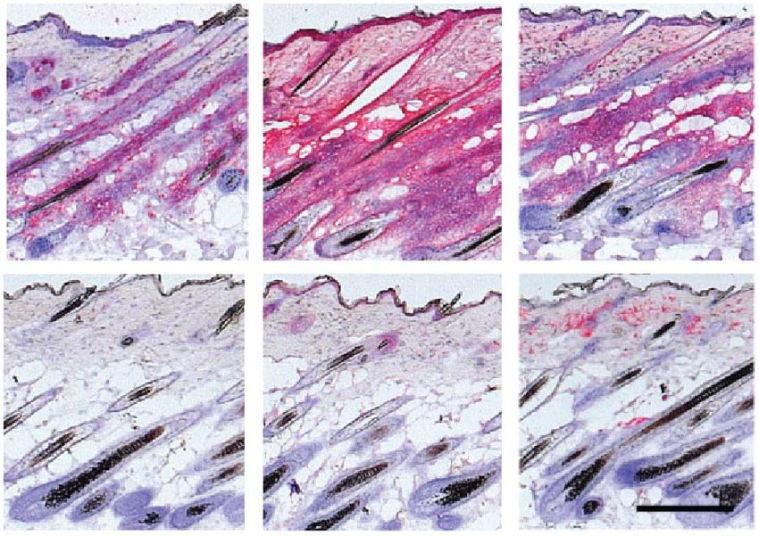
letters
RNA-seq demonstrated a transcriptional profile characteristic of To characterize the transcriptional profile of AA lesional skin
effector cytotoxic T lymphocytes (CTLs)9,10 and identified several from C3H/HeJ mice as well as human AA, we performed Affymetrix
additional NK-specific transcripts (Supplementary Table 1 and microarray analyses to identify differentially expressed genes in skin
Supplementary Fig. 2). between individuals with AA and skin from control individuals
We next evaluated the requirement of these CD8+ TEM cells in dis- without disease (Supplementary Fig. 3 and Supplementary Tables
ease pathogenesis. Transfer of cytotoxic CD8+NKG2D+ cells or total 2 and 3) Three gene expression signatures were identified in lesional
lymph node cells from diseased mice induced AA in all five healthy skin: IFN response genes, such as those encoding the IFN-inducible
C3H/HeJ recipients by 14 weeks after transfer, whereas lymph node chemokines CXCL-9, CXCL-10 and CXCL-11 (Supplementary
cell populations depleted of NKG2D+ cells were unable to transfer Figs. 3 and 4), several key CTL-specific transcripts, such as those
disease (Fig. 1g). Thus, CD8+NKG2D+ T cells are the dominant encoding CD8A and granzymes A and B, and γc cytokines and their
cell type in the dermal infiltrate and are necessary and sufficient for receptors, such as the transcripts for interleukin-2 (IL-2) and IL-15,
T cell–mediated transfer of AA. in both human and mouse AA skin. As IL-2Rα was previously shown
a H60 H60 H60
b CD8/NKG2D CD8/NKG2D CD8/NKG2D
c
C3H AA
C3H
80 ***
C57BL/6 C3H C3H AA C57BL/6 C3H C3H AA
Cell number (×106)
60
H60 K71 H60/K71 CD8 NKG2D CD8/NKG2D
40
20
0
C3H AA C3H AA C3H AA C3H AA C3H AA C3H AA
C3H AA C3H
d Skin e CD8+NKG2D– cell CD8+NKG2D+ cell
CD8+NKG2D+ T cells (%)
Skin Lymph node 25 ***
20 20
3.8
C3H AA
15
10
5
0
C3H AA C3H NKG2A/C/E DX5 CD44
CD8β
0.03 0.1 Lymph node
C3H
CD8+NKG2D+ T cells (%)
5 ***
NKG2D
4
3
% max
CD8α 2
1
0 CD69 CD103 CD62L IFN-γ
C3H AA C3H
g Total LN NKG2D-depleted
+
CD8 NKG2D
+ +
CD8 NKG2D
–
f Dermal sheath cell
40
Control
Anti-NKG2D
30
Specific lysis (%)
20
10
Isotype control 0
10:1 30:1 90:1
Anti–Rae-1
Effector/target ratio
Figure 1 CD8+NKG2D+ cytotoxic T lymphocytes accumulate in the skin and are necessary and sufficient to induce disease in AA mice.
(a) Immunofluorescence staining of NKG2D ligand (H60) in the hair follicle inner root sheath (marked by K71). Scale bar, 100 µm. (b) CD8+NKG2D+
cells in hair follicles of C57BL/6, healthy C3H/HeJ and C3H/HeJ AA mice. Top scale bar, 100 µm; bottom scale bar, 50 µm. (c) Cutaneous
lymphadenopathy and hypercellularity in C3H/HeJ AA mice. (d) Frequency (number shown above boxed area) of CD8+NKG2D+ T cells in the skin and
skin-draining lymph nodes in alopecic mice versus ungrafted mice. (e) Immunophenotype of CD8+NKG2D+ T cells in cutaneous lymph nodes of C3H/HeJ
alopecic mice. (f) Left, Rae-1t–expressing dermal sheath cells grown from C3H/HeJ hair follicles. Right, dose-dependent specific cell lysis induced by
CD8+NKG2D+ T cells isolated from AA mice cutaneous lymph nodes in the presence of blocking anti-NKG2D antibody or isotype control. Effector to
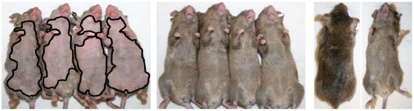
target ratio given as indicated. Data are expressed as means ± s.d. (g) Hair loss in C3H/HeJ mice injected subcutaneously with total lymph node (LN)
cells, CD8+NKG2D+ T cells alone, CD8+NKG2D− T cells or lymph node cells depleted of NKG2D+ (5 mice per group). Mice are representative of two
experiments. ***P < 0.001 (Fisher’s exact test). For c,d,f, n and number of repeats are detailed in the Supplementary Methods.
advance online publication nature medicine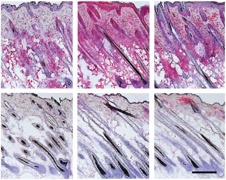
letters
to be expressed on infiltrating lymphocytes surrounding human AA found IL-15Rβ expressed on infiltrating CD8+ T cells in humans
hair follicles11, we performed immunofluorescence analysis for both (Supplementary Figs. 5 and 6).
IL-15 and its chaperone receptor IL-15Rα to identify the source IL-2 and IL-15 are well-known drivers of cytotoxic activity by
of IL-15 in the skin. We detected a marked upregulation of both IFN-γ–producing CD8+ effector T cells and NK cells12,13 and have
components in AA hair follicles in both human and mouse AA and been implicated in the induction and/or maintenance of autoreactive
a Control Anti–IFN-γ
c CD8 MHC class I MHC class II
Control
b 100 17 Skin
Control
CD8+NKG2D+ T cells (%)
Without hair loss (%)
80 25 **
Control (n = 5)
*
Anti–IFN-γ
Anti–IFN-γ (n = 5) 20
60
15
40 10
0.12
20 5
Anti–IFN-γ
0
NKG2D
0 Control Anti–IFN-γ
0 4 8 12 16
Time (weeks)
CD8
d Control Anti–IL-2
f CD8 MHC class I MHC class II
Control
e
100
Without hair loss (%)
22
80 Control (n = 5) Skin
CD8+NKG2D+ T cells (%)
Control
Anti–IL-2 (n = 5) * 50
***
60
40
Anti–IL-2
40
35
20 20
0.59
Anti–IL-2
0 10
NKG2D
0 4 8 12 16 0
Control Anti–IL-2
Time (weeks)
CD8
g Control Anti–IL-15Rβ i CD8 MHC class I MHC class II
Control
h 100
25
Skin
CD8+NKG2D+ T cells (%)
Control
Without hair loss (%)
80 40 ***
Anti–IL-15Rβ
Control (n = 12)
60 ***
Anti–IL-15Rβ
Anti–IL-15Rβ (n = 12) 20
40 0.03
20 0
NKG2D
Control Anti–IL-15Rβ
0
0 4 8 12 16 20 24
CD8
Time (weeks)
Figure 2 Prevention of AA by blocking antibodies to IFN-γ, IL-2 or IL-15Rβ. C3H/HeJ grafted mice were treated systemically from the time of grafting.
(a–h) AA development in C3H/HeJ grafted mice treated systemically from the time of grafting with antibodies to IFN-γ (a,b), IL-2 (d,e) and IL-15Rβ (g,h).
Frequency (number shown above boxed area) of CD8+NKG2D+ T cells in the skin of mice treated with antibodies to IFN-γ (b), IL-2 (e) and IL-15Rβ (h)
compared to PBS-treated mice. (*P < 0.05, **P < 0.01, ***P < 0.001, statistical methods described in the Supplementary Methods. Immunohistochemical
staining of skin biopsies showing CD8 and MHC class I and II expression in skin of mice treated or with isotype control antibody or with antibodies to
IFN-γ (c), IL-2 (f) or IL-15Rβ (i). Scale bars, 100 µm. For each experiment, n and number of repeats are detailed in the Supplementary Methods.
nature medicine advance online publication 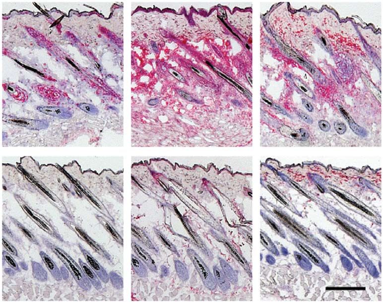
letters
CD8+ T cells14–16. To test the efficacy of IFN-γ– and γc-targeted for IL-2 in AA pathogenesis was previously established using genetic
therapies in vivo, we used the well-established graft model of AA, in experiments in which IL-2 haploinsufficiency on the C3H/HeJ back-
which skin grafts from mice with spontaneous AA are transferred ground conferred resistance to disease by about 50% using the graft
onto the backs of unaffected 10-week-old recipient C3H/HeJ mice. model19, and this role is supported by our genome-wide associa-
In this model, AA develops reliably in 95-100% of grafted recipients tion studies in humans2. Systemically administered blocking anti-
within 6–10 weeks7, allowing us to test interventions aimed at either bodies to either IL-2 (Fig. 2d–f) or IL-15Rβ (Fig. 2g–i) prevented
preventing or reversing disease. AA in grafted mice, blocked the accumulation of CD8+NKG2D+
The role of IFN-γ in AA was previously investigated using both T cells in the skin and abrogated MHC upregulation. However,
knockout studies and administration of IFN-γ, where IFN-γ–deficient IL-21 blockade failed to prevent the development of AA in grafted
mice were resistant and exogenous IFN-γ precipitated disease17,18. C3H/HeJ mice (Supplementary Fig. 7). Notably, none of these
Administration of neutralizing antibodies to IFN-γ at the time of blocking antibodies given alone was able to reverse established AA
grafting prevented AA development in grafted recipients and abro- (data not shown).
gated major histocompatibility complex (MHC) upregulation and We next asked whether we could recapitulate the effects of type I
CD8+NKG2D+ infiltration in the skin (Fig. 2a–c). Likewise, a role cytokine blockade by intervening downstream using small-molecule
a 12 weeks b 100
e
Control JAK1/2i after treatment
Without hair loss (%)
80
60 Control (n = 10) Sham
JAK1/2i (n = 10)
***
40 Control, week 5 JAK1/2i, week 5
Control, week 13 JAK1/2i, week 13
20
0
0 4 8 12 16 20 24 10
Time (weeks)
c Skin d CD8 MHC class I MHC class II
Skin Lymph node 25 ***
IFN score
CD8+NKG2D+ T cells (%)
22 1.7 5
Control
Control
0
Control JAK1/2i
Lymph node
0.04 0.38 *** 0
2
JAK1/2i
NKG2D
1
JAK1/2i
0 0 5 10 15
CD8 Control JAK1/2i CTL score
f g 100 j
Without hair loss (%)
12 weeks
80
Control JAK3i after treatment Sham
60 Control (n = 7)
JAK3i (n = 5) ** Control, week 5 JAK3i, week 5
40
Control, week 13 JAK3i, week 13
20
0
0 4 8 12 16 20 24
10
Time (weeks)
h Skin i CD8 MHC class I MHC class II
***
IFN score
Skin Lymph node 25 5
Control
CD8+NKG2D+ T cells (%)
15 3.3
Control
0
Control JAK3i 0
Lymph node
0.04 0.21 ***
6
JAK3i
NKG2D
JAK3i
0 5 10 15
0 CTL score
CD8 Control JAK3i
Figure 3 Systemic JAK1/2 or JAK3 inhibition prevents the onset of AA in grafted C3H/HeJ mice. (a–j) AA development in C3H/HeJ grafted mice treated
systemically from the time of grafting with ruxolitinib (JAK1/2i) (a,b) or tofacitinib (JAK3i) (f,g) (**P < 0.01). Frequency (number shown above boxed
area) of CD8+NKG2D+ T cells in skin and cutaneous lymph nodes of mice treated with PBS or with JAK1/2i (c) or JAK3i (h) (***P < 0.001, statistical
methods described in Supplementary Methods). Immunohistochemical staining of skin biopsies showing CD8 and MHC class I and II expression in skin
of mice treated with PBS or with JAK1/2i (d) or JAK3i (i). ALADIN score of transcriptional analysis from mice treated with PBS or with JAK1/2i (e) or
JAK3i (j), given as log2 mean expression Z-scores as indicated in the Supplementary Methods. Hair regrowth after an additional 12 weeks after treatment
withdrawal is also shown. (a,f). Scale bars, 100 µm. For each experiment, n and number of repeats are detailed in the Supplementary Methods.
advance online publication nature medicine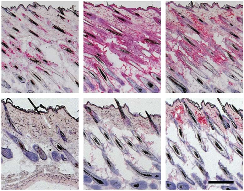
letters
inhibitors of JAK kinases, which signal downstream of a wide range Ruxolitinib, a US Food and Drug Administration (FDA)–approved
of cell surface receptors. In particular, IFN-γ receptors and γc fam- small-molecule inhibitor of the JAK1/2 kinases (JAK selectivity is
ily receptors signal through JAK1/2 and JAK1/3, respectively20. JAK1 = JAK2>Tyk2>>>JAK3)21 critical for IFN-γR signaling inhibited
JAK activation was shown by the presence of phosphorylated signal these responses (Supplementary Fig. 9a,b). In cultured CTL effec-
transducer and activator of transcription (STAT) proteins (pSTAT1, tors from C3H/HeJ mice, the FDA-approved small-molecule JAK3
pSTAT3 and to a lesser extent pSTAT5) in human and mouse alopecic inhibitor tofacitinib (JAK3>JAK1>>JAK2 selectivity)22 blocked IL-
hair follicles, but not in normal hair follicles (Supplementary Fig. 8). 15–triggered pSTAT5 activation (Supplementary Fig. 9c). Tofacitinib
In in vitro–cultured dermal sheath cells from C3H/HeJ mice, exog- also blocked killing of dermal sheath cell (Supplementary Fig. 9d)
enous IFN-γ increased STAT1 activation, whereas IFN-γ plus TNF- and IL-15-induced upregulation of granzyme B and IFN-γ expression
α increased surface IL-15 expression (Supplementary Fig. 9a,b). (Supplementary Fig. 9e).
Skin
a Before 12 weeks 8 weeks b c Skin Lymph node *
treatment after treatment after treatment
Control (n = 9)
25 *
*** *** 21 2.7
Vehicle
20
JAK1/2i (n = 9)
15
Vehicle
JAK3i (n = 9)
10
CD8 NKG2D T cells (%)
5
600 0
JA le
i
i
0.55 1.3
/2
K3
Hair growth index
c
JAK1/2i
K1
hi
JA
Ve
+
400
Lymph node
JAK1/2i
NS
200 *
+
3
0.28 1.7
2
JAK3i
NKG2D
0 1
JAK3i
0 2 4 6 8 10 12 0
Time (weeks)
e
JA i
i
/2
K3
cl
CD8
K1
hi
Ve
JA
d e Vehicle JAK1/2i JAK3i
Vehicle, week 0
0 JAK1/2i, week 0 CD8 MHC class I MHC class II CD8 MHC class I MHC class II CD8 MHC class I MHC class II
–2 JAK3i, week 0
treatment
IFN score
Before
–4 Vehicle, week 6
JAK1/2i, week 6
–6
JAK3i, week 6
–8 Vehicle, week 12
Treatment
–10 JAK1/2i, week 12
–6 –5 –4 –3 –2 –1 0 JAK3i, week 12
CTL score
f Baseline Week 12 g CD8 CD4 HLA-A/B/C HLA-DP/DQ/DR h Color key
Baseline
–2 –1 0 1 2
Row Z-score
PRF1
CD8A
GZMB
ICOS
Week 12
PRF1
STAT1
MX1
CXCL9
CXCL10
i STAT1
CXCL11
Figure 4 Reversal of established AA with topical small-molecule inhibitors CXCL11
DSG4
of the downstream effector kinases JAK1/2 or JAK3, and clinical results PKP1
of patients with AA. (a) Three mice per group with long-standing AA 8 KRT31
KRT32
IFN scor
(at least 12 weeks after grafting) treated topically on the dorsal back
KRT33B
with 0.5% JAK1/2i (center), 0.5% JAK3i (bottom) or vehicle alone 4 HOXC13
(Aquaphor, top) by daily application for 12 weeks. This experiment PKP1
score
e
0 KRT82
was repeated three times. Hair regrowth at an additional 8 weeks after 0
KRT
treatment withdrawal is also shown. (b) Time course of hair regrowth CTL sco –5
re
index shown as weeks after treatment. (c) The frequency (number shown 0 –10 Baseline Normal
5
above boxed area) of CD8+NKG2D+ T cells in the skin of mice treated with 10
Week 12
JAK1/2i or JAK3i compared to vehicle control mice (mean ± s.e.m., n = 3
per group, *P < 0.05, **P < 0.01, statistical methods described in the Supplementary Methods). NS, not significant. (d) The ALADIN score
shows treatment-related loss of CTL and IFN signatures, given as log2 mean expression Z-scores as indicated in the Supplementary Methods.
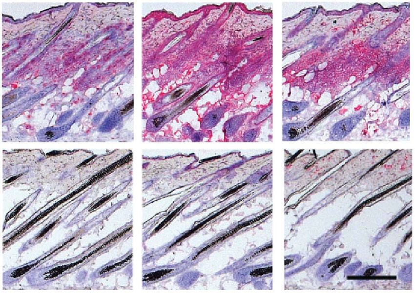
(e) Immunohistochemical staining of mouse skin biopsies shows treatment-related loss of expression of CD8 and MHC class I and II markers.
Scale bar, 100 µm. (f) Treatment of patient 3 with AA, who had hair loss involving >80% of his scalp at baseline, with ruxolitinib and hair regrowth after
12 weeks of oral treatment. (g) Clinical correlative studies of biopsies obtained before treatment (baseline) and after 12 weeks of treatment of patient 2,
including immunostains for CD4, CD8 and human leukocyte antigen (HLA) class I (A, B, C) and class II (DP, DQ, DR). Scale bar, 200 µm. (h,i) RNA
microarray analysis from treated patients 1 and 2 with AA (before treatment versus after treatment versus 3 normal subjects) presented as a heatmap (h)
and as a cumulative ALADIN index (i). KRT, hair follicle keratins.
nature medicine advance online publication 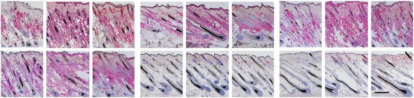
letters
To test whether inhibition of these signaling pathways would of a small number of patients with AA to treatment with the JAK1/2
be therapeutically effective in vivo, we systemically administered inhibitor ruxolitinib suggests future clinical evaluation of this com-
ruxolitinib (Fig. 3a–c) and tofacitinib (Fig. 3f–h) at the time of pound or other JAK protein tyrosine kinase inhibitors currently in
grafting and found that they prevented the development of AA and clinical development is warranted in AA29.
the expansion of CD8+NKG2D+ T cells in all grafted recipients.
The skin of mice treated with either drug showed no histological Methods
signs of inflammation (Fig. 3d,i). Global transcriptional analysis of Methods and any associated references are available in the online
whole-skin biopsies showed that both drugs also blocked the dermal version of the paper.
inflammatory signature, as measured by Alopecia Areata Disease
Activity Index (ALADIN, Fig. 3e,j), and Gene Expression Dynamic Accession codes. Microarray and RNA-seq data were deposited
Index (GEDI) analysis (Supplementary Fig. 10)23. in Gene Expression Omnibus with accession numbers GSE45657,
We next asked whether systemic tofacitinib treatment could reverse GSE45512, GSE45513, GSE45514, GSE45551 and GSE58573.
established disease by initiating therapy 7 weeks after grafting, a time
Note: Any Supplementary Information and Source Data files are available in the
point at which all mice had developed extensive AA. Systemic therapy online version of the paper.
resulted in substantial hair regrowth all over the body, reduced the
frequency of CD8+NKG2D+ T cells and reversed histological markers Acknowledgments
of disease (Supplementary Fig. 11), all of which persisted 2–3 months We thank the National Alopecia Areata Registry, as well as M. Duvic, V. Price,
M. Hordinsky and D. Norris, and the National Alopecia Areata Foundation.
after the cessation of treatment. We thank J. Sundberg, T. Behrens, D. Bickers, J. O’Shea, T. Waldmann, B. Jabri,
Next, to test a more clinically relevant route of delivery, we asked D. Raulet, L. Lanier, T. Spies, M. Hayden, R. Paus, P. Green, B. Lebwohl, D. Accili
whether topical administration of protein tyrosine kinase inhibitors and C. Jahoda for stimulating discussions. We are grateful for clinical support
could reverse established AA in mice with kinetics similar to those of from M. Furniss, C. Clark and G. Ulerio and expert technical assistance from
M. Zhang, E. Chang, H. Lam and J. Huang. This work was supported in part
systemic delivery. In established disease, we found that topical rux-
by US Public Health Service National Institutes of Health NIAMS grants
olitinib and topical tofacitinib were both highly effective in reversing R01AR056016 (to A.M.C.) and R21AR061881 (to A.M.C and R.C.), a Shared
disease in treated lesions (applied to back skin). A full coat of hair Instrumentation Grant for the LSR II Flow Cytometer (S10RR027050) to R.C.
emerged in the ruxolitinib- or tofacitinib-treated mice by 7 weeks of and the Columbia University Skin Disease Research Center (P30AR044535),
as well as the Locks of Love Foundation and the Alopecia Areata Initiative.
treatment (data not shown), and we observed complete hair regrowth
J.E.C. is supported by the T32GM082271 Medical Genetics Training Grant
within 12 weeks following topical therapy (Fig. 4a,b). Topical therapy (issued to A.M.C.). A.J., C.A.H., S.H. and A.d.J. are recipients of Career
was associated with a markedly reduced proportion of CD8+NKG2D+ Development Awards from the Dermatology Foundation, and A.J. is also
T cells in the treated skin and lymph node (Fig. 4c), normalization supported by the Louis V. Gerstner Jr Scholars Program.
of the ALADIN transcriptional signature (Fig. 4d), reversal of histo-
AUTHOR CONTRIBUTIONS
logical markers of disease (Fig. 4e) and correction of the GEDI in all L.X., Z.D. and A.J. were responsible in large part for performing the studies
treated mice (Supplementary Fig. 12). Notably, untreated areas on reported herein and participated in the design, execution and interpretation
the abdomen remained alopecic (Fig. 4a and Supplementary Fig. 13), of the data. C.A.H. was responsible for establishing the C3H/HeJ graft model.
demonstrating that topical therapy acted locally and that the observed A.d.J., S.H., G.M.D., L.R. and P.S. were involved in additional molecular and cell
biological experiments. W.G. performed immunofluorescence and morphometric
therapeutic effects were not the result of systemic absorption. These studies. L.P. and J.E.C. performed biostatistical analysis of all data sets. J.M.-W. was
effects were visible as early as 2–4 weeks after the onset of treatment instrumental in human sample acquisition and analysis. A.M.C. and R.C. were
(Supplementary Fig. 14) and persisted 2–3 months after the cessation responsible for conception, design, oversight, execution and interpretation of data
of treatment (Fig. 4a). for this study. All authors contributed to drafts, writing, figure preparation and
editing of the final manuscript.
To test the efficacy of JAK inhibitors in human subjects with AA,
we treated three patients with moderate to severe disease orally with COMPETING FINANCIAL INTERESTS
ruxolitinib, 20 mg twice daily. Ruxolitinib is currently FDA-approved The authors declare competing financial interests: details are available in the online
for the treatment of myelofibrosis24,25, a disease driven by wild-type version of the paper.
and mutant JAK2 signaling downstream of hematopoietic growth fac- Reprints and permissions information is available online at http://www.nature.com/
tor receptors. In addition, small clinical studies using topical ruxolitinib reprints/index.html.
in psoriasis26 have demonstrated anti-inflammatory activity that may
be due to interruption of the IL-17 signaling axis. All three ruxolitinib- 1. Gilhar, A., Etzioni, A. & Paus, R. Alopecia areata. N. Engl. J. Med. 366, 1515–1525
treated patients exhibited near-complete hair regrowth within 3 to (2012).
5 months of oral treatment (Fig. 4f and Supplementary Figs. 15 and 2. Petukhova, L. et al. Genome-wide association study in alopecia areata implicates
both innate and adaptive immunity. Nature 466, 113–117 (2010).
16). Comparison of biopsies obtained at baseline and after 12 weeks 3. Gilhar, A., Ullmann, Y., Berkutzki, T., Assy, B. & Kalish, R.S. Autoimmune hair loss
of treatment demonstrated reduced perifollicular T cell infiltration, (alopecia areata) transferred by T lymphocytes to human scalp explants on SCID
mice. J. Clin. Invest. 101, 62–67 (1998).
reduced follicular expression of human leukocyte antigen class I and 4. McElwee, K.J. et al. Transfer of CD8+ cells induces localized hair loss whereas CD4+/
class II expression (Fig. 4g) and normalization of the ALADIN inflam- CD25− cells promote systemic alopecia areata and CD4+/CD25+cells blockade disease
matory and hair keratin signatures following treatment (Fig. 4h,i). onset in the C3H/HeJ mouse model. J. Invest. Dermatol. 124, 947–957 (2005).
5. Ito, T. et al. Maintenance of hair follicle immune privilege is linked to prevention
Taken together, our data suggest CD8+NKG2D+ T cells promote of NK cell attack. J. Invest. Dermatol. 128, 1196–1206 (2008).
AA pathogenesis, acting as cytolytic effectors responsible for autoim- 6. Sundberg, J.P., Cordy, W.R. & King, L.E. Jr. Alopecia areata in aging C3H/HeJ mice.
J. Invest. Dermatol. 102, 847–856 (1994).
mune attack of the hair follicle (Supplementary Fig. 17). We postulate
7. McElwee, K.J., Boggess, D., King, L.E. Jr. & Sundberg, J.P. Experimental induction
that IFN-γ produced by CD8 T cells leads to the collapse of immune of alopecia areata-like hair loss in C3H/HeJ mice using full-thickness skin grafts.
privilege in the hair follicle27, inducing further production of IL-15 J. Invest. Dermatol. 111, 797–803 (1998).
8. Bertolini, M. et al. Abnormal interactions between perifollicular mast cells and CD8+
(ref. 28) (Supplementary Figs. 5 and 6) and a feed-forward loop T cells may contribute to the pathogenesis of alopecia areata. PLoS ONE 9, e94260
that promotes type I cellular autoimmunity. The clinical response (2014).
advance online publication nature medicine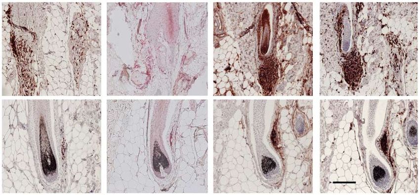
letters

9. Best, J.A. et al. Transcriptional insights into the CD8+ T cell response to infection    19. Freyschmidt-Paul, P. et al. Reduced expression of interleukin-2 decreases the
    and memory T cell formation. Nat. Immunol. 14, 404–412 (2013).                              frequency of alopecia areata onset in C3H/HeJ mice. J. Invest. Dermatol. 125,
10. Bezman, N.A. et al. Molecular definition of the identity and activation of natural          945–951 (2005).
    killer cells. Nat. Immunol. 13, 1000–1009 (2012).                                       20. O’Shea, J.J., Kontzias, A., Yamaoka, K., Tanaka, Y. & Laurence, A. Janus kinase
11. Brajac, I., Gruber, F., Petrovecki, M. & Malnar-Dragojevic, D. Interleukin-2 receptor       inhibitors in autoimmune diseases. Ann. Rheum. Dis. 72 (suppl. 2), ii111–ii115
    α-chain expression in patients with alopecia areata. Acta Dermatovenerol. Croat.            (2013).
    ADC 12, 154–156 (2004).                                                                 21. Quintás-Cardama, A. et al. Preclinical characterization of the selective JAK1/2
12. Fehniger, T.A. & Caligiuri, M.A. Interleukin 15: biology and relevance to human             inhibitor INCB018424: therapeutic implications for the treatment of myeloproliferative
    disease. Blood 97, 14–32 (2001).                                                            neoplasms. Blood 115, 3109–3117 (2010).
13. Ye, W., Young, J.D. & Liu, C.C. Interleukin-15 induces the expression of mRNAs          22. Ghoreschi, K. et al. Modulation of innate and adaptive immune responses by
    of cytolytic mediators and augments cytotoxic activities in primary murine                  tofacitinib (CP-690,550). J. Immunol. 186, 4234–4243 (2011).
    lymphocytes. Cell. Immunol. 174, 54–62 (1996).                                          23. Eichler, G.S., Huang, S. & Ingber, D.E. Gene Expression Dynamics Inspector (GEDI):
14. Meresse, B. et al. Reprogramming of CTLs into natural killer-like cells in celiac           for integrative analysis of expression profiles. Bioinformatics 19, 2321–2322 (2003).
    disease. J. Exp. Med. 203, 1343–1355 (2006).                                            24. Verstovsek, S. et al. Safety and efficacy of INCB018424, a JAK1 and JAK2 inhibitor,
15. Saikali, P., Antel, J.P., Pittet, C.L., Newcombe, J. & Arbour, N. Contribution of           in myelofibrosis. N. Engl. J. Med. 363, 1117–1127 (2010).
    astrocyte-derived IL-15 to CD8 T cell effector functions in multiple sclerosis.         25. Harrison, C. et al. JAK inhibition with ruxolitinib versus best available therapy for
    J. Immunol. 185, 5693–5703 (2010).                                                          myelofibrosis. N. Engl. J. Med. 366, 787–798 (2012).
16. Meresse, B. et al. Coordinated induction by IL15 of a TCR-independent NKG2D             26. Punwani, N. et al. Preliminary clinical activity of a topical JAK1/2 inhibitor in the
    signaling pathway converts CTL into lymphokine-activated killer cells in celiac             treatment of psoriasis. J. Am. Acad. Dermatol. 67, 658–664 (2012).
    disease. Immunity 21, 357–366 (2004).                                                   27. Paus, R., Nickoloff, B.J. & Ito, T.A. A ‘hairy’ privilege. Trends Immunol. 26, 32–40
17. Freyschmidt-Paul, P. et al. Interferon-γ-deficient mice are resistant to the                (2005).
    development of alopecia areata. Br. J. Dermatol. 155, 515–521 (2006).                   28. Waldmann, T.A. The biology of interleukin-2 and interleukin-15: implications for
18. Gilhar, A., Kam, Y., Assy, B. & Kalish, R.S. Alopecia areata induced in C3H/HeJ             cancer therapy and vaccine design. Nat. Rev. Immunol. 6, 595–601 (2006).
    mice by interferon-γ: evidence for loss of immune privilege. J. Invest. Dermatol.       29. Dolgin, E. Companies hope for kinase inhibitor JAKpot. Nat. Rev. Drug Discov. 10,
    124, 288–289 (2005).                                                                        717–718 (2011).

nature medicine advance online publication 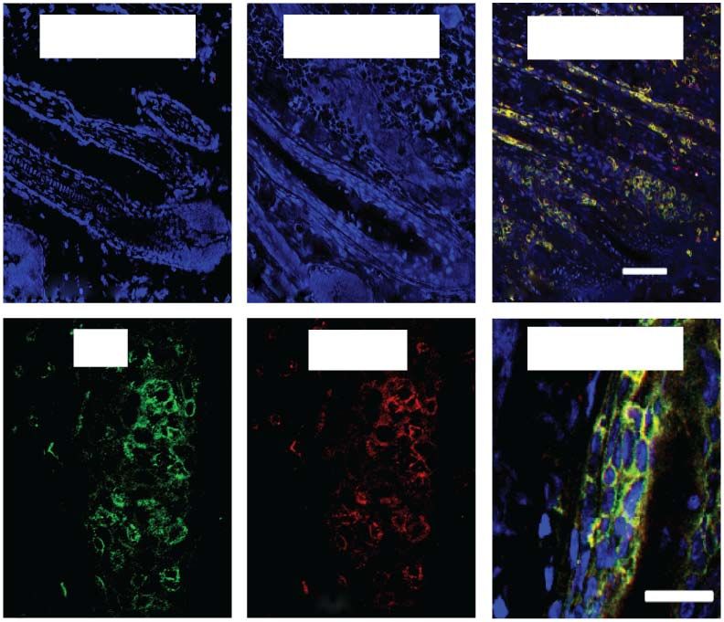
ONLINE METHODS surface of each mouse at interim time points, and skin samples were either
Mice. 7- to 10-week-old female C57BL/6 and C3H/HeJ mice (Jackson snap frozen in liquid nitrogen for RNA extraction or snap frozen in OCT for
Laboratories, Bar Harbor, ME) were used and maintained under specific immunostaining. Hair status was examined twice weekly.
pathogen–free conditions. Experiments were performed in compliance with
institutional guidelines as approved by the Institutional Animal Care and Use Human clinical studies. We initiated a single center, proof-of-concept clini-
Committee of Columbia University Medical Center. cal trial in the Clinical Trials Unit in the Department of Dermatology at the
Columbia University Medical Center entitled “An Open-Label Pilot Study to
Transfer of alopecia areata using grafted C3H/HeJ skin. Normal-haired Evaluate the Efficacy of ruxolitinib in Moderate to Severe Alopecia Areata,”
C3H/HeJ mice were grafted at 8 weeks of age (during the second telogen) with ClinicalTrials.gov identifier NCT01950780. The protocol for this interven-
skin from a C3H/HeJ mouse that developed AA spontaneously, as described tion trial was reviewed and approved by the Institutional Review Board at
previously7. In brief, mice spontaneously affected with AA were euthanized, Columbia University and conducted under the Declaration of Helsinki prin-
and full thickness skin grafts of approximately 2 cm in diameter were removed ciples. Informed consent was received before inclusion in the study. Eligibility
and grafted to normal-haired C3H/HeJ mice. All experiments included 10 criteria included >30% hair loss for at least 3 months in duration with no
grafted mice (5 treated and 5 untreated) and 3 sham grafted mice (mice grafted evidence for hair regrowth at the time of enrollment. The first three treated
with autologous grafts). Hair loss typically began at around 4–6 weeks after patients are described here. The ruxolitinib dose was 20 mg orally twice daily
grafting. No sham grafted mouse developed hair loss. for 3–6 months. Skin punch biopsies were obtained at baseline and after
12 weeks of treatment. Consent for photography was obtained for the patients
Flow cytometric analysis of skin and cutaneous lymph nodes. To make a shown in Figure 4 and in the Supplementary Figures 15 and 16.
single-cell suspension of mouse skin, fat was removed from the overlying
skin in cold PBS and then incubated in collagenase type I (2 mg/ml in PBS) Immunohistochemistry and immunofluorescence. 8 µM acetone-fixed
at 32 °C for 75 min. After digestion, the skin was minced in RPMI/10% FBS, frozen mouse skin or human skin sections were air-dried and stained
filtered through a 70-µM cell strainer and centrifuged at 1100g for 5 min. overnight with anti-mouse or anti-human antibodies (see Supplementary
The pellet was resuspended in RPMI/10% FBS, filtered through a 40-µM cell Methods) at 4 °C in a moist chamber. Human hair follicles were microdis-
strainer and spun at 400g for 5 min. The pellet was resuspended in FACS buffer sected and embedded in OCT compound before sectioning and staining
(PBS/5% BSA), DAPI to gate on live cells and staining antibodies (listed in (see Supplementary Methods).
Supplementary Methods). Cutaneous lymph nodes were pooled, minced in
RPMI, filtered through a 40-µM cell strainer, centrifuged at 400g for 5 min, Primary dermal sheath and lymphokine-activated killer (LAK) cell
stained and analyzed on a BD LSR II flow cytometer. culture. Dermal sheath (DS) cells were isolated from microdissected mouse
vibrissa follicles and cultured in 20% FBS DMEM with 5 ng/ml murine FGF
Transfer of T cell populations into recipient C3H/HeJ mice. For posi- (Pepro Tech). LAK cells were generated from bulk splenocytes plated at
tive selection of T cell populations, lymph node cells were obtained from 5 4 × 10⁶ in 6-well plates with 50 ng/ml murine IL-15 (Pepro Tech), 50 nM JAK3i
C3H/HeJ alopecic mice, stained with anti-CD4, anti-CD8 and anti-NKG2D (tofacitinib) or 50 ng/ml murine IL-15 plus 50 nM JAK3i and incubated at
antibodies) and then sorted (BD Influx) to obtain two fractions: CD8+NKG2D+ 37 °C in a 5% CO2 incubator for 96 h.
T cells and CD8+NKG2D− T cells. Antibody dilutions are given in the
Supplementary Methods. Three to five 7-week-old C3H/HeJ mice per group In vitro cytotoxicity assays. Determination of specific killing of target cells
were injected subcutaneously with two million sorted cells of each population. was performed using CFSE-labeled DS cells as targets mixed with different
For negatively selected populations, NKG2D+ cells were depleted by incubat- ratios of effector cells incubated for 5 h at 37 °C 5% CO2 with or without
ing total lymph node cells from 3 alopecic C3H/HeJ mice with biotinylated neutralizing rat anti-mouse NKG2D antibody (20 ug/ml) (Biolegend, CX5).
anti-NKG2D (CX5) and then with streptavidin-conjugated beads (Miltenyi) Specific lysis of DS cells was determined flow cytometrically by measuring cell
before removal on a Miltenyi magnetic column. Five million cells (either death of CFSE + DS cells using Annexin V/7-AAD.
CD8/NKG2D-depleted or total lymph node cells) were suspended in 100 ul
PBS and transferred into each of 5 mice by subcutaneous injection. Gene expression sample preparation in human and mouse skin and T cells.
Total RNA was isolated using the miRNeasy Mini Kit (Qiagen Inc., Valencia,
Prevention and treatment studies in mice. For prevention studies, mice were CA, USA) with on-column DNA digestion using the RNase-free DNase
treated beginning on the day of grafting (n = 5–10 mice per group). For anti- set (Qiagen, Inc.). For RNA-seq analysis CD3+CD8+CD44+NKG2D+ and
IFN-γ experiments, i.p. injections of hamster isotype control IgG or hamster CD3+CD8+CD44+NKG2D− cells were flow-sorted from lymph nodes
polyclonal anti-IFN-γ IgG (BioXCell) 300 µg in 100µl PBS were administered of alopecic C3H/HeJ mice. RNA was extracted as above and prepared for
ten times weekly for 12 weeks. For anti–IL-2 experiments, i.p. injections of RNA-seq using the TruSeq RNA Sample Prep Kit v2. Samples were sequenced
control rat isotype control IgG or simultaneous administration of two anti- on the HiSeq 2000 sequencer (Illumina, San Diego, CA) for 50 cycles.
IL-2 rat monoclonal antibodies (BioXCell) (250 µg clone S4B6 and 250 µg RNA-seq files were demultiplexed by the Rockefeller University Genomics
JES6-1 together in 100 µl PBS) were administered three times weekly for Core Facility.
12 weeks. For anti–IL-15-Rβ experiments, i.p. injections of rat isotype control For global transcriptional profiling in mouse skin, total extracted RNA was
IgG or anti-IL-15Rβ antibody (Biolegend, clone TM-β1) (200 µg in 100 µl processed using the 3′ IVT Express Kit from Affymetrix. Resulting biotinylated
PBS) were administered two times weekly for 12 weeks. For JAK1/2i experi- cDNA samples were hybridized to the Mouse Genome 430 2.0 gene chips and
ments, mice were administered vehicle (0.5% methylcellulose; Sigma-Aldrich) subsequently washed, stained with streptavidin-phycoerythrin and scanned on
or vehicle-containing 50 mg/kg of JAK1/2i ruxolitinib (ChemieTek) daily by an HP GeneArray Scanner (Hewlett-Packard Company, Palo Alto, CA). For gene
oral gavage for 12 weeks. For JAK3i experiments, mice were implanted sub- expression studies, mice grafted with autologous healthy skin were included as
cutaneously with Alzet osmotic mini-pumps (pumps, model 2004, Durect sham-operated controls.
Corporation) on the back of each mouse to deliver vehicle (poly(ethylene For human AA samples, perilesional punch biopsies from 5 patients with
glycol) (PEG)300) or vehicle containing the JAK3i tofacitinib (Abmole) at patchy alopecia areata who were not undergoing local or systemic treat-
15 mg/kg/day for 12 weeks. ments were collected and compared to scalp biopsies from 5 unrelated unaf-
For topical treatment studies, grafted mice with long-standing AA (more fected individuals. All procedures were performed under Institutional Review
than 8 weeks) were treated once daily for 12 weeks to affected skin on the dorsal Board–approved protocols at Columbia University and conducted under the
back with vehicle (10% DMSO in Aquaphor) or vehicle containing JAK inhibi- Declaration of Helsinki principles. Informed consent was received before inclu-
tors, initially dissolved in DMSO and then diluted 1:10 in Aquaphor, to achieve sion in the study. Extracted total RNA was reverse transcribed and amplified
0.5% JAKi ointment). Full-thickness skin biopsies were excised from the dorsal using the Ovation RNA Amplification V2 kit (NuGEN Technologies, Inc.,
nature medicine doi:10.1038/nm.3645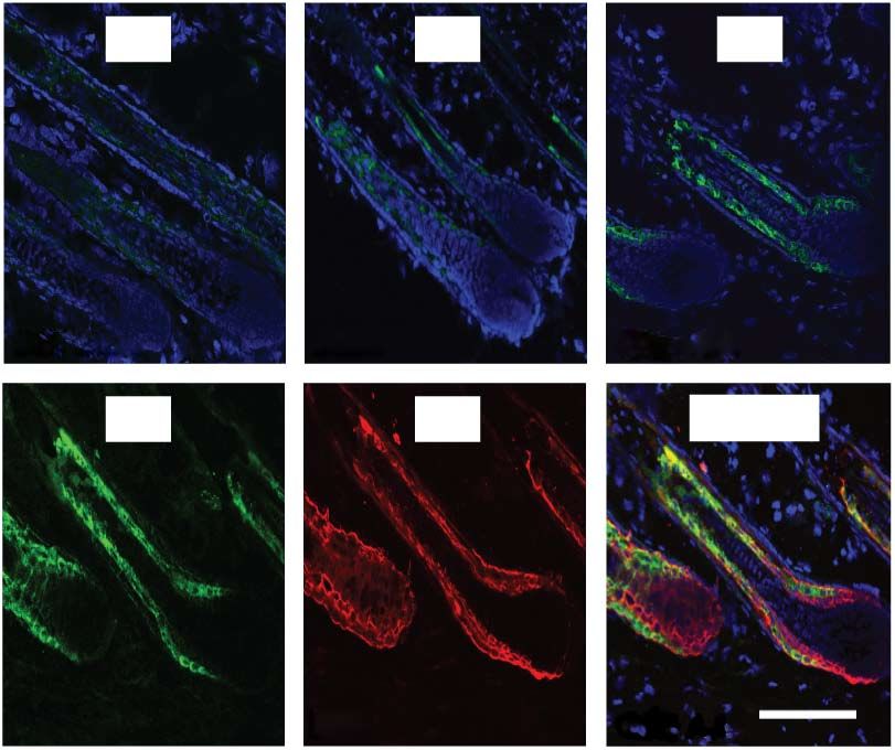
San Carlos, CA). Amplified cDNA was biotinylated with the Encore Biotin Module   Statistical analyses. Statistical methods for each figure are given in the
(NuGEN Technologies) and then hybridized to the U133 Plus 2.0 gene chips.        Supplementary Methods. No statistical method was used to predetermine
RT-PCR confirmations done as described in the Supplementary Methods.             sample size. The investigators were not blinded to allocation during experi-
Primer sequences are given in Supplementary Tables 4 and 5.                      ments and outcome assessment. The experiments were not randomized.

doi:10.1038/nm.3645                                                                                                                     nature medicine
You can also read