CXCR3 plays a critical role for host protection against Salmonellosis
←
→
Page content transcription
If your browser does not render page correctly, please read the page content below
www.nature.com/scientificreports

                        OPEN          CXCR3 plays a critical role for host
                                      protection against Salmonellosis
                                      Belal Chami1,2, Amanda Yeung2, Michael Buckland                   2
                                                                                                            , Hongjun Liu2, Genevieve M. Fong2,
                                      Kun Tao1 & Shisan Bao1,2
Received: 16 February 2017
                                      CXCR3 and its ligands are heavily associated with inflammation and have been implicated in numerous
Accepted: 25 May 2017                 inflammatory diseases. CXCR3 plays an important role in recruiting pro-inflammatory cells, specifically
Published: xx xx xxxx                 neutrophils, in a model of sterile colitis whereby CXCR3−/− mice showed an attenuated course of colitis
                                      with markedly reduced host-tissue damage in the inflamed caecum. The role of CXCR3 during infectious
                                      colitis, however, is unclear and therefore in this study, we investigated the role of CXCR3 in the
                                      regulation of the immune response during acute and chronic gastrointestinal infection, using a murine
                                      model of Salmonella enterica serovar Enteritidis. During acute infection with Salmonella, we observed
                                      significantly increased Salmonella loading in the caecum and dissemination to the spleen and liver in
                                      CXCR3−/− mice, but not in Wt counterparts. During chronic infection, increased pathological features
                                      of inflammation were noted in the spleen and liver, with significantly increased levels of apoptosis in
                                      the liver of CXCR3−/− mice, when compared to Wt counterparts. In addition, compromised intestinal
                                      IgA levels, CD4+ helper T cells and neutrophil recruitment were observed in CXCR3−/− challenged with
                                      Salmonella, when compared to Wt counterparts. Our data suggests that CXCR3 is a key molecule in host
                                      intestinal immunity against Salmonellosis via regulating neutrophils chemotaxis.

                                      Chemokines influence the recruitment of leukocytes to sites of inflammation and are implicated in the host pro-
                                      tection against numerous infectious agents. Infectious inflammation is usually self-resolving, whereby the reduced
                                      infectious stimuli regulates the chemokine response, allowing the appropriate cessation of pro-inflammatory cell
                                      influx into inflamed tissue. During sterile inflammation, however, the prolonged stimuli results in the continuous
                                      dysregulation of chemokines and subsequent leukocyte recruitment to the inflamed site, resulting in an accumu-
                                      lation of pro-inflammatory leukocytes, thereby exacerbating inflammation and associated host-tissue damage1, 2.
                                      As a result, chemokine blockage has become an attractive therapeutical approach for many inflammatory diseases
                                      including rheumatoid arthritis3, multiple sclerosis4 and inflammatory bowel disease (IBD)5.
                                          CXCR3 is a chemokine receptor implicated in pathogenesis in several chronic inflammatory disorders, e.g.
                                      rheumatoid arthritis6, multiple sclerosis, and inflammatory bowel disease7–9. CXCR3 is constitutively expressed
                                      on pro-inflammatory cells, including neutrophils10, macrophages11, 12, T cells13, B cells7, and natural killer (NK)
                                      cells14, while its ligands, CXCL9, CXCL10, and CXCL11 are induced by IFN-γ15. Our previous reports demon-
                                      strate the pro-inflammatory effects of CXCR3 in DSS-induced colitis – a model of IBD16, as well as, in hepatitis B
                                      viral chronic hepatitis17 or in drug induced liver failure18, suggesting an important inflammatory role of CXCR3
                                      in gastrointestinal track. Many investigators have also highlighted the deleterious role of CXCR3 in many inflam-
                                      matory conditions2, 5, 19, 20, implicating its potential as a novel therapeutic target in inflammation-mediated pathol-
                                      ogies. However, little is known of its role in host intestinal immunity, particularly against common gram-negative
                                      infectious agents.
                                          Salmonella species are facultative, gram-negative intra-cellular bacteria harbouring over 2500 serovars.
                                      Salmonella is the leading cause of bacterial gastroenteritis with morbidity ~1.3 billion cases worldwide21.
                                      Salmonella invades the gastrointestinal epithelium by transiting through M cells overlying Peyer’s patches and
                                      drain to the mesenteric lymph nodes (MLN) via the lymphatic pathway during development of salmonellosis.
                                      Dissemination of salmonella into the bloodstream occurs via the thoracic duct where Salmonella are mostly
                                      removed by macrophages of the reticuloendothelial system (RES) of mainly the spleen, liver, and bone mar-
                                      row22, 23. Immunocompromised individuals with specific defects or blockades of specific cytokines (particularly
                                      IL-12/IL-23/IL-17 and TNF) are highly susceptible to developing primary bacteraemic disease and have markedly

                                      1
                                       Department of Pathology, Tongren Hospital, Shanghai Jiaotong University, Shanghai, China. 2Discipline
                                      of Pathology, School of Medical Sciences and Bosch Institute, The University of Sydney, Sydney, Australia.
                                      Correspondence and requests for materials should be addressed to K.T. (email: taokun@shtrhospital.com) or S.B.
                                      (email: bob.bao@sydney.edu.au)

        SCIenTIfIC Reports | 7: 10181 | DOI:10.1038/s41598-017-09150-z                                                                                     1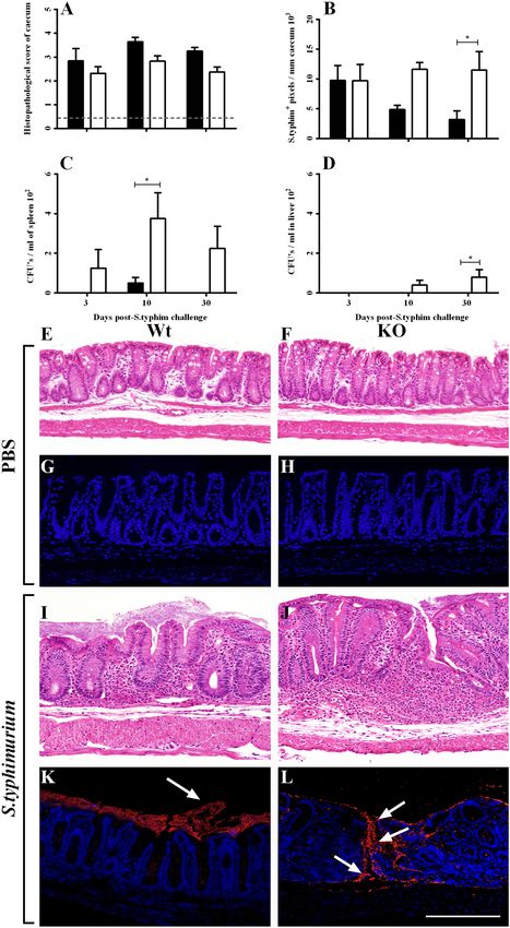
www.nature.com/scientificreports/
Figure 1. Clinical and post-mortem observations in Wt and KO mice at day 3, 10 and 30 post-S. typhimurium
challenge. Faecal consistency, hematochesia and rectal bleeding were scored individual and tallied to represent
the ratio of (A) caecum and (B) spleen weight (n = 4) against the respective body weight of mock-treated mice
at same time point. Dashed line represents baseline values from combined mock-treated Wt and KO mice.
*p < 0.05, and ***p < 0.001. W + S and K + S group values were significantly higher (p < 0.05) than baseline at
day 3, 10 & 30 in panel A). W + S and K + S group values were significantly higher (p < 0.05) than baseline only
at day 30 in panel B).
increased mortality rates, when infected with Salmonella24 highlighting the importance of specific inflammatory
mediators in preventing Salmonella dissemination.
Herein, we explored the role of CXCR3 in the clearance of live S. typhimurium bacteria and in the recruitment
of leukocytes during both acute and chronic salmonellosis.
Results
Clinical examination. There was no mortality observed in wild-type (Wt) or CXCR3 knockout (KO) mice
at days 3, 10, or 30 post-Aro−/− S. typhimurium challenge, consistent with our previous observations in this
model25. At day 3, 10 and 30, S. typhimurium-challenged Wt & KO mice showed significantly increased cae-
cum weight to total body weight ratios when compared to their mock-treated counterparts (Fig. 1A), suggesting
infectious colitis in the caecum. Interestingly S. typhimurium-challenged KO mice revealed significantly heavier
caecum at day 10, compared to its S. typhimurium-challenged Wt counterpart (p < 0.05), suggesting an increased
S. typhimurium bacterial load in the caecum of S. typhimurium-challenged KO. S. typhimurium disseminates to
the spleen and liver if intestinal mucosal immunity is compromised and/or virulence of the bacteria is strong,
where the intestinal infection cannot be contained22, 23. Therefore, spleen weight (as a function of body weight)
was measurement to assess whether S. typhimurium potentially migrated and caused local inflammation to the
tissue. Spleen weight increased significantly on day 30 post-S. typhimurium challenge in Wt and KO mice, com-
pared to PBS (mock)-challenged group (Fig. 1B). Furthermore, S. typhimurium-challenged KO mice had sig-
nificantly heavier spleens when compared to their Wt counterparts at day 30, suggesting that a more significant
degree of bacterial dissemination from the caecum occurred in KO mice. No significant difference was noted
between mock-treated Wt and KO mice at any time point.
Histopathological damage and bacterial load. It was established in our pilot study that salmonello-
sis did not significantly affect the colon as this region showed little to no pathology following S. typhimurium
challenge, even after 30 days26. However, large areas of inflammatory destruction were detected in the cae-
cum. Histopathological analysis showed that there was significant pathology at day 3, 10, and 30 in both
S. typhimurium-challenged Wt and KO mice when compared to their respective counterparts (Fig. 2A,I and J),
but no signs of pathology in the mock-treated groups (Fig. 2A,E and F). S. typhimurium-challenged KO mice
showed slightly decreased histopathology at day 3, day 10, and day 30, although this was not significant.
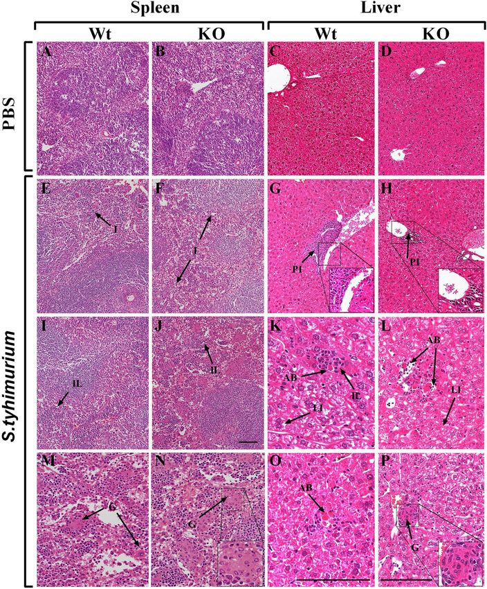
Quantification of S. typhimurium in the caecum established the degree of bacterial load in challenged mice.
As expected, no detectable salmonella was observed in mock challenged Wt or KO mice (Fig. 2G and H).
Furthermore, no differences in S. typhimurium+ intensity were noted on day 3 post-S. typhimurium challenge
in Wt and KO mice (Fig. 2B). However, day 10 and day 30 post-S. typhimurium challenge showed increased S.
typhimurium labelling in the caecum of KO mice, compared to their Wt counterparts (Fig. 2B). This suggests a
higher degree of S. typhimurium loading in the caecum of KO mice. In addition, most S. typhimurium was local-
ised to the caecum lumen in challenged Wt mice, whereas we observed increased transmural S. typhimurium
labelling in KO mice only, clearly signifying a breakdown in barrier function (Fig. 2K and L). Organs suscepti-
ble to bacterial dissemination of S. typhimurium, namely, the spleen and liver, were homogenized and cultured
for S. typhimurium. Cultured spleens homogenates of S. typhimurium-challenged KO mice exhibited increased
CFUs at day 3, day 10 and day 30 (Fig. 2C), while CFUs in the livers of S. typhimurium-challenged KO mice
were only found at day 10 and 30 (Fig. 2D). No S. typhimurium was detected in the spleen and liver samples of
S. typhimurium-challenged Wt mice, with the exception of the spleen at day 10 (Fig. 2C).
Next, we assessed the extent of spleen and liver inflammation and determined the histopathology of the
respective organs (Fig. 3). Mock-treated mice showed no histological evidence of pathology in the spleen or liver
of both groups (Fig. 3A–D). However S. typhimurium-challenged Wt and KO mice had regions of inflammation
SCIenTIfIC Reports | 7: 10181 | DOI:10.1038/s41598-017-09150-z 2
www.nature.com/scientificreports/

                              Figure 2. Histopathology and S. typhimurium loading in Wt and KO mice at day 3, 10 and 30 post-
                              S. typhimurium challenge. (A) Histopathological scoring (n = 8) and (B) quantification of S. typhimurium+
                              immunofluorescence labelling (n = 4) in the caecum following S. typhimuirum challenge. The entire length
                              of the caecum was imaged at 20× magnification and scored or quantified per field of view and later averaged.
                              Enumeration of S. typhimurium CFU’s cultured from (C) spleen and (D) liver homogenates on BBLchrom
                              agar plates (n = 4). Representative H&E and S. typhimurium+ immunofluorescence images at day 30 in the
                              caecum of Wt and KO mice treated with (E–H) PBS and (I–L) S. typhimurium, respectively. (L) Representative
                              image of S. typhimurium breaching the epithelium and lamina propria in S. typhimuirum-challenged KO mice.
                              Images were taken at 20× magnification and the scale bar represents 250 µm. Arrows indicate S. typhimurium+
                              labelling. Dashed line represents baseline values from combined mock-treated Wt and KO mice. *p < 0.05 and
                              ***p < 0.001. W + S and K + S group values were significantly higher (p < 0.05) than baseline at day 3, 10 & 30
                              in Fig. 2A.

SCIenTIfIC Reports | 7: 10181 | DOI:10.1038/s41598-017-09150-z                                                                                  3
www.nature.com/scientificreports/
Figure 3. Histopathology of the spleen and liver in mock- and S. typhimurium-challenged mice at day 30.
(A–D) H&E images of the spleen and liver in mock-treated Wt and KO mice, respectively. Representative
pathological features of inflammation (I) in the spleen of (E) Wt and (F) KO mice and portal inflammation
(PI) in the liver of (G) Wt and (H) KO mice, challenged with S. typhimurium. Lobular inflammation (LI) was
also noted in the spleen of S. typhimurium-challenged (I) Wt and (J) KO mice, while acidophil bodies (AB),
infiltrating leukocytes (IL) and LI were found in the S. typhimurium-challenged liver of (K) Wt and (L) KO
mice. Extensive granulomas (G) were noted in the S. typhimurium-challenged spleens of (M) Wt and more
particularly, (N) KO mice. No Gs were found in the liver of S. typhimuirum-challenged (O) Wt mice, though
evidence of Gs were found frequently in (P) KO mice. Images A–J were captured at ×20 magnification, L–N &
P at ×40 magnification and K & O at ×63 magnification. Scale bar represents 150 µm. All inserts were digitally
zoomed 200%.
consistent with bacterial dissemination. In the spleen, S. typhimurium-challenged KO mice displayed increased
regions of inflammation (Fig. 3F), infiltrating lymphocytes (Fig. 3F), and granulomas (Fig. 3N,E,I and M) when
compared to Wt counterparts (Fig. 3F,J and N). Histopathological analysis of the liver revealed similar find-
ings to that of the spleen. Granulomas were observed in the livers of S. typhimurium-challenged KO mice (P)
(Fig. 3G,K and O), which were not observed in the livers of S. typhimurium-challenged Wt mice (Fig. 3H,L and P).
Although both S. typhimurium-challenged Wt and KO mice showed lobular inflammation in the liver (Fig. 3K
and L, respectively), the extent of lobular inflammation was observed to be higher in the KO group. In addi-
tion, more instances of portal inflammation (Fig. 3H) and leukocyte infiltration were observed in the liver of
S. typhimurium-challenged KO mice than in livers of Wt counterparts. Acidophil bodies were also present in
S. typhimurium-challenged Wt and KO mice (Fig. 3K and L, respectively).
SCIenTIfIC Reports | 7: 10181 | DOI:10.1038/s41598-017-09150-z 4www.nature.com/scientificreports/
Figure 4. TUNEL assay in the spleen and liver of Wt and KO mice post-S. typhimurium challenge.
Quantification of positively stained fragmented nuclei in the (A) spleen and (B) liver of Wt and KO mice at day
30. An average of 10 random microscopic fields at ×20 magnification were captured in the spleen and liver and
were each subjected to software-based quantification and represented as mean ± SD. Representative images
(×20 magnification) in the spleen in mock-treated (C) Wt and (D) KO mice and S. typhimurium-challenged
(E) Wt and (F) KO mice. Representative images (×20 magnification) in the liver in mock-treated (G) Wt and
(H) KO mice and S. typhimurium-challenged (I) Wt and (J) KO mice. Scale bar represents 250 µm. Data are
presented as mean ± SD over 20 microscopic fields (×20 magnification) of each mouse.
Salmonella is a known to hijack macrophages and disseminate to organs in this manner before inducing
pyroptosis- A caspase-1 dependent cell death usually induced by intracellular bacteria27. We assessed the extent
the pyroptosis in the spleen and liver using a TUNEL assay in mice 30 days post-S. typhimurium-challenge. No
differences in TUNEL staining were noted in the spleens of all groups (Fig. 4A and C–F). However, there was a
significant increase in TUNEL staining in the liver of both S. typhimurium-challenged groups, when compared
to mock-challenged counterparts. (Fig. 4B,G and H). Furthermore, TUNEL staining was significantly increased
(p < 0.05) in the liver of S. typhimurium-challenged KO mice, when compared to the infected Wt mice (Fig. 4B,I
and J) – a finding consistent with increased S. typhimurium dissemination to the spleen. TUNEL staining in the
spleen and liver of mock-challenged Wt and KO mice was consistent and was sparsely observed, especially in the
liver (Fig. 4C,D, G and H, respectively).
Cellular infiltrates. Reduced migration of CXCR3+ B cells can exacerbate salmonellosis, as indicated by a
report stating that IgA antibodies against S. typhimurium O-antigen (a component of lipopolysaccharide) was
highly effective in preventing Salmonella infection28, 29. In light of this, plasma cells of several immunoglob-
ulins were quantified using immunohistochemistry. IgA+ plasma cells in the caecum significantly increased
following S. typhimurium-challenge in both Wt and KO mice relative to mock-treated controls, with the excep-
tion of Wt mice at day 3 post-infection (Fig. 5A). However, IgA+ plasma cells were significantly reduced in
S. typhimurium-challenged KO mice at day 30 compared to the Wt mice. Interestingly, IgG+ plasma cells in the
caecum showed a significant increase at day 30 in S. typhimurium-challenged KO mice (Fig. 5B). IgM+ plasma
SCIenTIfIC Reports | 7: 10181 | DOI:10.1038/s41598-017-09150-z 5www.nature.com/scientificreports/
Figure 5. Leukocyte influx to the caecum of S. typhimurium-challenged Wt and KO mice at day 3, 10 and 30.
Software-based enumeration of IHC labelled (A) IgA+, (B) IgG+ and (C) IgM+ plasma cells in the caecum.
Macrophage, neutrophil and T cell influx into the caecum was also software-enumerated by IHC labelling of
markers (D) F4/80+, (E) NIMP R14+ and (F) CD4+, respectively. Dashed line represents baseline values from
combined mock-treated Wt and KO mice. Data are presented as mean ± SD (n = 4) over 20 microscopic fields
(×20 magnification) of each mouse. *p < 0.05, **p < 0.01 and ***p < 0.001. W + S and K + S group values were
significantly higher (p < 0.05) than baseline at day 3, 10 & 30 in Fig. 2A, except for W + S day 3. δ Significantly
different (p < 0.05) than baseline levels.
cells in the caecum appeared to peak at day 10 in both the S. typhimurium-challenged groups and, as expected,
decreased towards baseline levels by day 30 as the immune response matured (Fig. 5C).
Macrophages, also express CXCR3, play an important role in controlling salmonellosis. Quantification of
F4/80+ macrophages in the caecum showed no significant differences in S. typhimurium-challenged Wt and KO
mice, despite the levels of F4/80+ macrophages being significantly increased in both groups when compared
to their mock-treated counterparts (Fig. 5D), alluding to compensatory mechanisms for macrophage chemot-
axis outside the CXCR3-axis. CXCR3 is expressed abundantly on neutrophils and has been previously shown to
influence recruitment of neutrophils during colitis16. It has also been previously shown that increased intestinal
bacterial loading of S. typhimurium is associated with compromised neutrophil response30, whereby neutrophil
recruitment is crucial in limiting S. typhimurium infection to the intestine and preventing dissemination31. The
influx of NIMP R14+ neutrophils was reduced in the caecum at day 3, 10, and significantly on day 30 post-
S. typhimurium challenge, compared to the Wt counterparts (Fig. 5E), suggesting that the CXCR3-axis is crucial
for chemotaxis of neutrophils during infectious inflammation. CD4+ T cell cells were also significantly mitigated
at day 30 in S. typhimurium-challenged KO mice relative to Wt counterparts (Fig. 5F).
Discussion
Dissemination of S. typhimurium from the gastrointestinal tract is a potentially life-threatening condition, medi-
ated by neutrophil breakdown barrier32 and modified of macrophage motility33 It has been reported that the
C57BL/6 strain (NRAMP1−/−) is susceptible to Salmonella challenge, due to dysfunction of Nrampl34. This is in
line with our current finding, showing that host response to Salmonella was over 30 days. Furthermore, the mice
challenged with the avirulent strain of Salmonella, BRD509, had zero mortality at day 30 post-challenge. Here, we
SCIenTIfIC Reports | 7: 10181 | DOI:10.1038/s41598-017-09150-z 6www.nature.com/scientificreports/
demonstrated CXCR3 is important in gut mucosal immunity to S. typhimurium challenge. The absence of CXCR3
promotes bacterial propagation and dissemination, significantly compromising host resistance to infection. This
was supported by worse histopathology in liver and spleen, with increased apoptosis. Previously, we have demon-
strated that lack of CXCR3 attenuates DSS-induced colitis, possibly owing to limited chemotaxis of proinflamma-
tory cells such as neutrophils and macrophages16. However, in the context of intracellular bacteria-induced colitis,
CXCR3 seems to be crucial in mounting an efficient immune response to limit and clear salmonellosis, which
may be due to host response to different pathogenic challenges.
The AroA/D mutant strain used in the manuscript was obtained from Prof Strugnell35, who has described in
details how the BRD509 sub-strain was constructed and performed a Southern blot to confirm that BRD509 is
deficient in AroA and AroD.
The Wt mice challenged with BRD509 were largely able to contain the infection to within the caecum, which is
characteristic of the BRD509 course of Salmonellosis in mice36. Moreover, Dunstan and colleagues did not report
any mortalities in mice infected with BRD509, even following 38 days post infection and 10 days post boost infec-
tion. However, others26, 37 have demonstrated that Wt mice infected with the non-attenuated form of Salmonella
(SL1344) rapidly succumb to Salmonella infection and do not survive to day 10 post-S. typhimurium challenge.
We reported zero mortality at day 30 in Wt mice challenged with BRD509, suggesting that the sub-strain did not
revert to the virulent SL1344 form.
There was compromised host mucosal immunity in CXCR3 KO mice to S. typhimurium challenge in our
current study, showing delayed bacterial clearance in caecum, as well as in the liver and spleen. The protective
role of CXCR3 during salmonellosis may be attributed to its neutrophil chemotactic function during infection
with gram-negative intracellular bacteria. Recently, S. typhimurium-induced intestinal injury38 and dissemina-
tion32 is associated with CXCR3 expressing neutrophils and mitigated neutrophil chemotaxis, respectively. In
addition, S.typhimuirum dissemination to the spleen occurred as early as day 3 in CXCR3 KO mice and was
associated with mitigated neutrophil chemotaxis. Neutrophils are a chief source of myeloperoxidase (MPO) in the
gut mucosa, which produces hypochlorous acid or other hypohalites from hydrogen peroxide, killing intra-and
extracellular bacteria. However, a recent study in MPO KO mice illustrates only a slight increase in S. typhimu-
rium survival in neutrophils39, suggesting that either MPO may be not a crucial for S. typhimurium killing, or
that neutrophils can compensate with other oxidative species, such as H2O2 via reactive oxygen burst to mediate
S. typhimurium clearance. Interestingly, Cheminary, 2004 purported that rapid neutrophil chemotaxis effectively
prohibited Salmonella dissemination from the gastrointestinal source, while a mitigated neutrophil response
resulted in invasion and destruction of M cells in the PP and subsequent dissemination to various organs40.
CXCR3-mediated neutrophil migration to the mucosa seems particularly important during the early stages of
infection for localising and preventing S. typhimurium dissemination, however its significance during chronic
Salmonellosis remains unclear.
Intestinal mucosal humoral immunity is critical in host defence against pathogenic invasion. Unlike
other immunoglobins, pathogens neutralised with secretory IgA are not subjected to a coordinated immune
response, and therefore the likely function of IgA is immune exclusion by preventing the adherence of path-
ogens to the mucosal surface. Secretory IgA also has a direct effect on the virulence of S. typhimurium, bind-
ing to the O-antigen component of membrane LPS, destabilising and decreasing flagellum-based motility41.
Our data showed a significant decrease in caecum IgA+ plasma cells in S. typhimurium-challenged KO mice
at day 30, compared to Wt counterparts. This is consistent histopathology and bacteriology observations in
S. typhimurium-challenged KO mice, supporting a critical role of IgA in eliminating Salmonella25. The compro-
mised mucosal IgA probably is compensated by over production of IgG in the CXCR3 KO mice, despite this, it is
not efficient in elimination of S. typhimurium. No significant differences of intestinal mucosal IgM+ plasma cells
between CXCR3 KO and Wt mice were noted, suggesting CXCR3 might not play an important role in regulating
host immunity at Ig class switching.
Notably, the possibility exists that the mice used in these experiments may have been exposed to a previous
Salmonella infection, despite being held in a specific-pathogen-free (SPF) facility. For example, the mother of the
experimental mice may have transferred some level of IgA-mediated immunity to their offspring. Nevertheless,
both Wt and KO mice were bred and held in the same SPF facility and any previous exposure to any strain of
Salmonella would have been equal.
There is differential regulation of neutrophil and macrophage chemotaxis during Salmonellosis in CXCR3 KO
mice, despite CXCR3 being expressed on both neutrophils and macrophages10, 12. Macrophages may use other
compensatory mechanism for activation and/or recruitment in the absence of CXCR3 receptor. Our explanation
for the current finding is that substantially reduced IgA but without compromised infiltrating number of mac-
rophage in CXCR3 KO mice, invites the speculation that macrophages alone may not be able to effectively clear
Salmonella. This is supported by our previous publication, illustrating that bactericidal activity of macrophage is
compromised in the absence of IFN-γ25.
Conclusion
Our results suggest that CXCR3 confers early protection against S. typhimurium infection which is crucial in pre-
venting bacterial dissemination to the spleen and liver of mice. Both the early recruitment of CXCR3-expressing
neutrophils and CXCR3-mediated IFN-γ secretion are instrumental in an effective immune response against
gram-negative S. typhimurium. Careful consideration of CXCR3’s role in host-immunity is warranted to ade-
quately make a benefit versus potential risk assessment before considering targeting CXCR3 or the ligands in the
clinical setting.
SCIenTIfIC Reports | 7: 10181 | DOI:10.1038/s41598-017-09150-z 7www.nature.com/scientificreports/
Materials and Methods
Mice. All experiments were approved and carried out in accordance to The University of Sydney Animal Ethics
Committee (AEC) (K20/4-2011/2/5369). The strain for both of Wt and KO mice used in this study was C57BL/6
stain (NRAMP1−/−). Wt and Ko mice were bred in the Animal facility, The University of Sydney, and housed in
environmentally enriched cages with ad libitum. The genetic background was routinely checked in the laboratory
animal facility at the time when we obtained the mice42. Clinical scores evaluation were performed daily to mon-
itor disease progression and animal wellbeing. Four age- and sex-matched mice were allocated to each group and
experiments performed 3 times.
Inoculation with S. typhimurium and Salmonellosis. The recent development of the streptomycin
pre-treated murine model of Salmonella enterica serovar typhimurium (S. typhimurium) has enabled investiga-
tion into the molecular mechanisms of invasion, evasion, intracellular survival, and persistence of S. typhimurium
in host tissue43. The attenuated sub-strain, BRD509 (aroA/D deleted mutant) of S. typhimurium (strain SL1344)
was kindly provided by Professor Richard Strugnell (Department of Microbiology, University of Melbourne,
™
Australia)35. Salmonella was positively identified via growth on selective and differential BBL CHROMagar
Salmonella medium (BD, Australia), which specifically colours Salmonella colonies mauve. The strain of
™
Salmonella used was the streptomycin-resistant BRD509 AroA/D mutant, a substrain of SL1344 and grown in
streptomycin supplemented lysogeny broth (w/v, 100 µg/mL). C57BL/6 mice (NRAMP1−/−) were anaesthetized
with inhaled isoflurane (Lyppard, Australia) and orally gavaged with 200 μL 0.75% sodium bicarbonate solu-
tion to ensure neutralisation of stomach acid34. Both Wt and KO mice were on a C57BL/6 background42 and
were gavaged with streptomycin (120 µg/1600 uL w/v)43 30 min prior to S. typhimurium inoculation. Mice in the
S. typhimurium-challenged groups were gavaged with 2 × 10⁹/150 µL CFU S. typhimurium. Wt and KO mock
challenged mice were gavaged with 150 μL of sterile PBS.
Microbiological Methods. To determine bacterial load within susceptible tissues, spleen and liver from
both Wt and KO groups were collected aseptically and pushed through a 0.75-μm cell strainer (BD Biosciences,
USA) into 2 mL of sterile PBS. The resulting homogenate was collected, serially diluted (10-fold) in sterile PBS
and plated onto LB agar. LB agar plates were incubated overnight in a 37 °C incubator and bacterial growths were
counted the following day. Bacterial counts were expressed as CFU per mL of homogenate.
Caecum Homogenate. The caecum was excised from the ascending colon and small intestine immedi-
ately following culling of mice on day 3, 10 and 30 post S. typhimurium- or mock-challenge Wt and KO mice.
Caecums were examined grossly, weighed and sectioned coronally into two equal sized portions which were
either snap frozen in liquid nitrogen or fixed in 70% ethanol for histological analysis. Snap-frozen caecums were
later homogenized.
Histological Examination. The caecum was perfused with cold PBS, weighted, sectioned coronally and
stained with haematoxylin and eosin for assessment of histopathological changes. The caecum was significantly
affected following S. typhimurium challenge at days 3, 10 and 30. Twenty fields of view (×20 magnification) were
randomly captured in the caecum under an Olympus BX40 microscope. The images were scored in a blinded
fashion and the values averaged. The histopathological scoring method was adopted from previously described
methods44. Each histological feature, including crypts, epithelia, goblet cells, cellular infiltration and oedema was
individually scored to reflect either normal morphology (0–1), mild (2–4), moderate (5–7) or severe (8–10) colitis
and tallied.
Immunohistochemistry. Briefly, specimens were dewaxed, rehydrated, endogenous peroxided activity
blocked and incubated with either polyclonal anti-IgA, -IgG, -IgM (1:200 v/v, Merck Millipore), -F4/80 (1:250 v/v,
BM8, Life Technologies, Australia), -NIMP R14 (Abcam, England), or anti-CD4 (1:200 v/v, GK1.5, Biolegend,
USA) for 1 h at room temperature. Labelling was visualised by peroxidase activity of HRP-conjugated secondary
antibody with DAB substrate to form a brown precipitate. Sections were imaged and quantified as previously
described45.
Apoptotic Assay. ™
Apoptotic cells in the spleen and liver was assessed using the DeadEnd Fluorometric
TUNEL System (Promega, USA) in both S. typhimurium and mock challenged Wt and KO. Fragmented nuclear
DNA of apoptotic cells is measured by catalytically incorporating fluorescein-12-dUTP at 3′-OH DNA ends using
the enzyme Terminal Deoxynucleotidyl Transferase (TdT), which forms a polymeric tail using the principle of
the TUNEL (TdT-mediated dUTP Nick-End Labeling) assay. The fluorescein-12-dUTP-labeled DNA is then
visualised directly by fluorescence microscopy (Olympus BX40 microscope, attached to a digital camera (DP70,
Olympus)). Following dewaxing and rehydrating, specimens were permeabilised in 0.2% Triton X-100 in PBS
for 5 min. Slides were then rinsed and tissue specimens incubated with 100 μL of Equilibration Buffer 10 min at
room temperature (RT). Next, the Equilibration Buffer was removed and 50 µl of TdT reaction mix added to tissue
specimens and allowed to incubate for 60 min at 37 °C. Excess TdT reaction mix was removed and slides were
immersed in 2X SSC solution for 15 min. Slides were then washed, counterstained in DAPI and coverslipped.
Photography. An Olympus BX40 microscope attached to a digital camera (DP70, Olympus) was used to
photograph slides at 20× and 40× magnification. Images were recorded using Olympus DP controller.
Statistics. All data are presented as mean ± SD. Statistical analysis was performed using a two-way ANOVA,
one-way ANOVA or t-test as appropriate. P-values less than 0.05 were regarded as statistically significant.
Asterisks denote various p values as follows: *p < 0.05, **p < 0.01 and ***p < 0.001.
SCIenTIfIC Reports | 7: 10181 | DOI:10.1038/s41598-017-09150-z 8www.nature.com/scientificreports/
References
1. McDonald, B. et al. Intravascular danger signals guide neutrophils to sites of sterile inflammation. Science 330, 362–366, doi:10.1126/
science.1195491 (2010).
2. Panzer, U. et al. Chemokine receptor CXCR3 mediates T cell recruitment and tissue injury in nephrotoxic nephritis in mice. Journal
of the American Society of Nephrology: JASN 18, 2071–2084, doi:10.1681/ASN.2006111237 (2007).
3. Haringman, J. J., Kraan, M. C., Smeets, T. J., Zwinderman, K. H. & Tak, P. P. Chemokine blockade and chronic inflammatory disease:
proof of concept in patients with rheumatoid arthritis. Annals of the rheumatic diseases 62, 715–721 (2003).
4. Szczucinski, A. & Losy, J. Chemokines and chemokine receptors in multiple sclerosis. Potential targets for new therapies. Acta
neurologica Scandinavica 115, 137–146, doi:10.1111/j.1600-0404.2006.00749.x (2007).
5. Tokuyama, H. et al. The simultaneous blockade of chemokine receptors CCR2, CCR5 and CXCR3 by a non-peptide chemokine
receptor antagonist protects mice from dextran sodium sulfate-mediated colitis. International immunology 17, 1023–1034,
doi:10.1093/intimm/dxh284 (2005).
6. Patel, D. D., Zachariah, J. P. & Whichard, L. P. CXCR3 and CCR5 ligands in rheumatoid arthritis synovium. Clinical immunology 98,
39–45, doi:10.1006/clim.2000.4957 (2001).
7. Nanki, T. et al. Chemokine receptor expression and functional effects of chemokines on B cells: implication in the pathogenesis of
rheumatoid arthritis. Arthritis research & therapy 11, R149, doi:10.1186/ar2823 (2009).
8. Goldberg, S. H. et al. CXCR3 expression in human central nervous system diseases. Neuropathology and applied neurobiology 27,
127–138 (2001).
9. Yuan, Y. H. et al. Chemokine receptor CXCR3 expression in inflammatory bowel disease. Inflammatory bowel diseases 7, 281–286
(2001).
10. Hartl, D. et al. Infiltrated neutrophils acquire novel chemokine receptor expression and chemokine responsiveness in chronic
inflammatory lung diseases. Journal of immunology 181, 8053–8067 (2008).
11. Luster, A. D. & Leder, P. IP-10, a -C-X-C- chemokine, elicits a potent thymus-dependent antitumor response in vivo. The Journal of
experimental medicine 178, 1057–1065 (1993).
12. Luster, A. D., Greenberg, S. M. & Leder, P. The IP-10 chemokine binds to a specific cell surface heparan sulfate site shared with
platelet factor 4 and inhibits endothelial cell proliferation. The Journal of experimental medicine 182, 219–231 (1995).
13. Qin, S. et al. The chemokine receptors CXCR3 and CCR5 mark subsets of T cells associated with certain inflammatory reactions. The
Journal of clinical investigation 101, 746–754, doi:10.1172/JCI1422 (1998).
14. Thomas, S. Y. et al. CD1d-restricted NKT cells express a chemokine receptor profile indicative of Th1-type inflammatory homing
cells. Journal of immunology 171, 2571–2580 (2003).
15. Cole, K. E. et al. Interferon-inducible T cell alpha chemoattractant (I-TAC): a novel non-ELR CXC chemokine with potent activity
on activated T cells through selective high affinity binding to CXCR3. The Journal of experimental medicine 187, 2009–2021 (1998).
16. Chami, B., Yeung, A. W., van Vreden, C., King, N. J. & Bao, S. The Role of CXCR3 in DSS-Induced Colitis. PloS one 9, e101622,
doi:10.1371/journal.pone.0101622 (2014).
17. Xiang, X. et al. IL-22 and non-ELR-CXC chemokine expression in chronic hepatitis B virus-infected liver. Immunology and cell
biology 90, 611–619, doi:10.1038/icb.2011.79 (2012).
18. Lai, R. et al. Protective effect of Th22 cells and intrahepatic IL-22 in drug induced hepatocellular injury. Journal of hepatology 63,
148–155, doi:10.1016/j.jhep.2015.02.004 (2015).
19. Nie, L. et al. Acute pulmonary inflammation is inhibited in CXCR3 knockout mice after short-term cigarette smoke exposure. Acta
pharmacologica Sinica 29, 1432–1439, doi:10.1111/j.1745-7254.2008.00899.x (2008).
20. van Wanrooij, E. J. et al. CXCR3 antagonist NBI-74330 attenuates atherosclerotic plaque formation in LDL receptor-deficient mice.
Arteriosclerosis, thrombosis, and vascular biology 28, 251–257, doi:10.1161/ATVBAHA.107.147827 (2008).
21. McGhie, E. J., Brawn, L. C., Hume, P. J., Humphreys, D. & Koronakis, V. Salmonella takes control: effector-driven manipulation of
the host. Current opinion in microbiology 12, 117–124, doi:10.1016/j.mib.2008.12.001 (2009).
22. Carter, P. B. & Collins, F. M. The route of enteric infection in normal mice. The Journal of experimental medicine 139, 1189–1203
(1974).
23. Jensen, V. B., Harty, J. T. & Jones, B. D. Interactions of the invasive pathogens Salmonella typhimurium, Listeria monocytogenes, and
Shigella flexneri with M cells and murine Peyer’s patches. Infection and immunity 66, 3758–3766 (1998).
24. Gordon, M. A. Salmonella infections in immunocompromised adults. The Journal of infection 56, 413–422, doi:10.1016/j.
jinf.2008.03.012 (2008).
25. Bao, S., Beagley, K. W., France, M. P., Shen, J. & Husband, A. J. Interferon-gamma plays a critical role in intestinal immunity against
Salmonella typhimurium infection. Immunology 99, 464–472 (2000).
26. Hapfelmeier, S. & Hardt, W. D. A mouse model for S. typhimurium-induced enterocolitis. Trends in microbiology 13, 497–503,
doi:10.1016/j.tim.2005.08.008 (2005).
27. Fink, S. L. & Cookson, B. T. Pyroptosis and host cell death responses during Salmonella infection. Cellular microbiology 9,
2562–2570, doi:10.1111/j.1462-5822.2007.01036.x (2007).
28. Michetti, P. et al. Monoclonal immunoglobulin A prevents adherence and invasion of polarized epithelial cell monolayers by
Salmonella typhimurium. Gastroenterology 107, 915–923 (1994).
29. Iankov, I. D., Petrov, D. P., Mladenov, I. V., Haralambieva, I. H. & Mitov, I. G. Lipopolysaccharide-specific but not anti-flagellar
immunoglobulin A monoclonal antibodies prevent Salmonella enterica serotype enteritidis invasion and replication within HEp-2
cell monolayers. Infection and immunity 70, 1615–1618 (2002).
30. Dejager, L., Pinheiro, I., Bogaert, P., Huys, L. & Libert, C. Role for neutrophils in host immune responses and genetic factors that
modulate resistance to Salmonella enterica serovar typhimurium in the inbred mouse strain SPRET/Ei. Infection and immunity 78,
3848–3860, doi:10.1128/IAI.00044-10 (2010).
31. Tsolis, R. M., Young, G. M., Solnick, J. V. & Baumler, A. J. From bench to bedside: stealth of enteroinvasive pathogens. Nature
reviews. Microbiology 6, 883–892, doi:10.1038/nrmicro2012 (2008).
32. Yang, K. K. et al. Neutrophil influx in response to a peritoneal infection with Salmonella is delayed in lipopolysaccharide-binding
protein or CD14-deficient mice. Journal of immunology 169, 4475–4480 (2002).
33. Worley, M. J., Nieman, G. S., Geddes, K. & Heffron, F. Salmonella typhimurium disseminates within its host by manipulating the
motility of infected cells. Proceedings of the National Academy of Sciences of the United States of America 103, 17915–17920,
doi:10.1073/pnas.0604054103 (2006).
34. Valdez, Y. et al. Nramp1 drives an accelerated inflammatory response during Salmonella-induced colitis in mice. Cellular
microbiology 11, 351–362, doi:10.1111/j.1462-5822.2008.01258.x (2009).
35. Strugnell, R. et al. Characterization of a Salmonella typhimurium aro vaccine strain expressing the P.69 antigen of Bordetella
pertussis. Infection and immunity 60, 3994–4002 (1992).
36. Dunstan, S. J., Ramsay, A. J. & Strugnell, R. A. Studies of immunity and bacterial invasiveness in mice given a recombinant
salmonella vector encoding murine interleukin-6. Infection and immunity 64, 2730–2736 (1996).
37. Woo, H., Okamoto, S., Guiney, D., Gunn, J. S. & Fierer, J. A model of Salmonella colitis with features of diarrhea in SLC11A1 wild-
type mice. PloS one 3, e1603, doi:10.1371/journal.pone.0001603 (2008).
SCIenTIfIC Reports | 7: 10181 | DOI:10.1038/s41598-017-09150-z 9www.nature.com/scientificreports/
38. Su, L. et al. Coinfection with an Intestinal Helminth Impairs Host Innate Immunity against Salmonella enterica Serovar
Typhimurium and Exacerbates Intestinal Inflammation in Mice. Infection and immunity 82, 3855–3866, doi:10.1128/IAI.02023-14
(2014).
39. Fenlon, L. A. & Slauch, J. M. Phagocyte roulette in Salmonella killing. Cell host & microbe 15, 7–8, doi:10.1016/j.chom.2014.01.001
(2014).
40. Cheminay, C., Chakravortty, D. & Hensel, M. Role of neutrophils in murine salmonellosis. Infection and immunity 72, 468–477
(2004).
41. Forbes, S. J. et al. Association of a protective monoclonal IgA with the O antigen of Salmonella enterica serovar Typhimurium
impacts type 3 secretion and outer membrane integrity. Infection and immunity 80, 2454–2463, doi:10.1128/IAI.00018-12 (2012).
42. Hancock, W. W. et al. Requirement of the chemokine receptor CXCR3 for acute allograft rejection. The Journal of experimental
medicine 192, 1515–1520 (2000).
43. Barthel, M. et al. Pretreatment of mice with streptomycin provides a Salmonella enterica serovar Typhimurium colitis model that
allows analysis of both pathogen and host. Infection and immunity 71, 2839–2858 (2003).
44. Obermeier, F. et al. Interferon-gamma (IFN-gamma)- and tumour necrosis factor (TNF)-induced nitric oxide as toxic effector
molecule in chronic dextran sulphate sodium (DSS)-induced colitis in mice. Clinical and experimental immunology 116, 238–245
(1999).
45. Xu, Y., Hunt, N. H. & Bao, S. The role of granulocyte macrophage-colony-stimulating factor in acute intestinal inflammation. Cell
research 18, 1220–1229, doi:10.1038/cr.2008.310 (2008).
Acknowledgements
We appreciated the support from the Bosch Small Equipment grant and from the Shanghai Natural Science
Foundation, China (Grant No. 16ZR1432600).
Author Contributions
B.C., performed the experiment, A.Y. assisted manuscript writing, M.B. provided pathological assistance, H.L.
and G.F. assisted the experimental work, K.T. and S.B. designed experiment and provide financial support.
Additional Information
Competing Interests: The authors declare that they have no competing interests.
Publisher's note: Springer Nature remains neutral with regard to jurisdictional claims in published maps and
institutional affiliations.
Open Access This article is licensed under a Creative Commons Attribution 4.0 International
License, which permits use, sharing, adaptation, distribution and reproduction in any medium or
format, as long as you give appropriate credit to the original author(s) and the source, provide a link to the Cre-
ative Commons license, and indicate if changes were made. The images or other third party material in this
article are included in the article’s Creative Commons license, unless indicated otherwise in a credit line to the
material. If material is not included in the article’s Creative Commons license and your intended use is not per-
mitted by statutory regulation or exceeds the permitted use, you will need to obtain permission directly from the
copyright holder. To view a copy of this license, visit http://creativecommons.org/licenses/by/4.0/.
© The Author(s) 2017
SCIenTIfIC Reports | 7: 10181 | DOI:10.1038/s41598-017-09150-z 10You can also read