Diversity and bioprospecting of filamentous fungi isolated from Nausitora fusticulus (Bivalvia: Teredinidae) digestive organs for ...
←
→
Page content transcription
If your browser does not render page correctly, please read the page content below
Brazilian Journal of Development 10114
                                                                                                ISSN: 2525-8761

    Diversity and bioprospecting of filamentous fungi isolated from
    Nausitora fusticulus (Bivalvia: Teredinidae) digestive organs for
                        lignocellulolytic enzymes

   Diversidade e bioprospecção de fungos filamentosos isolados dos
 órgãos digestivos de Nausitora fusticulus (Bivalvia:Teredinidae) para
                 obtenção de enzimas lignocelulolíticas

DOI:10.34117/bjdv7n1-685

Recebimento dos originais: 26/12/2020
Aceitação para publicação: 26/01/2021

                             Gabriela S. Kronemberger
Laboratory of Tissue Bioengineering, Directory of Metrology Applied to Life Sciences,
         National Institute of Metrology, Quality and Technology (Inmetro)
                             Duque de Caxias, RJ, Brazil

                                 Cárol Cabral Terrone
 Institute for Researcher in Bioenergy (IPBEN) – São Paulo State University (UNESP)
                                  Rio Claro, SP, Brazil

                         Daniela Toma de Moraes Akamine
    Microscopy Laboratory of Life Sciences, Directory of Metrology Applied to Life
     Sciences, National Institute of Metrology, Quality and Technology (Inmetro)
                              Duque de Caxias, RJ, Brazil

                                    Michel Brienzo
 Institute for Researcher in Bioenergy (IPBEN) – São Paulo State University (UNESP)
                                  Rio Claro, SP, Brazil
                            E-mail: michel.brienzo@unesp.br

ABSTRACT
The conversion of cellulose into fermentable sugars is a process of great interest to the
industry and biotechnological research. The search for new sources of enzymes capable
of hydrolyzing these polymers becomes urgent because of the numerous applications for
energy generation. The depolymerization of the cellulose can be carried out by an
enzymatic complex of cellulases capable of hydrolyzing the cellulose fractions to their
glucose monomers. These enzymes are produced by microorganisms, such as filamentous
fungi, that live in several types of habitats, including inside the digestive system of
animals’ wood consuming, as is the case of shipworms. The objective of this work was
to investigate the presence of microorganisms in the digestive organs of Nausitora
fusticulus shipworm and to evaluate the production of cellulases by these microspecies.
From the digestive tract of N. fusticulus specimens, fungi and bacteria were isolated, and
from the total of isolates, some fungi presented cellulase production. Enzyme-producing
fungi were separated by enzyme index tests and the ones with the best performance were
selected to produce enzymes in liquid medium in the presence of carboxy-methyl-
cellulose and sugar cane bagasse as substrates. Cultures with sugar cane bagasse showed
higher production of cellulases, indicating that these fungi can be induced to increase their

                Brazilian Journal of Development, Curitiba, v.7, n.1, p.10114-10135 Jan. 2021
Brazilian Journal of Development 10115
ISSN: 2525-8761
production. This work shows the symbiotic interaction between the shipworms and the
microorganisms that inhabit it and proves that these microorganisms aid them in the
digestion of wood producing cellulolytic enzymes.
Keywords: Cellulases, Digestive microbiota, Teredinidae; Symbiosis, Enzyme index,
Sugarcane bagasse.
RESUMO
A conversão de celulose em açúcar fermentáveis é um processo de grande interesse para
a indústria e para a pesquisa biotecnológica. A busca por novas fontes de enzimas capazes
de hidrolisar polímeros começa a ser urgente devido a necessidade crescente de novas
fontes geradoras de energia. A despolimerização da celulose pode ser realizada por um
complexo de enzimas, as celulases, capazes de hidrolisar a celulose em monômeros de
glicose. Estas enzimas podem ser produzidas por micro-organismos, como os fungos
filamentos encontrados em diferentes habitats, inclusive no sistema digestivo de animais
que se alimentam de madeira, como é o caso dos teredo. O objetivo deste trabalho foi
investigar a presença de micro-organismos nos órgãos digestivos da espécie de teredo
Nausitora fusticulus e avaliar sua produção de celulases. Do trato digestivo de N.
fusticulus foram isolados fungos e bactérias, e destes isolados, alguns fungos
demonstraram ser produtores de celulases. Estes fungos foram classificados por testes de
índice de enzimas e os que apresentaram os maiores índices foram selecionados para a
produção das enzimas em meio líquido, na presença de carboximetilcelulose e bagaço de
cana-de-açúcar como substratos. As culturas com bagaço de cana-de-açúcar produziram
maior quantidade de celulases, indicando que estes fungos são induzíveis para a produção
de celulases. Este trabalho também relata a interação simbiótica entre teredos e os micro-
organismos que habitam seu trato digestório e confirma que estes microorganismos
auxiliam essas espécies de molusco na digestão da madeira pela produção de enzimas
celulolíticas.
Palavras-chave: Celulases, Microbiota, Teredinidae, Simbiose, Índice enzimático,
Bagaço de cana-de-açucar.
1 INTRODUCTION
The Teredinidae family consists of bivalves that inhabit marine environments and
brackish water, from temperate and tropical regions (Borges et al. 2014). The organisms
of this family are specialized in the drilling and digestion of wood (Turner 1966; Borges
et al. 2014; Brito et al. 2018). Teredinids have a specialized and modified digestive system
for wood digestion, the only bivalves that have digestive glands effective in this function,
as well as specific glands to digest suspended particles (Lopes & Narchi 1998). Members
of this family are abundantly found on the Brazilian coast, including the coast of the state
of Rio de Janeiro. The species Nausitora fusticulus (Jeffreys,1860) is economically
important because they accelerate the recycling of organic matter in the environment.
Several microorganisms have been found in the digestive system of these animals. These
micro species have been described for several species of teredinids (Distel et al. 1991).
Brazilian Journal of Development, Curitiba, v.7, n.1, p.10114-10135 Jan. 2021Brazilian Journal of Development 10116
ISSN: 2525-8761
Betcher et al (2012) found a population of bacteria in the gut of diverse species of
teredinids, suggesting that these microorganisms may be involved in the degradation of
lignocellulosic biomass. Microorganisms have been shown to coexist with others as a
component of an endosymbiotic microbial consortium within the teredinids cells (Distel
et al. 2002; Yang et al., 2009). These microorganisms act in the production of cellulolytic
enzymes that help the teredinids in the degradation of the ingested wood (Brito et al.
2018).
The cellulolytic complex produced by these microbes is composed of specific
glycoside hydrolases (EC 3.2.1.-). This is a group of enzymes which hydrolyzes the
glycosidic bond between two or more carbohydrates or between a carbohydrate and a
non-carbohydrate moiety. To convert cellulose to glucose it is necessary a synergistic
action of endoglucanases (EC 3.2.1.4), exoglucanases (EC 3.2.1.91), and β-glucosidases
(EC 3.2.1.21) (Marques et al. 2018). Endoglucanases randomly hydrolyze the internal
regions of the amorphous structure of the cellulosic fiber, cleaving β-1,4 bonds and
releasing oligo and monosaccharides. This cleavage results in new reducing and non-
reducing terminals (Maeda et al. 2013). Endoglucanases are responsible for the cellulose
molecule polymerization degree reduction (Dienes et al. 2004). Carboxy-methyl-
cellulose (CMC) is the preferred substrate to its activity because CMC has a high
polymerization degree and low crystallinity (Narra et al. 2014). Exoglucanases act at the
end of the microcrystalline cellulose polymers releasing cellobiose units. This enzyme
family presents enzymes that can hydrolyze reducing ends and enzymes that can
hydrolyze non-reducing ends (Narra et al. 2014). β-glucosidases are able to hydrolyze
cellobioses and some glucose-soluble oligosaccharides into glucose monomers. They are
the last enzyme acting in the cellulose polymer degradation. Its activity reduces the
cellobiose concentration in the reaction, reducing the inhibition of endoglucanases and
exoglucanases by the substrate (Narra et al. 2014).
Akamine et al. (2018) investigated the production of cellulolytic enzymes by
digestive organs cells of Neoteredo reynei, a different species of shipworm that occurs in
Brazilian mangroves. They found that these organisms produce endoglucanases in their
cells, but the volume of enzymes produced by these cells could be not enough to digestion
and wood degradation. Thus, the research by other producers of these enzymes in the
shipworms digestive system becomes important for the understanding of the functioning
of these organisms. This study aimed to verify the presence of microorganisms in the
Nausitora fusticulus digestive organs, to relate this to the production of cellulases
Brazilian Journal of Development, Curitiba, v.7, n.1, p.10114-10135 Jan. 2021Brazilian Journal of Development 10117
ISSN: 2525-8761
necessary to the wood degradation, as well as to select some microspecies to explore the
production of cellulases using different material as a substrate. This study aimed verify
microorganisms capable of producing the three cellulases at the same time, in large
quantity, to apply it later in cellulolytic hydrolyzes process for saccharification and other
biotechnological applications.
2 MATERIALS AND METHODS
2.1 COLLECTING TEREDINIDS SPECIMENS OF NAUSITORA FUSTICULUS
(JEFFREYS, 1860)
The specimens of N. fusticulus were collected in the mangrove of Barra de
Guaratiba, Rio de Janeiro, Brazil (22º59’S, 43º36’W) (Akamine et al. 2018). After
collection, the trunks containing the teredinids were kept in an aquarium with constant
aeration and controlled salinity until the specimens were taken to the Laboratory of
Microscopy at the National Institute of Metrology, Quality, and Technology (Inmetro).
Six whole animals were carefully removed from the wood and washed for removal of any
microorganisms on the outside of the mollusks. Then they were placed in Petri dish
containing sterile 1 % phosphate-buffered saline (PBS) and taken for dissection under a
stereoscope microscope (Labomed Luxeo 4D).
2.2 ISOLATION OF MICROORGANISMS FROM DIGESTIVE ORGANS AND
GILLS OF N. FUSTICULUS
After dissection, the digestive organs were carefully separated to avoid
contamination. Each organ had its contents separated from the tissue. It was established
that organ is the entire tissue (content-free) and content is the liquid and particles inside
the organ. The stomach, esophagus, and appendix tissues could not be separated from
their contents because they were small and fragile. The organ tissues and contents were
macerated separately in PBS. The following experiments were performed with raw
extracts of the anal canal; stomach, appendix, and esophagus; gills; intestine; normal
digestive diverticula and specialized digestive diverticula.
For bioprospecting, 1 mL aliquots of the macerates were placed in tubes
containing Nutrient Broth Medium (HiMedia) and the antifungal Amphotericin B
(Sigma-Aldrich) and in tubes containing Sabouraud Dextrose Broth (HiMedia) and the
antibiotics Streptomycin and Penicillin (Sigma-Aldrich). Serial dilution was used to
dilute the content of the tube from 1:10 to 1: 100. Each dilution was plated on Sabouraud
Brazilian Journal of Development, Curitiba, v.7, n.1, p.10114-10135 Jan. 2021Brazilian Journal of Development 10118
ISSN: 2525-8761
Agar Medium (for fungi) and Nutrient Agar Medium (for bacteria), with the same
antibiotic and antifungal mentioned above. After five days, the grown colonies were
isolated in Petri dishes (60 mm diameter) containing Nutrient Agar Medium and
Sabouraud Agar Medium. The microorganisms were isolated from the observation and
identification of distinct macroscopic characteristics, such as morphology (texture and
form of the colonies) and color.
The isolated microorganisms were cryopreserved in an ultra-freezer at a
Macromolecules Laboratory at Inmetro. For this, each isolated microorganism colonies
were inoculated into 2 mL volume cryotubes, in the proportion of 80% culture: 20%
glycerol as cryopreservative.
2.3 FUNGI MORPHOLOGY AND GROWTH AT DIFFERENT TEMPERATURES
To describe the macroscopic morphological characteristics of the fungi colonies
according to their color, texture, and topography, the isolated strains were grown on
Potato Dextrose Agar Medium (HiMedia) at 30 °C for five days. To verify the difference
in the growth of some isolated filamentous fungi at a different temperature, these were
grown in Sabouraud Agar Medium at 20º C, 30º C, 40º C, and 45º C for seven days. After
this period, the microorganism’s colonies growth was measured.
2.4 QUALITATIVE EVALUATION OF THE ENZYMATIC PRODUCTION
The isolated fungal strains were cultured on Sabouraud Dextrose Agar medium at
30 ºC for seven days. After this period, fungal plugs of these colonies were transferred as
inoculum on Petri dishes containing test medium. Each test was performed in duplicate.
Reference strains of Trichoderma harzianum IOC3844 (TH1) and Trichoderma
harzianum IOC4038 (TH2) (Castro et al. 2010) were used as positive controls. To
evaluate qualitatively the enzymatic activity of the isolated fungi and the control strains
a minimum solid medium [MgSO4.7H2O (2.5 g), KH2PO4 (4 g), Glycine (1 g), Agar (20
g) and distilled water (1000 mL)] was used for strains cultivation. To evaluate different
enzymes production each kind of medium was supplemented with a sole carbon source:
for cellulases was used 0.5% (m/v) of carboxymethylcellulose (CMC) (ISOFAR Inc.);
for ligninases the medium was supplemented with 0.5% (m/v) of lignin (Sigma-Aldrich);
for xylanases 0.5% (m/v) beechwood xylan (Sigma-Aldrich) was added. These samples
were incubated at 30 ºC for four days. The strains growth was also evaluated in the same
minimum medium but containing 0.5% (m/v) of sugar cane bagasse as sole carbon source.
Brazilian Journal of Development, Curitiba, v.7, n.1, p.10114-10135 Jan. 2021Brazilian Journal of Development 10119
ISSN: 2525-8761
This sugar cane bagasse was previously pretreated with 20% sulfuric acid (m/v) at 121
°C for 30 min. The plates were incubated at 30 ºC for 54 hours. In all the tests, the
filamentous fungi that have grown were stained with the Gram's Iodine dye (Kasana et
al. 2008). The clear zones around the colonies were considered as indicative of the
enzymes production. To determine the clear zones diameter, the length of the colony was
measured plus the length of the clear zone around the colony. Enzymatic index (EI) is the
microorganism’s capacity of producing extracellular enzymes. The EI was measured by
the ratio between the average diameter of the clear zone around the colony and the average
diameter of the colony growth (Hankin and Anagnostakis 1975; Sharma and Sumbali
2013).
2.5 HISTOLOGICAL ANALYSIS OF SELECTED FILAMENTOUS FUNGI
Five selected fungi and both strains of Trichoderma harzianum (TH1 and TH2)
were cultured in a humid chamber and histologically analyzed. Each microorganism was
grown on histological slides containing the Malt Extract Agar medium (HiMedia). They
were kept inside a Petri dish containing humidified filter paper and were incubated at 30
°C for 10 days. The fungal growth was monitored daily by stereoscopic microscope and
after the period growth, the fungi were stained with lactophenol blue (Sigma-Aldrich) and
visualized by optical microscope (Zeiss).
2.6 MORPHOLOGICAL IDENTIFICATION OF FILAMENTOUS FUNGI
The filamentous fungi were fixed on slides with 2.5% (m/v) glutaraldehyde (EMS)
in 0.1M sodium cacodylate buffer (EMS) pH 7.2 at 4 °C for 72 hours. Then, they were
washed in 0.1 M sodium cacodylate buffer and post-fixed in 1% (m/v) osmium tetroxide
in a buffer for 30 minutes at room temperature and protected from light. After the post-
fixation, the samples were dehydrated in an increasing ethanol concentration (30%, 50%,
70%, 90%, and 100%). After dehydration, the samples were dried in a critical point dryer
(Leica CPD030) and then metalized with gold or platinum (10 nm thickness) in a
metallizer (Leica EMSCD 500). The samples were observed in an FEI Scanning Electron
Microscope.
2.7 CULTURE CONDITIONS FOR ENZYMATIC PRODUCTION
The five isolated fungal strains and the TH1 strain were selected to produce
endoglucanases, exoglucanases, and β-1,4-glycosidases. The inoculums consisted of 105
Brazilian Journal of Development, Curitiba, v.7, n.1, p.10114-10135 Jan. 2021Brazilian Journal of Development 10120
ISSN: 2525-8761
to 106 UFC/mL of each fungus, considering the total volume of the medium. Flasks
containing liquid minimum mediums supplemented with 0.5% (m/v) of CMC or sugar
cane bagasse were inoculated and incubated at 30 ºC in a rotary shaker (Innova 42 –
Eppendorf) at 200 rpm for six days. Aliquots of 14 mL were taken every day, filtered
through a paper filter, and used as crude cell-free enzymes extract.
2.8 ENZYME ACTIVITIES AND PROTEIN CONTENT ASSAYS
Endoglucanases, exoglucanases, and β-1,4-glycosidases activities assays were
performed for the filtered of each day of each fungal. The substrate for each enzyme
activity was prepared with 50 mM sodium citrate buffer pH 5.4 and CMC at 0.44% (m/v)
to endoglucanases, Avicel as 1.00 % (m/v) to exoglucanases and 0.10% of ρ-nitrophenyl-
β-D-glucopyranoside to β-1,4-glycosidases. The assays of endoglucanases and
exoglucanases were based on the method described by Tanaka et al. (1981), followed by
quantification of reducing sugars determined by the DNS method (Miller 1959). The
assays of β-glucosidases follow the same method described by Tanaka et al. (1981) but
the reaction was stopped with sodium bicarbonate 10% (m/v). The absorbances of the
resulting solutions in the reaction tube were measured in a spectrophotometer (Spectra
Max/190) at 540 nm to endo and exoglucanases and at 410 nm to β-glucosidases. One
unit of enzyme activity was defined as the amount of enzyme that releases 1 μmol of
reducing sugars per minute under the experimental conditions.
To quantify proteins was used the Bradford Protein Assay quantification kit (Bio-
Rad). Bovine serum albumin was used as a standard. The absorbances of the resultant
solutions of the reaction were read in a spectrophotometer at 545 nm.
2.9 ZYMOGRAPHY AND SDS-PAGE-ELECTROPHORESIS
For zymograms were used the substrates 4-methylumbelliferyl-β-D-
glucopyranoside (MUG) (Sigma) at 0.01% for β-glycosidase and carboxymethylcellulose
at 0.1% for endoglucanase. The crude filtrate was obtained by the five strains selected
cultured in submerged fermentation in a minimal medium just containing sugar cane
bagasse as substrate. The electrophoretic separation was performed at a constant
temperature of 4 °C for 120 min at 100 V. Then, the obtained gels were rinsed with 20%
isopropanol and sodium citrate for 10 min. The procedures were repeated twice for each
reagent. Gels were then incubated in sodium citrate at 37 °C for 120 min. Then, gel CMC
containing was stained with 0.1% Congo red dye and destained with 1 mol/L NaCl
Brazilian Journal of Development, Curitiba, v.7, n.1, p.10114-10135 Jan. 2021Brazilian Journal of Development 10121
ISSN: 2525-8761
solution for 30 minutes or until clear bands were visible. The activities in the gels with
MUG as substrate were detected by the clear zones revealed under ultraviolet light. All
gels were finally fixed with a destained solution (30% methanol and 10% acetic acid) ten
times diluted.
3 RESULTS
3.1 AEROBIC MICROBIOTA FROM DIGESTIVE ORGAN CONTENT AND GILLS
OF N. FUSTICULUS
From the tissues of N. fusticulus were isolated 98 microorganisms: 46 filamentous
fungi, 6 yeasts, and 46 bacteria. The filamentous fungi were cultivated in solid minimal
medium containing CMC, lignin, xylan, and sugar cane bagasse as sole carbon source
and 37 strains showed the ability to produce extracellular enzymes. The isolated bacteria
did not present degradation of the culture medium, indicating a lower cellulolytic
potential in relation to the selected fungi, so they were not used in the later tests.
The diameter of the colonies and the enzymatic index of the 37 isolated
filamentous fungi and the two Trichoderma harzianum strains are given in Table 1. The
data showed cellulase, ligninase, and xylanase activities in 28 (75.7%), 33 (89.2%), and
33 (89.2%) of 37 isolate fungi from N. fusticulus, respectively. From each digestive organ,
were isolated 11 fungi from anal canal of which 9 produced cellulases and 8 produced
ligninases and xylanases. From the intestine 4 fungi were isolated and 2 produced
cellulases and 4 produced ligninases and xylanases. From the normal digestive diverticula
were isolated 7 fungi being 4 cellulases producers and 7 ligninases and xylanases
producers. From the specialized digestive diverticula 3 fungi, 1 cellulase producer and 3
ligninase and xylanase producers were isolated. From the stomach, appendix and
esophagus were isolated 3 fungi cellulase producers but only 2 of them were ligninases
and xylanase producers.
Brazilian Journal of Development, Curitiba, v.7, n.1, p.10114-10135 Jan. 2021Brazilian Journal of Development 10122
ISSN: 2525-8761
Table 1: Enzymatic activity of isolated fungi from Nausitora fusticulus shipworm expressed by the clear
zone diameter around the colonies.
From the gills were isolated 9 fungi and all of them produced cellulases,
ligninases, and xylanases. Of the total 37 isolated fungi, all of which produced ligninases
also produced xylanase. Those that did not produce ligninases also did not produce
xylanases. Furthermore, those that did not produce cellulases produced ligninases and
xylanases, and those that did not produce ligninases nor xylanases produced cellulases.
As shown in Table 1, the enzymatic index was higher for the ligninases and xylanases
producers than cellulases producers. In comparison to Trichoderma harzianum strains,
most of the isolates showed a higher enzymatic index for all media tested. The T.
harzianum strains presented enzymatic index equal to one for all enzymes evaluated
because the clear zones formed were the same size as the colonies (Hankin and
Anagnostakis 1975; Sharma and Sumbali 2013).
3.2 QUANTITATIVE TESTS OF CELLULOLYTIC ENZYMES PRODUCTION
The extracellular enzymatic activities were established through crude enzyme
extract assay after fungi cultivation. The strains (and the isolated digestive organ) selected
to be cultured in submerged fermentation were fungi code 10 (intestine), 12 (normal
digestive diverticula), 13 (anal canal), 17 (normal digestive diverticula) and 19 (stomach,
appendix, and esophagus). Trichoderma harzianum IOC3844 strain (TH1) was cultured
as a control. Figure 1 shows the production of endoglucanases (a) and exoglucanases (b)
by the strains over six days of culture in medium containing CMC.
Brazilian Journal of Development, Curitiba, v.7, n.1, p.10114-10135 Jan. 2021Brazilian Journal of Development 10123
ISSN: 2525-8761
Most of the fungi showed a low rate for endoglucanase activity in all days of
culture when compared to the enzymatic activity produced by TH1 (Figure 1a). The TH1
extract had a maximum rate close to 0.07 U/mL of endoglucanase activity on the fourth
day of cultivation, and no extract of the selected microorganisms showed higher rates.
The selected microorganisms presented an increase in production from the third day of
cultivation. In figure 1b are shown the results of exoglucanase activity for the selected
strains. The production was ten times higher than that of endoglucanase by most strains.
The largest producer was still the TH1 strain that reached an exoglucanase activity rate
of 0.5 U/mL. It was also from the third day of cultivation that there was an increase in
exoglucanase production by the selected strains. The β-glucosidase production was also
tested (Figure 1c).
Figure 1: Time course of endoglucanase (a), exoglucanase (b) and β-glucosidase (c) production by fungi
isolated from Nausitora fusticulus digestive organs and by Trichoderma harzianum IOC3844 strain.
Cultivation in minimum medium with 0.5% (w/v) CMC at 30 ºC, in a rotary shaker at 200 rpm for six days.
Brazilian Journal of Development, Curitiba, v.7, n.1, p.10114-10135 Jan. 2021Brazilian Journal of Development 10124
ISSN: 2525-8761
In the crude extracts of strains 12 and 13 β-glucosidase activity was not detected
on any day of culture. For this enzyme, the production of the other strains increased from
the fourth day of cultivation. The TH1 crude extract presented a maximum enzymatic
activity rate on the fifth day of culture (0.02 U/mL). The production of the enzymes tested
was higher, in relation to the culture with CMC when the microorganisms were cultivated
in minimal medium containing sugarcane bagasse. Figure 2 shows the results of the
production of endoglucanases, exoglucanases, and β-glucosidases by the selected strains
cultivated in this medium. The production of endoglucanase was doubled and that of β-
glucosidase was triplicated in the medium containing sugarcane bagasse.
Endoglucanase production was higher for fungus code 10 than TH1 from day 5 of
culture (Figure 2a). Strain 13 did not produce endoglucanases in this culture medium
within the time evaluated. The other strains showed an increase in production from the
4th day of cultivation.
Figure 2: Time course of endoglucanase (a), exoglucanase (b) and β-glucosidase (c) production by fungi
isolated from Nausitora fusticulus digestive organs and by Trichoderma harzianum IOC3844 strain.
Cultivation in minimum medium with 0.5% (w/v) of sugar cane bagasse at 30 ºC, in a rotary shaker at 200
rpm for six days.
Brazilian Journal of Development, Curitiba, v.7, n.1, p.10114-10135 Jan. 2021Brazilian Journal of Development 10125
ISSN: 2525-8761
All strains produced exoglucanases in the medium containing sugarcane bagasse
(Figure 2b). Strains 10 and 19 were highlighted because they produced more enzymes
than TH1 strain during the period evaluated. In most experiments, the production of
exoglucanase remained constant during the collection days. Only strain 17 increased
production, from the 3rd day of cultivation.
The Figure 2c shows the production of β-glucosidase by the selected strains,
except for the strain 12 that did not produce this enzyme in the medium containing
sugarcane bagasse. For this enzyme, the TH1 strain was the largest producer, and with all
strains, the production increased from the fourth day of cultivation.
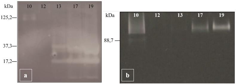
3.3 ZYMOGRAPHIC DETECTION OF CELLULASES
All extracts (except for fungus 12) showed enzymatic activity, evidenced by the
presence of clear bands in the gel (Figure 3). In the gel revelation for endoglucanase
activity, the extract of fungus 10 presented a band of activity with a molecular weight of
125.2 kDa, the extracts of fungi 13 and 17 presented two bands, one of molecular weight
37.3 kDa and one of molecular weight of 17.2 kDa. Fungus 19 extract had a molecular
weight band of 17.2 kDa.
For β-glucosidase activity, extracts of fungi 10, 17, and 19 showed bands of
molecular weights greater than 90 kDa. The extract of fungus 10 also presented a band of
88.7 kDa. The extracts of fungi 12 and 13 did not present bands for this enzyme.
SDS-PAGE gels did not yield as expected because it was not possible to verify
any protein bands for any of the extracts.
Brazilian Journal of Development, Curitiba, v.7, n.1, p.10114-10135 Jan. 2021Brazilian Journal of Development 10126
ISSN: 2525-8761
Figure 3: Zymograms of endoglucanases (a) and β-glucosidases (b) from five selected filamentous fungi
from Nausitora fusticulus digestive organs. The samples were filtrate extracts from fungi cultivation in
minimum medium containing sugar cane bagasse. Gels were prepared with 0.1% of carboxy-methyl-
cellulose (a) and 0.01% of 4-methylumbelliferyl-β-D-glucopyranoside (b).
4 DISCUSSION
The presence of microorganisms in digestive organs of different Teredinidae
species was reported in some studies (Deschamps 1957; Rosenberg & Cutter 1972; Sipe
et al. 2000, Elshahawi et al. 2013; Betcher et al. 2012 and O'Connor et al. 2014), however,
none reported the presence of symbionts in the Nausitora fusticulus species. Akamine et
al. (2018) investigated cellulolytic enzymes production by digestive organs of
Teredinidae species and they found that they produce these enzymes in the gills and in
the digestive organs, but in low quantity, which would not be enough to degrade the wood
ingested by these bivalves. They concluded that the wood degradation is improved
through symbiotic association with groups of microorganisms. Our study demonstrated
that there are microorganisms inside the digestive organs of Nausitora fusticulus
shipworm and they can produce hemicellulolytic enzymes.
In this work was described the presence of fungi that act symbiotically with N.
fusticulus in their digestive organs, producing cellulases, xylanases, and ligninases to
break down the cell wall of the wood ingested, providing the nutrients needed for these
animal's development. The organs that presented the largest number of cellulolytic fungi
were canal anal, gills, and normal digestive diverticula, but we found at least one
cellulolytic microorganism in each organ of the N. fusticulus digestive system. The
microorganism distribution in the digestive system suggests that in each organ occurs the
degradation of the wood by the symbionts. Our results agree with a hypothetical
distribution of cellulolytic microorganisms in the digestive system based on organ size.
Depending on the size or anatomical shape of the digestive organ, the wood can be
retained for longer, which allows the development of more microorganisms to perform
Brazilian Journal of Development, Curitiba, v.7, n.1, p.10114-10135 Jan. 2021Brazilian Journal of Development 10127
ISSN: 2525-8761
complete digestion (Lopes & Narchi 1998). Due to research in each region of the
shipworms' digestive system, our work is innovative compared to other recent reports,
which name the appendix as the site of digestion in Teredinidae species, and do not
describe the microbial behavior in each part of the digestive tract (Brito et al. 2018;
Sabbadin et al. 2018).
Analyzes were performed with Gram's Iodine stain, which dyes polysaccharides
released in the extracellular medium. We chose this technique because it presents a lower
degree of toxicity than Congo red, in addition to revealing the clear zone of degradation
with more prominent staining (Kasana et al. 2008). These methods indicate qualitatively
the enzyme production by Enzymatic Index, a correlation between the diameters of the
degradation clear zone and the colony growth. The Enzymatic Index is an applied tool
that simplifies the isolation of microorganism enzyme producers and allows the
comparison of their enzymatic production (Carrim et al. 2006). Florencio et al. (2012)
studied endoglucanases production of several fungi, cultivating those in Petri dishes and
with solid-state fermentation. In CMC containing medium, all the filamentous fungi,
isolated for us, presented EI higher than or equal to the T. harzianum IOC3844 strain.
This strain is a great producer of endoglucanases and produces significant levels of β-
glucosidases and FPase (Castro et al. 2010). These data indicate that some fungi, isolated
from N. fusticulus organs, have the potential to produce cellulolytic enzymes in high
levels besides xylanases and ligninases.
The identification of filamentous fungi has been performed based on the analysis
of their microscopic structures and morphologic aspects. Most of the fungi isolated from
different organs of Nausitora fusticulus presented a morphology in Sabouraud medium
with white color, cottony texture, and flat topography. Microscopic observations allow
identifying characteristics of hyphae, shape, arrangement, reproductive structures,
conidia, and the formation of spores. Only the genus of the microorganism 19 was
identified as Aspergillus sp. by the analysis of their reproductive structures. It presented
unbranched conidiophore, globular vesicles with uniserial phialides, and globular conidia
(Figure 4). The other filamentous fungi cannot be identified by microscopy since the
absence of reproductive characters. The five selected filamentous fungi will be identified
in future works by molecular biology techniques.
Brazilian Journal of Development, Curitiba, v.7, n.1, p.10114-10135 Jan. 2021Brazilian Journal of Development 10128
ISSN: 2525-8761
Figure 4: Optical micrograph (a) and scanning electron micrograph (b) of filamentous fungi 19 identified
as Aspergillus genus.
In the submerged fermentation the production of extracellular enzymes by the five
selected fungal strains was evaluated. In general, the strains produced more cellulolytic
enzymes in culture media containing sugarcane bagasse, as sole carbon source, in relation
to cultures containing CMC. We noted that the rates of enzyme production were
influenced by the substrate. The use of purified substrates, such as CMC, for the enzymes
production is very expensive, so, the cost of producing enzymes can be reduced by using
waste substrates such as sugar cane bagasse. Several authors have investigated substrates
that enable the anchoring and aggregation of fungus while providing enough nutrients to
produce enzymes (Yoon et al. 2014). This justifies the use of sugarcane bagasse as a
source of carbon in the production of lignocellulolytic enzymes. The highest production
occurred by strain 10, mainly of endoglucanases and exoglucanases, which presented
rates comparable to that produced by TH1 strain. These results corroborate with the
results obtained in the solid culture that determined the enzymatic index for these strains.
The TH1 strain, being a wild strain, not modified genetically, but which is used as a
reference in the production of cellulolytic enzymes (Castro et al. 2010), presented a
production of the enzymes in levels sometimes inferior to that released by the other
strains. The TH1 strain showed little variation in the production of the three enzymes
evaluated indicating that the strain is less sensitive to the variation of the culture medium.
With the data of enzymatic activity shown by the strains it can be confirmed the
presence of good fungi producing cellulases inhabiting the digestive system of N.
fusticulus. Other studies investigating the production of cellulases by wild fungal strains
have been reported (Teng et al. 2010; Grigorevski-Lima et al. 2011; Li et al. 2013;
Manavalan et al. 2015; Sabaddin et al. 2018). For the extracts obtained from filtration of
the minimal medium containing sugarcane bagasse, the endoglucanase zymogram
Brazilian Journal of Development, Curitiba, v.7, n.1, p.10114-10135 Jan. 2021Brazilian Journal of Development 10129
ISSN: 2525-8761
revealed clear bands of 12 and 37 kDa for fungi 13, 17, and 19 and a slightly cleared band
for extract 10 with a weight of 125 kDa (Figure 3a). Several authors reported fungal
endoglucanases with weights between 13 and 50 kDa (Tong et al. 1980; Beldman et al.
1985; Okada 1985; Sprey and Uelker 1992; Akiba et al. 1995; Sul et al. 2004; Naika et
al. 2007; Begum and Absar 2009; Rawat et al. 2015). But fungal endoglucanases with
larger weights have been reported, like endoglucanases from Trichoderma koningii with
78,1 kDa (Ge et al. 2015) and Thermothelomyces thermophila with 100 kDa (Roy et al.
1990). Grigorevski-Lima and co-workers (2013) performed a zymography with an extract
from Trichoderma atroviride culture containing endoglucanase activity and detected in
the zymogram two clear bands with molecular weights of 104 and 204 kDa, like that
obtained by extract 10 in this work. Asha and co-workers (2016) identified Aspergillus
ochraceus cellulases by a zymogram. For endoglucanase, using the CMC substrate, was
detected a band with a molecular weight of 78 kDa, and in the zymogram with the
substrate for the β-glycosidase enzyme was detected a band with a molecular weight of
43 kDa (Asha et al. 2016).
A robust enzymatic cocktail is fundamentals for application such as biomass
conversion into high added products (Chiyanzu et al., 2014). Moreover, combination of
different enzymes is positive, improving the cocktail application allowing for example
hemicellulases enzymes uses (Freitas et al., 2020; Bueno et al., 2020; Calore et al., 2020).
5 CONCLUSION
Nausitora fusticulus shipworms present cellulolytic microorganisms in their
digestive organs, that help them with wood degradation for their nutrition. Among
isolated microorganisms, 5 fungi stood out in the production of cellulases. These strains
cultivated in minimum medium containing sugar cane bagasse shown higher production
of endoglucanases, exoglucanases e β-glucosidases when compared with the cultivation
in minimum medium containing carboxymethyl-cellulose. The zymographic tests showed
cellulolytic activity of each strain and allowed to detect the molecular weight of the
enzymes, that matches with other cellulases in the literature. Our results suggest that
shipworms are dependent on microorganisms, in a symbiotic relationship, for the
degradation of cellulosic material. The next steps of this research could be related to fungi
identification and enzyme separation to study their biochemistry properties.
Brazilian Journal of Development, Curitiba, v.7, n.1, p.10114-10135 Jan. 2021Brazilian Journal of Development 10130
ISSN: 2525-8761
ACKNOWLEDGEMENTS
This work was supported by Faperj (E-26/260.001/2014 and E-26/190.180/2013)
and São Paulo Research Foundation (2017/22401-8; 2019/12997-6). Authors thanks the
Laboratory of Microbiology and the Laboratory of Microscopy of Directory of Metrology
Applied to Life Sciences, both from the National Institute of Metrology, Quality and
Technology (Inmetro), at Xerém, Rio de Janeiro, Brazil. We also thank Dr. Bernardo
Yépez and the Fundação Oswaldo Cruz Filamentous Fungi Culture Collection for making
available a sample of the Trichoderma harzianum strain (IOC-3844).
Brazilian Journal of Development, Curitiba, v.7, n.1, p.10114-10135 Jan. 2021Brazilian Journal of Development 10131
ISSN: 2525-8761
REFERENCES
Akamine DTM, Silva DAC, Câmara GL, Carvalho TV, Brienzo M (2018).
Endoglucanase activity in Neoteredo reynei (Bivalvia, Teredinidae) digestive organs and
its content. World J Microbiol Biotechnol. https://doi.org/10.1007/s11274-018-2468-x
Akiba S, Kimura Y, Yamamoto K, Kumagai H (1995). Purification and characterization
of a protease-resistant cellulase from Aspergillus niger. J Biosci Bioeng.
https://doi.org/10.1016/0922-338X(95)94078-6
Asha P, Divya J, Bright Singh IS (2016). Purification and characterization of processive-
type endoglucanase and β-glucosidase from Aspergillus ochraceus MTCC 1810 through
saccharification of delignified coir pith to glucose. Bioresour Technol.
https://doi.org/10.1016/j.biortech.2016.03.013
Begum MF, Absar N (2009). Purification and Characterization of Intracellular Cellulase
from Aspergillus oryzae ITCC-4857.01. MYCOBIOLOGY.
https://doi.org/10.4489/MYCO.2009.37.2.121
Beldman G, Searle-Van Leeuwen MF, Rombouts FM, Voragen FG (1985). The cellulase
of Trichoderma viride. Purification, characterization and comparison of all detectable
endoglucanases, exoglucanases and beta-glucosidases. Eur J Biochem, 146 (2):301-308.
Betcher MA, Fung JM, Han AW, O’Connor R, Seronay R, Concepcion GP, Distel DL,
Haygood MG (2012). Microbial distribution and abundance in the digestive system of
five shipworm species (Bivalvia: Teredinidae). PloS ONE. https
://doi.org/10.1371/journal.pone.00453 09
Borges LMS, Merckelbach LM, Sampaio I, Cragg SM (2014). Diversity, environmental
requirements, and biogeography of bivalve wood-borers (Teredinidae) in European
coastal waters. Front Zool. https://doi.org/10.1186/1742-9994-11-13
Brito TL, Campos AB, Bastiaan von Meijenfeldt FA, Daniel JP, Ribeiro GB, Silva GGZ,
Wilke DV, Moraes DT, Dutilh BE, Meirelles PM, Trindade-Silva AE (2018) The gill-
associated microbiome is the main source of wood plant polysaccharide hydrolases and
secondary metabolite gene clusters in the mangrove shipworm Neoteredo reynei. PLoS
ONE. https://doi.org/10.1371/journal.pone.0200437
Bueno D, Brienzo M (2020). Xylan solubilization and use as carbon source/inductor for
microbial xylanase production. Brazilian Journal of Development 6 (10), 78426-78433
Calore RH, Bueno D, Brienzo M (2020). Production of substrates for determination of
xylanase activity. Brazilian Journal of Development 6 (10), 78404-78409
Chiyanzu I, Brienzo M, García-Aparicio MP, Görgens JF (2014). Application of endo-β-
1, 4, d-mannanase and cellulase for the release of mannooligosaccharides from steam-
pretreated spent coffee ground. Applied biochemistry and biotechnology 172 (7), 3538-
3557.
Brazilian Journal of Development, Curitiba, v.7, n.1, p.10114-10135 Jan. 2021Brazilian Journal of Development 10132
ISSN: 2525-8761
Carrim AJI, Barbosa EC, Vieira JDG (2006). Enzymatic activity of endophytic bacterial
isolates of Jacaranda decurrens Cham. (Carobinha-do-campo). Braz Arch Biol Technol.
http://dx.doi.org/10.1590/S1516-89132006000400001.
Castro AM, Pedro KC, Cruz JC, Ferreira MC, Leite SG, Pereira Júnior N (2010).
Trichoderma harzianum IOC-4038: A promising strain for the production of a cellulolytic
complex with significant β-glucosidase activity from sugarcane bagasse cellulignin. Appl
Biochem Biotechnol. https://doi.org/10.1007/s12010-010-8986-0
Deschamps P (1957). La nutrition chez les molusques térédinides: possibilité de rétraction
et fermeture de l’étui calcaire. Chromatographie quantitative des sucres. Bull. Soc. Zol.
France, 82: 72-81.
Dienes D, Egyhazi A, Reczey K (2004). Treatment of recycled fiber with Trichoderma
cellulases. Ind Crop Prod. https://doi.org/10.1016/j.indcrop.2003.12.009
Distel DL, Beaudoin DJ, Morrill W (2002) Coexistence of multiple proteobacterial
endosymbionts in the gills of the wood-boring Bivalve Lyrodus pedicellatus (Bivalvia:
Teredinidae). Appl Environ Microbiol https://doi.org/10.1128/AEM.68.12.6292-
6299.2002
Distel DL, Delong EF, Waterbury JB (1991). Phylogenetic characterization and in situ
localization of the bacterial symbiont of shipworms (Teredinidae: Bivalvia) by using 16S
rRNA sequence analysis and oligodeoxynucleotide probe hybridization. Appl Environ.
Microbiol, 57(8): 2376-82.
Elshahawi SI, Trindade-Silva AE, Hanora A, Han AW, Flores MS, Vizzoni V, Schrago
CG, Soares CA, Concepcion GP, Distel DL, Schmidt EW, Haygood MG (2013)
Boronated tartrolon antibiotic produced by symbiotic cellulose-degrading bacteria in
shipworm gills. Proc Natl Acad Sci USA. https://doi.org/10.1073/pnas.1213892110
Florencio C, Couri S, Farinas SC (2012). Correlation between Agar Plate Screening and
Solid-State Fermentation for the Prediction of Cellulase Production by Trichoderma
Strains. Enzyme Res. http://dx.doi.org/10.1155/2012/793708
Freitas C, Terrone CC, Carmona EC, Brienzo M (2020). Evaluation of
xylooligosaccharides effect on the growth of probiotic microorganisms
Brazilian Journal of Development 6 (9), 73400-73411
Ge F, Shi B, Tang Y, Zhu L, Li WZ, Tao Y (2015). Isolation, purification, and enzymatic
properties of cellulase system in mutant strain Trichoderma koningii SG0026. Mod Food
Sci Technol. https://doi.org/10.13982/j.mfst.1673-9078.2015.12.022.
Grigorevski-Lima AL, Oliveira MM, Nascimento RP, Bon EP, Coelho RR (2011).
Production and partial characterization of cellulases and Xylanases from Trichoderma
atroviride 676 using lignocellulosic residual biomass. Appl Biochem Biotechnol.
https://doi.org/10.1007/s12010-012-0053-6
Grigorevski-Lima AL, Oliveira MM, Nascimento RP, Bon EP, Coelho RR (2013).
Production and partial characterization of cellulases and xylanases from Trichoderma
Brazilian Journal of Development, Curitiba, v.7, n.1, p.10114-10135 Jan. 2021Brazilian Journal of Development 10133
ISSN: 2525-8761
atroviride 676 using lignocellulosic residual biomass. Appl Biochem Biotechnol.
https://doi.org/10.1007/s12010-012-0053-6.
Hankin L, Anagnostakis SL (1975). The Use of Solid Media for Detection of Enzyme
Production by Fungi. Mycologia. https://doi.org/10.2307/3758395
Jeffreys JG (1860). A synoptical list of the British species of Teredo with a notice of the
exotic species. Annals and Magazine of Natural History. 3 (6): 121-127. Available online
at http://biodiversitylibrary.org/page/22218533. Teredo fusticulus - pg.: 125.
Kasana RC, Salwan R, Dhar H, Dutt S, Gulati A (2008). A Rapid and Easy Method for
the Detection of Microbial Cellulases on Agar Plates Using Gram’s Iodine. Curr
Microbiol. https://doi.org/10.1007/s00284-008-9276-8
Li C, Yang Z, He R, Zhang D, Chen S, Ma L (2013). Effect of pH on cellulase production
and morphology of Trichoderma reesei and the application in cellulosic material
hydrolysis. J Biotechnol. https://doi.org/10.1016/j.jbiotec.2013.10.003
Lopes SGBC, Narchi W (1998). Functional anatomy of Nausitora fusticulus (Jeffreys,
1860) (Mollusca, Bivalvia). Veliger. 41:274–288
Maeda RN, Barcelos CA, SantaAnna LMM, Pereira Junior N (2013). Cellulase
production by Penicillium funiculosum and its application in the hydrolysis of sugar cane
bagasse for second generation ethanol production by fed batch operation. J Biotechnol.
https://doi.org/10.1016/j.jbiotec.2012.10.014
Manavalan T, Manavalan A, Thangavelu KP, Heese K (2015). Characterization of a novel
endoglucanase from Ganoderma lucidum. J Basic Microbiol.
https://doi.org/10.1002/jobm.201400808
Marques N P, Cassia Pereira J, Gomes E, Silva R, Araújo AR, Ferreira H, Rodrigues A,
Dussán KJ, Bocchini DA (2018). Cellulases and xylanases production by endophytic
fungi by solid state fermentation using lignocellulosic substrates and enzymatic
saccharification of pretreated sugarcane bagasse. Ind Crop Prod.
https://doi.org/10.1016/j.indcrop.2018.05.022
Miller GL (1959) Use of dinitrosalicylic acid reagent for determination of reducing sugar.
Anal Chem 31:426–428
Narra M, Dixit G, Divecha J, Kumar K, Madamwar D, Shah AR (2014). Production,
purification and characterization of a novel GH 12 family endoglucanase from
Aspergillus terreus and its application in enzymatic degradation of delignified rice straw.
Int Biodeterior Biodegradation. https://doi.org/10.1016/j.ibiod.2013.12.016
Naika GS, Kaul P, Prakash V (2007). Purification and characterization of a new
endoglucanase from Aspergillus aculeatus. J Agric Food Chem.
https://doi.org/10.1021/jf070710p
Brazilian Journal of Development, Curitiba, v.7, n.1, p.10114-10135 Jan. 2021Brazilian Journal of Development 10134
ISSN: 2525-8761
O’Connor RM, Fung JM, Sharp KH, Benner JS, McClung C, Cushing S, … Distel DL
(2014). Gill bacteria enable a novel digestive strategy in a wood-feeding mollusk. Proc
Natl Acad Sci USA. https://doi.org/10.1073/pnas.1413110111
Okada G (1985). Purification and Properties of a Cellulase from Aspergillus niger. Agric
Biol Chem. https://doi.org/10.1080/00021369.1985.10866894
Rawat R, Kumar S, Chadha BS, Kumar D, Oberoi HS (2015). An acidothermophilic
functionally active novel GH12 family endoglucanase from Aspergillus niger HO:
purification, characterization and molecular interaction studies. Antoine Van
Leeuwenhoek. https://doi.org/10.1007/s10482-014-0308-z
Rosenberg FA, Cutter J (1972). The role of cellulolytic bacteria in the digestive processes
of the Shipworm. In: Acker RF (ed) Materials in the sea. Evanston, Northwestern, pp 778-
796.
Roy SK, Dey SK, Raha SK, Chakrabarty SL (1990). Purification and properties of an
extracellular endoglucanase from Myceliophthora thermophila D-14 (ATCC 48104). J
Gen Microbiol. https://doi.org/10.1099/00221287-136-10-1967.
Sabbadin F, Pesante G, Elias L, Besser K, Li Y, Steele‑King C, Stark M, Rathbone DA,
Dowle AA, Bates R, Shipway JR, Cragg SM, Bruce NC and McQueen‑Mason SJ (2018).
Uncovering the molecular mechanisms of lignocellulose digestion in shipworms.
Biotechnol Biofuels. https://doi.org/10.1186/s13068-018-1058-3
Sharma V, Sumbali G (2013). Diversity of cellulase active micromycetes in the ant-hill
soil environment of parks and gardens of Jammu city (India). Int J Pharm Bio, 4(2): B541-
548.
Sipe AR, Wilbur AE, Cary SC (2000). Bacterial symbiont transmission in the wood-
boring shipworm Bankia setacea (Bivalvia: Teredinidae). Appl Environ. Microbiol, 66:
1685-1691.
Sprey B, Uelker A (1992). Isolation and properties of a low molecular mass
endoglucanase from Trichoderma reesei. FEMS Microbiol Lett.
https://doi.org/10.1016/0378-1097(92)90718-4
Sul OJ, Kim JH, Park SJ, Son YJ, Park BR, Chung DK, Jeong CS, Han IS (2004).
Characterization and molecular cloning of a novel endoglucanase from Trichoderma sp.
C-4. Appl Microbiol Biotechnol. https://doi.org/10.1007/s00253-004-1713-4
Tanaka M, Taniguchi MA, Matsumo R, Kamikubo T (1981). Purification and properties
of cellulases from Eupenicillium javanicum. Journal Fermentation Technology, 59: 177-
183.
Teng Y, Yin Q, Ding M, Zhao F (2010). Purification and characterization of a novel endo-
β-1,4-glucanase, AfEG22, from the giant snail, Achatina fulica frussac. Acta Biochim
Biophys Sinv. https://doi.org/10.1093/abbs/gmq083
Brazilian Journal of Development, Curitiba, v.7, n.1, p.10114-10135 Jan. 2021Brazilian Journal of Development 10135
ISSN: 2525-8761
Tong CC, Cole AL, Shephard MG (1980). Purification and properties of the cellulases
from the thermophilic fungus Thermoascus auriantiacus. Biochem J, 191(1):83-94
Turner RD (1966). A survey and illustrated catalogue of the Teredinidae (Mollusca:
Bivalvia). Museum of Comparative Zoology. Cambridge (Massachusetts): Harvard
University, page: 102.
Yang JC, Madupu R, Durkin AS, Ekborg NA, Pedamallu CS, Hostetler JB, … Distel DL
(2009). The Complete Genome of Teredinibacter turnerae T7901: An Intracellular
Endosymbiont of Marine Wood-Boring Bivalves (Shipworms). PLoS One.
https://doi.org/10.1371/journal.pone.0006085
Yoon LW, Ang TN, Ngoh GC, Chua ASM (2014). Fungal solid-state fermentation and
various methods of enhancement in cellulase production. Biomass Bioenergy.
https://doi.org/10.1016/j.biombioe.2014.05.013.
Brazilian Journal of Development, Curitiba, v.7, n.1, p.10114-10135 Jan. 2021You can also read