Molecular Characterization of from Ferret Badgers, Taiwan
←
→
Page content transcription
If your browser does not render page correctly, please read the page content below

RESEARCH

    Molecular Characterization of
 Cryptically Circulating Rabies Virus
    from Ferret Badgers, Taiwan
                     Hue-Ying Chiou, Chia-Hung Hsieh, Chian-Ren Jeng, Fang-Tse Chan,
                                   Hurng-Yi Wang,1 and Victor Fei Pang1

      After the last reported cases of rabies in a human in        in those countries of Europe and North America that have
1959 and a nonhuman animal in 1961, Taiwan was consid-             well-established vaccination programs (3). Mustelids, in-
ered free from rabies. However, during 2012–2013, an out-          cluding various species of the genera Melogale, Meles,
break occurred among ferret badgers in Taiwan. To exam-            and Mellivora of the weasel family Mustelidae, can carry
ine the origin of this virus strain, we sequenced 3 complete       RABV (4–6). In southeastern China, Chinese ferret badgers
genomes and acquired multiple rabies virus (RABV) nu-
                                                                   (CNFB; Melogale moschata moschata) have been associ-
cleoprotein and glycoprotein sequences. Phylogeographic
analyses demonstrated that the RABV affecting the Taiwan
                                                                   ated with human rabies for many years and are considered
ferret badgers (RABV-TWFB) is a distinct lineage within the        to be a primary host in this region (7–9).
group of lineages from Asia and that it has been differentiat-           After what were considered to be the last reported
ed from its closest lineages, China I (including isolates from     cases of rabies in a human and a nonhuman animal in 1959
Chinese ferret badgers) and the Philippines, 158–210 years         and 1961, respectively, Taiwan was rabies free for >50
ago. The most recent common ancestor of RABV-TWFB                  years until the 2012–2013 outbreak of ferret badger–asso-
originated 91–113 years ago. Our findings indicate that            ciated rabies. During May 2012–January 2013, through a
RABV could be cryptically circulating in the environment.          government-supported program of routine disease surveil-
An understanding of the underlying mechanism might shed            lance of free-range dead wild animals that had been killed
light on the complex interaction between RABV and its host.        by vehicles or were receiving treatment for injuries and/
                                                                   or illness at the wildlife first aid station, 3 dead Taiwan

R    abies is possibly one of the oldest zoonotic diseases.
     It is caused by the rabies virus (RABV), a neurotropic
virus in the family Rhabdoviridae, genus Lyssavirus. Ex-
                                                                   ferret badgers (TWFB; M. moschata subaurantiaca) were
                                                                   submitted to the School of Veterinary Medicine, National
                                                                   Taiwan University, for further examination.
cept for a small number of countries and regions, particu-               Pathologic examination revealed nonsuppurative me-
larly islands, RABV is found worldwide. The virus infects          ningoencephalomyelitis with formation of eosinophilic
nearly all warm-blooded animals and causes severe neu-             intracytoplasmic inclusion bodies in all 3 animals; reverse
rologic signs, which almost invariably lead to death (1). It       transcription PCR and immunohistochemical staining ex-
was estimated that worldwide in 2010, the disease caused           cluded the possibility of infection with the canine distem-
>60,000 human deaths, primarily in Africa and Asia (2).            per virus. However, the results of fluorescence antibody
Although dogs are considered the principal host of RABV            testing, immunohistochemical staining, and reverse tran-
in developing countries, the virus is also dispersed among         scription PCR, followed by sequencing for RABV, were
many species of wild carnivora and chiroptera, especially          positive (H.-Y. Chiou, unpub. data). After the rabies diag-
                                                                   noses for the initial 3 ferret badgers were confirmed, by
Author affiliations: National Taiwan University, Taipei, Taiwan,
                                                                   the end of August of 2013, rabies had been diagnosed by
Republic of China (H.-Y. Chiou, C.-H. Hsieh, C.-R. Jeng, H.-Y.
                                                                   fluorescence antibody testing for an additional 105 dead or
Wang, V.F. Pang); and Council of Agriculture, Executive Yuan,
                                                                   ill and euthanized ferret badgers and 1 shrew.
Nantou County, Taiwan, Republic of China (F.-T. Chan)

DOI: http://dx.doi.org/10.3201/eid2005.131389
                                                                   1
                                                                       Joint senior authors who contributed equally to this article.

790                          Emerging Infectious Diseases • www.cdc.gov/eid • Vol. 20, No. 5, May 2014
Rabies Virus from Ferret Badgers, Taiwan

     Our objective in this study was to clarify whether                 signs: emaciation, coma, paddling, loss of pain response,
the current outbreak of the TWFB–associated rabies is an                reduced body temperature, and a 2-cm skin wound on the
emerging, a reemerging, or a cryptically circulating disease.           chin; extreme weakness and inability to move; and signs of
We investigated the possible origin of this outbreak and its            weakness and respiratory signs, including labored breath-
relations with CNFB-associated rabies in mainland China                 ing and increased breath sounds with hypersalivation and
via genomic organization and characterization and analysis              exudation of foamy fluid from the mouth and nose. Initial
of genetic diversity and phylogeographic origin of RABV-                supportive treatment was provided at the wildlife first aid
TWFB. In addition, we propose a mechanism that might be                 station, but the ferret badgers died within 1–3 days, and
contributing to the limited host range of RABV-TWFB.                    their carcasses were submitted to the School of Veterinary
                                                                        Medicine, National Taiwan University, for routine disease
Materials and Methods                                                   surveillance. Full necropsy was performed, during which
                                                                        half of the left cerebral hemisphere was collected from each
Animals and Specimen Collection                                         animal and stored at –80°C for subsequent nucleic acid ex-
    During May 2012–January 2013, three ill TWFB                        traction. Representative tissue samples were taken from all
were collected from different regions of central Taiwan                 major organs and fixed in 10% neutral buffered formalin
(Figure 1). One was in the Xitou nature education area at               for histopathologic examination.
Lugu Township, Nantou County (R2012–26); one was in
Gukeng Township, Yunlin County (R2012–88); and one                      Sample Preparation and Genome Sequencing
was in Yuchih Township, Nantou County (R2013–01).                            Approximately 25 mg of brain specimen from each
These 3 TWFB, respectively, showed the following clinical               animal was homogenized, and 1 mL of TRIzol reagent
                                                                        (Invitrogen, Carlsbad, CA, USA) was added. Total RNA
                                                                        was extracted by using an RNeasy Mini Kit (QIAGEN,
                                                                        Valencia, CA, USA), and cDNA was synthesized by
                                                                        using a Transcriptor First Strand cDNA Synthesis Kit
                                                                        (Roche Diagnostics, Indianapolis, IN, USA) according
                                                                        to the manufacturer’s instructions. To amplify the whole
                                                                        genome, we used 19 pairs of primers (Table 1), including
                                                                        the forward primer for the 5′ end and the reverse primer
                                                                        for the 3′ end designed to be complementary to the respec-
                                                                        tive ends of the genome, as described (10).

                                                                        Sequence Analyses and Phylogenetic Reconstruction
                                                                             Sequences were assembled by using the Seqman pro-
                                                                        gram (Lasergene 8, Madison, WI, USA) (GenBank acces-
                                                                        sion nos. KF620487–KF620489) and then aligned by using
                                                                        the ClustalW program (11). The genetic distance was esti-
                                                                        mated by using the Kimura 2-parameter substitution model
                                                                        implemented in MEGA version 5.0 (12). The nucleotide di-
                                                                        versity within populations was calculated by using DnaSP
                                                                        version 5.0 (13). To test for the deviation of neutral ex-
                                                                        pectation, we conducted the Tajima D (14) and the Fu and
                                                                        Li D* (15) tests implemented in DnaSP. Significance was
                                                                        assessed by 104 coalescent simulations (13).
                                                                             To investigate the phylogenetic position of RABV-
                                                                        TWFB isolates, we included 24 complete RABV genomes
                                                                        representing the 3 major phylogenetic groups (16). For
Figure 1. Collection sites of rabies-positive Taiwan ferret badgers
(TWFB), Taiwan. Solid circles marked with 1–3 represent the
                                                                        global phylogeny of RABV, we analyzed 218 full-length
collection sites of the first 3 rabies-positive animals. Triangles      (1,335-nt) sequences of the nucleoprotein (N) gene, in-
represent the collection sites of other rabies virus (RABV) sequences   cluding 11 sequences from Taiwan. We also analyzed 125
included in this study. Crosses represent the most diverged lineages    full-length (1,575-nt) sequences of the glycoprotein (G)
of rabies virus from Taiwan ferret badgers (TWFB, TW1614, and           gene, including 13 sequences from Taiwan (17). For each
TW1955), shown in Figure 5, panel B, Appendix (wwwnc.cdc.
gov/EID/article/20/5/13-1389-F5.htm), and the easternmost cross
                                                                        gene, phylogenetic trees were inferred by using maximum-
represents the isolate from a shrew, TW1955.                            likelihood and Bayesian inference methods.

                               Emerging Infectious Diseases • www.cdc.gov/eid • Vol. 20, No. 5, May 2014                        791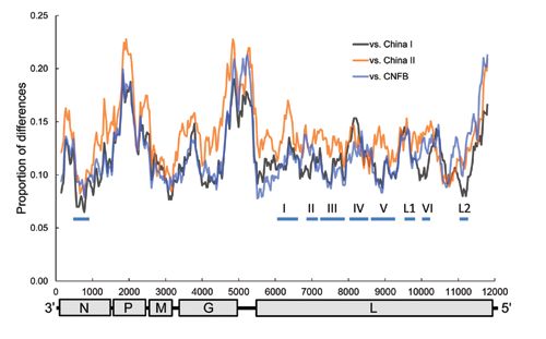
RESEARCH

Table 1. Primers used for the amplification and sequencing of rabies virus genome*
Primer                                    Sequence, 5′→3′                                Amplicon size, nt         Position, nt
3′F                               GTACCTAGACGCTTAACAAC                                         499                   1–499
3′R                               AAGACCGACTAAGGACGCAT
NF                                ATGTAACACCTCTACAATGG                                        1,533                 55–1587
NR                                  CAGTCTCYTCNGCCATCT
PF                                 GAACCAYCCCAAAYATGAG                                        1,001                1500–2500
PR                               TTCATTTTRTYAGTGGTGTTGC
MF                                 AAAAACRGGCAACACCACT                                         641                 2479–3119
MR                               TCCTCYAGAGGTAWACAAGTG
G1F                                TGGTGTATAACATGRAYTC                                        1,097                3000–4096
G1R                                ACCCATGTYCCRTCATAAG
G2F                             TGGATTTGTGGATKAAAGAGGC                                        1,542                3995–5536
G2R                              GAGTTNAGRTTGTARTCAGAG
L1F                              TGGRGAGGTYTATGATGACCC                                         726                 5430–6155
L1R                            CAGCATNAGTGTRTAGTTTCTGTC
L2F                            GGTCGATTATGATAAKGCATTTGG                                        704                 5885–6588
L2R                              TTGACAGACCCTTTCGATAATC
L3F                             GGATCAATTCGACAACATACATG                                        550                 6473–7024
L3R                             AAGTCTTCATCHGGCARTCCTCC
L4F                             AGACTAGCTTCHTGGYTGTCAG                                         708                 6882–7589
L4R                              TACTTTGGTTCTTGTGTTCCTG
L5F                            AGTGTTTGGATTGAAGAGAGTGTT                                        662                 7337–7998
L5R                             GAAAGACTGCCTGCACTGACAT
L6F                            AATAGTCAACCTCGCCAATATAATG                                       767                 7897–8645
L6R                           GGATCTCTGAGTTGTAGAAGGATTC
L7F                            CCGAGTCAATCATTGGATTGATAC                                        621                 8517–9137
L7R                            GAATACCCTCCTTCGCTGTATCTG
L8F                             GAGAAGGTCACCAATGTTGATG                                        1,045               8958–10002
L8R                              AGATCCAYARCCAGTCATTCTC
L9F                              ACATAATGCTCAGAGAACCGT                                         503                9820–10322
L9R                               CCATTCTGAACATCCTACCTT
L10F                             TGTTCAGAATGGGTCTGCTCT                                         509                10302–10811
L10R                              TGCATCGCAAATAATGAGGT
L11F                              ATTATTTGCGATGCAGAAGT                                         524                10797–11320
L11R                            ATGATAGCCACTTTAGACAGAGT
L12F                            GTTACAGAGGGGAACTCTGTCT                                         386                11285–11670
L12R                           TCTTCACTATCTTGTAAATCAACCT
5′F                           TGGATCAGGTTGATTTACAAGATAGT                                       293                11640–11932
5′R                           ACGCTTAACAAATAAACAACAAAAAT
*Primers were from Lei et al. (10).

     The maximum-likelihood analysis was conducted by                  of virus isolates were estimated by using an established
using PhyML 3.0 online (18); the starting tree was derived             Bayesian MCMC approach implemented in BEAST version
from the neighbor-joining method, and the nearest neigh-               1.7 (21). The analysis was performed by using the general
bor interchange topology search option was used. The                   time-reversible model of nucleotide substitution assuming
nucleotide substitution model for phylogenetic reconstruc-             an uncorrelated lognormal molecular clock (22). We linked
tion was determined by using the Akaike information crite-             substitution rates for the first and second codon positions
rion implemented in jModeltest 0.1.1 (19). The method of               and allowed independent rates in the third codon position.
Bayesian inference was performed by using MrBayes ver-                 The molecular clocks were 2.3 × 10⁻⁴ (range 1.1–3.6 × 10⁻⁴)
sion 3.1.2 (20). Analyses were initiated with random start-            and 3.9 × 10⁻⁴ (1.2–6.5 × 10⁻⁴) substitutions/site/year for N
ing trees, and Metropolis-coupled Markov chain Monte                   and G genes, respectively (17). A slightly faster clock, 4.3 ×
Carlo (MCMC) analyses were run for 1 × 10⁶ generations                 10–4 (3.1–5.6 × 10⁻⁴) substitutions/site/year for N gene (23),
and sampled every 100 generations. The steady state of the             was also used in a separate analysis.
log-likelihood was reached at ≈20,000 generations. Subse-                   Because a previous study revealed that the population
quently, the first 201 trees were excluded and the remaining           dynamics of RABV supported a model of constant popula-
9,800 trees were retained to compute a 50% majority-rule               tion size through time (17), we restricted our analysis to
consensus tree.                                                        this demographic model. For each analysis, we performed
                                                                       2 independent runs with 2 × 10⁷ MCMC steps, of which
Divergence Dating                                                      the first 10% were discarded as burn-in. To confirm that
     The divergence time between different viral lineages              both were sampling the same distribution, we compared
and the time to the most recent common ancestor (TMRCA)                and then combined the results. Log files were checked by

792                               Emerging Infectious Diseases • www.cdc.gov/eid • Vol. 20, No. 5, May 2014
Rabies Virus from Ferret Badgers, Taiwan
using Tracer (http://beast.bio.ed.ac.uk/Tracer), and the ef- lineages, the most conserved protein is M, followed by N,
fective sample size for each parameter was >300, which is the virion-associated RNA polymerase (L), and G; the least
adequate according to the authors of BEAST software. conserved is phosphoprotein (P) (Table 3). Among the
RABV groups, however, N becomes the most conserved
Results followed by L, M, G, and P. The RABV-TWFB is closest
to China I lineage in the N, P, and L gene regions, but it is
Genomic Organization and Characterization closest to RABV-CNFB in the M and G gene regions.
of RABV-TWFB The genetic variations across the whole genome among
Similar to previous analyses (16,17), our phyloge- different lineages can be viewed in a sliding window anal-
netic analysis that used whole RABV genomes revealed 3 ysis (Figure 3). Within N, there seems to be a conserved
major groups with high bootstrap support (Figure 2). Al- central domain previously identified in RABV at residues
though the 3 RABV-TWFB isolates are clustered within 182–328 (24), which is also conserved in RABV-TWFB.
the Asia group, composed of 3 distinct lineages, (China The P, the last quarter of G and G–L intergenic regions, and
I [including CNFB], China II [16], and Southeast Asia), the last part of L are more variable among different lineag-
they do not appear close to any of the 3 lineages. More es of RABV than is the rest of the genome. The conserved
noteworthy, the 3 isolates of RABV-TWFB are not close M is functional in viral assembly and budding (25); is in-
to those of RABV-CNFB, indicating that they may have volved in the regulation of transcription and replication of
originated independently. viral RNA (26); and has been reported to induce apoptosis
The genome of RABV-TWFB is 11,923 nt long and (27), suggesting its role in host-cell interplay. The involve-
encodes 5 proteins. The nucleotide lengths of different ge- ment of M in multiple interactions explains its conserva-
nomic regions are within the range of variations in different tion among lineages. G is responsible for cell attachment
Asia lineages (Table 2), except the matrix protein (M)-G and fusion and is the main viral protein responsible for the
intergenic region, which is 1 nt longer than in the rest of the induction of neutralization antibodies and cell-mediated
lineages from Asia (212 vs. 211). Within the group of Asia immune responses. The region between aa 189 and aa 214,
Figure 2. Phylogenetic relationships
of 27 rabies virus (RABV) genomes
constructed by maximum-likelihood
method. Numbers close to the
nodes were from 1,000 bootstrap
replications. The tree was rooted with
RABV from bats and raccoons. Three
major groups, Asia, Cosmopolitan,
and India, are strongly supported,
as indicated (17). There are 4 major
lineages within the group from Asia,
including previously recognized
China I, China II (16), Southeast
Asia, and RABV from Taiwan ferret
badgers (TWFB). RABVs derived
from Chinese ferret badgers (CNFB)
are clustered with China I, indicating
that RABVs of TWFB and CNFB
are of independent origin. Scale bar
indicates nucleotide substitutions
per site.
Emerging Infectious Diseases • www.cdc.gov/eid • Vol. 20, No. 5, May 2014 793RESEARCH
Table 2. Genomic organization and nucleotide lengths of terminal and intergenic regions and viral genes of rabies virus from Taiwan
and other lineages from Asia*
Gene, nucleotide length
Isolate 3′-UTR N N–P P P–M M M–G G G–L L 5′-UTR Genome size
TWFB 70 1,353 91 894 86 609 212 1,575 519 6,384 130 11,923
CNFB 70 1,353 91 894 86 609 211 1,575 519 6,384 130–131 11,922–11,923
China I† 70 1,353 90–91 894 87–88 609 211 1,575 519 6,384 130 11,923
China II† 70 1,353 91 894 87 609 211 1,575 518–519 6,384 131 11,923–11,924
*UTR, untranslated region; TWFB, Taiwan ferret badgers; CNFB, Chinese ferret badgers.
†The definitions of China I and II are based on He et al. (16). China I does not include CNFB.
proposed to be needed for G binding to the nicotinic ace- RABV-TWFB was separated from China I and the Phil-
tylcholine receptor (28), is relatively conserved in the da- ippines isolates 158 years ago with 95% highest posterior
taset. Nevertheless, 3 substitutions (N194Y, R196K, and density (HPD) ranging from 110 to 225 years (Figure 5,
G203E) are found exclusively in RABV-TWFB G. In L, panel A, Appendix, wwwnc.cdc.gov/EID/article/20/5/13-
Poch et al. (29) recognized 6 conserved blocks, including 1389-F5.htm). The divergence between China I and the
B1 (233–424), B2 (504–608), B3 (609–832), B4 (890– Philippines isolates occurred 132 (95% HPD, 90–192)
1061), B5 (1091–1326), and B6 (1674–1749) (29). In ad- years ago, which is similar to previous estimations (23,32).
dition, 2 regions, L1 (1418–1515) and L2 (1884–1961), are The TMRCA of isolates from Taiwan was 91 years (95%
also conserved across lyssaviruses (30). In RABV-TWFB, HPD, 57–137). A similar timescale, with overlapping 95%
the B4 and L1 regions in L are variable, and the rest of the HPD, was derived by using the molecular clock of 2.3 ×
blocks are conserved (Figure 3). 10–4/site/year for N gene (17) (Figure 5, panel A, hatched
numbers, Appendix). The mean substitution rate for G gene
Genetic Diversity and Phylogeographic Origin sequences was 3.5 × 10⁻⁴/site/year (7.8 × 10⁻⁴ for the third
of RABV-TWFB codon position), and the divergence of RABV-TWFB, Chi-
The data shown in Figure 2 indicate that RABV- na I, and the Philippines isolates was initiated 210 (107–
TWFB is a distinct lineage within the Asia group of virus- 553) years ago, and the TMRCA of isolates from Taiwan
es. To further explore the detailed origin of RABV-TWFB, was 113 (53–296) years (Figure 5, panel B, Appendix). It
we included the representative N and G sequences of is notable that the TMRCA of RABV-TWFB was more
RABV from human and various animal species for analy- ancient than that of several distinct lineages in Figure 5,
sis (17,31). Because maximum-likelihood and Bayesian in- Appendix. For example, the TMRCA was 62–116 years for
ference methods yielded similar topologies, we report only the Southeast Asia lineage and 54–102 years for RABV of
the results derived from the former. Both N and G gene the Philippines. The origin of RABV-CNFB was relatively
trees support the conclusion that RABV-TWFB is a distinct recent; TMRCA was 13–25 years.
lineage within the Asia group, clustered with the China I The nucleotide diversities of RABV-TWFB are 3.14%
lineage, including RABV-CNFB, and sequences from the for the N and 4.21% for the G genes (Table 4), which are
Philippines (Figure 4, Appendix, wwwnc.cdc.gov/EID/ almost 5 times higher than those of RABV-CNFB, which
article/20/5/13-1389-F4.htm). are 0.67% for the N and 0.87% for G genes. For com-
Divergence time was estimated by using a Bayesian parison purposes, the 65 N and 232 G gene sequences of
coalescent approach. In this analysis, we included only se- RABV isolates from the Philippines were also included for
quences of the Asia group. On the basis of the molecular analysis (33). The nucleotide diversities are 2.00% for the
clock of 4.3 × 10⁻⁴ /site/year for N gene (23), the substitution N gene and 2.57% for the G gene. The results of both the
rate at the third codon position is 1.1 × 10⁻³/site/year, and Tajima D and the Fu and Li D* tests are not significant for
Table 3. Genetic distances between Taiwan isolates and other rabies virus lineages or groups in different genomic regions*
Gene
Rabies virus group† N P M G L Genome
Asia
CNFB 0.115 0.172 0.105 0.129 0.130 0.134
China I‡ 0.104 0.157 0.107 0.133 0.123 0.127
China II 0.130 0.186 0.132 0.172 0.142 0.152
Southeast Asia 0.140 0.189 0.125 0.169 0.147 0.155
Cosmopolitan 0.161 0.216 0.173 0.209 0.177 0.192
India 0.152 0.229 0.183 0.211 0.175 0.191
Outgroup 0.200 0.284 0.212 0.237 0.209 0.232
*CNFB, Chinese ferret badgers; outgroup, rabies virus derived from bat and raccoon.
†Groups of rabies virus are based on the work of Bourhy et al. (17).
‡In this analysis, China I does not include CNFB.
794 Emerging Infectious Diseases • www.cdc.gov/eid • Vol. 20, No. 5, May 2014Rabies Virus from Ferret Badgers, Taiwan
Figure 3. Sliding window analysis
of rabies virus (RABV) genetic
variations between Taiwan ferret
badgers and China I, China II, and
Chinese ferret badgers (CNFB). The
genomic organization of RABV is
shown at the bottom with nucleotide
positions on the x-axis. The thick
horizontal lines indicate conserved
regions across lyssaviruses. N,
nucleoprotein; P, phosphoprotein;
M, matrix protein; G, glycoprotein; L,
virion-associated RNA polymerase.
RABV-TWFB, indicating that the viral population is under differentiated virus strains. Nevertheless, all isolates from
neutral equilibrium, which in turn suggests that RABV was Taiwan formed a monophyletic lineage distinct from other
not recently introduced to TWFB. In contrast, the results of virus isolates. Unless several undetected virus strains were
the Tajima D and the Fu and Li D* tests are significantly circulating around Taiwan, which is highly unlikely, the
negative for the sequences of RABV-CNFB and sequences phylogenetic analyses support the existence of only 1 ori-
from the Philippines isolates, which are caused by an ex- gin of RABV-TWFB.
cessive of low-frequency mutation or by differentiation Second, the ancient estimates could have resulted
among populations. from the application of an inadequate molecular clock.
However, the nucleotide substitution (mutation) rates of
Discussion 2.3–4.3 × 10⁻⁴ and 3.9 × 10⁻⁴/site/year, for the N and G
genes, respectively, used in the study reported here are
The Ancient Origin of RABV-TWFB in agreement with findings of other studies of lyssavirus
We sequenced and characterized a RABV strain, evolution (17,23,32,34,35). In a study of RABV in bats,
RABV-TWFB, recently isolated from ferret badgers in Streicker et al. (34) found that the nucleotide substitution
Taiwan. Our data showed that RABV-TWFB is clustered rates in the third codon position, which are predominately
with sequences from the Philippines, China I, and RABV- silent (synonymous) substitutions, among viral lineages
CNFB. This relationship is strongly supported on the ba- in different bat species spanned 8.3 × 10⁻⁵–2.1 × 10⁻³/site/
sis of multiple sequences of the N and G genes and of the year. Our estimations of mutations of 1.1 × 10⁻³ and 7.8 ×
complete genome (Figures 2 and 4, Appendix). Of ferret 10–4/site/year for the third codon position of the N and G
badger isolates, RABV-TWFB and RABV-CNFB come genes, respectively, are actually close to the upper bound-
from phylogenetically distinct lineages, indicating that ary of their estimations. Therefore, our results should be
multiple RABV colonization events in this species prob- conservative.
ably occurred. A major question addressed in this study is Third, RABV-TWFB exhibits high nucleotide diver-
whether RABV was recently introduced into the population sity in the N and G genes. Notably, 232 G gene sequenc-
of TWFB or perpetuated in TWFB without revealing its es collected from a large area of the Philippines showed
presence pathogenically after it was first introduced in the nucleotide diversity that was two thirds that of RABV-
ancient past. Our divergence dating showed that the RABV TWFB. Taken together, all current genetic evidence sup-
has been circulating in TWFB for ≈100 years. ports the hypothesis of the ancient origin of RABV-TWFB.
Our divergence and TMRCA estimations have a few In addition, RABV-TWFB has been maintained in a large
potential sources of error. First, the RABV isolates from population for a long time.
Taiwan might have originated from several introduction Last, our recent retrospective study that used the ar-
events, including the probability that multiple viral lineages chived formalin-fixed and paraffin-embedded brain tissues
occurred in the recent past and that the inflated TM- of ferret badgers, kindly provided by various institutes,
RCA resulted from the combination of different, highly demonstrated that the current earliest TWFB-associated
Emerging Infectious Diseases • www.cdc.gov/eid • Vol. 20, No. 5, May 2014 795RESEARCH Table 4. Nucleotide diversity of rabies virus from TWFB, CNFB, and the Philippines* Virus, gene Length, nt Sample size, no. Π, % Tajima D Fu and Li D* TWFB N 1,335 11 3.14 0.886 0.735 G 1,575 12 4.21 0.264 0.740 CNFB N 1,350 12 0.67 1.294† 1.918† G 1,575 14 0.87 1.168† 1.336† Philippines N 1,124 65 2.00 1.380† 1.759† G 1,575 232 2.57 1.676† 3.80‡ *TWFB, Taiwan ferret badgers; CNFB, Chinese ferret badgers. †p
Rabies Virus from Ferret Badgers, Taiwan

NSC102-2313-B-002-062 from the Ministry of Science and                         17.   Bourhy H, Reynes JM, Dunham EJ, Dacheux L, Larrous F,
Technology, Taiwan (ROC) (to H.-Y.W.).                                               Huong VT, et al. The origin and phylogeography of dog rabies
                                                                                     virus. J Gen Virol. 2008;89:2673–81. http://dx.doi.org/10.1099/
      Dr Chiou is a chief pathology resident and a PhD candidate                     vir.0.2008/003913-0
                                                                               18.   Guindon S, Gascuel O. A simple, fast, and accurate algorithm
in the School of Veterinary Medicine, National Taiwan Univer-
                                                                                     to estimate large phylogenies by maximum likelihood. Syst Biol.
sity. Her primary research interests include disease surveillance                    2003;52:696–704. http://dx.doi.org/10.1080/10635150390235520
and monitoring of wildlife and zoonotic diseases.                              19.   Posada D. jModelTest: phylogenetic model averaging. Mol Biol
                                                                                     Evol. 2008;25:1253–6. http://dx.doi.org/10.1093/molbev/msn083
                                                                               20.   Ronquist F, Huelsenbeck JP. MrBayes 3: Bayesian phylogenetic
References                                                                           inference under mixed models. Bioinformatics. 2003;19:1572–4.
                                                                                     http://dx.doi.org/10.1093/bioinformatics/btg180
 1.   Jackson AC. Pathogenesis. In: Jackson AC, Wunner WH, editors.            21.   Drummond AJ, Suchard MA, Xie D, Rambaut A. Bayesian
      Rabies. London: Elsevier Academic Press; 2007.                                 phylogenetics with BEAUti and the BEAST 1.7. Mol Biol Evol.
 2.   World Health Organization. WHO Expert Consultation on Rabies:                  2012;29:1969–73. http://dx.doi.org/10.1093/molbev/mss075
      second report. Geneva: The Organization; 2013. p. 1–139.                 22.   Drummond AJ, Ho SY, Phillips MJ, Rambaut A. Relaxed phyloge-
 3.   Wandeler A. Virus infections of non-domestic carnivores: rabies                netics and dating with confidence. PLoS Biol. 2006;4:e88. http://
      virus. In: Appel MJ, editor. Virus infections of carnivores.                   dx.doi.org/10.1371/journal.pbio.0040088
      Amsterdam: Elsevier Science Publishers; 1987. p. 449–61.                 23.   Meng S, Sun Y, Wu X, Tang J, Xu G, Lei Y, et al. Evolutionary
 4.   Barnard BJH. The role played by wildlife in the epizootiology of               dynamics of rabies viruses highlights the importance of China
      rabies in South Africa and South-West Africa. Onderstepoort J Vet              rabies transmission in Asia. Virology. 2011;410:403–9. http://dx.doi.
      Res. 1979;46:155–63.                                                           org/10.1016/j.virol.2010.12.011
 5.   Smith GC, Wilkinson D. Modelling disease spread in a novel               24.   Kissi B, Tordo N, Bourhy H. Genetic polymorphism in the rabies
      host: rabies in the European badger Meles meles. Journal of                    virus nucleoprotein gene. Virology. 1995;209:526–37. http://dx.doi.
      Applied Ecology. 2002;39:865–74. http://dx.doi.org/10.1046/                    org/10.1006/viro.1995.1285
      j.1365-2664.2002.00773.x                                                 25.   Mebatsion T, Weiland F, Conzelmann KK. Matrix protein of rabies
 6.   Wandeler A, Wachendorfer G, Forster U, Krekel H, Muller J, Steck F.            virus is responsible for the assembly and budding of bullet-shaped
      Rabies in wild carnivores in central Europe. II. Virological and               particles and interacts with the transmembrane spike glycoprotein G.
      serological examinations. Zentralbl Veterinarmed B. 1974;21:                   J Virol. 1999;73:242–50.
      757–64. http://dx.doi.org/10.1111/j.1439-0450.1974.tb00479.x             26.   Finke S, Mueller-Waldeck R, Conzelmann KK. Rabies virus matrix
 7.   Liu Y, Zhang SF, Wu XF, Zhao JH, Hou YL, Zhang F, et al.                       protein regulates the balance of virus transcription and replication.
      Ferret badger rabies origin and its revisited importance as potential          J Gen Virol. 2003;84:1613–21. http://dx.doi.org/10.1099/vir.0.
      source of rabies transmission in southeast China. BMC Infect Dis.              19128-0
      2010;10:234. http://dx.doi.org/10.1186/1471-2334-10-234                  27.   Kassis R, Larrous F, Estaquier J, Bourhy H. Lyssavirus matrix
 8.   Zhenyu G, Zhen W, Enfu C, Fan H, Junfen L, Yixin L, et al. Human ra-           protein induces apoptosis by a TRAIL-dependent mechanism
      bies cluster following badger bites, People’s Republic of China. Emerg         involving caspase-8 activation. J Virol. 2004;78:6543–55.
      Infect Dis. 2007;13:1956–7. http://dx.doi.org/10.3201/eid1312.070465           http://dx.doi.org/10.1128/JVI.78.12.6543-6555.2004
 9.   Zhang S, Tang Q, Wu XF, Liu Y, Zhang F, Rupprecht CE, et al.             28.   Lentz TL, Wilson PT, Hawrot E, Speicher DW. Amino acid sequence
      Rabies in ferret badgers, southeastern China. Emerg Infect Dis.                similarity between rabies virus glycoprotein and snake venom
      2009;15:946–9. http://dx.doi.org/10.3201/eid1506.081485                        curaremimetic neurotoxins. Science. 1984;226:847–8. http://dx.doi.
10.   Lei YL, Wang XG, Liu FM, Chen XY, Ye BF, Mei JH, et al.                        org/10.1126/science.6494916
      Complete genome sequencing and analyses of rabies viruses isolated       29.   Poch O, Blumberg BM, Bougueleret L, Tordo N. Sequence
      from wild animals (Chinese ferret-badger) in Zhejiang Province                 comparison of five polymerases (L proteins) of unsegmented negative-
      [in Chinese]. Zhonghua Liu Xing Bing Xue Za Zhi. 2009;30:                      strand RNA viruses: theoretical assignment of functional domains.
      824–8.                                                                         J Gen Virol. 1990;71:1153–62. http://dx.doi.org/10.1099/0022-
11.   Thompson JD, Higgins DG, Gibson TJ. CLUSTAL W: improving                       1317-71-5-1153
      the sensitivity of progressive multiple sequence alignment through       30.   Kuzmin IV, Wu X, Tordo N, Rupprecht CE. Complete genomes of
      sequence weighting, position-specific gap penalties and weight                 Aravan, Khujand, Irkut and West Caucasian bat viruses, with special
      matrix choice. Nucleic Acids Res. 1994;22:4673–80. http://dx.doi.              attention to the polymerase gene and non-coding regions. Virus Res.
      org/10.1093/nar/22.22.4673                                                     2008;136:81–90. http://dx.doi.org/10.1016/j.virusres.2008.04.021
12.   Tamura K, Peterson D, Peterson N, Stecher G, Nei M, Kumar S.             31.   Zhang S, Zhao J, Liu Y, Fooks AR, Zhang F, Hu R. Character-
      MEGA5: Molecular Evolutionary Genetics Analysis using                          ization of a rabies virus isolate from a ferret badger (Melogale
      maximum likelihood, evolutionary distance, and maximum                         moschata) with unique molecular differences in glycoprotein
      parsimony methods. Mol Biol Evol. 2011;28:2731–9. http://dx.doi.               antigenic site III. Virus Res. 2010;149:143–51. http://dx.doi.
      org/10.1093/molbev/msr121                                                      org/10.1016/j.virusres.2010.01.010
13.   Librado P, Rozas J. DnaSP v5: a software for comprehensive analysis      32.   Gong W, Jiang Y, Za Y, Zeng Z, Shao M, Fan J, et al. Temporal
      of DNA polymorphism data. Bioinformatics. 2009;25:1451–2.                      and spatial dynamics of rabies viruses in China and Southeast
      http://dx.doi.org/10.1093/bioinformatics/btp187                                Asia. Virus Res. 2010;150:111–8. http://dx.doi.org/10.1016/
14.   Tajima F. Statistical method for testing the neutral mutation                  j.virusres.2010.02.019
      hypothesis by DNA polymorphism. Genetics. 1989;123:585–95.               33.   Saito M, Oshitani H, Orbina JR, Tohma K, de Guzman AS,
15.   Fu YX, Li WH. Statistical tests of neutrality of mutations. Genetics.          Kamigaki T, et al. Genetic diversity and geographic distribution of
      1993;133:693–709.                                                              genetically distinct rabies viruses in the Philippines. PLoS Negl Trop
16.   He CQ, Meng SL, Yan HY, Ding NZ, He HB, Yan JX, et al.                         Dis. 2013;7:e2144. http://dx.doi.org/10.1371/journal.pntd.0002144
      Isolation and identification of a novel rabies virus lineage in China    34.   Streicker DG, Lemey P, Velasco-Villa A, Rupprecht CE. Rates of
      with natural recombinant nucleoprotein gene. PLoS ONE.                         viral evolution are linked to host geography in bat rabies. PLoS Pathog.
      2012;7:e49992. http://dx.doi.org/10.1371/journal.pone.0049992                  2012;8:e1002720. http://dx.doi.org/10.1371/journal.ppat.1002720

                                  Emerging Infectious Diseases • www.cdc.gov/eid • Vol. 20, No. 5, May 2014                                             797
RESEARCH
35. Biek R, Henderson JC, Waller LA, Rupprecht CE, Real LA. 39. Tuffereau C, Schmidt K, Langevin C, Lafay F, Dechant G,
A high-resolution genetic signature of demographic and spatial Koltzenburg M. The rabies virus glycoprotein receptor p75NTR is
expansion in epizootic rabies virus. Proc Natl Acad Sci U S A. not essential for rabies virus infection. J Virol. 2007;81:13622–30.
2007;104:7993–8. http://dx.doi.org/10.1073/pnas.0700741104 http://dx.doi.org/10.1128/JVI.02368-06
36. Faber M, Faber ML, Papaneri A, Bette M, Weihe E, Dietzschold 40. Kuzmin IV, Shi M, Orciari LA, Yager PA, Velasco-Villa A,
B, et al. A single amino acid change in rabies virus glycoprotein Kuzmina NA, et al. Molecular inferences suggest multiple host
increases virus spread and enhances virus pathogenicity. J Virol. shifts of rabies viruses from bats to mesocarnivores in Arizona
2005;79:14141–8. http://dx.doi.org/10.1128/JVI.79.22.14141-14148. during 2001–2009. PLoS Pathog. 2012;8:e1002786. http://dx.doi.
2005 org/10.1371/journal.ppat.1002786
37. Faber M, Li J, Kean RB, Hooper DC, Alugupalli KR, Dietzschold B.
Effective preexposure and postexposure prophylaxis of rabies with a Address for correspondence: Victor Fei Pang, Graduate Institute of
highly attenuated recombinant rabies virus. Proc Natl Acad Sci U S
Molecular and Comparative Pathobiology, School of Veterinary Medicine,
A. 2009;106:11300–5. http://dx.doi.org/10.1073/pnas.0905640106
38. Lafon M. Rabies virus receptors. J Neurovirol. 2005;11:82–7. National Taiwan University, No. 1, Sec. 4, Roosevelt Rd., Taipei 10617,
http://dx.doi.org/10.1080/13550280590900427 Taiwan, ROC; email: pang@ntu.edu.tw
798 Emerging Infectious Diseases • www.cdc.gov/eid • Vol. 20, No. 5, May 2014You can also read