Iron overload in aging Bmp6 / mice induces exocrine pancreatic injury and fibrosis due to acinar cell loss
←
→
Page content transcription
If your browser does not render page correctly, please read the page content below
INTERNATIONAL JOURNAL OF MOlecular medicine 47: 60, 2021

               Iron overload in aging Bmp6‑/‑ mice induces exocrine
                pancreatic injury and fibrosis due to acinar cell loss
  Martina Pauk1, Vera Kufner1, Viktorija Rumenovic1, Ivo Dumic‑Cule1,4, Vladimir Farkas2,
          Milan Milosevic3, Tatjana Bordukalo‑Niksic1 and Slobodan Vukicevic1

           1
            Laboratory for Mineralized Tissues, Center for Translational and Clinical Research, School of Medicine,
 University of Zagreb; 2Molecular Biology Department, Rudjer Boskovic Institute; 3Andrija Stampar School of Public Health,
                            School of Medicine, University of Zagreb, HR‑10000 Zagreb, Croatia

                                       Received May 26, 2020; Accepted January 19, 2021

                                                  DOI: 10.3892/ijmm.2021.4893

Abstract. The relationship between hemochromatosis and              structure, normal insulin secretion and moderately increased
diabetes has been well established, as excessive iron deposi‑       α‑cell mass compared with those in the age‑matched WT
tion has been reported to result in impaired function of the        mice. Additionally, iron overload and pancreatic damage were
endocrine and exocrine pancreas. Therefore, the objective           not observed in the aging WT mice. These results supported a
of the present study was to analyze the effects of iron accu‑       pathogenic role of iron overload in aging Bmp6 ‑/‑ mice leading
mulation on the pancreata and glucose homeostasis in a              to iron‑induced exocrine pancreatic deficiency, whereas the
bone morphogenetic protein 6‑knockout (Bmp6 ‑/‑) mouse              endocrine pancreas retained normal function.
model of hemochromatosis. The sera and pancreatic tissues
of wild‑type (WT) and Bmp6 ‑/‑ mice (age, 3 and 10 months)          Introduction
were subjected to biochemical and histological analyses. In
addition, 18F‑fluorodeoxyglucose biodistribution was evaluated      Hereditary hemochromatosis (HH) is a heterogeneous group of
in the liver, muscle, heart, kidney and adipose tissue of both      genetic disorders characterized by the deficiency or dysregula‑
animal groups. The results demonstrated that 3‑month‑old            tion of the liver hormone hepcidin, a key regulator of systemic
Bmp6 ‑/‑ mice exhibited iron accumulation preferentially in         iron homeostasis (1). Hepcidin acts via binding to the iron
the exocrine pancreas, with no signs of pancreatic injury or        exporter ferroportin, inducing its degradation and subsequently
fibrosis. No changes were observed in the glucose metabolism,       inhibiting intestinal iron absorption and macrophage iron
as pancreatic islet diameter, insulin and glucagon secretion,       release (2). Insufficient hepcidin production results in exces‑
blood glucose levels and glucose uptake in the liver, muscle        sive iron accumulation in the parenchymal cells of the liver,
and adipose tissue remained comparable with those in the WT         heart, pancreas and other organs, leading to tissue damage and
mice. Aging Bmp6 ‑/‑ mice presented with progressive iron           fibrosis (3). HH is caused by mutations in the genes encoding
deposits in the exocrine pancreas, leading to pancreatic degen‑     hemochromatosis protein (HFE), transferrin receptor 2 (Tfr2),
eration and injury that was characterized by acinar atrophy,        hemojuvelin (HJV), ferroportin (SLC40A1) and hepcidin
fibrosis and the infiltration of inflammatory cells. However, the   (Hamp); however, for a limited subset of patients with HH‑like
aging mice exhibited unaltered blood glucose levels and islet       phenotypes, mutations have been identified in bone morphoge‑
                                                                    netic protein 6 (BMP6) (4,5).
                                                                        Bone morphogenetic proteins (BMPs) belong to the
                                                                    transforming growth factor‑β (TGF‑β) superfamily (6,7) and
Correspondence to: Professor Slobodan Vukicevic, Laboratory         serve distinct roles in various biological processes, ranging
for Mineralized Tissues, Center for Translational and Clinical      from embryogenesis and development to adult tissue homeo‑
Research, School of Medicine, University of Zagreb, 11 Salata       stasis (8,9). Our previous studies have reported that the loss of
Street, HR‑10000 Zagreb, Croatia                                    endogenous BMP6 in animal models leads to iron overload and
E‑mail: slobodan.vukicevic@mef.hr                                   hemochromatosis with low levels of serum hepcidin, suggesting
                                                                    a key role of BMP6 in iron metabolism (10,11). The administra‑
Present address: 4Children's Hospital Srebrnjak, HR‑10000 Zagreb,
                                                                    tion of BMP6 increases hepcidin expression and consequently
Croatia
                                                                    reduces serum iron levels, whereas BMP inhibitors inhibit
Abbreviations: BMP, bone morphogenetic protein; HH, hereditary      hepcidin synthesis, mobilize reticuloendothelial iron cell stores
hemochromatosis; HFE, hemochromatosis protein; Hamp, hepcidin;      and increase the circulating iron levels (10,12,13).
WT, wild‑type; 18‑FDG, 18F‑fluorodeoxyglucose                           Although the pathogenesis of diabetes associated with
                                                                    hemochromatosis has not been fully elucidated, it is considered
Key words: bone morphogenetic protein 6, glucose homeostasis,       to be multifactorial; it has been suggested that both insulin
pancreas, iron metabolism, diabetes                                 deficiency and resistance are contributing factors for glucose
                                                                    intolerance and diabetes, which have a high prevalence among
2                     Pauk et al: Iron overload and exocrine pancreaTIC INSUFficiency in Bmp6 -/- mice

patients with hemochromatosis (14‑16). Previous studies on             obtained from Roche Diagnostics. Serum amylase and lipase
mouse models of HH have demonstrated excessive iron accu‑              activity levels were measured as previously described (27).
mulation predominantly in the exocrine pancreas in Hamp‑/‑,
HJV‑/‑, Bmp6 ‑/‑ and Trf‑/‑ mice (10,17‑21). In aging Hamp‑/‑ and      Histology and immunohistochemistry. Pancreatic tissues from
SLC40A1C326S/C326S mice, iron overload in the pancreatic acinar        Bmp6 ‑/‑ and WT mice were fixed in 10% formalin at room
cells leads to chronic pancreatitis and exocrine pancreatic            temperature for 24 h and embedded in paraffin. Sections were
failure without an effect on glucose homeostasis (22,23). In           cut at 5 µm, deparaffinized in xylene and hydrated in distilled
HFE ‑/‑ mice, which is another mouse model of hemochroma‑              water. To identify morphological changes, the sections were
tosis, excess iron in β cells results in pancreatic islet apoptosis,   stained with hematoxylin and eosin according to standard
leading to a decrease in insulin secretory capacity and an             methods. To determine the iron levels, the sections were placed
age‑dependent decrease in glucose tolerance, without devel‑            in Perl's solution (5% potassium ferrocyanide and 5% HCl) for
oping diabetes (24).                                                   30 min at room temperature and counterstained with nuclear fast
    Considering the numerous studies on the association                red (Sigma‑Aldrich; Merck KGaA) according to the manufac‑
between iron metabolism and glucose homeostasis in multiple            turer's instructions. For the measurement of collagen deposition,
transgenic mouse lines, the present study aimed to further             the sections were placed in 0.1% Sirius Red solution (Fluka;
characterize another mouse model of hemochromatosis. Since             Honeywell International, Inc.) dissolved in aqueous saturated
Bmp6 ‑/‑ mice exhibit an iron overload phenotype with increased        1.2% picric acid, pH 2.0, for 1 h, washed twice with acidified
iron accumulation in the liver and pancreas, the present study         water (0.5% acetic acid) and passed through 100% ethanol thrice
aimed to analyze glucose homeostasis in Bmp6 ‑/‑ mice and              using standard procedures. Quantitative analysis of collagen
characterize the pathogenic consequences of iron overload on           deposition was performed using ImageJ software (version 1.51r;
the pancreatic tissue of aging Bmp6 ‑/‑ mice.                          National Institutes of Health) as previously described (28). The
                                                                       amount of collagen was expressed as a percentage of the total
Materials and methods                                                  pancreatic surface. The pancreatic islet diameter was measured
                                                                       using ImageJ software. For immunohistochemistry, rabbit
Animals. The use and care of animals used in the present               anti‑insulin (cat. no. ab181547; dilution, 1:64,000; Abcam),
study was in compliance with the standard operating proce‑             mouse anti‑glucagon (cat. no. sc‑71152; dilution 1:25; Santa Cruz
dures of the animal facility and the European Convention for           Biotechnology, Inc.), mouse anti‑ macrophage (Clone KiM2R;
the Protection of Vertebrate Animals Used for Experimental             cat. no. ABIN284638; dilution, 1:40; Antibodies Online) and
and Other Scientific Purposes (ETS 123) (25). Animals were             rabbit anti‑CD15 (Clone FUT4/1478R; Novusbio NBP2‑53367,
housed in conventional laboratory conditions with standard             dilution 1:10) antibodies were added, and the samples were incu‑
good laboratory practice diet (Mucedola S.R.L.) and water              bated at 4˚C overnight in a humidified chamber. Micro‑polymer
ad libitum. Bmp6 ‑/‑ mice with a mixed 129Sv/C57 background            IHC Detection kit (cat. no. ab236467; Abcam) was used according
were obtained by courtesy of Professor Elisabeth Robertson             to manufacturer's instructions with a goat anti‑rabbit secondary
(University of Oxford, Oxford, UK) (26). Animals were moni‑            antibody incubation for 1 h at room temperature. Images were
tored daily for general health and signs of distress or pain, as       captured using an Olympus BX51 light microscope (Olympus
evidenced by decreased or no appetite, weight loss, little or          Corporation) under x10 and x20 magnification. A minimum of
no movement, or lethargy. Male Bmp6 ‑/‑ mice and background            five unique fields of view were analyzed per sample of pancreatic
strain‑matched wild‑type (WT) mice were subjected to                   tissue obtained from four mice per group.
analyses at 3 and 10 months of age (n=6 mice/group). After
blood sampling, all animals were re‑anesthetized and sacri‑            Small‑animal positron emission tomography (PET) study. PET
ficed by cervical dislocation, and pancreatic tissue samples           studies were performed with a small animal PET scanner (Raytest
were collected. The sera and pancreatic tissues were subjected         ClearPET; Elysia‑Raytest GmbH) (29). Briefly, 3‑month‑old
to biochemical and histological analysis, respectively. The            WT and Bmp6 ‑/‑ mice (n=4 mice/group) were injected intra‑
present study was approved by the Ethical Committee of The             peritoneally with 10‑18 MBq 18F‑fluorodeoxyglucose (18‑FDG)
University of Zagreb, Faculty of Sciences (Zagreb, Croatia;            following a short anesthesia period with 200 mg/kg ketamine
approval no. 251‑58‑508‑12‑49).                                        and 10 mg/kg xylazine. PET was started 60 min after 18‑FDG
                                                                       injection. The biodistribution of 18‑FDG in the target tissues
Biochemical parameters. Mice were anesthetized intraperito‑            (heart, quadriceps, liver, kidney, urinary bladder and adipose
neally with a ketamine/xylazine solution (200 mg/kg ketamine           tissue) was compared between non‑fasted WT and Bmp‑/‑ mice.
and 10 mg/kg xylazine). Blood samples (200‑500 µl) were                After the experiment, the mice were allowed to recover from
collected from the retro‑orbital sinus of the mice using capil‑        anesthesia and returned to their cages.
lary tubes following overnight (16 h) fasting. Within 1 h of
collection, the blood samples were centrifuged at 1,000 x g for        Quantitative image analysis. Regions of interest (ROIs) were
15 min at 4˚C for serum separation. The serum was frozen               manually drawn over the following organs: Liver, heart,
at ‑80˚C until analysis within 1 week of collection. Blood             kidney, quadriceps, bladder and adipose tissue. The ROIs
glucose levels were measured using an Accu‑Chek® glucose               were applied to the automatically co‑registered PET images
assay (Roche Diabetes Care, Ltd.). Serum alanine transami‑             to measure the corresponding 18‑FDG standard uptake value
nase and aspartate transaminase levels were determined using           (SUV; mean and maximum). 18‑FDG uptake was quantified
the Roche Cobas® 6000 clinical chemical analysis machine               using the following formula: SUV=tissue activity concentra‑
(F. Hoffmann‑La Roche, Ltd.). All original reagents were               tion (Bq/ml)/injected dose (Bq) x body weight (g).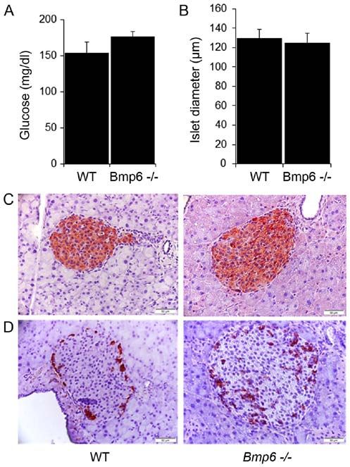
INTERNATIONAL JOURNAL OF MOlecular medicine 47: 60, 2021                                                          3

                                                                               Figure 2. Young Bmp6 ‑/‑ mice exhibit no signs of pancreatic fibrosis.
                                                                               (A) Pancreata from 3‑month‑old WT and Bmp6 ‑/‑ mice (n=6 mice/group) were
                                                                               analyzed for collagen deposition by Sirius Red staining. Original magnifi‑
                                                                               cation, x10. Scale bar, 100 µm. (B) Serum amylase and lipase levels were
                                                                               assessed as pancreatic injury markers. Data are presented as the mean ± SD.
                                                                               Bmp6 ‑/‑, bone morphogenetic protein 6‑knockout; WT, wild‑type.

                                                                               and lipase compared with those in WT mice (Fig. 2A and B).
Figure 1. Young Bmp6 ‑/‑ mice exhibit iron overload in the exocrine pancreas   Taken together, these results demonstrated that iron overload
with no histological changes. (A‑D) Pancreata from 3‑month‑old WT and          did not induce any morphological alterations of the exocrine
Bmp6 ‑/‑ mice (n=6 mice/group) were analyzed for morphological changes         pancreas in 3‑month‑old Bmp6 ‑/‑ mice.
by (A) hematoxylin and eosin staining, (B) iron measurement by Perl's
Prussian blue staining, (C) degree of pancreatic inflammation by neutro‑
phil marker CD15 and (D) macrophage marker KiM2R immunostaining.               Young Bmp6 ‑/‑ mice exhibit normal morphology and function
Original magnification, x20. Scale bar, 50 µm. Bmp6 ‑/‑, bone morphogenetic    of the endocrine pancreas. The effects of iron overload on
protein 6‑knockout; WT, wild‑type.                                             glucose homeostasis were next analyzed in 3‑month‑old
                                                                               Bmp6 ‑/‑ mice. The blood glucose levels did not differ between
                                                                               Bmp6 ‑/‑ and WT mice (Fig. 3A). Morphometric analysis of
Statistical analysis. The data are presented as the mean ± stan‑               pancreatic sections revealed no significant differences in the
dard deviation. Changes in gene expression and serum                           mean diameter of pancreatic islets compared with that in
parameter levels were evaluated using the unpaired two‑tailed                  the WT mice (Fig. 3B). Immunohistochemical staining also
Student's t‑test in Microsoft Office Excel 2016 (Microsoft                     demonstrated no changes in the distribution of β insulin cells
Corporation). P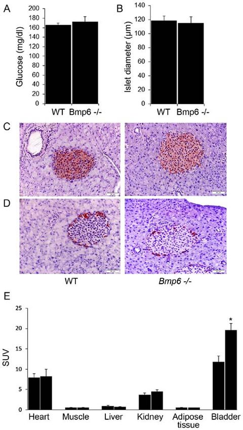
4 Pauk et al: Iron overload and exocrine pancreaTIC INSUFficiency in Bmp6 -/- mice
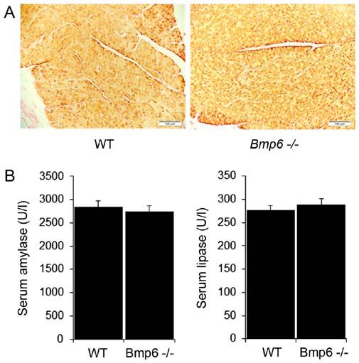
Figure 4. Aging Bmp6 ‑/‑ mice exhibit severe iron overload in the exocrine
pancreas, leading to acinar cell loss and macrophage infiltration.
(A‑D) Pancreata from 10‑month‑old WT and Bmp6 ‑/‑ mice (n=6 mice/group)
were analyzed for (A) morphological changes by hematoxylin and eosin
staining, (B) iron measurement by Perl's Prussian blue staining, (C) degree
of pancreatic inflammation by neutrophil marker CD15 (white arrows) and
Figure 3. Young Bmp6 ‑/‑ mice exhibit normal glucose metabolism. (A) Blood (D) macrophage marker KiM2R (black arrows) immunostaining. Original
glucose levels were measured following overnight fasting in 3‑month‑old WT magnification, x20. Scale bar, 50 µm. Bmp6 ‑/‑, bone morphogenetic
and Bmp6 ‑/‑ mice (n=6 mice/group). (B‑D) Pancreatic sections were analyzed protein 6‑knockout; WT, wild‑type.
for (B) mean diameter of pancreatic islets, (C) insulin and (D) glucagon secre‑
tion by immunohistochemistry. Original magnification, x20. Scale bar, 50 µm.
(E) Biodistribution of 18‑FDG in target tissues was assessed 60 min post‑i.p.
injection of 10‑18 MBq 18‑FDG (n=4 mice/group). 18‑FDG uptake was
and periacinar areas and around the pancreatic ducts (Fig. 5A).
expressed as SUV. Data are presented as the mean ± SD. *P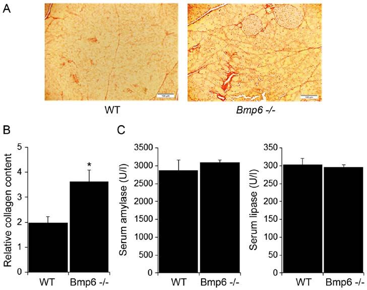
INTERNATIONAL JOURNAL OF MOlecular medicine 47: 60, 2021                                                              5

Figure 5. Aging Bmp6 ‑/‑ mice develop pancreatic fibrosis. (A and B) Pancreata from 10‑month‑old WT and Bmp6 ‑/‑ mice (n=6 mice/group) were analyzed for
(A) collagen deposition by Sirius Red staining and (B) relative amount of pancreatic collagen quantified by morphometric analysis. Original magnification, x10.
Scale bar, 100 µm. (C) Serum amylase and lipase levels were assessed as pancreatic injury markers. Data are presented as the mean ± SD. *P
6                       Pauk et al: Iron overload and exocrine pancreaTIC INSUFficiency in Bmp6 -/- mice

                                                                              axis contributed to iron accumulation in the exocrine pancreas.
                                                                              Similar sensitivity of the exocrine pancreas to iron over‑
                                                                              load has been observed in mice with a ferroportin mutation
                                                                              (SLC40A1C326S/C326S), where pancreatic failure leads to prema‑
                                                                              ture death between 7 and 14 months of age (22). These mice
                                                                              display profound weight loss attributed to malabsorption as a
                                                                              result of exocrine pancreatic insufficiency and a lack of diges‑
                                                                              tive enzymes (22). Compared with these mice, Hamp‑/‑ mice
                                                                              exhibit iron overload‑induced chronic pancreatitis, but the
                                                                              pancreatic damage is not associated with any changes in serum
                                                                              lipase levels or premature lethality (23). Chronic pancreatitis
                                                                              is characterized by inflammatory cell infiltration, acinar cell
                                                                              degeneration and development of fibrosis, which may lead to the
                                                                              impairment of exocrine and endocrine pancreatic function (41).
                                                                              In the present study, following the progression of acinar cell
                                                                              damage, 10‑month‑old Bmp6 ‑/‑ mice exhibited no changes in
                                                                              serum amylase and lipase levels compared with those in the WT
                                                                              mice, suggesting limited acinar damage that was not reflected
                                                                              by histological changes. In addition, these mice had a normal
                                                                              lifespan without exhibiting any weight loss or diarrhea (data
                                                                              not shown), suggesting that the extent of pancreatic injury and
                                                                              acinar cell loss was not sufficient for iron‑mediated lethality.
                                                                              Further studies are needed to investigate why these differences
                                                                              in mortality occur among mouse models of hemochroma‑
                                                                              tosis with similar patterns of iron deposition and consequent
                                                                              exocrine pancreatic insufficiency.
                                                                                  As demonstrated by acinar cell loss, fibrosis and infiltration
Figure 6. Damaged exocrine pancreas does not affect glucose metabolism        of inflammatory cells, the exocrine pancreas in Bmp6‑/‑ mice was
in aging Bmp6 ‑/‑ mice. (A) Blood glucose levels were measured following      severely affected in the present study. Our recent study indicated
overnight fasting in 10‑month‑old WT and Bmp6 ‑/‑ mice (n=6 mice/group).
(B‑D) Pancreatic sections were analyzed for (B) mean diameter of pancreatic
                                                                              the possible role of BMP6 in glucose homeostasis (42). The role
islets, (C) insulin and (D) glucagon secretion by immunohistochemistry.       of BMP6 in development of diabetes, although reported in the
Original magnification, x20. Scale bar, 50 µm. Data are presented as the      literature, is still not fully understood. Recently, delayed frac‑
mean ± SD. Bmp6 ‑/‑, bone morphogenetic protein 6‑knockout; WT, wild‑type.    ture healing due to the BMP6 downregulation has been reported
                                                                              in a streptozotocin‑induced rat diabetes model (43). The low
                                                                              Bmp6 expression levels in smooth muscle progenitor cells in a
hepcidin regulation has been confirmed in mice with a condi‑                  mouse diabetes model (44) and in myofibroblast progenitor cells
tional knockout of BMP2, which exhibit a hemochromatosis                      of patients with diabetes (45) has suggested the role of BMP6
phenotype similar to that observed in Bmp6 ‑/‑ mice (36).                     in vascular tissue remodeling, which may promote the genera‑
Furthermore, similar hepatocellular iron overload without                     tion of cells with antiangiogenic and profibrotic properties (46).
developing liver fibrosis has also been reported in other mouse               The present study aimed to investigate whether exocrine
models of HH such as HFE‑/‑, SLC40A1C326S/C326S, Hamp‑/‑ and                  pancreatic damage may impact glucose metabolism in aging
Hjv‑/‑ mice (22,23,38,39). Hjv‑/‑ mice have been demonstrated to              Bmp6‑/‑ mice. The islet diameters in Bmp6‑/‑ and WT mice were
be resistant to liver fibrosis even after consuming a high‑fat diet           similar during aging, suggesting no changes in β‑cell mass. In
supplemented with iron (40). The livers of Hamp‑/‑ mice exhibit               addition, no changes were observed in the insulin content by
low mRNA levels of divalent metal transporter 1 (DMT1)                        immunohistochemistry in the pancreatic tissues during aging in
and Tfr1, which mediate the uptake of non‑transferrin‑bound                   both animal groups. Blood glucose levels and 18‑FDG uptake
and transferrin‑bound iron, respectively (23). By contrast, the               in the liver, muscle and adipose tissues were comparable in
DMT1 mRNA levels are slightly increased in Hamp‑knockout                      Bmp6‑/‑ and WT mice, suggesting normal glucose metabolism.
pancreata compared with those in pancreatic tissues from WT                   By contrast, aging Bmp6‑/‑ mice exhibited moderately increased
animals, suggesting that Hamp‑/‑ mice have a transcriptional                  islet glucagon content compared with that in the WT mice,
response promoting iron uptake in the pancreas. Other studies on              indicating increased α‑cell mass. The role of altered glucagon
HH mouse models, such as HFE‑/‑, SLC40A1C326S/C326S and Hjv‑/‑                content in aging Bmp6‑/‑ mice should be additionally studied in
mice, have concluded that despite severe tissue iron overload,                animals >10 months.
these mice are protected from liver damage by yet unknown                         HFE‑/‑ mice exhibit iron accumulation in β cells, resulting
mechanisms (22,38‑40). Further studies should clarify whether                 in decreased insulin secretion compared with that in WT
hepatic iron regulation in Bmp6 ‑/‑ mice is modulated by other                animals, secondary to β cell oxidant stress and apoptosis
BMP ligands or other mechanisms, including transcriptional                    without developing diabetes (24). However, other mouse models
responses of iron transporters. The results of the present study              of HH such as Hamp‑/‑ and Hjv‑/‑ mice present with preferential
were in accordance with those of previous studies (10,17‑20),                 iron loading in the exocrine pancreas without impacting β cells
in which malfunctions of the hepcidin‑ferroportin regulatory                  and glucose homeostasis (17,38). SLC40A1C326S/C326S mice also
INTERNATIONAL JOURNAL OF MOlecular medicine 47: 60, 2021 7
display excessive iron accumulation in the pancreatic acinar Availability of data and materials
cells but differ from the other models by failure of the exocrine
pancreas (22). Despite degeneration of the pancreatic acini, All data generated or analyzed during this study are included
aged Hamp‑/‑ mice exhibit normal glucose homeostasis (17). A in this published article.
previous study has suggested an important role of zinc transporter
ZIP14, a member of the ZIP family of metal ion transporters, Authors' contributions
in contributing to non‑transferrin‑bound iron uptake and iron
accumulation by hepatocytes and pancreatic acinar cells in MP and SV conceived the study and designed the method‑
iron overload disorders (47). Since DMT1 and Tfr1 expression ology. MP, VK, VR, IDC, VF, MM and TBN performed the
levels are low in SLC40A1C326S/C326S mice, iron accumulation in experiments and analyzed the data. MP wrote the original
the exocrine pancreas may be attributed to the increased uptake draft. MP, VK, TBN and SV revised the manuscript. SV
of non‑transferrin‑bound iron via ZIP14 (22). In addition, acquired the funding and supervised the study. MP, TBN and
SLC40A1C326S/C326S mice present with a subset of acinar cells that SV confirm the authenticity of all the raw data. All authors
lack ferroportin expression, which may be prone to extensive read and approved the final manuscript.
iron accumulation and degeneration (22). Additionally, Hamp‑/‑
mice exhibit the same pattern of ferroportin expression in the Ethics approval and consent to participate
pancreas (23). Although the endocrine pancreas of Hamp‑/‑ mice
contains high levels of ferroportin, a limited number of acinar All applicable international, national, and/or institutional
cells that undergo severe iron overload have relatively low ferro‑ guidelines for the care and use of animals were followed. The
portin levels; however, it remains unclear why a number of the present study was approved by the Ethical Committee of The
acinar cells do not express ferroportin, and whether this may University of Zagreb, Faculty of Sciences (Zagreb, Croatia;
be the reason for preferential iron accumulation in the exocrine approval no. 251‑58‑508‑12‑49).
pancreas in these animals (23). Notably, SLC40A1C326S/C326S and
Hamp‑/‑ mice exhibit high hepatic levels of Bmp6 mRNA and a Patient consent for publication
functional BMP/SMAD signaling pathway (48,49), indicating
that acinar cell loss leading to exocrine pancreatic injury in Not applicable.
these mice is a direct effect of iron loading and is not attributed
to any effects of BMP6. Competing interests
The present study had certain limitations. The results of
the study are preliminary, as only two age groups of mice The authors declare that they have no competing interests.
were evaluated. To further understand the glucose metabolism
of Bmp6‑/‑ mice, glucose and insulin tolerance tests, as well as References
analysis of serum insulin and glucagon levels will be performed
in future studies. In addition, more age groups, in particular mice 1. Fleming RE and Ponka P: Iron overload in human disease.
N Engl J Med 366: 348‑359, 2012.
>10 months, may provide further insight into the changes in the 2. Nemeth E, Tuttle MS, Powelson J, Vaughn MB, Donovan A,
pancreas and other organs in this animal model during aging. Ward DM, Ganz T and Kaplan J: Hepcidin regulates cellular iron
In conclusion, the results of the present study demonstrated efflux by binding to ferroportin and inducing its internalization.
Science 306: 2090‑2093, 2004.
that Bmp6 ‑/‑ mice exhibited features of chronic pancreatitis due 3. Pietrangelo A: Hereditary hemochromatosis: Pathogenesis,
to age‑dependent iron accumulation in the exocrine pancreas. diagnosis, and treatment. Gastroenterology 139: 393‑408, 408.
However, acinar cell atrophy and exocrine pancreatic injury did e1‑e2, 2010.
4. Piubelli C, Castagna A, Marchi G, Rizzi M, Busti F, Badar S,
not induce diabetes in Bmp6 ‑/‑ mice, as these animals exhibited Marchetti M, De Gobbi M, Roetto A, Xumerle L, et al:
normal islet structure with unaltered levels of insulin produc‑ Identification of new BMP6 pro‑peptide mutations in patients
tion and blood glucose. Future studies are needed to determine with iron overload. Am J Hematol 92: 562‑568, 2017.
5. Daher R, Kannengiesser C, Houamel D, Lefebvre T,
why iron predominately accumulated in the exocrine pancreas Bardou‑Jacquet E, Ducrot N, de Kerguenec C, Jouanolle AM,
and thereby protected pancreatic islets against iron accumula‑ Robreau AM, Oudin C, et al: Heterozygous mutations in BMP6
tion and oxidative damage in Bmp6 ‑/‑ mice. pro‑peptide lead to inappropriate hepcidin synthesis and moderate
iron overload in humans. Gastroenterology 150: 672‑683,e4, 2016.
6. Urist MR: Bone: Formation by autoinduction. Science 150:
Acknowledgements 893‑899, 1965.
7. Reddi AH: Role of morphogenetic proteins in skeletal tissue
engineering and regeneration. Nat Biotechnol 16: 247‑252, 1998.
The authors would like to thank Mrs. Djurdjica Car and Mrs. 8. Wagner DO, Sieber C, Bhushan R, Börgermann JH, Graf D and
Mirjana Marija Renic (University of Zagreb, Zagreb, Croatia) Knaus P: BMPs: From bone to body morphogenetic proteins. Sci
for their technical support in animal experiments. Signal 3: mr1, 2010.
9. Sampath KT: The systems biology of bone morphogenetic
proteins. In: Bone Morphogenetic Proteins: Systems Biology
Funding Regulators. Vukicevic S and Sampath KT (eds). Springer
International Publishing, pp15‑38, 2017.
10. Andriopoulos B Jr, Corradini E, Xia Y, Faasse SA, Chen S,
The present study was supported by the Scientific Center Grgurevic L, Knutson MD, Pietrangelo A, Vukicevic S, Lin HY
of Excellence for Reproductive and Regenerative Medicine and Babitt JL: BMP6 is a key endogenous regulator of hepcidin
(project ‘Reproductive and regenerative medicine‑explo‑ expression and iron metabolism. Nat Genet 41: 482‑487, 2009.
11. Meynard D, Kautz L, Darnaud V, Canonne‑Hergaux F, Coppin H
ration of new platforms and potentials’; grant. no. GA and Roth MP: Lack of the bone morphogenetic protein BMP6
KK.01.1.1.01.0008) funded by the EU through the ERDF. induces massive iron overload. Nat Genet 41: 478‑481, 2009.8 Pauk et al: Iron overload and exocrine pancreaTIC INSUFficiency in Bmp6 -/- mice
12. Corradini E, Schmidt PJ, Meynard D, Garuti C, Montosi G, 32. Hatunic M, Finucane FM, Brennan AM, Norris S, Pacini G
Chen S, Vukicevic S, Pietrangelo A, Lin HY and Babitt JL: BMP6 and Nolan JJ: Effect of iron overload on glucose metabolism
treatment compensates for the molecular defect and ameliorates in patients with hereditary hemochromatosis. Metabolism 59:
hemochromatosis in Hfe knockout mice. Gastroenterology 139: 380‑384, 2010.
1721‑1729, 2010. 33. Dichmann DS, Miller CP, Jensen J, Scott Heller R and Serup P:
13. Yu PB, Hong CC, Sachidanandan C, Babitt JL, Deng DY, Expression and misexpression of members of the FGF and
Hoyng SA, Lin HY, Bloch KD and Peterson RT: Dorsomorphin TGFbeta families of growth factors in the developing mouse
inhibits BMP signals required for embryogenesis and iron pancreas. Dev Dyn 226: 663‑674, 2003.
metabolism. Nat Chem Biol 4: 33‑41, 2008. 34. Pratt DS and Kaplan MM: Evaluation of abnormal liver‑enzyme
14. McClain DA, Abraham D, Rogers J, Brady R, Gault P, Ajioka R results in asymptomatic patients. N Engl J Med 342: 1266‑1271,
and Kushner JP: High prevalence of abnormal glucose homeo‑ 2000.
stasis secondary to decreased insulin secretion in individuals 35. Ramos E, Kautz L, Rodriguez R, Hansen M, Gabayan V,
with hereditary haemochromatosis. Diabetologia 49: 1661‑1669, Ginzburg Y, Roth MP, Nemeth E and Ganz T: Evidence for
2006. distinct pathways of hepcidin regulation by acute and chronic
15. Mendler MH, Turlin B, Moirand R, Jouanolle AM, Sapey T, iron loading in mice. Hepatology 53: 1333‑1341, 2011.
Guyader D, Le Gall JY, Brissot P, David V and Deugnier Y: Insulin 36. Canali S, Wang CY, Zumbrennen‑Bullough KB, Bayer A
resistance‑associated hepatic iron overload. Gastroenterology 117: and Babitt JL: Bone morphogenetic protein 2 controls iron
1155‑1163, 1999. homeostasis in mice independent of Bmp6. Am J Hematol 92:
16. Hramiak IM, Finegood DT and Adams PC: Factors affecting 1204‑1213, 2017.
glucose tolerance in hereditary hemochromatosis. Clin Invest 37. Xiao X, Dev S, Canali S, Bayer A, Xu Y, Agarwal A, Wang CY
Med 20: 110‑118, 1997. and Babitt JL: Endothelial bone morphogenetic protein 2 (Bmp2)
17. Ramey G, Faye A, Durel B, Viollet B and Vaulont S: Iron over‑ knockout exacerbates hemochromatosis in homeostatic iron
load in Hepc1(‑/‑) mice is not impairing glucose homeostasis. regulator (Hfe) knockout mice but not bmp6 knockout mice.
FEBS Lett 581: 1053‑1057, 2007. Hepatology 72: 642‑655, 2020.
18. Latour C, Besson‑Fournier C, Meynard D, Silvestri L, 38. Huang FW, Pinkus JL, Pinkus GS, Fleming MD and Andrews NC:
Gourbeyre O, Aguilar‑Martinez P, Schmidt PJ, Fleming MD, A mouse model of juvenile hemochromatosis. J Clin Invest 115:
Roth MP and Coppin H: Differing impact of the deletion of 2187‑2191, 2005.
hemochromatosis‑associated molecules HFE and transferrin 39. Wagner J, Fillebeen C, Haliotis T, Charlebois E, Katsarou A,
receptor‑2 on the iron phenotype of mice lacking bone morpho‑ Mui J, Vali H and Pantopoulos K: Mouse models of hereditary
genetic protein 6 or hemojuvelin. Hepatology 63: 126‑137, 2016. hemochromatosis do not develop early liver fibrosis in response
19. Latour C, Besson‑Fournier C, Gourbeyre O, Meynard D, Roth MP to a high fat diet. PLoS One 14: e0221455, 2019.
and Coppin H: Deletion of BMP6 worsens the phenotype of 40. Padda RS, Gkouvatsos K, Guido M, Mui J, Vali H and
HJV‑deficient mice and attenuates hepcidin levels reached after Pantopoulos K: A high‑fat diet modulates iron metabolism but
LPS challenge. Blood 130: 2339‑2343, 2017. does not promote liver fibrosis in hemochromatotic Hjv‑/‑ mice.
20. Pauk M, Grgurevic L, Brkljacic J, Kufner V, Bordukalo‑Niksic T, Am J Physiol Gastrointest Liver Physiol 308: G251‑G261, 2015.
Grabusic K, Razdorov G, Rogic D, Zuvic M, Oppermann H, et al: 41. Mareninova OA, Sung KF, Hong P, Lugea A, Pandol SJ,
Exogenous BMP7 corrects plasma iron overload and bone loss in Gukovsky I and Gukovskaya AS: Cell death in pancreatitis:
Bmp6‑/‑ mice. Int Orthop 39: 161‑172, 2015. Caspases protect from necrotizing pancreatitis. J Biol Chem 281:
21. Meynard D, Vaja V, Sun CC, Corradini E, Chen S, López‑Otín C, 3370‑3381, 2006.
Grgurevic L, Hong CC, Stirnberg M, Gütschow M, et al: 42. Pauk M, Bordukalo‑Niksic T, Brkljacic J, Paralkar VM,
Regulation of TMPRSS6 by BMP6 and iron in human cells and Brault AL, Dumic‑Cule I, Borovecki F, Grgurevic L and
mice. Blood 118: 747‑756, 2011. Vukicevic S: A novel role of bone morphogenetic protein 6
22. Altamura S, Kessler R, Gröne HJ, Gretz N, Hentze MW, Galy B (BMP6) in glucose homeostasis. Acta Diabetol 56: 365‑371, 2019.
and Muckenthaler MU: Resistance of ferroportin to hepcidin 43. Guo Q, Wang W, Abboud R and Guo Z: Impairment of matura‑
binding causes exocrine pancreatic failure and fatal iron over‑ tion of BMP‑6 (35 kDa) correlates with delayed fracture healing
load. Cell Metab 20: 359‑367, 2014. in experimental diabetes. J Orthop Surg Res 15: 186, 2020.
23. Lunova M, Schwarz P, Nuraldeen R, Levada K, Kuscuoglu D, 44. Westerweel PE, van Velthoven CT, Nguyen TQ, den Ouden K,
Stützle M, Vujić Spasić M, Haybaeck J, Ruchala P, Jirsa M, et al: de Kleijn DP, Goumans MJ, Goldschmeding R and Verhaar MC:
Hepcidin knockout mice spontaneously develop chronic Modulation of TGF‑β/BMP‑6 expression and increased levels of
pancreatitis owing to cytoplasmic iron overload in acinar cells. circulating smooth muscle progenitor cells in a type I diabetes
J Pathol 241: 104‑114, 2017. mouse model. Cardiovasc Diabetol 9: 55, 2010.
24. Cooksey RC, Jouihan HA, Ajioka RS, Hazel MW, Jones DL, 45. Nguyen TQ, Chon H, van Nieuwenhoven FA, Braam B,
Kushner JP and McClain DA: Oxidative stress, beta‑cell apop‑ Verhaar MC and Goldschmeding R: Myofibroblast progenitor
tosis, and decreased insulin secretory capacity in mouse models cells are increased in number in patients with type 1 diabetes
of hemochromatosis. Endocrinology 145: 5305‑5312, 2004. and express less bone morphogenetic protein 6: A novel clue to
25. Council of Europe: European Convention for the Protection of adverse tissue remodelling? Diabetologia 49: 1039‑1048, 2006.
Vertebrate Animals used for Experimental and Other Scientific 46. Vinci MC, Gambini E, Bassetti B, Genovese S and Pompilio G:
Purposes. ETS 123, Strasbourg, 1986. When good guys turn bad: Bone marrow's and hematopoietic
26. Solloway MJ, Dudley AT, Bikoff EK, Lyons KM, Hogan BL stem cells' role in the pathobiology of diabetic complications. Int
and Robertson EJ: Mice lacking Bmp6 function. Dev Genet 22: J Mol Sci 21: 3864, 2020.
321‑339, 1998. 47. Jenkitkasemwong S, Wang CY, Coffey R, Zhang W, Chan A,
27. Bhatia M, Saluja AK, Hofbauer B, Frossard JL, Lee HS, Biel T, Kim JS, Hojyo S, Fukada T and Knutson MD: SLC39A14
Castagliuolo I, Wang CC, Gerard N, Pothoulakis C and is required for the development of hepatocellular iron overload in
Steer ML: Role of substance P and the neurokinin 1 receptor in murine models of hereditary hemochromatosis. Cell Metab 22:
acute pancreatitis and pancreatitis‑associated lung injury. Proc 138‑150, 2015.
Natl Acad Sci USA 95: 4760‑4765, 1998. 48. Kautz L, Meynard D, Monnier A, Darnaud V, Bouvet R, Wang RH,
28. Rangan GK and Tesch GH: Quantification of renal pathology by Deng C, Vaulont S, Mosser J, Coppin H and Roth MP: Iron regulates
image analysis. Nephrology (Carlton) 12: 553‑558, 2007. phosphorylation of Smad1/5/8 and gene expression of Bmp6, Smad7,
29. Roldan PS, Chereul E, Dietzel O, Magnier L, Pautrot C, Id1, and Atoh8 in the mouse liver. Blood 112: 1503‑1509, 2008.
Rbah‑Vidal L, Sappey‑Marinier D, Wagner A Zimmer L, 49. Vujić Spasić M, Sparla R, Mleczko‑Sanecka K, Migas MC,
Janier MF, et al: Raytest ClearPET (TM), a new generation small Breitkopf‑Heinlein K, Dooley S, Vaulont S, Fleming RE and
animal PET scanner. Nucl Instrum Methods Phys Res A Accel Muckenthaler MU: Smad6 and Smad7 are co‑regulated with
Spectrom Detect Assoc Equip 571: 498‑501, 2007. hepcidin in mouse models of iron overload. Biochim Biophys
30. Hansen JB, Moen IW and Mandrup‑Poulsen T: Iron: The hard Acta 1832: 76‑84, 2013.
player in diabetes pathophysiology. Acta Physiol (Oxf) 210:
717‑732, 2014. This work is licensed under a Creative Commons
31. Buysschaert M, Paris I, Selvais P and Hermans MP: Clinical Attribution-NonCommercial-NoDerivatives 4.0
aspects of diabetes secondary to idiopathic haemochromatosis International (CC BY-NC-ND 4.0) License.
in French‑speaking Belgium. Diabetes Metab 23: 308‑313, 1997.You can also read