Atopic Dermatitis - An Update - South African Pharmaceutical Journal
←
→
Page content transcription
If your browser does not render page correctly, please read the page content below
REVIEW

                     Atopic Dermatitis – An Update
                                       Engler D, BPharm, BSc Hons (Pharmacology), MSc Med (Clinical Pharmacy), Lecturer
                                                    Makola F, BPharm, Academic Intern (Masters in Pharmacy)
                                                 Magongwa NM, BPharm, Academic Intern (Masters in Pharmacy)
                                  School of Pharmacy, Faculty of Health Sciences, Sefako Makgatho Health Sciences University
                                              Correspondence to: Deirdré Engler, e-mail: deirdre.engler@smu.ac.za
                                        Keywords: Atopic dermatitis, pruritus, filaggrin, FLG gene, microbiome, Th2 cells

  Abstract
  The aetiology of atopic dermatitis is multi-faceted and affects our first line host defence, the skin. Atopic dermatitis has a significant
  influence on a patients’ social and occupational functioning and can have long-lasting effects. The signs and symptoms of AD includes
  pruritus, erythema, fissuring, and lichenification, which can be reduced by the use of moisturizing agents. Guidelines on how to
  manage atopic dermatitis aims to improve symptoms and achieve long-term disease control. Patient education remains as important
  as other treatment strategies and the pharmacist plays an integral role in educating patients on the management of their condition
  and adherence to therapy.
  Republished with updates from S Afr Pharm J 2017;84(3):36-41                                                              S Afr Pharm J 2018;85(5):29-36

Introduction
The skin is our largest organ and acts as a protective barrier
between the host organism and its external environment. Except
for preventing entry of pathogens and allergens, water loss from
the body is also minimized.1

Atopic dermatitis, also referred to as eczema, is a chronic
inflammatory skin disease that commonly affects children younger
than five years, but onset can be at any age.2 It is characterized by
                                                                                            Figure 2. Common sites of AD outbreaks5
pruritic, erythematous and scaly skin lesions that are in most cases
localized to the flexural surfaces of the body. The areas mainly                    of life, but also on patients’ mental health, and on their social and
affected include the face, scalp and extensor surfaces, especially                  emotional functioning.7 The condition is recognized as a lifelong
in infants and its onset is usually from 3 months of age.2,3                        disposition with variable clinical manifestation and expressivity, in
                                                                                    which defects of the epidermal barrier play a pivotal role.8
Atopic dermatitis (AD) is the first manifestation of allergy to present
in “the atopic march” and precedes food allergy, asthma, and                        Types of atopic dermatitis
allergic rhinitis.6 A family history of AD, asthma or allergic rhinitis
often prevails.2 The disease is debilitating and impairs a patient’s                The clinical manifestation of AD is distinct, but due to numerous
quality of life.5 Not only does AD impact on health-related quality                 differences in other aspects, AD can be categorized in two forms:
                                                                                    intrinsic (non-allergic) and extrinsic (allergic).9,10 An intrinsic form
                                                                                    of AD not associated with IgE mediated sensitization contradicts
                                                                                    the classic definition of an atopic disease and should be better
                                                                                    referred to as non-atopic AD11

                                                                                    Epidemiology
                                                                                    Atopic dermatitis is the most common chronic inflammatory
                                                                                    skin disease. The prevalence of AD has plateaued at 10-20% in
                                                                                    developed countries but continues to increase in low income
                                                                                    countries.8,12,13 Although the disease can become apparent at any
       Figure 1. Atopic dermatitis - flexural4                                      age, it manifests at an early age in approximately 60% of cases8

                                                          S Afr Pharm J     29     2018 Vol 85 No 5
REVIEW

 Table 1. Categories of atopic dermatitis9,10                                      complex and the factors triggering the disease are diverse.21 An
 Types                    Non-atopic            Atopic
                                                                                   interaction between genetics, immunologic and environmental
                                                                                   factors contribute to the pathogenesis of AD.15Genetic studies
 Onset                    Later onset           Early childhood
                                                                                   have mainly focussed on immunological mechanisms, but a
 Frequency                15% – 30%             70% - 85%
                                                                                   defect in the primary epithelial barrier has been anticipated.14It
 IgE serum levels         Normal                High
                                                                                   is important to have a good understanding of the interaction
 Specific IgE             Absent                Present for aeroallergens
                                                and foods                          between the various factors to enable effective management of
 Skin prick reactions     Negative              Positive
                                                                                   the condition.
 Cytokines: IL-4, IL-13   Low levels            High levels                        Genetics
 Skin barrier             Normal                Defect
                                                                                   Genetic factors play an important role: monozygotic twins
 Filaggrin gene           No                    Yes
 mutations                                                                         showed a consistent higher concordance rate (0.77) compared to
 Other atopic diseases    Absent                Present                            dizygotic twins (0.15). A positive parental history is furthermore
                                                                                   the strongest risk factor for AD; if the disease is present in one
with 10-20% lifetime prevalence in children.14 According to a 2013                 parent, the incidence rate is doubled or tripled should both
report, the worldwide incidence of AD averaged at 7.9% in the                      patents suffer from AD.22
6 to 7 year age group but varied a lot between regions; from 3% in                 Filaggrin, a key protein in terminal differentiation of the epidermis
the Indian subcontinent and 4.8% in the Eastern Mediterranean to                   and development of the skin barrier, protects the body from the
10.2% in Asia-Pacific and 10.3% in North America.15 In South Africa                entry of foreign environmental substances that can otherwise
the prevalence of AD in children was found to be around 17%.16                     trigger immune responses. It is synthesised as a giant precursor
While the majority of patients usually develop the disease during                  protein, profilaggrin. The latter is found within the granular
early infancy, it sometimes persists into or starts in adulthood.15                layer of the epidermis and is encoded by the FLG gene. This
Adult AD has been recognized with a prevalence of between                          gene is located within the epidermal differentiation complex on
                                                                                   chromosome 1q21.1 It has been shown that two independent
2-10%.17 The prevalence of AD among different races is not known,
                                                                                   loss-of-function genetic variants (R510X and 2282de14) in the FLG
but due to AD being a heterogeneous disorder with its different
                                                                                   gene are important pre-disposing factors for atopic dermatitis.22
genetic mechanisms, some races may be more prone to develop
the disease. Asians have increased TH17 and TH22 responses when                    Based on a patients’ ethnic background, several differences have
compared to Caucasians, while the immune response in the                           been noted in AD phenotypes. Lower rates of FLG mutations,
African population still needs to be determined.18                                 higher prevalence, and more severe, treatment-resistant AD
                                                                                   appears in African Americans compared to Caucasians. A lower
                                                                                   ceramide-to-cholesterol ratio is characteristic in normal skin of
                                                                                   African Americans and with greater trans-epidermal water loss.17

                                                                                   Several other candidate genes have been suggested to play a role
                                                                                   in AD e.g. chromosome 5q31-33, the locus containing genes for

    Figure 3. Different races and AD18

With regards to gender, various studies show the difference to be
either insignificant or male preponderant in preschool children,
while more females suffer from AD in adulthood.19,20

Pathogenesis of AD
Although the clinical picture of AD is homogenous, presenting                      Figure 4. Th2 cytokines and AD: a schematic representation11
with acute flare-ups of eczematous, pruritic lesions on dry skin                   TSLP, thymic stromal lymphopoietin; LC, Langerhans cells;
at distinct areas of the body, the pathophysiologic network is                     FLG, filaggrin; INV, involucrin; LOR, loricrin

                                                              S Afr Pharm J   30      2018 Vol 85 No 5
REVIEW
the Th2 cytokines interleukin (IL)-3, IL-4, IL-5, IL-13, and granulocyte increase the risk of AD.15 No evidence has however been reported
macrophage colony stimulating factor.14 Variants of an encoding in favour of AD management with regards to probiotics, dietary
region or functional mutations of promoter regions could be supplements, botanical extracts and homoeopathy.8
linked to the incidence of non-atopic dermatitis. Furthermore,
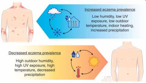
Figure 5 depicts factors such as temperature, indoor heating,
polymorphisms of the IL-18 gene may be the cause of the
humidity, and UV-light exposure influencing the prevalence of AD.
dysbalance between Th1 and Th2-immune responses, resulting
A combination of high humidity and precipitation are associated
in Th2 predominance23 The functions of Th2 cytokines include
with an increase in the disease, while high temperatures and
increased epidermal thickening, sensitization, inflammation,
exposure to UV-light has shown to have protective effects specific
pruritus, decreased expression of antimicrobial peptides and the
to AD.19
barrier proteins filaggrin, loricrin and involcrin.11
The acidic environment of the skin contributes to its barrier
Figure 4 represents a schematic illustration pertaining to Th2
function as it has a strong antibacterial effect and controls the
cytokines and AD: allergens, microbes and mechanical injury (e.g.
desquamation of corneocytes. Soaps and other detergents are
scratching) activate keratinocytes. A defective skin barrier, due to
common environmental agents that increase the skin pH. In
decreased filaggrin, is an important contributing factor. Thymic
addition, these agents emulsify skin surface lipids, change skin
stromal lymphopoietin (TSLP), IL-25 and IL-33 act on mast cells
proteases, and consequently thin the stratum corneum.15
and antigen presenting cells e.g. dendritic or Langerhans cells
with a subsequent secretion of several Th2 cytokines.11 Immunology
Another possible contributing factor to the genetic susceptibility Both the adaptive and innate immune systems are implicated in
for AD is a genetic variant of mast cell chymase, a serine protease the development of AD. A complex interaction of immune cells
secreted by skin mast cells, which may have organ specific effects.14 mediates AD skin lesions. T cells play a major role in adaptive
immunity and pathogenesis of AD. A relative imbalance of different
Environmental factors
types of T helper cells e.g. Th1, Th2, Th17 cells, is considered in
The incidence of AD worldwide and the variations thereof suggest the pathomechanism of many immune-mediated diseases. AD
that environmental factors play a pivotal role in the expression of lesions contain an increased amount of Th2 cytokines during both
AD. Some of the environmental factors implicated include climate, acute and chronic phases of the disease compared to normal skin.
diet, obesity, smoking rate, and microbial exposure.15 Chronic lesions are however associated with a reduced production
of IL-4 and IL-13 and an increased production of IL-5 and IL-12.15
Skin microbiota is involved in the homeostasis as well as
pathogenic conditions of the skin. Both Staphylococcus aureus Langerhans cells (LC) and inflammatory dendritic epidermal cells
and Streptococcus epidermidis significantly increases during (IDEC) are two types of epidermal dendritic cell populations that
exacerbation of AD.15 These bacteria release allergenic compounds are crucial elements of the immune system, bridging innate and
and superantigens (toxins)24 and can act as effective immunological adaptive immunity. These cells express increased levels of IgE high
adjuvants for increased IgE response to aeroallergens. Intense affinity receptor, FcεRI, on their surface and have the potential to
pruritus is a hallmark of AD, and skin damage due to scratching respond to numerous antigens in an antigen-specific manner. It
enhances the progress and continuance of the disease.25 was shown that Langerhans cells, activated by FcεRI, drive naïve
T cells into Th2 cells. They further highly express the receptor for
Gut microbiota might also be involved in the pathogenesis of AD thymic stromal lymphopoietin. The latter plays a critical role in
as it has been shown that children who present with AD later on in Th2 skewing and mediation of AD development.15 Refer back to
life have different early gut microbiota compared to children who Figure 5.
do not develop AD, referring both to composition and diversity.
Furthermore, systemic antibiotic treatment was reported to Inflammatory AD skin furthermore contains, except for LC, IDEC
and various T cell subsets, vast numbers of neutrophils, basophils,
eosinophils, innate lymphoid cells, natural killer cells and
fibroblasts.21
Skin barrier
Skin barrier dysfunction is a major pathogenic factor for AD.21
Causes of skin barrier dysfunction include a defect in expression of
the filaggrin gene, decrease in skin ceramides, and overactivation
of epidermal proteases. Several genetic risk loci relating to
epidermal barrier function have been identified in genome-wide
association studies.15 Filaggrin plays a pivotal role in skin barrier
Figure 5. Impact of climate on AD prevalence during childhood19 integrity: 15,21
S Afr Pharm J 32 2018 Vol 85 No 5REVIEW
• it aggregates keratin filaments into tight bundles approximately half of children affected by AD reach disease
• modifies the composition of keratinocytes and the granular cell resolution into their adult phase.28 Guidelines on how to manage
layer AD aims to improve symptoms and achieve long-term disease
control. Prior to development of novel treatments, a multistep
• moisturizes the stratum corneum
approach has included avoidance of trigger factors, continuous
Reduced availability of filaggrin metabolites alters hydration and epidermal barrier repair with emollients, and anti-inflammatory
pH of the skin.21 Patients diagnosed with non-atopic AD, lack therapy with topical corticosteroids or calcineurin inhibitors. The
barrier dysfunction and/or FLG gene mutation and it is therefore use of phototherapy or systemic immunosuppressant therapy
a feature of atopic AD. is indicated in severe and refractory cases.8Other options to
consider in the management of AD include novel treatments, e.g.
Although not an inherent factor in patients suffering from AD,
dupilumab.29 The management of AD should however always
ceramide is a lipid that is important for water retention in the
be adapted according to disease severity. Patient education
stratum corneum. The significance of ceramide is evident as an should not be neglected by health care providers and remains as
inversed correlation between transepidermal water loss and the important as other treatment strategies.23
level of ceramides in the stratum corneum of AD patients exists. A
decreased level of ceramide in patients with AD is thought to be a Non-Pharmacological management
post-inflammatory effect.15
Avoidance of trigger factors
Human kallikrein-related peptidases are key proteases for
Several factors, usually individualized and based on a previous
desquamation of corneocytes. The activity of these proteases is
reaction to an identified provoking agent, can trigger or worsen
pH dependent with enhanced activity when the pH in the stratum
the symptoms of AD.30 These factors should be avoided in order
corneum is elevated. Activation of epidermal proteases and
to reduce disease exacerbations and flare-ups. Based on clinical
subsequent increased corneocytes desquamation can induce AD-
experience, the following factors are widely assumed to worsen
like dermatitis.15
this disease: food, inhalant, or contact allergens, detergents,
Figure 6 depicts the interplay among contributing factors in the wool fabrics, climate, infections and stress. Based on evidence,
pathogenesis of AD and pruritus as a characteristic feature of aeroallergens (e.g. dust mites) and food allergens (e.g. cow’s
the disease. It is important to remember that histamine has little milk) worsens the symptoms of AD in both children and adults.
relevance in the pruritic pathway of AD and is therefore poorly Avoiding these trigger factors should be individualized and based
effective in the management of the disease. First generation on a definite history of worsening of disease symptoms after
antihistamines are indicated for their sedative effect in order to exposure.8
facilitate sleep which might be impaired due to itching.26 Second
generation antihistamines seem to have little or no value in the
Examples include:
treatment of AD, as concluded by most studies.23 Mechanical,
Non-specific wool, acids,
chemical, biological
provocating factors bleaches, water,
agents
microbes
Barrier dysfunction Evironmental Factors
Ceramide reduction Climate Smoking Examples include:
Specific
Over-desquamation Skin pH dust mites, cow's
provocating Aeroallergens and
milk, soy, wheat,
factors (evidence food allergens
Ceramide reduction Microbiome hen's egg and
based)
pollen related food
Pruritus
Figure 7: Different factors causing AD flare-ups30
Th2 cells Th17 cells Tregs Patients should avoid ingesting food that contains potential
allergens, ensure optimum skin care, and most importantly
ILC 2 Eosinophils Mast cells attain educational information on the disease itself. Avoiding
Immunological Factors
provocation factors has shown to contribute to the long-term and
effective management of AD.30
Figure 6. Interplay among contributing factors and pruritus in Moisturizing
pathogenesis of AD15
The main aim of using moisturizing agents is to combat xerosis
(the cardinal clinical feature of AD that results from a dysfunctional
AD and its management
barrier layer). Moisturizing agents predominantly reduce trans-
Atopic dermatitis is a skin condition, considered by experts to epidermal water loss and have shown to lessen symptoms and signs
be incurable.27 Contrary to this, some experts conclude that of AD, including pruritus, erythema, fissuring, and lichenification.30
S Afr Pharm J 33 2018 Vol 85 No 5REVIEW
The various available products include emollients, humectants short term and intermittent treatment.8 These agents selectively
and other miscellaneous agents. Frequent use of emollients is inhibit the production and release of pro-inflammatory cytokines
of key importance in maintaining homeostasis of the epidermal and mediators by T cells and mast cells.33 The advantage of
barrier. Emollients supply exogenous lipids and thereby soften calcineurin inhibitors is that they do not cause skin atrophy and
the skin and reduce water loss by forming an occlusive layer. is therefore of particular value in areas with delicate skin e.g. the
Emollients are mostly composed of different ingredients such as face and groin. Topical corticosteroids and calcineurin inhibitors
glycerol, petrolatum, mineral oil, purified water (e.g. Dexeryl®). should be applied proactively for two consecutive days per week
These products ensure that the skin is softened and lubricated.31 to help reduce exacerbations of the disease.8
Humectants such as urea (avoid in infants; ≤ 4% in children; up to
10% in adults), glycerine, and lactic acid added to the emollient, Table 2. Different topical corticosteroids used in AD, including their
further increase water binding in the stratum corneum.8,23 potency and trade names28
POTENCY EXAMPLES
Products containing perfumes, colourants etc. could induce
Weak
an allergic reaction or act as an irritant and best avoided. Non-
Hydrocortisone 0.5% Dilucort®, Skincalm®
soap fragrance free cleaners with a neutral to low pH are highly
recommended for patients suffering from AD. The use of wet-wrap Hydrocortisone 1% Procutan®, Mylocort ®, Biocort®
therapy, with or without a topical corticosteroid, has also been Moderately potent
seen as a favourable alternative in the management of AD. The Betamethasone Half-Strength 0.05% Sekpharma®
latter is recommended for patients with moderate to severe AD (as valerate)
in an attempt to decrease disease severity and water loss during Potent
flares.8 Beclometasone dipropionate 0.025% Beclate®
Betamethasone 0.1% (as valerate) Sekpharma®, Lenovate®, Persivate®
It is imperative to remember that Aqueous Cream BP, commonly
Betamethasone 0.05% (as Diprosone®
prescribed to relieve skin dryness, contains the surfactant dipropionate)
sodium lauryl sulphate (SLS). Sodium lauryl sulphate affects the
Diflucortolone 0.1% (as valerate) Nerisone®
effectiveness of the skin barrier. Studies have shown that SLS
Fluocinolone acetonide 0.025% Synalar®, Cortoderm®
significantly reduces the thickness of the stratum corneum with
Fluticasone propionate 0.05% Cutivate®
an overall increase in baseline transepidermal water loss (TEWL).32
Hydrocortisone butyrate Locoid®
Phototherapy Methylprednisolone aceponate 0.1% Advantan®
In cases where AD cannot be controlled with topical treatment, Mometasone Elocon®
short-term phototherapy should be considered. It has been Very potent
shown that narrow-band ultraviolet B radiation and medium-dose Clobetasol propionate 0.05% Dermovate®, Dovate®, Xenovate®
ultraviolet A1 radiation are the most effective. This therapy should
Systemic immunosuppressive therapy
not be combined with topical calcineurin inhibitors and systemic
cyclosporin treatment due to a potentially increased cumulative Several immunomodulatory agents that have been investigated
risk of skin cancer.8 for its use in AD including cyclosporine, azathioprine,
methotrexate, oral corticosteroids and mycophenolate mofetil.
Pharmacological management The immunosuppressive agents are usually used in refractory
Anti-inflammatory therapy conditions where treatment failure with topical and phototherapy
options have been observed. Most of these agents are used off-
Topical corticosteroids are the mainstay anti-inflammatory label with the exception of cyclosporine and oral corticosteroids.
treatment to control acute outbreaks of AD and has a low risk profile
when used appropriately and intermittently.8 The mechanism Novel therapy
of action of this class of medicine is linked to their potential to
Dupilumab is a novel therapeutic option that has been approved
affect various immune cells. The latter include T lymphocytes,
for use in AD. It is a fully human monoclonal antibody that exerts its
macrophages, monocytes and dendritic cells interfering with
action through binding to interleukin 4Ra which is a component
antigen processing and the suppression of pro-inflammatory
of the IL-4 and IL-13 receptors. Binding to these receptors inhibits
cytokines release.30 Low-potency corticosteroids are preferred to
their signaling and results in the downregulation of type-2
use on the face, on areas with thinner skin, and in children. Short-
immunity.29 The breakthrough of dupilumab was achieved in 2012
term treatment of severe exacerbations is an exception.8 Thicker
when results from a phase 2 trial showed safety and efficacy as
skinned areas should initially be treated with moderate to high
monotherapy for use in moderate-to-severe AD.34 The US Food
potent steroids followed by a dose reduction or an exchange to a
and Drug Administration (FDA) approved dupilumab, the first
lower potency preparation.23Refer to Table 2.
biological therapy for use in AD, in March 2017.29 The use of
Calcineurin inhibitors for topical application include tacrolimus dupilumab has enhanced confidence in the long-term control of
and pimecrolimus and are regarded as a second-line option for AD, especially in patients with resistant, extensive disease.29
S Afr Pharm J 34 2018 Vol 85 No 5REVIEW
Conclusion 16. Zhao CY, Wijayanti A, Doria MC, Harris AG, et al. The reliability and validity of outcome
measures for atopic dermatitis in patients with pigmented skin: A grey area. International
Journal of Women’s Dermatology. 2015. 1:150–154.
Atopic dermatitis affects people of all ages but children are 17. Mansouri Y, Guttman-Yassky E. Immune pathways in atopic dermatitis, and definition of
sometimes able to outgrow the condition. The physical effects biomarkers through broad and targeted therapies. J Clin Med. 2015; 4
18. Leung DYM. Atopic dermatitis: Age and race do matter! J Allergy Clin Immunol, 2015; 136:
of AD are unpleasant, but even more worrisome is the link to 1265-7. https://www.jacionline.org/article/S0091-6749(15)01348-2/pdf (Accessed 22 Oc-
psychological and emotional distress. Treatment focuses on tober 2018)
the use of topical therapies such as corticosteroids and/or 19. Silverberg JI, Hanifin J, & Simpson EL. Clinical factors associated with childhood eczema
prevalence in the United States. 2013. https://www.ncbi.nlm.nih.gov/pmc/articles/
calcineurin inhibitors to reduce the immunological response. PMC3646081 (Accessed 7 May 2016)
The use of systemic therapies is usually reserved for AD resistant 20. Chen W, Mempel M, Schober W, Behrendt H and Ring J. Gender difference, sex hormones,
and immediate type hypersensitivity reactions. Allergy. 2008; 63
to conventional therapy. Exciting new treatment options for the 21. Peng W and Novak W. Pathogenesis of atopic dermatitis. 2015. http://www.thelancet.com/
long-term control of AD, especially in patients with resistant, pdfs/journals/lancet/PIIS0140-6736(15)00149-X.pdf (Accessed 27 April 2017)
22. Palmer CN, Irvine AD, Terron-Kwiatkowski A, Zhoa Y, Liao H, Lee SP et al. Common loss of
extensive disease has recently become available and needs to be function variants of the epidermal barrier protein filaggrin are a major predisposing fac-
considered. tor for atopic dermatitis. Nat Genet. 2006; 38(4): 441-446 https://www.ncbi.nlm.nih.gov/
pubmed/16550169 (Accessed 27 April 2017)
References 23. Bieber, T. Atopic dermatitis. Ann Dermatol. 2010; 22(2)
24. Langley RJ and Toufic R. Superantigens. 2011. http://www.els.net/WileyCDA/ElsArticle/
1. Sandilands A, Sutherland C, Irvine AD, McLean WHI. 2009. Filaggrin in the frontline:
role in skin barrier function and disease. https://www.ncbi.nlm.nih.gov/pmc/articles/ refId-a0001216.html (Accessed 6 May 2017)
PMC2721001/?report=printable (Accessed 5 May 2017) 25. Pastore S, Mascia F, Giustizieri ML, Gianetti A and Girolomoni, G. Pathogenetic mechanisms
2. Stewart D, Pruvis D. Eczema in children - a topical issue. 2014. https://www.starship.org.nz/ of atopic dermatitis. 2000. https://www.iitd.pan.wroc.pl/files/AITEFullText/48z609.pdf (Ac-
media/269782/saferx_eczema_for_pharmacists_.pdf (Accessed 26 April 2017) cessed 25 April 2017)
3. Berke R, Singh A, Guralnick M. Atopic dermatitis: an overview. Am Fam Physician. 2012; 26. Alomar A and Yélamos O. Guidelines review on atopic dermatitis management. 2013.
86(1): 35-42 http://www.openaccessjournals.com/articles/guidelines-review-on-atopic-dermatitis-
4. Cunliffe T. 2018. Eczema – atopic eczema. http://www.pcds.org.uk/clinical-guidance/atop- management.pdf (Accessed 7 May 2017)
ic-eczema (Accessed 22 October 2018) 27. Sinclair W, Aboobaker J, Jordaan F, Modi D and Todd G. Management of atopic dermatitis
5. Anonymous. What is atopic dermatitis/eczema? n.d. http://www.skindermatologists.com/ in adolescents and adults in South Africa. SAMJ. 2008; 98(4)
eczema.html (Accessed 26 April 2017) 28. Rossiter D, editor. South African medicine formulary. 12th ed, Cape Town: Health and Medi-
6. Kim BS. Atopic Dermatitis. 2017. http://emedicine.medscape.com/article/1049085-over- cal Publishing Group. 2016.
view (Accessed 25 April 2017) 29. Blauvelt A, de Bruin-Weller M, Gooderham M, et al. Long-term management of moder-
7. Holm EA, Wulf HC, Stegmann H, Jemec GBE. Life quality assessment among patients with ate-to-severe atopic dermatitis with dupilumab and concomitant topical corticosteroids
atopic dermatitis. Br J Dermatol. 2006; 154(4):719–725. (LIBERTY AD CHRONOS CHRONOS): a 1-year, randomised, double-blinded, placebo-con-
8. Weidinger S, Novak N. Atopic dermatitis. 2016. http://www.thelancet.com/pdfs/journals/ trolled, phase 3. Lancet 2017; published online May 4. http://dx.doi.org/10.1016/S0140-
lancet/PIIS0140-6736(15)00149-X.pdf (Accessed 27 April 2017) 6736(17)31191-1.
9. Tokura Y. Extrinsic and Intrinsic types of atopic dermatitis. 2010. http://www.sciencedirect. 30. Ring, J., Alomar, A., Bieber, T., Deleuran, M., Fink‐Wagner, A., Gelmetti, C., Gieler, U., Lipozen-
com/science/article/pii/S0923181110000526 (Accessed 5 May 2017) cic, J., Luger, T., Oranje, A.P. and Schäfer, T., 2012. Guidelines for treatment of atopic eczema
10. Peterson JD, Lawrence SC. A comprehensive management guide for atopic dermatitis. (atopic dermatitis) part I. Journal of the European Academy of Dermatology and Venereol-
2006. http://www.medscape.com/viewarticle/551352 (Accessed 5 May 2017) ogy, 26(8), pp.1045-1060.
11. Brandst EB, Sivaprasad U. Th2 Cytokines and Atopic Dermatitis. 2011. https://www.ncbi. 31. Eichenfield, L.F., Tom, W.L., Berger, T.G., Krol, A., Paller, A.S., Schwarzenberger, K., Bergman,
nlm.nih.gov/pmc/articles/PMC3189506/ (Accessed 6 May 2017) J.N., Chamlin, S.L., Cohen, D.E., Cooper, K.D. and Cordoro, K.M., 2014. Guidelines of care for
12. Deckers IA, McLean S, Linssen S, Mommers M, van Schayck CP, Sheikh A. Investigating in- the management of atopic dermatitis: section 2. Management and treatment of atopic
ternational time trends in the incidence and prevalence of atopic dermatitis 1990–2010: dermatitis with topical therapies. Journal of the American Academy of Dermatology, 71(1),
a systematic review of epidemiological studies. PLoS One. 2012; 7(7): e39803. (Accessed pp.116-132.
26 April 2017) 32. Tsang M and Guy RH. Effect of aqueous cream BP on human stratum corneum in vivo. 2010.
13. Wedgeworth EK, Sharp H, Powell A, Flohr C. What’s news in eczema? Part 1: epidemiology https://www.ncbi.nlm.nih.gov/pubmed/20649794 (Accessed January 2016)
and pathophysiology. Dermatological Nursing. 2012; 11(4): 9-15 33. Czarnowicki, T., Krueger, J.G. and Guttman-Yassky, E., 2017. Novel concepts of prevention
14. Karagiannidou A, Botskariova S, Farmaki E, Imvrios G, Mavroudi A. Atopic dermatitis: in- and treatment of atopic dermatitis through barrier and immune manipulations with impli-
sights on pathogenesis, evaluation and management. J Allergy Ther. 2014; 5(6) cations for the atopic march. Journal of Allergy and Clinical Immunology, 139(6), pp.1723-
15. Egawa G and Weninger W. Pathogenesis of atopic dermatitis: a short review. 2015. http:// 1734.
www.tandfonline.com/doi/full/10.1080/23312025.2015.1103459 (Accessed 26 April 2017) 34. Chang, H.Y. and Nadeau, K.C., 2017. IL-4Rα inhibitor for atopic disease. Cell, 170(2), p.222.
S Afr Pharm J 36 2018 Vol 85 No 5You can also read