Ultraviolet radiation protection potentials of Methylene Blue for human skin and coral reef health
←
→
Page content transcription
If your browser does not render page correctly, please read the page content below
www.nature.com/scientificreports
OPEN Ultraviolet radiation protection
potentials of Methylene Blue
for human skin and coral reef
health
Zheng‑Mei Xiong1,3, Xiaojing Mao1,3, Mason Trappio1, Chanda Arya1,2, Jasmin el Kordi2 &
Kan Cao1,2*
Methylene blue (MB) is a century-old medicine, a laboratory dye, and recently shown as a premier
antioxidant that combats ROS-induced cellular aging in human skins. Given MB’s molecular structure
and light absorption properties, we hypothesize that MB has the potential to be considered as a
sunscreen active for UV radiation protection. In this study, we tested the effects of MB on UVB ray-
induced DNA double-strand breaks in primary human keratinocytes. We found that MB treatment
reduced DNA damages caused by UVB irradiation and subsequent cell death. Next, we compared
MB with Oxybenzone, which is the most commonly used chemical active ingredient in sunscreens
but recently proven to be hazardous to aquatic ecosystems, in particular to coral reefs. At the same
concentrations, MB showed more effective UVB absorption ability than Oxybenzone and significantly
outperformed Oxybenzone in the prevention of UVB-induced DNA damage and the clearance of UVA-
induced cellular ROS. Furthermore, unlike Oxybenzone, MB-containing seawater did not affect the
growth of the coral species Xenia umbellata. Altogether, our study suggests that MB has the potential
to be a coral reef-friendly sunscreen active ingredient that can provide broad-spectrum protection
against UVA and UVB.
Methylene blue (MB) is a common laboratory dye and widely used medicine. Since its first synthesis in 1876, MB
has been reported to have broad applications in clinical treatments of methemoglobinemia, malaria, vasoplegia,
septic shock, and Alzheimer’s disease, e tc1–3. As a diaminophenothiazine, MB has a low redox potential of 11
mV. This antioxidant property allows for efficient cycling between the oxidized form (MB) and the reduced form
leucomethylene blue (MBH2), which facilitates electron transport in the mitochondrial inner membrane and
reduces mitochondrial superoxide p roduction4,5. The use of MB in skincare helps combat ROS-induced cellular
6
aging . We have previously demonstrated that MB outperforms other leading antioxidants (e.g., N-Acetyl-L-
Cysteine (NAC), MitoQ, and mitoTEMPO) in neutralizing free radicals and improving skin longevity6. MB
also shows promising effects in stimulating the production of healthy skin fibers (e.g., Collagen and Elastin),
improving skin hydration, and promoting wound healing. Additionally, MB is environmentally friendly and is
highly compatible with marine ecosystems. It is commonly used as an aquaculture supplement to treat injured
or sick fish. The properties of MB as an oxygen transporter make it useful in the treatment of known cyanide
and nitrite poisoning of aquarium fish.
The two types of UV radiation that reach our skin are UVA and UVB waves, which cause different types of
skin damage. UVA waves are longer in wavelength (320–400 nm) and penetrate deeper into the skin, generating
high levels of ROS in the dermis and cause photoaging. UVB waves are shorter (280–320 nm) in wavelength and
carry higher energy—they cause erythema (i.e., sunburn), and DNA damage/mutations within skin cells (e.g.,
melanocytes, keratinocytes) that may lead to skin cancer.
Oxybenzone (BP-3) is one of the most commonly used chemical blockers in sunscreen products as a chemical
blocker—over 80% of chemical sunscreens contain Oxybenzone or its derivatives7. However, multiple studies
have shown that Oxybenzone is a significant hazard for aquatic ecosystems and expedites the destruction of
coral reefs8,9, which are essential for aquatic biodiversity and are considered “the rain forest of the sea”. These
compounds have been found entering the ocean environment through wastewater effluent or directly from
1
Department of Cell Biology and Molecular Genetics, University of Maryland, College Park, MD, USA. 2Mblue Labs,
Bethesda, MD, USA. 3These authors contributed equally: Zheng-Mei Xiong and Xiaojing Mao. *email: kcao@
umd.edu
Scientific Reports | (2021) 11:10871 | https://doi.org/10.1038/s41598-021-89970-2 1
Vol.:(0123456789)
www.nature.com/scientificreports/
swimmers wearing sunscreens. In particular, a study published recently showed four major toxic effects of Oxy-
benzone in early, developing coral: increased susceptibility to bleaching, DNA damage, abnormal skeleton growth
(via endocrine disruption), and gross deformities of coral nubbins8. Thus, searching for nontoxic Oxybenzone
alternatives is critical for protecting reefs and the exacerbating effects posed by climate change and bleaching. In
May 2018, Hawaii became the first state to ban chemical sunscreens containing Oxybenzone. Since then, Florida,
California, the US Virgin Islands, Australia, Mexico Bonaire, Palau, and Aruba have joined the ever-growing list
of concerned states and countries.
Given MB’s phenothiazine ring structure, solubility, and antioxidant p roperty10, we speculate that MB can be
served as a potential Oxybenzone alternative. In this study, we measured the UV absorption properties of MB
in comparison with Oxybenzone and tested the effects of MB on DNA damage and cell death induced by UVB
in primary human keratinocytes. To determine the effects of MB on UVA-induced photoaging, we compared
the cellular ROS and cell proliferation in skin cells treated with MB, Oxybenzone, or as well as two popular
antioxidants in skincare, Vitamin A and Vitamin C. Finally, we compared the effects of MB exposure on a soft
coral species Xenia umbellata with Oxybenzone.
Results
MB, but not Oxybenzone (BP‑3), mitigates DNA double‑strand breaks induced by UVB irra‑
diation in human keratinocytes. Our previous study demonstrated that MB could stimulate the DNA
repair process by upregulating DNA damage repair pathway factors, such as PARP1, which potentially can undo
the DNA damage caused by UVB radiation11. To directly analyze the impact of MB on DNA damage caused by
UVB irradiation, we conducted a dosage escalation study. Primary human skin keratinocytes were pre-treated
with 100 nM MB for two weeks and then exposed to 0, 5, 10, and 20 s of UVB rays at 1 W/cm2, which resulted
in irradiation dosages of 0, 5, 10, and 20 J/cm2 (Fig. 1A). This range of UVB irradiation intensity was chosen
empirically based on the UVB intensities detected on hot summer days at the University of Maryland campus
(Supplemental Figure S1A-C).
To quantify the levels of DNA double-strand breaks, we used a well-established DNA damage marker
γH2AX12. The histone H2AX is phosphorylated to a gamma form (γH2AX) upon DNA double-strand breakage,
and the levels of γH2AX positively correlate with the presence of DNA breaks, which is commonly generated by
UVB13. β-actin was also probed as an internal loading control of each sample, and the total expression of H2AX
was measured as well. The DNA damage could be detected as early as two hours post UVB exposure, indicated
by elevated γH2AX levels (Fig. 1B). Compared to non-treated controls, skin keratinocytes that were pre-treated
with MB showed reduced γH2AX expression upon UVB irradiation at each dosage (Fig. 1C), suggesting that MB
provides protection of the skin cells from the UVB damage. Furthermore, to determine the long-term recovery
potential in these irradiated cells, we measured γH2AX levels at 2 and 24 h after UVB irradiation (Fig. 1D–G).
We found that under a lower dosage (5 J/cm2), it was possible for the control cells (PBS pre-treatment) to recover
and express the relatively normal level of γH2AX after 24 h (Fig. 1D,E). Under the higher dosage of 10 J/cm2 UVB
irradiation, the expression of γH2AX continued to increase until at least 24 h after UVB exposure (Fig. 1F,G). In
MB pre-treated cells, the magnitude of DNA damage was significantly reduced (Fig. 1D–G).
MB prevents human keratinocytes from UVB‑induced cell death. Following the induction of
unfixable amounts of DNA damage by UVB, major routes of cell inactivation are either cell senescence or cell
death14. To further evaluate MB’s ability to protect skin cells from UVB damage, we examined the cell senescence
and viability by senescence-associated β-galactosidase (SA-β-Gal) staining and AlamarBlue cell viability assay,
respectively. Keratinocytes were first treated with MB or control PBS for two weeks, then subjected to 0, 5, 10, or
20 J/cm2 of UVB irradiation and allowed to recover for 5 days. At each UVB dosage, we found that there were
an overall healthier appearance and reduced β-Gal staining in the MB-treated cells than in control. A higher
percentage of dead keratinocytes were found in the untreated controls, as indicated by the dark staining of the
cells (Fig. 2A). Consistently, the MB-treated groups showed significantly higher survival rates after five days of
recovery (Fig. 2B).
MB shows a broad UV absorption capability. One of the major characteristics of a chemical sunscreen
blocker is its ability to absorb UV rays and convert the rays into heat. Thus, we next measured the absorption
characteristics of MB using spectrophotometric analysis and compared the results with BP-3 at the same con-
centrations of 100uM and 1 mM (Fig. 3A). The result indicated that MB has a broad-spectrum absorption range
across UVA, UVB, and UVC wavelengths. It is a more effective UV blocker than BP-3 at the high energy, short
wavelength UVB/C range (< 310 nm). For the longer wavelength lights (> 310 nm, UVA), BP-3 absorbs better
than MB.
MB is a more robust ROS scavenger than BP‑3, Vitamin A, and Vitamin C in skin cells. UVA
rays are longer in wavelength and penetrate deeper into the skin, generating high levels of ROS, leading to skin
cell aging and death. To compare the MB’s and BP-3’s effects on combating ROS, next, we conducted the cell
proliferation and ROS assays in keratinocytes after two-week treatment with either MB or BP-3 at the 100 nM
and 1 µM concentrations (Fig. 3B,C). We observed an increase in cell proliferation in MB-treated human pri-
mary keratinocytes, indicating MB is beneficial to overall skin cell longevity. Although BP-3 has an EWG hazard
score of 8 (toxic) on the human body, the proliferation study did not reveal any significant inhibitory effects of
BP-3 (Fig. 3B), which suggests that at the 100 nM and 1 µM concentrations, BP-3 is probably safe to human skin
cells. Moreover, consistent with our previous study6, an evident dosage-dependent reduction in ROS levels in
Scientific Reports | (2021) 11:10871 | https://doi.org/10.1038/s41598-021-89970-2 2
Vol:.(1234567890)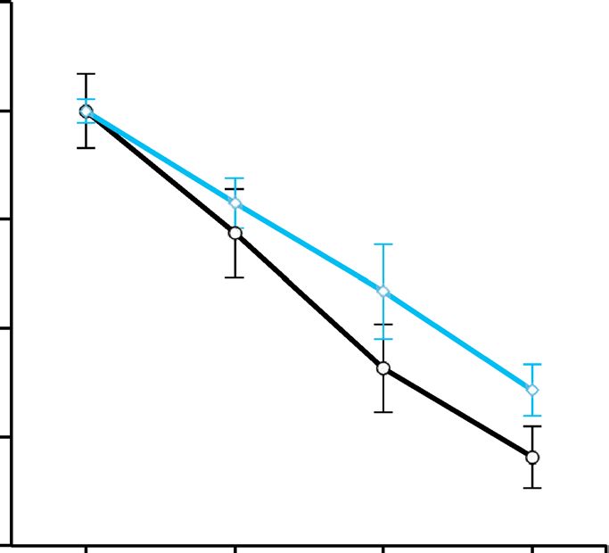
www.nature.com/scientificreports/
Figure 1. MB alleviates DNA damage induced by UVB irradiation in human keratinocytes. (A) Schematic
diagram showing the experimental timeline starting from pre-incubation of keratinocytes with 100 nM MB or
PBS for 2 weeks, then exposure to UVB light, and ending with cell pellets collection before irradiation (time
0), and 2 or 24 h post-irradiation for Western blotting analysis. (B) Representative Western blotting images
with anti-γH2AX, H2AX, and β-actin antibodies showing DNA damage response after 2 h of UVB irradiation
at different dosages. No UVB control is designated as “⎯”. (C) Quantification of γH2AX band intensities
shown in (B), which is normalized to total H2AX. * p < 0.05; ** p < 0.01 MB versus Control at the same dose.
(D,F) Representative Western blotting images with anti-γH2AX, H2AX, and β-actin antibodies in human
keratinocytes. The cell pellet samples were collected at 0, 2, or 24 h after UVB irradiation at 5 J/cm2 (D) or 10
J/cm2 (F). Zero hour control is designated as “⎯”. (E, G) Quantification of γH2AX band intensities shown in
(D,F), which is normalized to total H2AX. * p < 0.05; MB versus Control at the same time point.
MB-treated cells was found (Fig. 3C). In contrast, no significant changes in ROS levels were observed in the BP-3
treated cells compared to Dimethyl Sulfoxide (DMSO)-treated controls, indicating BP-3 is not a ROS scavenger.
To further investigate MB’s capability as a ROS scavenger and UVA protective agent for the skin, we obtained
four human skin fibroblast cell lines from young male, old male, young female, and old female donors (Detailed
cell line information in Supplemental Table S1). We evaluated the effectiveness of MB in comparison with Vita-
min A and Vitamin C, both of which are popular antioxidants in skincare p roducts15. We first monitored the cell
proliferation for a 2-week time frame. In this experiment, we observed that MB enhanced the cell proliferation
greatly in all four lines with comparable effectiveness to Vitamin C (Fig. 4A,B). Vitamin A, conversely, did not
affect the cell proliferation rates (Fig. , B). The combination of MB and Vitamin C synergistically boosted the
cell proliferation capacities even higher in all four lines (Fig. 4A,B). Next, we measured the final mitochondrial
ROS after a four-week treatment period. Surprisingly, MB was the only treatment that consistently reduced the
mitochondrial ROS in all four cell lines. The combination of MB and Vitamin C worked more effectively in old
fibroblasts in ROS clearance, while in young cell lines, this combination has less of an effect (Fig. 4C,D).
MB is non‑toxic to coral health as compared to Oxybenzone (BP‑3). The results of the previous
experiments support that MB’s potential as an effective protectant for both UVA and UVB. To determine MB’s
Scientific Reports | (2021) 11:10871 | https://doi.org/10.1038/s41598-021-89970-2 3
Vol.:(0123456789)www.nature.com/scientificreports/
Figure 2. MB prevents human keratinocytes from UVB-induced cell death. (A) Representative images of
senescence-associated β-galactosidase (SA-β-gal) staining in human keratinocytes 5 days after UVB irradiation
at 0, 5, 10, and 20 J/cm2. The cells were pretreated with Control (PBS) or 100 nM MB for two weeks before
UVB irradiation. Scale bar = 200 μm. (B) AlarmaBlue cell viability assay in Control and MB-treated human
keratinocytes at day five after UVB exposure at denoted dosages. The cells were pretreated with Control (PBS) or
100 nM MB for two weeks before UVB irradiation. * p < 0.05 MB versus Control.
effects on coral reefs, we grew the soft coral species Xenia umbellata in seawater containing either 1 μM MB
or 1 μM Oxybenzone for one week (Fig. 5A). We observed that by Day 7 in the Oxybenzone -treated coral,
tissue debris was found at the bottom of the coral incubating bag (Fig. 5B). Also, following the collection of
the tissue debris, we observed the lack of pulsing from the treated coral. Pulsing in the Xenia coral completely
ceased (Supplementary videos). The coral colony appeared limp, disintegrated, and fell away from the light above
(Fig. 5B), possibly indicating an expulsion of symbiotic zooxanthellae species from within the coral to the treated
seawater16. None of these observations were found within the MB-treated corals (Supplementary videos).
Discussion
The two types of sunscreens on the market include physical blockers that scatter UV rays, i.e., Zinc Oxide, Tita-
nium Dioxide, and chemical absorbers, such as Oxybenzone, that absorb UVA and UVB rays; both are designed
to prevent UV rays from penetrating the skin. Although this traditional method for sun protection is mostly
effective, even the sunscreens with the highest SPFs can only stop up to 98% of UVB rays from penetrating the
skin17. The remaining UV waves that penetrate the skin can still cause damage. Moreover, penetration of UV
waves into the skin can have positive health benefits, such as the generation of Vitamin D. Thus, we argue that
new sun protection products should also be geared towards reversing the negative effects of UV radiations in
addition to blocking UV waves.
MB is the first man-made compound that was synthesized in 187618. It has been approved for medical use
by the FDA, and it is also on the WHO list of safe and essential medicines. Our previous work indicated that
MB activates the DNA damage repair pathway inside the cell, possibly through activating DNA damage repair
pathways11. In this study, we tested the ability of MB on mitigating UVB -induced DNA damages in primary
Scientific Reports | (2021) 11:10871 | https://doi.org/10.1038/s41598-021-89970-2 4
Vol:.(1234567890)www.nature.com/scientificreports/
Figure 3. MB is a potent ROS scavenger with stronger UVB absorption than Oxybenzone (BP-3). (A) Graphs
of UV absorption spectrum of MB and BP-3 at 100 μM and 1 mM. PBS, the solvent of MB, was used to establish
blank reading for UV spectrum measurement. DMSO, the solvent of BP-3, at a final concentration of 0.01%
in PBS, was measured for adjustment of BP-3 absorbance. (**p < 0.01, *** p < 0.001 MB 1 mM vs. BP-3 1 mM).
(B) Keratinocytes were seeded at the same cell number on day 0 and maintained in culture medium containing
PBS, 100 nM or 1 µM MB, 0.01% DMSO (solvent for BP-3), 100 nM or 1 µM BP-3, respectively. Cell counting
was conducted on day 10 and day 13 to compare the cell proliferation under different treatments. *** p < 0.001;
****p < 0.0001 comparing with PBS on the same day ; ## p < 0.01 #### p < 0.0001 MB versus BP-3 on the same
day and same concentration. (C) Keratinocytes pretreated with PBS, 100 nM MB, 1 µM MB, 0.01% DMSO, 100
nM BP-3 and 1 µM BP-3 for 2 weeks were examined for mitochondrial specific superoxide (MitoSOX) levels by
FACS on day 14. * p < 0.05 comparing with PBS; # p < 0.05 MB 1 µM versus BP-3 1 µM.
human keratinocytes. We found that MB treatment reduced the overall levels of DNA double strand breaks
caused by UVB irradiation and increased cell survival rate (Figs. 1, 2). Absorption spectroscopic analysis revealed
that MB has a broad-spectrum light absorption range covering from UVA, UVB to UVC wavelength (Fig. 3A).
Thus, MB has the desired abilities of both UVB blocking and DNA damage mitigation. It not only absorbs UVB
but also reduces the DNA damages induced by UVB, thereby promoting cell survival post UVB irradiation.
UVA radiation significantly increases levels of ROS inside the skin. These high levels of ROS result in damage
to skin cells and their surrounding connective tissue, which leads to the physical signs (e.g., wrinkles, reductions
in skin elasticity) of premature photoaging. Endogenous antioxidants within the skin are depleted during elevated
levels of UVA generated ROS. As a result, many anti-aging skincare creams deliver exogenous antioxidants to
help combat ROS. Vitamin A (commonly known as Retinol in skincare) and Vitamin C are popular active anti-
oxidants used in skincare. In the comparative studies demonstrated in Figs. 3B, C and 4, both MB and Vitamin
C promoted cell proliferation, but BP-3 and Vitamin A had little effects at the tested concentrations (Figs. 3B
and 4A,B). Among these four compounds, MB is no doubt the most effective ROS scavenger in human skin cells
(Figs. 3C and 4C,D). Additionally, we found that when MB is combined with Vitamin C, a positive synergistic
benefit was observed. The MB plus Vitamin C treated cells showed the highest skin cell proliferation rate and low-
est cellular stress as determined by mitochondrial ROS assay. Furthermore, this synergistic effect on combating
ROS is more prominent in old skin cells than in young cells (Fig. 4C,D). These results indicate the combination
of MB and Vitamin C could be used in skincare to deliver optimal anti-aging effects.
Besides MB’s UVA and UVB blocking and mitigating effects, the soft coral experiment indicated that at a high
concentration (1uM), MB posed no negative effects on coral health and growth (Fig. 5), which was not a surprise
Scientific Reports | (2021) 11:10871 | https://doi.org/10.1038/s41598-021-89970-2 5
Vol.:(0123456789)www.nature.com/scientificreports/
Figure 4. MB is a more effective ROS scavenger than Vitamin A and Vitamin C in human skin fibroblasts.
(A) Young (Left) and old (Right) male fibroblasts proliferation during the fourteen-day treatment with control
PBS, 0.01% DMSO, 100 nM MB, 100 nM Vitamin A, 100 μM Vitamin C, and 100 nM MB + 100 μM Vitamin
C, respectively. p < 0.05, **p < 0.01 comparing with Control PBS on the same day; # p < 0.05 or ## p < 0.01
comparing with MB + Vitamin C-treated cells on the same day. (B) Young (Left) and old (Right) female
fibroblasts proliferation during the fourteen-day treatment with control PBS, 0.01% DMSO, 100 nM MB, 100
nM Vitamin A, 100 μM Vitamin C, and 100 nM MB + 100 μM Vitamin C, respectively. p < 0.05, **p < 0.01
comparing with Control PBS on the same day; # p < 0.05 or ## p < 0.01 comparing with MB + Vitamin C-treated
cells on the same day. (C) Comparison of the mitochondrial specific superoxide (MitoSOX) levels in young
and old male fibroblasts after the treatment of control PBS, 0.01% DMSO, 100 nM MB, 100 nM Vitamin A, 100
μM Vitamin C, or 100 nM MB + 100 μM Vitamin C for 4 weeks. p < 0.05, **p < 0.01 comparing with Control
PBS. (D) Comparison of the final mitochondrial specific superoxide (MitoSOX) levels in young and old female
fibroblasts after the treatment of PBS control, 0.01% DMSO, 100 nM MB, 100 nM Vitamin A, 100 μM Vitamin
C, and 100 nM MB + 100 μM Vitamin C for 4 weeks. *p < 0.05, **p < 0.01 comparing with Control PBS.
as MB has been commonly used by fish hobbyists as an aquaculture supplement. In contrast, Oxybenzone, which
was reported to pollute the marine ecosystem previously8,9, impaired the growth of coral reefs and caused coral
bleaching and death in our experiments.
Altogether, these results suggest that MB has great potential to serve as a coral reef-friendly, Oxybenzone
alternative in sunscreen products. It will bring many benefits that outweigh most of the current sunscreen actives,
particularly relative to Oxybenzone. Firstly, as a potent antioxidant, the MB-based sunscreen may protect skin
from ROS and photoaging caused by UVA rays6, which is lacking in current sunscreen products19. Secondly, MB
has a broad-spectrum absorption covering UVA and UVB and is able to lessen the UVB-induced DNA damages
and protect cells from cell death, possibly by activating DNA repair pathways. Therefore, MB has the abilities
of both UVB blocking and UVA/B damage mitigation. Lastly and most importantly, the MB-based sunscreen is
safe for marine life and ecosystems, especially for the already endangered coral reefs. In the US, the FDA regu-
lates sunscreens as an over-the-counter (OTC) and has established a monograph for active ingredients. New
ingredients have to undergo rigorous and costly safety testing to be considered for inclusion in the sunscreen
monograph. Future work will include conducting the required FDA studies in order to submit an application
for full inclusion of Methylene Blue in the sunscreen monograph.
Scientific Reports | (2021) 11:10871 | https://doi.org/10.1038/s41598-021-89970-2 6
Vol:.(1234567890)www.nature.com/scientificreports/
Figure 5. MB is non-toxic to coral health compared to oxybenzone (BP-3). (A) Schematic experimental
timeline showing the two administrations of PBS control, DMSO 0.005%, 1 μM MB, and 1 μM Oxybenzone
on day 1 and day 4, respectively. (B) Representative images of coral colonies growing on the stone base in the
seawater containing different chemicals on day 0 (before adding chemicals), day 1, day 3, and day 7. Red arrows
indicate the death of corals in the Oxybenzone-treated experiment on day 7.
Methods
Cell culture and drug treatment. The normal human primary epidermal keratinocyte line (PCS-
200-010) was obtained from ATCC. Keratinocytes were thawed and cultured in Dermal Cell Basal Medium
(PCS200030, ATCC) supplemented with Keratinocyte Growth Kit (PCS200040, ATCC) at 37 °C with 5% C O2.
Four of normal human skin fibroblasts were purchased from Coriell Institute (detailed information described in
Table S1). All fibroblast cells were cultured in MEM (Life Sciences) supplemented with 15% FBS (Gemini Bio-
Products) and 2 mM L-glutamine (Life Sciences) at 37 °C with 5% CO2.
Methylene blue (1%, Akron) and L-Ascorbic acid (Vitamin C, A4544, Sigma-Aldrich) were prepared in PBS.
Retinoid acid (Vitamin A, R2500, Sigma-Aldrich), Oxybenzone (BP-3, H36206, Sigma-Aldrich), and Octinoxate
(PHR1080, Sigma-Aldrich) were dissolved in DMSO (final concentration 0.01% in culture medium or 0.005%
in seawater).
Drug treatment and UVB irradiation. Keratinocytes were seeded in culture dishes at a density of 2500
cell/cm2 and fed with fresh medium containing MB or vehicle PBS on next morning to start the treatment for 1
or 2 weeks before UVB irradiation. The cells were subcultured every 2 or 3 days when the culture has reached
about 80% confluency.
UVB treatment on human keratinocytes was conducted in a tissue culture hood with a handheld UV lamp
emitting a peak wavelength of 302 nm (UVM-57, Analytik Jena). The UVB intensity was measured each time
before irradiation with a digital Radiometer (UVP UVX™, Analytik Jena) that is attached to a radiometer sensor
(UVX-31 300NM, Analytik Jena). Briefly, the cells were gently rinsed out once with PBS then exposed to UVB
at the intensity of 1 W/cm2 for 0 s, 5 s, or 10 s, which is equal to various doses at 0, 5, or 10 J/cm2, respectively.
The cells were then immediately fed with fresh medium containing MB or PBS and collected after 2- or 24-h for
western blotting analysis.
Western blotting. Western blotting was done following the same protocol as described in Xiong et al.14
Antibodies used in this study included: γH2AX (Ab11174, Abcam), β-actin (A3854, Sigma-Aldrich), H2AX
(600201, Biolegend).
Scientific Reports | (2021) 11:10871 | https://doi.org/10.1038/s41598-021-89970-2 7
Vol.:(0123456789)www.nature.com/scientificreports/
Senescence associated β‑Galactosidase activity assay. We followed the manufacturer’s protocol
(#9860; Cell Signaling). Briefly, keratinocytes cells grown on a six-well plate were fixed in 1X fixative solution
for 10 min and then stained overnight at 37 °C with the β-galactosidase staining solution at pH 6.0 for 15 h.
Bright-field and phase contrast images were taken with a Zeiss AX10 microscope and a SPOT PURSUIT camera.
AlamarBlue cell viability assay. Cells growing in a culture plate were incubated in complete medium
containing 10% alamarBlue reagent (DAL1025, ThermoFisher Scientific) for 3 h and the plates were read by
the fluorescence spectrophotometer using 570/590 nm (excitation/emission) filter settings (Molecular Devices
Spectramax M5).
UV spectra measurement. The absorbance of MB or BP-3 to broad UV–VIS spectrum was measured in
triplicate using NanoDrop 2000 spectrophotometer (ThermoFisher Scientific). Briefly, 2 μL of MB or BP-3 was
pipetted onto the pedestal for measurement after the establishment of a blank using PBS under the setting of
UV–Vis application. The solvent of BP-3, DMSO, at a final concentration of 0.01% in PBS, was also measured for
adjustment of BP-3 absorbance.
Flow cytometry analysis for mitochondrial ROS. Mitochondrial superoxide was measured following
the same protocol as described in Xiong et al.14 Briefly, cells cultured on 60-mm dishes were incubated with fresh
complete medium containing 5 μM MitoSOX Red (Life Technologies, M36008) at 37 °C. After 30 min, stained
cells were harvested by trypsin digestion and rinsed twice with PBS. Single cell suspensions in 400 ml PBS were
prepared for FACS analysis (FACS Canto II; BD). MitoSOX Red was excited by a laser at 488 nm, and the data
was collected at 582 ± 21 nm.
Coral growth and treatment. Soft Corals (Xenia umbellata) frags were purchased from ORA. The cor-
als were cultivated in a 12 gallon NanoCube aquarium with a 12 h light cycle (JBJ). Reef Salts (Instant Ocean)
were used to bring the specific gravity of the aquarium and treatment water to 1.024. Corals were fed with liquid
and dry feeds (Polyp Lab) and supplemented with potassium iodide (SeaChem). Water temperature was held
constant at 25.5 °C by a submersible HT10 thermometer (Tetra). Chemical tests on coral health started after
one month acclimation period. On day 0, each coral colony was gently transferred into a 2 mil thick resealable
bag (VWR) containing 600 mL seawater. The bag was then magnetically mounted on the wall of the aquar-
ium (Siam). The coral colony was imaged two hours after settlement and various chemicals were administered
directly to the bag water on day 0 and day 4.
Statistical analysis. Results are presented as the mean ± standard deviation. Data statistical analysis was
performed using one-way ANOVA with GraphPad Prism 9, and a p value less than 0.05 was considered signifi-
cant.
Received: 19 January 2021; Accepted: 19 April 2021
References
1. Schirmer, R. H., Adler, H., Pickhardt, M. & Mandelkow, E. Lest we forget you—methylene blue. Neurobiol. Aging 32(2325), e7-2325.
e16 (2011).
2. Paciullo, C. A., McMahon Horner, D., Hatton, K. W. & Flynn, J. D. Methylene blue for the treatment of septic shock. Pharmaco-
therapy 30, 702–715 (2010).
3. Atamna, H. & Kumar, R. Protective role of methylene blue in Alzheimer’s disease via mitochondria and cytochrome c oxidase. J.
Alzheimers. Dis. 20(Suppl 2), S439–S452 (2010).
4. Atamna, H. et al. Methylene blue delays cellular senescence and enhances key mitochondrial biochemical pathways. FASEB J. 22,
703–712 (2007).
5. Wu, J. J. et al. Mitochondrial dysfunction and oxidative stress mediate the physiological impairment induced by the disruption of
autophagy. Aging (Albany. NY.) 1, 425–437 (2009).
6. Xiong, Z. M. et al. Anti-aging potentials of methylene blue for human skin longevity. Sci. Rep. 7, 1–12 (2017).
7. Nash, J., Tanner, P., Grosick, T. & Zimnawoda, M. Sunscreen market analysis: the evolution and use of UVA-1 actives. J. Am. Acad.
Dermatol. 50, P34 (2004).
8. Danovaro, R. et al. Sunscreens cause coral bleaching by promoting viral infections. Environ. Health Perspect. 116, 441–447 (2008).
9. Downs, C. A. et al. Toxicopathological effects of the sunscreen UV filter, oxybenzone (Benzophenone-3), on coral planulae and
cultured primary cells and its environmental contamination in Hawaii and the U.S. Virgin Islands. Arch. Environ. Contam. Toxicol.
70, 265–288 (2016).
10. Cenens, J. & Schoonheydt, R. A. Visible spectroscopy of methylene blue on hectorite, laponite B, and Barasym in aqueous suspen-
sion. Clays Clay Miner. 36, 214–224 (1988).
11. Zhang, H. et al. Loss of H3K9me3 correlates with ATM activation and histone H2AX phosphorylation deficiencies in Hutchin-
son–Gilford progeria syndrome. PLoS ONE 11, e0167454 (2016).
12. Kuo, L. J. & Yang, L. X. γ-H2AX—a novel biomarker for DNA DOUBLE-STRAND BREAKS. Vivo 22, 305–309 (2008).
13. Garcia-Canton, C., Anadón, A. & Meredith, C. γH2AX as a novel endpoint to detect DNA damage: Applications for the assessment
of the in vitro genotoxicity of cigarette smoke. Toxicol. Vitro 26, 1075–1086 (2012).
14. Xiong, Z. M. et al. Methylene blue alleviates nuclear and mitochondrial abnormalities in progeria. Aging Cell 15, 279–290 (2016).
15. Lee, C.-M. Fifty years of research and development of cosmeceuticals: a contemporary review. J. Cosmet. Dermatol. 15, 527–539
(2016).
Scientific Reports | (2021) 11:10871 | https://doi.org/10.1038/s41598-021-89970-2 8
Vol:.(1234567890)www.nature.com/scientificreports/
16. Studivan, M. S., Hatch, W. I. & Mitchelmore, C. L. Responses of the soft coral Xenia elongata following acute exposure to a chemical
dispersant. Springerplus 4, 80 (2015).
17. Dale Wilson, B., Moon, S. & Armstrong, F. Comprehensive review of ultraviolet radiation and the current status on sunscreens. J.
Clin. Aesthet. Dermatol. 5, 18–23 (2012).
18. Guttmann, P. & Ehrlich, P. Über die Wirkung des Methylenblau bei Malaria. Berliner Klinische Wochenschr. 28, 953–956 (1891).
19. Gasparro, F. P. Sunscreens, skin photobiology, and skin cancer: the need for UVA protection and evaluation of efficacy. Environ.
Health Perspect. 108, 71–78 (2000).
Acknowledgements
Drs. Yixian Zheng and Chen-Ming Fan (Carnegie) for helpful discussions, Mr. Soheil Jafari for Coral Reef tech-
nical support, and Drs. David Mosser and Joseph Smith for COI grant management. This research is funded by
an NSF STTR Grant (JEK).
Author contributions
K.C., J.E.K. and C.A. synthesized the idea, Z.X. and M.T. did the experiments, Z.X., X.M. and K.C. analyzed the
data, wrote the main manuscript text and prepared figures. All authors reviewed the manuscript.
Funding
National Foundation for Science and Technology Development, Grand No. 1842745.
Competing interests
Cao is the founder of Mblue Labs and a professor at the University of Maryland, College Park. El Kordi is the CEO
of Mblue Labs. The University of Maryland College Park and Mblue Labs have submitted a joint PCT application
on the potential use of methylene blue as an active ingredient in sunscreen products. Xiong, Mao, Trappio, and
Arya have no competing interests as defined by Nature Research, or other interests that might be perceived to
influence the results and/or discussion reported in this paper.
Additional information
Supplementary Information The online version contains supplementary material available at https://doi.org/
10.1038/s41598-021-89970-2.
Correspondence and requests for materials should be addressed to K.C.
Reprints and permissions information is available at www.nature.com/reprints.
Publisher’s note Springer Nature remains neutral with regard to jurisdictional claims in published maps and
institutional affiliations.
Open Access This article is licensed under a Creative Commons Attribution 4.0 International
License, which permits use, sharing, adaptation, distribution and reproduction in any medium or
format, as long as you give appropriate credit to the original author(s) and the source, provide a link to the
Creative Commons licence, and indicate if changes were made. The images or other third party material in this
article are included in the article’s Creative Commons licence, unless indicated otherwise in a credit line to the
material. If material is not included in the article’s Creative Commons licence and your intended use is not
permitted by statutory regulation or exceeds the permitted use, you will need to obtain permission directly from
the copyright holder. To view a copy of this licence, visit http://creativecommons.org/licenses/by/4.0/.
© The Author(s) 2021
Scientific Reports | (2021) 11:10871 | https://doi.org/10.1038/s41598-021-89970-2 9
Vol.:(0123456789)You can also read