Novel canine isocitrate dehydrogenase 1 mutation Y208C attenuates dimerization ability
←
→
Page content transcription
If your browser does not render page correctly, please read the page content below
ONCOLOGY LETTERS 20: 351, 2020

                Novel canine isocitrate dehydrogenase 1 mutation
                     Y208C attenuates dimerization ability
          SHOTA KAWAKAMI1, MASAKI MICHISHITA2,3, MOTOHARU SAKAUE4, MASAMI MORIMATSU5,
            MITSUKI UEMURA1,6, NOBUAKI KASHIWAGI1,6, MARIKA MAEDA6, YUKINO MACHIDA2,
          DAIGO AZAKAMI7, AI S. EGUSA8, ERI ONOZAWA1, KATSUMI ISHIOKA1, MASAMI WATANABE9,
                   YOSHIKAZU TANAKA3,6, TOSHINORI OMI1,3 and KAZUHIKO OCHIAI1,3,6

      1
     School of Veterinary Nursing and Technology, Faculty of Veterinary Science; 2Laboratory of Veterinary Pathology,
 School of Veterinary Science; 3Research Center for Animal Life Science, Nippon Veterinary and Life Science University,
Musashino, Tokyo 180‑8602; 4Laboratory of Anatomy II, Department of Veterinary Medicine, School of Veterinary Medicine,
             Azabu University, Sagamihara, Kanagawa 252‑5201; 5Laboratory of Animal Science and Medicine,
    Department of Disease Control, Graduate School of Veterinary Medicine, Hokkaido University, Sapporo 060‑0818;
     6
       Laboratory of Veterinary Hygiene, School of Veterinary Science, Nippon Veterinary and Life Science University,
     Musashino, Tokyo 180‑8602; 7Laboratory of Clinical Oncology, Cooperative Department of Veterinary Medicine,
              Faculty of Agriculture, Tokyo University of Agriculture and Technology, Fuchu, Tokyo 183‑8538;
       8
         Department of Applied Life Science, Faculty of Food Science, Nippon Veterinary and Life Science University,
                    Musashino, Tokyo 180‑8602; 9Department of Urology, Graduate School of Medicine,
                  Dentistry and Pharmaceutical Sciences, Okayama University, Okayama 700‑8558, Japan

                                     Received June 12, 2020; Accepted September 11, 2020

                                                    DOI: 10.3892/ol.2020.12214

Abstract. Isocitrate dehydrogenase 1 (IDH1) mutations are           with that of wild‑type (WT) cIDH1, but the attenuation of
common in gliomas, acute myeloid leukemia, and chondro‑             Y208C was less intense than that of the R132H mutation. The
sarcoma. The mutation ‘hotspot’ is a single arginine residue,       induction of HIF‑1α response element activity and cell reten‑
R132. The R132H mutant of IDH1 produces the 2‑hydroxy‑              tion of HIF‑1α were not increased by Y208C overexpression.
glutarate (2‑HG) carcinogen from α‑ketoglutarate (α‑KG). The        In silico and cell biological analysis of IDH1 dimerization
reduction of α‑KG induces the accumulation of hypoxia‑induc‑        revealed that the Y208C mutation, but not the R132H muta‑
ible factor‑1α subunit (HIF‑1α) in the cytosol, which is a          tion, attenuated binding activity with WT cIDH1. These data
predisposing factor for carcinogenesis. R132H is the most           suggested that the attenuation of dimerization by the Y208C
common IDH1 mutation in humans, but mutations at the R132           mutation may cause tumorigenesis through different mecha‑
residue can also occur in tumor tissues of dogs. The current        nisms other than via 2‑HG production by the IDH1 R132
study reported the discovery of a novel Tyr208Cys (Y208C)           mutation.
mutation in canine IDH1 (cIDH1), which was isolated from
2 of 45 canine chondrosarcoma cases. As the genomic DNA             Introduction
isolated from chondrosarcoma tissue was mutated, but that
isolated from blood was not, Y208C mutations were considered        Gliomas are intracranial tumors that are thought to develop
to be spontaneous somatic mutations. The isocitrate dehydro‑        from astrocytes or oligodendrocytes (1,2). A genome‑wide
genase activity of the Y208C mutant was attenuated compared         mutation analysis of human brain tumors revealed predomi‑
                                                                    nant somatic mutations in the gene encoding isocitrate
                                                                    dehydrogenase 1 (IDH1) in glioblastoma (GBM) (3,4). Further
                                                                    analysis of the IDH1 gene structure confirmed these findings,
                                                                    identifying IDH1 mutations in over 70% of secondary GBM
Correspondence to: Dr Kazuhiko Ochiai, Laboratory of Veterinary
                                                                    or low‑grade gliomas, but infrequently in primary GBM (5,6).
Hygiene, School of Veterinary Science, Nippon Veterinary and Life
Science University, 1‑7‑1 Kyonan‑cho Musashino, Tokyo 180‑8602,     Almost all IDH1 mutations were identified at the 132th argi‑
Japan                                                               nine residue (R132). In addition, R132 mutations of IDH1
E‑mail: kochiai@nvlu.ac.jp                                          have been identified in acute myeloid leukemia (AML) and
                                                                    chondrosarcoma (CS) (7‑9). R132 mutants of IDH1 induced
Key words: canine, chondrosarcoma, dimerization, isocitrate         the reduction of α‑ketoglutarate (α‑KG) and increased the
dehydrogenase 1, glioma, mutation                                   2‑hydroxyglutarate (2‑HG) production, whereas converting
                                                                    nicotinamide adenine dinucleotide phosphate (NADPH) to
                                                                    NADP+ (10‑12). The 2‑HG increase in the brain propagates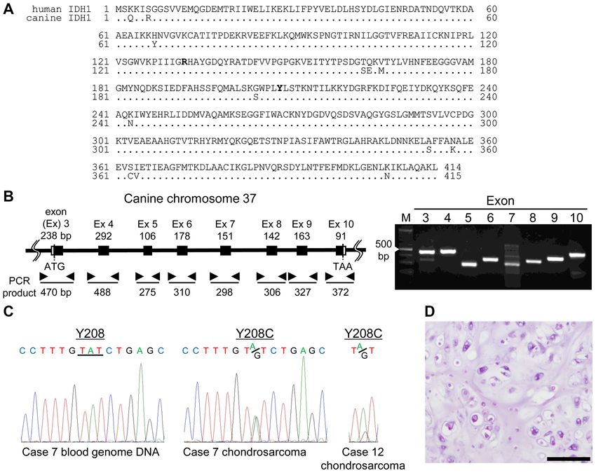
2                      KAWAKAMI et al: Y208C MUTATION IN IDH1 ATTENUATES THE DIMERIZATION ABILITY

reactive oxygen species, leading to a variety of after‑effects (13).   Cell cultures. HeLa and MDCK cells were purchased from
A decrease in α‑KG with an increase in 2‑HG causes the reduc‑          the American Type Culture Collection (ATCC). The cell lines
tion of α‑KG‑dependent prolyl hydroxylases, such as those              were maintained in Dulbecco's modified Eagle's medium
that regulate hypoxia‑inducible factor‑1α subunit (HIF‑1α)             (Wako) supplemented with 10% fetal bovine serum, penicillin,
levels. Alterations in HIF‑1α have been reported to result             and streptomycin (Applied Biosystems) and incubated at 37˚C
from mutant IDH1 protein expression (14), causing oncogenic            in a 5% CO2 atmosphere.
transformation. In addition, the loss or gain of IDH1 function
without R132 mutation is also related to cancer progression            Measurement of the isocitrate dehydrogenase activity.
and resistance to chemotherapy as mediated by NADPH                    To measure the production of NADH and NADPH,
biosynthesis (15‑17). Therefore, it is important to focus on           cIDH1‑transfected cells (5x10 4) were processed using the
mutations other than the R132H mutation, in order to further           Isocitrate Dehydrogenase Activity Colorimetric Assay
investigate IDH1 molecular function and cancer development.            Kit (BioVision) according to the manufacturer's instructions.
     Although intracranial tumors in dogs, such as meningi‑            The reaction mix was treated for 10 min, and the optical
omas and gliomas, are relatively common brain diseases (18),           density at 450 nm was measured using an iMark microplate
partial sequencing analysis targeting R132 in IDH1 has                 reader (Bio‑Rad Laboratories).
been performed, but no mutations have been identified in
canine gliomas (19). We recently cloned full‑length cDNA               Measurement of the HIF‑1α promoter activity. HeLa cell
of cIDH1 and performed artificial R132 mutation analysis of            transfection was performed in a 96‑well plate at 80%
cIDH1 (20). The antibodies used to detect the specific R132            confluency. The vector containing the HIF‑1α response element
mutation in humans could also be used to detect R132 muta‑             pGL4.42[luc2P/HRE/Hygro] (Promega) was co‑transfected
tions in cIDH1 (21,22), and the production ability of NADPH            with the control vector phRL‑TK (Promega) as a transfection
was attenuated by the R132 mutation of cIDH1. Furthermore,             efficiency control. Forty‑eight hours later, luciferase activity
the R132H mutant of cIDH1 intensified HIF‑1α expression,               was measured using the Dual‑Glo Luciferase Assay System
showing that the R132 mutation in cIDH1 plays a potential role         (Promega). Luciferase activity was normalized to that of
in tumor predisposition. The most recent study to show that the        Renilla luciferase activity.
R132C mutation was found in canine glioma cases (23) did not
found any other mutations in cIDH1.                                    α‑KG assays. HeLa cells were harvested in a 24‑well plate
    In this study, we found the novel mutation Y208C in cIDH1          at a density of 1x10⁵ cells/well and transfected with 250 ng
by sequencing formalin‑fixed paraffin‑embedded (FFPE)                  of HA‑tagged, full‑length WT, R132H, or Y208C mutant of
canine chondrosarcoma tissues. We compared the production              cIDH1 in pMACS Kk.HA‑C (Miltenyi Biotec). The assay
ability of NADPH and induction of HIF‑1α between the                   was performed using the coupled enzymatic assay method
wild‑type (WT) and cIDH1 mutants. Furthermore, the                     according to the manufacturer's instructions (Sigma‑Aldrich;
dimerization ability necessary to exert the enzyme activity of         Merck KGaA, catalog no. MAK054). In this method, α‑KG
IDH1 was estimated in silico and measured by cell biology              concentration is determined by a coupled enzyme assay,
analysis.                                                              which results in a colorimetric (570 nm) product that, in turn,
                                                                       is proportional to the amount of α‑KG present in the sample.
Materials and methods
                                                                       Induction of HIF‑1α expression by CoCl2. For the CoCl 2
Sample preparation and sequencing. The genomic DNA of                  (Wako) experiments to induce HIF‑1α expression, 2x10⁵ HeLa
the FFPE tissue from paraffin scrolls (Table SI) was extracted         cells were seeded in 6‑well plates for 24 h before being treated
from canine tumor samples using the QIAamp DNA FFPE                    with 100 µM CoCl2 for an additional 24 h.
Tissue Kit (Qiagen) according to the manufacturer's instruc‑
tions. PCR amplification was performed using PrimeSTAR                 Immunoblotting. Immunoblotting was performed using the
(Takara). Primer pairs used for amplifying cIDH1 exons are             following primary antibodies: Rabbit polyclonal anti‑HA
listed in Table I. Sequence data were directly determined using        (561, 1:1,000; MBL), anti‑β ‑actin (PM053, 1:2,000; MBL),
an ABI 3100‑Avant Genetic Analyzer (Applied Biosystems).               rabbit polyclonal anti‑HIF‑1α (#3716, 1:1,000; Cell Signaling
For the sequence analysis, human IDH1 (GenBank accession               Technology), and anti‑Halo antibody (G9281, 1:1000;
no. NP_005887.2) and cIDH1 (BBC43078.1) were compared                  Promega). Horseradish peroxidase‑conjugated secondary anti‑
accordingly (Fig. 1A).                                                 bodies and EzWestLumi plus (ATTO) were used for detecting
                                                                       antibody‑bound proteins.
Histological analysis. With permission from the University
Ethics Committee, we obtained tissue samples from the                  Crystal structure modeling. We retrieved the crystal structure
Department of Veterinary Pathology, School of Veterinary               of the human IDH1 dimer from the Research Collaboratory
Science, Nippon Veterinary and Life Science University                 for Structural Bioinformatics Protein Data Bank at
(approval no. 11‑50, 27 May 2018). All samples were classi‑            http://www.rcsb.org/ (PDB ID: 5YFM) and analyzed it using
fied by veterinary pathologists according to the World Health          the University of California, San Francisco Chimera software
Organization classification (24) (Table SⅠ). FFPE cancer               (http://www.cgl.ucsf.edu/chimera/) (25).
tissues were sliced at a thickness of 4 µm, the sections were
placed on slides, and hematoxylin and eosin (H&E) staining             Mammalian cell two‑hybrid assay. For the mammalian cell
was performed.                                                         two‑hybrid assay (MTH), WT and Y208C cIDH1 cDNA
ONCOLOGY LETTERS 20: 351, 2020 3
Table Ⅰ. Primer pairs to amplify canine Isocitrate dehydroge‑ Sample preparation, cross‑linking procedure and detection of
nase 1 exons. cIDH1 dimer. HeLa cells in 6‑well plates were transfected with
either WT, R132H or Y208C of cIDH1 expression plasmids
Exon Primer sequences (1 µg/well). After 48 h of transfection, the cells were lysed
with mammalian lysis buffer (Promega) supplemented with a
3 F: 5'‑GCAGCCTCAAAAGCCACACACGC‑3' protease inhibitor cocktail (Promega). Post lysis, the samples
R: 5'‑TGTACTTATCTTTAAGCATCCC‑3' were centrifuged (15,000 x g for 15 min at 4˚C) to obtain the
4 F: 5'‑CGTTGTGCGCCATCACACAG‑3' supernatant accordingly. Total protein levels were measured
R: 5'‑CACTTAAAGGGAGTAGTCAC‑3' using BCA (Nacalai Tesque). Equal amounts of proteins
5 F: 5'‑TGATCTTGAGTCTATACCAG‑3' (100 µg/condition) were then incubated with glutaraldehyde at
R: 5'‑TGGCTAGTTCCCTTTGTGTC‑3' different concentrations (0, 0.04, 0.1, 0.25 or 0.5%) and incu‑
6 F: 5'‑GACTTTCTTCCAATCACGTG‑3 bated on ice for 30 min accordingly. To make a working solution
of glutaraldehyde, commercially available 25% glutaraldehyde
R: 5'‑TATGCCCTTAACTTTATGGG‑3'
solution was diluted in PBS and discarded after use. To quench
7 F: 5'‑GCCTGATGCAAGACTCGATC‑3'
the reaction, sample buffer was added to obtain the following
R: 5'‑TTCATTGATGACTACACATGC‑3' final concentrations: 250 mM Tris‑HCl, pH 8.5; 2% lithium
8 F: 5'‑GGACCCTGCTTCCTGAGAGG‑3' dodecyl sulfate; 100 mM DTT; 0.4 mM EDTA; 10% glycerol;
R: 5'‑GGACCCTGCTTCCTGAGAGG‑3' and 0.2 mM bromophenol blue. Samples were then separated by
9 F: 5'‑TCTGCTCAACAGCAAGACAG‑3' SDS‑PAGE and the monomer and dimer of cIDH1 was detected
R: 5'‑TGACTGTGCTCCTTCCACAG‑3' using anti‑HA antibody accordingly.
10 F: 5'‑GTGGCCGAGCTGCCAGTGCAGGC‑3'
R: 5'‑CCTGCCACGTTCACGAGGGTG‑3' NADPH oxidase (NOX) activity assay. NOX activity
was evaluated by assessing the superoxide production by
F, forward; R, reverse. lucigenin‑enhanced chemiluminescence (26). WT or mutant
cIDH1‑transfected HeLa cells were disrupted in 0.2 ml of
extraction buffer (20 mM sodium phosphate buffer (pH 7.0),
1 mM EDTA, 0.5 mM PMSF, 1 µg/ml Aprotinin, 0.5 µg/ml
were cloned into the EcoRI and MluI sites of the pM GAL4 leupeptin) using a homogenizer on ice. After the homogenates
DNA‑binding domain of the GAL4‑DBD plasmid (pM) were centrifuged at 1,000 x g for 10 min at 4˚C, the supernatant
(Clontech Laboratories) and the pVP16 transactivation domain (20 µl) was added to 0.2 ml of assay buffer (50 mM sodium
of the VP16‑AD plasmid (pVP16) (Clontech Laboratories), phosphate buffer (pH 7.0), 1 mM EDTA, 150 mM sucrose,
respectively. Approximately 2x10⁵ HeLa cells were placed in and 50 mM lucigenin). After the addition of 0.1 mM NADPH,
a 24‑well plate and were co‑transfected with 100 ng of pM, luminescence was measured as relative light units (RLUs) at
100 ng of pVP16, 100 ng of pFR‑Luc firefly luciferase reporter 15‑sec intervals for 1,000 msec in a luminometer (GLOMAX;
plasmid (Promega), and 2 ng of phRL‑TK Renilla luciferase Promega). NOX activity was indicated as RLUs per minute per
reporter plasmid (Promega). The cells were harvested 48 h mg of protein.
after transfection, and luciferase activity was measured using
a Dual‑Glo Luciferase Assay System (Promega). Luciferase Statistical analysis. Data are expressed as mean ± standard
activity was normalized to that of Renilla luciferase activity. deviation (SD). Analysis of variance (ANOVA) with a Tukey's
post‑hoc test was used when multiple comparisons were
Pull‑down assay. We cloned the Halo‑ or HA‑tagged, required. P4 KAWAKAMI et al: Y208C MUTATION IN IDH1 ATTENUATES THE DIMERIZATION ABILITY
Figure 1. Detection and characterization of the canine IDH1 Y208C mutation. (A) The amino acid sequence comparison of human (NP_005887.2) and canine
(BBC43078.1) IDH1. A total of 401/414 residues were identical. The bold residues represent R132 and Y208, respectively. (B) Eight exons were amplified
from genomic DNA isolated from canine CS tissues (left). Electropherogram results demonstrated a single PCR band amplification (right). Amplicon sizes
are provided under the left panel. (C) Electropherograms of Sanger sequencing conducted on CS cases 7 and 12. (D) Photomicrographs of canine CS in
case number 7, which demonstrates representative CS pathogenesis, as indicated by hematoxylin and eosin staining (scale bar, 50 µm). IDH1, isocitrate
dehydrogenase 1; CS, chondrosarcoma; Ex, exon.
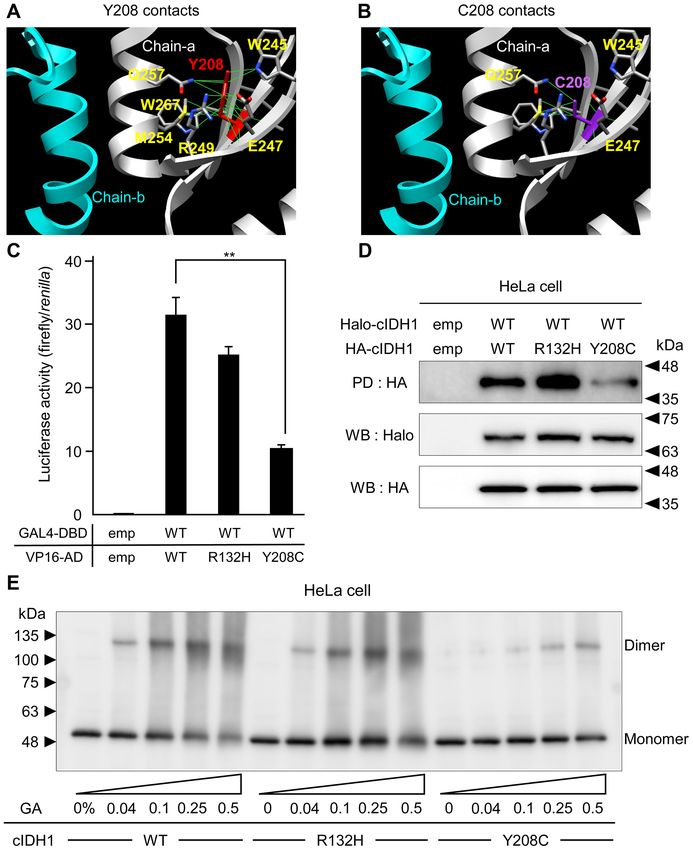
R132H mutant of cIDH1 was significantly lower than that in acids belonging to the intra‑strand W245, E247, R249, M254,
WT, and Y208C showed moderate productivity in both HeLa Q257, and W267 (Fig. 3A). The Rotamers tool allows amino
and MDCK cells (Fig. 2A). Production of α‑KG in HeLa cells acid side chain rotamers to be viewed and evaluated (25). The
was weakened in both R132H and Y208C mutant transfection best rotamers for C208 were selected based on their side‑chain
compared to in WT transfection (Fig. 2B). torsion as well as on the probability values in the rotamer
library and in the context of the structural environment. These
Overexpression of the Y208C mutant did not lead to HIF‑1α calculations revealed that the Y208C substitution disrupted or
induction. To evaluate HIF‑1α induction by overexpression of the reduced the inter‑strand hydrogen bond with the side chains of
cIDH1 mutant, a reporter assay was performed to measure under W245, E247, and Q257 (Fig. 3B). MTH and PD assays, which
the control of a promoter containing hypoxic response element examined the binding activities between WT and mutant
sites (27). Hypoxia caused by 10 µM CoCl2 stimulation and over‑ cIDH1, showed no significant change in WT‑R132H heterodi‑
expression of the R132H mutant led to the induction of HIF‑1α merization and attenuated binding activity in the WT‑Y208C
reporter activities, but the Y208C mutant did not (Fig. 2C). mutant (Fig. 3C, D). The dimerization ability of cIDH1 trans‑
HIF‑1α protein expression was also induced by CoCl2 stimula‑ fected into HeLa cells was assessed using a glutaraldehyde
tion and R132H expression, but not Y208C (Fig. 2D). cross‑linking assay. Cell lysates from HeLa cells expressing
HA‑tagged WT, R132H or Y208 mutant were treated first
Y208C mutation results in a conformational change in the with glutaraldehyde crosslinker and then analyzed by western
dimerization form of IDH1. To predict the functional alteration blotting method. Both WT and R132H mutants formed dimers
based on the IDH1 mutation, the protein structure editing tool following the glutaraldehyde treatment, however, the Y208C
in the UCSF Chimera software package was used to analyze mutant formed a weak dimer (Fig. 3E).
the possible structural outcomes of Y208C substitutions.
Y208 is located adjacent to the binding surface of the IDH1 R132H and Y208C mutations did not affect the alteration
dimerization form. Y208 showed hydrogen bonds with amino of NOX activities. NOX activity in WT or mutantONCOLOGY LETTERS 20: 351, 2020 5 Figure 2. Measurement of the functional alterations between WT and mutant cIDH1. (A) Upper graphs indicate the production of NADPH in HeLa (left) and MDCK (right) cells transfected with WT, R132H or Y208C cIDH1, as determined through a colorimetric assay. Western blotting of the lower panel indicates the even expression of transfected cIDH1s, which was fused with HA. **P
6                         KAWAKAMI et al: Y208C MUTATION IN IDH1 ATTENUATES THE DIMERIZATION ABILITY

Figure 3. In silico and cell biology analysis of the effect of Y208C mutation dimerization ability. Two cIDH1 proteins are depicted as magenta (chain‑a) and
gray (chain‑b) ribbons. The Y208 residue (red) was mutated to Cys (purple). The contacts between residues at the (A) Y208 or (B) C208 positions in the chain
were calculated. The amino acid residues linking Y208 and C208 are colored gray. The solid green lines denote stable contacts, as determined using the
Chimera program. (C) Binding intensities between GAL4‑DBD and VP16‑AD fused with the WT and cIDH1 mutants. Data are presented as the mean ± SD
(n=4). **P
ONCOLOGY LETTERS 20: 351, 2020                                                     7

                                                                           NADPH, a source of ROS, but IDH1 mutants lose the ability
                                                                           to produce more NADPH; therefore, mutant transfected cells
                                                                           could not produce ROS. Since there was no difference in the
                                                                           results of ROS productivity between R132H and Y208C, the
                                                                           mechanism of carcinogenesis of the Y208C mutation remains
                                                                           unclear. This study is the first experimental report to describe
                                                                           the relationship between canine IDH1 mutation and NOX
                                                                           activity. Future studies will need to elucidate the mechanism
                                                                           of tumorigenesis of the Y208C mutation. Furthermore, future
                                                                           studies will have to look for Y208C mutations in various tumors.
                                                                                In conclusion, we identified for the first time Y208C
                                                                           spontaneous somatic mutations of canine IDH1 in chon‑
                                                                           drosarcomas and assessed the impact of these mutations on
                                                                           IDH1 functions. Y208C mutation attenuated the NADPH
                                                                           production ability but did not enhance HIF‑1α retention in
                                                                           CoCl 2‑treated cells. This phenomenon was caused by the
                                                                           attenuation of the dimerization ability of the Y208C muta‑
                                                                           tion. We hope that the precise analysis of IDH1 functional
                                                                           changes can help elucidate the tumorigenesis involvement of
Figure 4. Generation of cellular ROS by NOX in cIDH1‑transfected cells.    the Y208C IDH1 mutation.
Cellular NOX activity in WT and mutant cIDH1‑transfected cells. Temporal
ROS generation (RLU/mg protein) by NOX was detected using lucigenin
chemiluminescence. Data are presented as the mean ± SD (n=3). **P
8 KAWAKAMI et al: Y208C MUTATION IN IDH1 ATTENUATES THE DIMERIZATION ABILITY
References 19. Reitman ZJ, Olby NJ, Mariani CL, Thomas R, Breen M, Bigner DD,
McLendon RE and Yan H: IDH1 and IDH2 hotspot mutations are
1. Sahm F, Reuss D, Koelsche C, Capper D, Schittenhelm J, not found in canine glioma. Int J Cancer 127: 245‑246, 2010.
Heim S, Jones DT, Pfister SM, Herold‑Mende C, Wick W, et al: 20. Kawakami S, Ochiai K, Azakami D, Kato Y, Michishita M,
Farewell to oligoastrocytoma: In situ molecular genetics favor Morimatsu M, Ishiguro‑Oonuma T, Onozawa E, Watanabe M and
classification as either oligodendroglioma or astrocytoma. Acta Omi T: R132 mutations in canine isocitrate dehydrogenase 1 (IDH1)
Neuropathol 128: 551‑559, 2014. lead to functional changes. Vet Res Commun 42: 49‑56, 2018.
2. Lapointe S, Perry A and Butowski NA: Primary brain tumours in 21. Kaneko MK, Tian W, Takano S, Suzuki H, Sawa Y, Hozumi Y,
adults. Lancet 392: 432‑446, 2018. Goto K, Yamazaki K, Kitanaka C and Kato Y: Establishment of
3. Parsons DW, Jones S, Zhang X, Lin JC, Leary RJ, Angenendt P, a novel monoclonal antibody SMab‑1 specific for IDH1‑R132S
Mankoo P, Carter H, Siu IM, Gallia GL, et al: An inte‑ mutation. Biochem Biophys Res Commun 406: 608‑613, 2011.
grated genomic analysis of human glioblastoma multiforme. 22. Kato Y: Specific monoclonal antibodies against IDH1/2 mutations
Science 321: 1807‑1812, 2008. as diagnostic tools for gliomas. Brain Tumor Pathol 32: 3‑11, 2015.
4. Reitman ZJ and Yan H: Isocitrate dehydrogenase 1 and 2 muta‑ 23. Amin SB, Anderson KJ, Boudreau CE, Martinez‑Ledesma E,
tions in cancer: Alterations at a crossroads of cellular metabolism. Kocakavuk E, Johnson KC, Barthel FP, Varn FS, Kassab C,
J Natl Cancer Inst 102: 932‑941, 2010. Ling X, et al: Comparative molecular life history of spontaneous
5. Bleeker FE, Lamba S, Rodolfo M, Scarpa A, Leenstra S, canine and human gliomas. Cancer Cell 37: 243‑257.e7, 2020.
Vandertop WP and Bardelli A: Mutational profiling of cancer 24. Misdorp W, Else RW, Hellmen E and Lipscomb TP: Histological
candidate genes in glioblastoma, melanoma and pancreatic classification of mammary tumors of the dog and the cat. In: World
carcinoma reveals a snapshot of their genomic landscapes. Hum Health Organization International Histological Classification of
Mutat 30: E451‑E459, 2009. Tumors of Domestic Animals. Schulman FY (ed). Vol 7. 2nd Series.
6. Yan H, Parsons DW, Jin G, McLendon R, Rasheed BA, Yuan W, Armed Forces Institute of Pathology, Washington, DC, p58, 1999.
Kos I, Batinic‑Haberle I, Jones S, Riggins GJ, et al: IDH1 and 25. Pettersen EF, Goddard TD, Huang CC, Couch GS, Greenblatt DM,
IDH2 mutations in gliomas. N Engl J Med 360: 765‑773, 2009. Meng EC and Ferrin TE: UCSF Chimera‑a visualization system
7. Mardis ER, Ding L, Dooling DJ, Larson DE, McLellan MD, Chen K, for exploratory research and analysis. J Comput Chem 25:
Koboldt DC, Fulton RS, Delehaunty KD, McGrath SD, et al: 1605‑1612, 2004.
Recurring mutations found by sequencing an acute myeloid 26. Jalil JE, Perez A, Ocaranza MP, Bargetto J, Galaz A and
leukemia genome. N Engl J Med 361: 1058‑1066, 2009. Lavandero S: Increased aortic NADPH oxidase activity in rats
8. Lugowska I, Teterycz P, Mikula M, Kulecka M, Kluska A, with genetically high angiotensin‑converting enzyme levels.
Balabas A, Piatkowska M, Wagrodzki M, Pienkowski A, Hypertension 46: 1362‑1367, 2005.
Rutkowski P and Ostrowski J: IDH1/2 mutations predict shorter 27. Emerling BM, Platanias LC, Black E, Nebreda AR, Davis RJ and
survival in chondrosarcoma. J Cancer 9: 998‑1005, 2018. Chandel NS: Mitochondrial reactive oxygen species activation
9. Waitkus MS, Diplas BH and Yan H: Biological role and therapeutic of p38 mitogen‑activated protein kinase is required for hypoxia
potential of IDH mutations in cancer. Cancer Cell 34: 186‑195, 2018. signaling. Mol Cell Biol 25: 4853‑4862, 2005.
10. Dang L, White DW, Gross S, Bennett BD, Bittinger MA, 28. Preusser M, Wohrer A, Stary S, Hoftberger R, Streubel B and
Driggers EM, Fantin VR, Jang HG, Jin S, Keenan MC, et al: Hainfellner JA: Value and limitations of immunohistochem‑
Cancer‑associated IDH1 mutations produce 2‑hydroxyglutarate. istry and gene sequencing for detection of the IDH1‑R132H
Nature 462: 739‑744, 2009. mutation in diffuse glioma biopsy specimens. J Neuropathol Exp
11. Gross S, Cairns RA, Minden MD, Driggers EM, Bittinger MA, Neurol 70: 715‑723, 2011.
Jang HG, Sasaki M, Jin S, Schenkein DP, Su SM, et al: 29. Ashraf S, Noguera NI, Di Giandomenico J, Zaza S, Hasan SK
Cancer‑associated metabolite 2‑hydroxyglutarate accumulates in and Lo‑Coco F: Rapid detection of IDH2 (R140Q and R172K)
acute myelogenous leukemia with isocitrate dehydrogenase 1 and mutations in acute myeloid leukemia. Ann Hematol 92: 1319‑1323,
2 mutations. J Exp Med 207: 339‑344, 2010. 2013.
12. Schnittger S, Haferlach C, Ulke M, Alpermann T, Kern W and 30. Catteau A, Girardi H, Monville F, Poggionovo C, Carpentier S,
Haferlach T: IDH1 mutations are detected in 6.6% of 1414 AML Frayssinet V, Voss J, Jenkins R Boisselier B, Mokhtari K, et al: A new
patients and are associated with intermediate risk karyotype sensitive PCR assay for one‑step detection of 12 IDH1/2 mutations
and unfavorable prognosis in adults younger than 60 years and in glioma. Acta Neuropathol Commun 2: 58, 2014.
unmutated NPM1 status. Blood 116: 5486‑5496, 2010. 31. Truve K, Dickinson P, Xiong A, York D, Jayashankar K, Pielberg G,
Koltookian M, Muren E, Fuxelius HH, Weishaupt H, et al:
13. Wajner M, Latini A, Wyse AT and Dutra‑Filho CS: The role of Utilizing the dog genome in the search for novel candidate
oxidative damage in the neuropathology of organic acidurias: genes involved in glioma development‑genome wide association
insights from animal studies. J Inherit Metab Dis 27: 427‑448, mapping followed by targeted massive parallel sequencing identi‑
2004. fies a strongly associated locus. PLoS Genet 12: e1006000, 2016.
14. Zhao S, Lin Y, Xu W, Jiang W, Zha Z, Wang P, Yu W, Li Z, 32. Tate JG, Bamford S, Jubb HC, Sondka Z, Beare DM, Bindal N,
Gong L, Peng Y, et al: Glioma‑derived mutations in IDH1 domi‑ Boutselakis H, Cole CG, Creatore C, Dawson E, et al: COSMIC:
nantly inhibit IDH1 catalytic activity and induce HIF‑1alpha. The catalogue of somatic mutations in cancer. Nucleic Acids
Science 324: 261‑265, 2009. Res 47: D941‑D947, 2019.
15. Calvert AE, Chalastanis A, Wu Y, Hurley LA, Kouri FM, Bi Y, 33. Huang LE: Friend or foe‑IDH1 mutations in glioma 10 years on.
Kachman M, May JL, Bartom E, Hua Y, et al: Cancer‑associated Carcinogenesis 40: 1299‑1307, 2019.
IDH1 promotes growth and resistance to targeted therapies in the 34. Koyasu S, Shimizu Y, Morinibu A, Saga T, Nakamoto Y,
absence of mutation. Cell Rep 19: 1858‑1873, 2017. Togashi K and Harada H: Increased 14C‑acetate accumulation
16. Wahl DR, Dresser J, Wilder‑Romans K, Parsels JD, Zhao SG, in IDH‑mutated human glioblastoma: Implications for detecting
Davis M, Zhao L, Kachman M, Wernisch S, Burant CF, et al: IDH‑mutated glioblastoma with 11C‑acetate PET imaging.
Glioblastoma therapy can be augmented by targeting IDH1‑mediated J Neuro Oncol 145: 441‑447, 2019.
NADPH biosynthesis. Cancer Res 77: 960‑970, 2017. 35. Zhao S and Guan KL: IDH1 mutant structures reveal a mecha‑
17. Zarei M, Lal S, Parker SJ, Nevler A, Vaziri‑Gohar A, Dukleska K, nism of dominant inhibition. Cell Res 20: 1279‑1281, 2010.
Mambelli‑Lisboa NC, Moffat C, Blanco FF, Chand SN, et al: 36. Vinekar R, Verma C and Ghosh I: Functional relevance
Posttranscriptional upregulation of IDH1 by HuR establishes a of dynam ic proper ties of Dimer ic NA DP‑dependent
powerful survival phenotype in pancreatic cancer cells. Cancer isocitrate dehydrogenases. BMC Bioinformatics 13 (Suppl 17):
Res 77: 4460‑4471, 2017. S2, 2012.
18. Dobson JM, Samuel S, Milstein H, Rogers K and Wood JL: This work is licensed under a Creative Commons
Canine neoplasia in the UK: Estimates of incidence rates from a
Attribution-NonCommercial-NoDerivatives 4.0
population of insured dogs. J Small Anim Pract 43: 240‑246, 2002.
International (CC BY-NC-ND 4.0) License.You can also read