Rapid screening of UPB1 gene variations by high resolution melting curve analysis
←
→
Page content transcription
If your browser does not render page correctly, please read the page content below
EXPERIMENTAL AND THERAPEUTIC MEDICINE 21: 403, 2021

                              Rapid screening of UPB1 gene variations by
                                high resolution melting curve analysis
                                   XIAOWEI XU1,2*, JIE ZHENG3*, QIANQIAN ZOU3, CHAO WANG1,2,
                                  XINJIE ZHANG1,2, XUETAO WANG1,2, YANG LIU4 and JIANBO SHU1,2

                              1
                           Tianjin Pediatric Research Institute, Tianjin Children's Hospital, Tianjin 300134;
                          2
                       Tianjin Key Laboratory of Prevention and Treatment of Birth Defects, Tianjin Children's
                      Hospital, Tianjin 300134; 3Graduate College, Tianjin Medical University, Tianjin 300070;
                        4
                          Department of Neonatology, Tianjin Children's Hospital, Tianjin 300134, P.R. China

                                             Received February 12, 2020; Accepted October 8, 2020

                                                          DOI: 10.3892/etm.2021.9834

Abstract. The present study aimed to analyze gene mutations                Introduction
in patients with β‑ureidopropinoase deficiency and establish a
rapid detection method for β‑ureidopropinoase (UPB1) patho‑                β ‑u reidopropinoase def iciency (On line Mendelia n
genic variations by high resolution melting (HRM) analysis.                Inheritance in Man no. 613161) is a rare autosomal
DNA samples with known UPB1 mutations in three patients                    recessive disease which is caused by mutations in the
with β‑ureidopropinoase deficiency were utilized to establish              β ‑ureidopropinoase (UPB1) gene. The clinical phenotypes
a rapid detection method for UPB1 pathogenic variations by                 of reported cases are complicated. Several patients presented
HRM analysis. Further rapid screening was performed on two                 with severe neurological involvement or mild psychomotor
patients diagnosed with β‑ureidopropinoase deficiency and 50               development delay, while other patients do not show any
healthy control individuals. The results showed that all known             symptoms (1). Most patients are diagnosed before the age of
UPB1 gene mutations can be analyzed by a specially designed                two years, while numerous patients who do not present with
HRM assay. Each mutation has specific HRM profiles which                   any symptoms may not have been diagnosed (1). Since the
could be used in rapid screening. The HRM method could                     disease lacks clinical specificity, auxiliary examination plays
correctly identify all genetic mutations in two children with              an important role in diagnosis. Gas chromatography‑mass
β ‑ureidopropinoase deficiency. In addition, the HRM assay                 spectrometry (GC‑MS) is widely used to diagnose this
also recognized four unknown mutations. To conclude, the                   disease, however, analysis of gene mutation is the main tool
results support future studies of applying HRM analysis as a               used to confirm diagnosis (2,3).
diagnostic approach for β‑ureidopropinoase deficiency and a                    High‑resolution melting analysis (HRM), a novel tech‑
rapid screening method for UPB1 mutation carriers.                         nology for mutation, polymorphism and epigenetic alteration
                                                                           detection in double‑stranded DNAs, has been one of the most
                                                                           widely used molecular diagnostic techniques in clinical and
                                                                           research settings (4). The basic principle of HRM is that
                                                                           DNA‑saturated fluorescent dye is added into the PCR reaction
                                                                           system (5). The PCR amplicon is denatured by heating in a
                                                                           certain temperature range (6). Specialized instruments detect
                                                                           the degenerated double‑stranded DNA fluorescent signal and
Correspondence to: Mr. Yang Liu, Department of Neonatology,                a melting curve is plotted (7). The combination of fluorescent
Tianjin Children's Hospital,           238    Longyan   Road,   Beichen,   dye with PCR products is monitored in real‑time during the
Tianjin 300134, P.R. China                                                 heating process (5). A single‑nucleotide mismatch leads to
E‑mail: etyyly@163.com                                                     double‑stranded DNA unwinding in the heating process (8).
                                                                           Fluorescent dyes are released from DNA molecules that are
Mr. Jianbo Shu, Tianjin Pediatric Research Institute, Tianjin
Children's Hospital, 238 Longyan Road, Beichen, Tianjin 300134,
                                                                           locally opened up, and the presence of mutations can be
P.R. China                                                                 determined from the fluorescence intensity and time curves
E‑mail: shjb1981@sina.com                                                  according to different melting temperature and shape of the
                                                                           melting curve, which allows wild‑type and heterozygous
*
    Contributed equally                                                    mutation types to be effectively distinguished (9).
                                                                               Considering the advantages of HRM in gene screening
Key words: β‑ureidopropinoase, β‑ureidopropinoase deficiency,              and detection, HRM in combination with other molecular
high resolution melting analysis, diagnosis                                diagnostic techniques can expand the application scope and
                                                                           improve the efficiency of HRM. For example, by using compet‑
                                                                           itive multiplex PCR and HRM, copy number variations can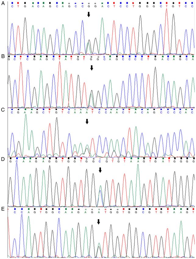
2                                               XU et al: HRM ANALYSIS FOR UPB1

be detected accurately (10). Recently, the rapid development       was isolated using a Blood Genomic DNA Mini kit (CoWin
of instruments, DNA dyes and analysis software significantly       Biosciences) according to the manufacturer's instructions.
enhanced the sensitivity, specificity and accuracy of HRM (11)     DNA quality and concentration were assessed using a
This provides a fast, efficient and economic molecular diag‑       NanoDrop™ 2000 UV‑Vis Spectrophotometer (Thermo
nostic platform for molecular diagnosis of inherited disease,      Fisher Scientific, Inc.).
molecular profiling and target therapy of cancer, identification
of pathogens and individualized medicine (12‑15). The present      PCR primer selection and amplification. Primers for PCR
study evaluated the potential application of HRM analysis          amplification of UPB1 genes with a size range of 271‑490 bp
for identifying gene mutations in patients and carriers with       were selected (16). The entire UPB1 coding region were ampli‑
β‑ureidopropinoase deficiency.                                     fied by PCR using these primers. Primers were synthesized by
                                                                   Genewiz, Inc. PCR reactions were performed in a total reac‑
Materials and methods                                              tion volume of 25 µl (16). The amplicons were detected using
                                                                   1.5% agarose gel electrophoresis and visualized by ChemiDoc
Clinical details. The methods used in the present study were       XRS+ Imaging system (Bio‑Rad Laboratories, Inc.). The DNA
performed in accordance with approved guidelines by the            products were sent to Genewiz, Inc. for Sanger sequencing.
Committee for the Ethical Review of Research Involving             Sequences from amplified UPB1 were compared to known
Human Subjects at Tianjin Children's Hospital. A total of          sequences using Chromas Lite and Gene Runner (Frank
50 healthy individuals were recruited as controls in this study    Buquicchio and Michael Spruyt; version 6.5.51x64).
from October 2018 to August 2019 in Tianjin Children's
Hospital (Tianjin, China), including 27 males and 23 females,      HRM primer selection. Primers for HRM amplification of
with a mean age of 2 years (range, 6 months to 12 years old).      UPB1 genes with a size range of 77‑249 bp were designed
Physical examination and GC‑MS results of these controls were      (Table I). Primers were synthesized by Suzhou Jinweizhi
normal. The controls had no relationship with any of these five    Biotechnology Co., Ltd. The primers for HRM are shorter
patients. Peripheral blood specimens were collected from all       than those used for PCR.
subjects after informed consent was obtained from their parents.
    During the screening of suspected children by urine            HRM reaction. HRM was performed using a Forget‑Me‑Not
GC/MS, those identified with abnormal increase in pyrimidine       EvaG reen qPCR Master M i x k it ( Biotium, I nc.;
degradation metabolites, including N‑carbamyl‑ β ‑alanine          cat. no. 31042‑1). The total volume of the reaction was
(β‑UP), N‑carbamyl‑β‑aminoisobutyric acid (β‑UIB), uracil,         20 µl, including 10 µl premix (2x), 1 µl forward and 1 µl
thymine, dihydouracil and dihydothymine, were diagnosed as         reverse primers (10 µM) and 25 ng genomic DNA template.
β‑ureidopropinoase deficiency. Five patients were recruited to     Amplification was performed using a LightCycler 480 High
the present study. The first three families have been previously   Throughput Real‑Time Fluorescence Quantitative PCR system
reported (16). Case 1, a 2‑month‑old male, was hospitalized        (Roche Diagnostics GmbH; 96-wells). The following thermo‑
with jaundice for nearly 2 months and progressive aggravation      cycling conditions were used for the PCR: Initial denaturation
with occasional clay‑like tool. The male was diagnosed with        at 95˚C for 5 min; 48 cycles of denaturation at 95˚C for 5 sec,
β‑ureidopropinoase deficiency by GC‑MS (16).                       annealing at 60˚C for 10 sec and extension at 72˚C for 20 sec
    Case 2, a 3‑day‑old male, was admitted due to temporary        with fluorescence reading and single point acquisition mode.
convulsion. His father also suffered from β‑ureidopropinoase       The subsequent melting analysis process of PCR amplification
deficiency. Elevated pyrimidines and metabolites in urine          products included three steps: Denaturation at 95˚C for 1 min;
were detected by GC‑MS. The male was diagnosed with                renaturation at 40˚C for 1 min followed by continuous fluores‑
β‑ureidopropinoase deficiency (16).                                cence reading mode at 65‑95˚C. The rise rate was 0.02˚C/sec,
    Case 3, a 2‑year‑old male, was admitted because of language    and data acquisition was 25 times/˚C (once every 0.04˚C
delay. The male was able to sit by himself in 7 months, climb      rise in temperature). The melting curve was analyzed using
in 10 months and walk until 14 months. The male was unable         LightCycler 480 Gene Scanning software (version 1.5; Roche
to speak until 2 years and 4 months. The results of GC‑MS          Diagnostics GmbH). Each sample was performed in duplicate.
urine screening showed that the male has β‑ureidopropinoase        All fragments showing HRM aberrant patterns in controls
deficiency (16).                                                   (cases 1, 2 and 3) and the two families (cases 4 and 5) were
    Case 4, an 8‑month‑old male, entered with a one‑day            amplified by PCR analysis. All PCR products were purified
history of dizziness, vomiting and stroking twice. The boy         and sequenced by Genewiz, Inc. Subsequently, the PCR
was delivered with a birth weight of 3.65 kg. No obvious           products were sent to Genewiz, Inc. for Sanger sequencing.
abnormalities were detected in neurological examinations.
The result of GC‑MS urine screening showed the boy has             Results
β‑ureidopropinoase deficiency.
    Case 5, a 20‑month‑old boy, was admitted due to motor          HRM development. DNA samples of the first three families
retardation. No obvious abnormalities were detected in exami‑      (cases 1, 2 and 3) were detected using whole exome sequencing.
nations. β‑ureidopropinoase deficiency was initially identified    These families with known mutations were used to determine
by GC‑MS performed in the clinical setting.                        proper parameters and normalized HRM profile (Table II).
                                                                   PCR amplifications of exon fragments 1, 7, 9 and 10 of the
Genomic DNA extraction. A total of 1 ml peripheral blood           UPB1 gene followed by HRM analysis were performed. All
was collected with EDTA anticoagulant. Genomic DNA                 selected mutations were confirmed by Sanger sequencing.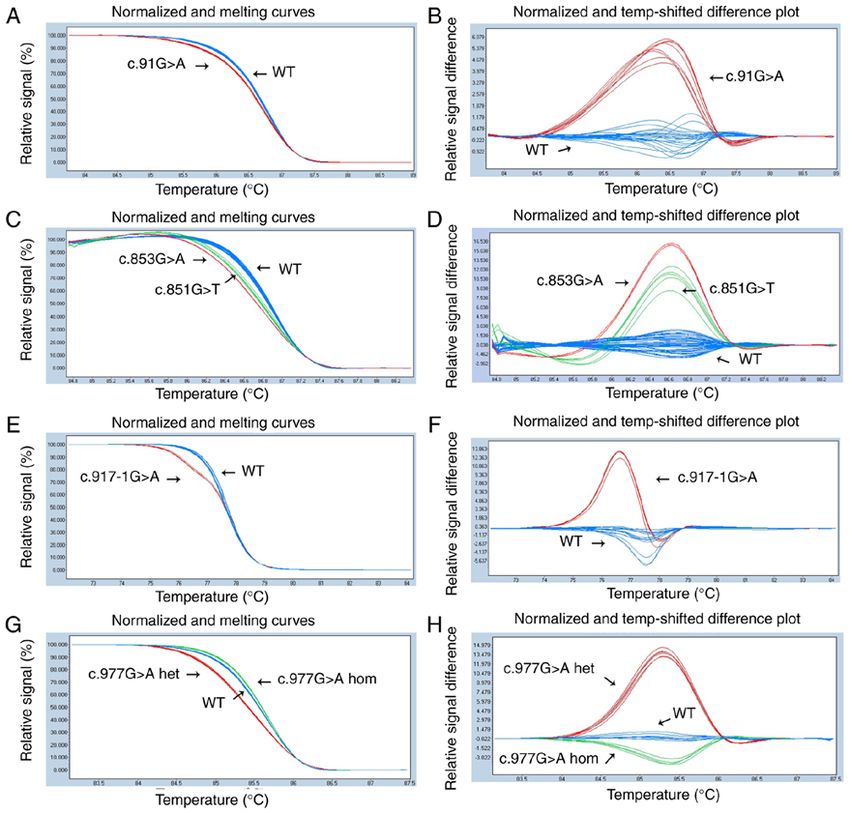
EXPERIMENTAL AND THERAPEUTIC MEDICINE 21: 403, 2021                                     3

Table I. UPB1 gene primers of high resolution melting curve analysis.

			                                                                                                          Length of
Name Direction                                                          Sequence (5'‑3')                  amplification (bp)

EX1‑F                         Forward                       GTGCGCGGACACAAGCACTG                                 165
EX1‑R                         Reverse                       CCATTAGCTTAGCCTCTTGG
EX2‑F                         Forward                       AAGTGGAGCAGACTGCATCA                                 246
EX2‑R                         Reverse                       ATGGTCAAAAGAGTCTGCAC
EX3‑F                         Forward                       CCATATGCATTAGGATGAGAT                                201
EX3‑R                         Reverse                       AGGCAGCAGAGAAAGCAC
EX4‑F                         Forward                       TGCCCTTTCTCCTATTTATAG                                140
EX4‑R                         Reverse                       CCAGAGGCACCGTTAATG
EX5‑F                         Forward                       ATAAGTGGGACTCTGCCA                                   216
EX5‑R                         Reverse                       ATCTTATGGGTGGCTCAC
EX6‑F                         Forward                       GAGTCTAAGGAAATCTTGAAGG                               249
EX6‑R                         Reverse                       CAGACCCCACCAACTGA
EX7‑F                         Forward                       GCTGAGCATCCACTGAGTC                                  228
EX7‑R                         Reverse                       CATGTGTGCAGAGGGAAC
EX8‑F                         Forward                       TAACTGTTTCCTGGCTGAC                                  235
EX8‑R                         Reverse                       GAAGCAGACAGAGAAGGG
EX9A‑F                        Forward                       AGCCCACAGTGCATCTAC                                    77
EX9A‑R                        Reverse                       AAAGTAGCCAAAGTCCTG
EX9B‑F                        Forward                       AAGCTCACAGATGTGTTTCTT                                226
EX9B‑R                        Reverse                       AGAGGAGCAGGCAACAACAG
EX10‑F                        Forward                       CGAGCCTTCCTGATGCGTTC                                 246
EX10‑R                        Reverse                       CAGAGGTGTCTTCCTCACCC

UPB1, β‑ureidopropinoase; 9A, the front part of exon 9; 9B, the latter part of exon 9.

Table II. Mutations of the UPB1 gene verified in the five families.

Case                                          Polymorphism                                   Mutation                  Exon

Case 1                                        c.91G>A                                      Heterozygous                 1
                                              c.977G>A                                     Homozygous                   9
Case 1's father                               c.977G>A                                     Heterozygous                 9
Case 1's mother                               c.91G>A                                      Heterozygous                 1
                                              c.977G>A                                     Heterozygous                 9
Case 2                                        c.851G>T                                     Heterozygous                 7
                                              c.977G>A                                     Heterozygous                 9
Case 2's father                               c.977G>A                                     Homozygous                   9
Case 2's mother                               c.851G>T                                     Heterozygous                 7
Case 3                                        c.853G>A                                     Heterozygous                 7
                                              c.917‑1 G>A                                  Heterozygous             Intron 8
Case 3's father                               c.853G>A                                     Heterozygous                 7
Case 3's mother                               c.917‑1 G>A                                  Heterozygous             Intron 8
Case 4                                        c.977 G>A                                    Homozygous                   9
Case 4's father                               c.977 G>A                                    Heterozygous                 9
Case 4's mother                               c.977 G>A                                    Heterozygous                 9
Case 5                                        c.91G>A                                      Heterozygous                 1
                                              c.977 G>A                                    Homozygous                   9
Case 5's father                               c.977 G>A                                    Heterozygous                 9
Case 5's mother                               c.91G>A                                      Heterozygous                 1
                                              c.977 G>A                                    Heterozygous                 9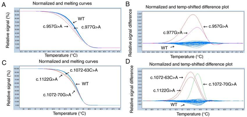
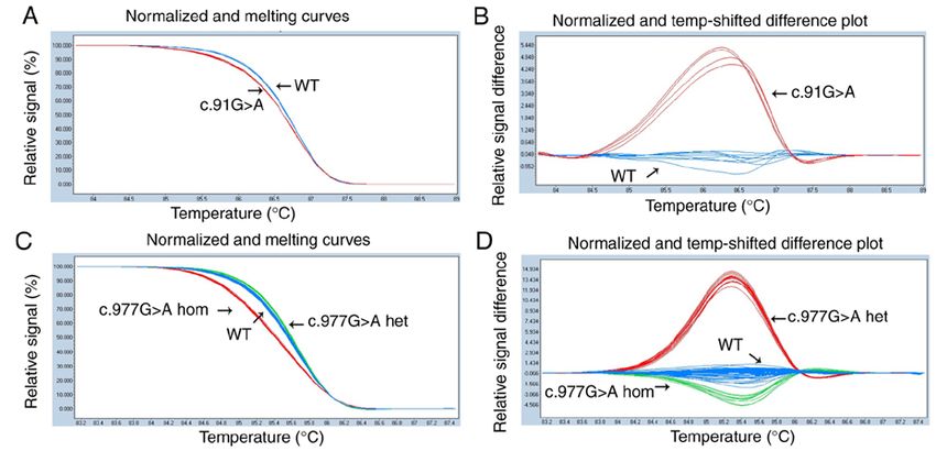
4                                                         XU et al: HRM ANALYSIS FOR UPB1

Figure 1. High resolution melting analysis of the UPB1 gene in the three families (cases 1, 2 and 3). The (A) melting curves and (B) difference plot of exon 1.
The (C) melting curves and (D) difference plot of exon 7. The (E) melting curves and (F) difference plot of exon 9A. The (G) melting curves and (H) difference
plot of exon 9B. WT, wild‑type; het, heterozygous; hom, homozygous; UPB1, β‑ureidopropinoase.

Figure 2. High resolution melting analysis of the UPB1 gene in the two families (cases 4 and 5) and 50 controls. The (A) melting curves and (B) difference
plot of exon 1. a, c.91G>A heterozygous mutation. The (C) melting curves and (D) difference plot of exon 9B in the two families. b, c.977G>A heterozygous
mutation. c, c.977G>A homozygous mutation. WT, wild‑type; het, heterozygous; hom, homozygous; UPB1, β‑ureidopropinoase.
EXPERIMENTAL AND THERAPEUTIC MEDICINE 21: 403, 2021 5
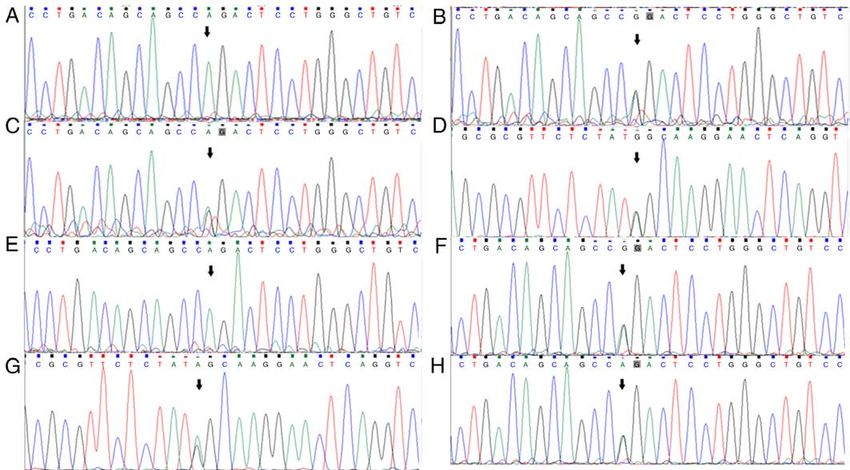
Figure 3. UPB1 gene mutations in the last two families (cases 4 and 5). (A) Case 4 carries a c.977G>A (p.R326Q) homozygous mutation. (B and C) The parents
of case 4 carry a c.977G>A heterozygous mutation. (D and E) case 5 carries a c.91G>A heterozygous mutation and a c.977G>A (p.R326Q) homozygous
mutation. (F) The father of case 5 carries a c.977G>A heterozygous mutation. (G and H) The mother of case 5 carries c.91G>A and c.977G>A heterozygous
mutations. The mutations are indicated by the black arrows. UPB1, β‑ureidopropinoase.
As shown in Fig. 1, each mutation had specific melting curve deficiency, 50 randomly chosen healthy children samples
profiles which significantly differed from the wild‑type. HRM were first tested by HRM. Among the 50 samples negative
also clearly divided heterozygote variants with homozygote for β‑ureidopropinoase deficiency, 46 samples were identified
variants in each sample. However, when the c.917‑1G>A as wild‑type and 4 samples were identified to have harbored
mutation was analyzed, the melting curve profile showed that mutations. All melting curves of 46 healthy children were
there was no curve variation with wild‑type controls. Hence, a consistent with those of normal melting profiles (Fig. 4).
primer checking was performed and it was found that although The four mutant samples carried c.977G>A, c.957G>A,
the primers uncovered the mutation site, the placement of c.1072‑63C>A, c.1072‑70G>A and c.1122G>A heterozygous
primers were not appropriate enough for HRM splice‑site mutations.
variation analysis. A set of primers were then redesigned to Following HRM analysis, all samples were genotyped by
differentiate this mutation. This data indicated that for patho‑ Sanger sequencing. For each of these mutation types and geno‑
genic splice‑site mutation HRM, the primer needs to extend types, HRM and Sanger sequencing gave consistent results
into part of the intronic region until the splice donor/acceptor (Fig. 5).
site is incorporated.
Discussion
HRM to identify UPB1 mutations in β ‑ureidopropinoase
deficiency. To evaluate the diagnostic utility of HRM to detect β ‑ureidopropinoase deficiency is an autosomal recessive
UPB1 mutations, two β ‑ureidopropinoase deficiency cases disease. The pathogenic gene, UPB1, is located at chromosome
(cases 4 and 5) with unknown mutations were selected. The 22q11.2 and consists of 10 exons. The cDNA is 1,152 bases
melting curves of different UPB1 mutations were used as posi‑ long and encodes 384 amino acids (1,17). β‑ureidopropinoase
tive controls, and the wild‑type UPB1 gene was used as the is the ultimate reactive enzyme in the metabolic path‑
normal control. The results are as follows. Case 4 carried a ways of uracil and thymine (18). Defects in the activity of
c.977G>A p.R326Q homozygous mutation from his parents. β‑ureidopropinoase directly affects the degradation of β‑UP
Case 5 carried a maternal c.91G>A p.G31S heterozygous and β‑UIB, resulting in the abnormal increase of β‑UP and
mutation and a c.977G>A p.R326Q homozygous mutation β‑UIB in body fluids (2,19). Concurrently, the uracil, thymine,
from his parents (Fig. 2). Sanger sequencing was performed to 5,6‑dihydrouracil (DHU) and 5,6‑dihydrothymine (DHT) of
verify the genotyping results (Fig. 3). its upstream metabolic substrates also accumulate to varying
degrees (2). It eventually leads to a series of clinical symptoms
Application to carrier screening. To validate the use of mainly of nervous system abnormalities and dysplasia, known
HRM for mutation carrier scanning in β ‑ureidopropinoase as β‑ureidopropinoase deficiency (18).6 XU et al: HRM ANALYSIS FOR UPB1
Figure 4. High resolution melting of the UPB1 gene in 50 controls. The (A) melting curves and (B) difference plot of exon 9B in the 50 controls, including
c.957G>A heterozygous mutation and c.977G>A heterozygous mutation. The (C) melting curves and (D) difference plot of exon 10, including c.1072‑63C>A
heterozygous mutation, c.1072‑70G>A heterozygous mutation and c.1122G>A heterozygous mutation. WT, wild‑type; UPB1, β‑ureidopropinoase.
Routine blood and biochemical tests showed no specific high throughput, cost‑effective and rapid DNA assays offers
changes in patients with β ‑ureidopropinoase deficiency (2). the possibility of avoiding missed diagnosis in susceptible
The MRI results of some patients are abnormal without speci‑ individuals. HRM allows for large‑scale carrier testing
ficity (1). Therefore, β ‑ureidopropinoase deficiency cannot screening, which can provide sufficient capacity for the
be diagnosed by routine examination. Nowadays, GC‑MS detection of known mutations in the UPB1 gene, playing a
metabolic screening of pyrimidine metabolism evaluation major role in accurate diagnosis.
of the β‑ureidopropinoase catalytic activity has been widely Since the introduction of HRM, its advantages and disadvan‑
used (2). Although GC‑MS is routinely used for the diagnosis tages have been the focus of a number of researches (4,21,22).
of β‑ureidopropinoase deficiency, genetic testing has become HRM technology is widely used in mutation scanning due
increasingly important, particularly in the identification of to its simplicity, accuracy, rapidity, low cost, short cycle time
asymptomatic patients and healthy carriers. and high throughput (23). Because each DNA segment has
With the development of gene detection technology, the its own unique sequence, similar to DNA fingerprinting, it
mutant sites of the UPB1 gene that causes β ‑ureidopropinoase has a unique melting curve shape during heat denaturation,
deficiency have been gradually discovered. According to the which makes HRM analysis highly specific, stable and repeat‑
ClinVar database, >50 mutations have been found in exons able (8). In addition, compared with other mutation screening
1‑10 and their introns of UPB1 (https://www.ncbi.nlm. methods, HRM analysis is a real closed‑tube method, which
nih.gov/clinvar/?term= UPB1%5Bgene%5D). Previously, indicates that the amplification product could be directly
we reported seven Chinese patients with five mutations in analyzed by the melting curve without any other treatment,
the UPB1 gene (11). Our study showed that the c.977G>A make screening more convenient (24). At present, mutation
mutation was the most common mutation of UPB1 in the detection methods have been relatively established, such as
Northern Chinese population, which suggested that a TaqMan probe‑based PCR and the molecular beacon method,
significant percentage of individuals with Chinese ethnicity which have higher requirements for probe design (25). Besides,
may have β ‑ureidopropinoase deficiency (20). Therefore, molecular hybridization‑based methods, such as dynamic
β ‑ureidopropinoase deficiency may be more prevalent allele‑specific hybridization and oligonucleotide ligation
than the current estimates, owing to a number of undiag‑ assays, which also involve expensive sequence‑specific
nosed patients. Moreover, β ‑ureidopropinoase deficiency probes, increases the cost of detection (18,19,26,27). Other
is associated with a variable clinical phenotype. Although PCR‑combined molecular methods are also available, such
only a low number of cases have been reported, patients as single‑strand conformation polymorphism and denaturing
have presented with such symptoms as seizures, delayed gradient gel electrophoresis (28,29). These techniques have
myelination, growth delay, microcephaly, mental retarda‑ their own distinctiveness, but the experimental design is rela‑
tion, brainstem hypoplasia, atrophia cerebri and cortical tively complex. Compared with them, HRM does not require
dysplasia (16). Children with β ‑ureidopropinoase deficiency specific probes, is relatively simple in the experimental design
may be difficult to identify due to the variable clinical and detection is not limited by mutation type. Furthermore,
phenotypic presentation, the subtlety of symptoms or the HRM is inexpensive, which do not require fluorescent
costly and time‑consuming nature of gene screening. To probes, with a cost of approximately $0.6 per reaction, when
meet the demands of clinical application, the development of compared to a cost of approximately $3 per reaction for SangerEXPERIMENTAL AND THERAPEUTIC MEDICINE 21: 403, 2021 7 Figure 5. UPB1 gene mutations (indicated by the black arrows) in the 50 controls. Sample with heterozygous mutations in (A) c.977G>A, (B) c.957G>A, (C) c.1122G>A, (D) c.1072‑63C>A and (E) c.1072‑70G>A. UPB1, β‑ureidopropinoase. sequencing (30,31). Since the price of genetic testing is still mutations (polymorphisms) or GC‑rich templates, the analysis generally high, HRM is a more convenient and low‑cost rapid sensitivity is limited by the length of amplification products detection technology. and the detection ability of large fragments is lower compared However, HRM still has several limitations, such as low with small fragments (4,11). The requirements for the quality of ability to identify complex gene fragments with multiple nucleic acid samples, experimental apparatus and fluorescent
8                                                    XU et al: HRM ANALYSIS FOR UPB1

Table III. Sensitivity and specificity of this procedure.                population. Following HRM analysis, all samples were geno‑
                                                                         typed by Sanger sequencing. The results showed that HRM
                       Gold standard                                     has the same accuracy as Sanger sequencing in detecting
	-------------------------------------
                                                                         UPB1 mutations (Table III). However, several types of vari‑
                     +               _                          Total
                                                                         ants cannot be distinguished using the HRM method, such
                                                                         as A/T or G/C, where melting profiles may become indis‑
Procedure
                                                                         tinguishable (36). In the present study, the UPB1 mutation
+                         29                  0                  29
                                                                         presented here did not harbor any A/T or G/C nucleotide
_                          0                101                 101      change, which may not lead to erroneous results. There might
Total                     29                101                 130      be a need for further optimization of HRM parameters that
                                                                         are specific for UPB1 mutations. Although Sanger sequencing
Sensitivity, 100%; specificity, 100%; positive predictive value, 100%;   is still the gold standard to detect mutations in the clinical
negative predictive value, 100%.
                                                                         setting, for patients diagnosed with β‑ureidopropinoase defi‑
                                                                         ciency, HRM can be used to scan the entire UPB1 gene, and
                                                                         subsequently sequence suspicious fragments to determine
                                                                         the pathogenic mutation, which can decrease the sequencing
dye are higher, making HRM genotyping to have a certain                  workload.
degree of uncertainty (30,31). Therefore, the optimization of                A previous study revealed an association between muta‑
HRM methods is key for successful experiments. Firstly, the              tions in the UPB1 gene and 5‑fluorouracil (5‑FU) toxicity
DNA template should be homogeneous. The present study                    in subjects of Chinese/Asian ethnicity (35). 5‑FU is widely
unified the concentration and the purity (A 260/A 280 =1.8‑2.0)          used for therapy of cancers, however, some patients may
of the DNA template prior to PCR. Secondly is the design of              occur symptoms of severe 5‑FU toxicity (36). The entire
primers. The sensitivity and specificity of HRM analysis for             molecular mechanisms underlying 5‑FU‑related toxicity
PCR products within 400 bp were 100% (32). Therefore, the                remains unclear. It was suggested that UPB1 variants may
length of each amplified fragment was set to A heterozygous mutation and                 Foundation of Tianjin City (grant no. 16JCQNJC11900),
c.1072‑70G>A heterozygous mutation without any clinical                  the National Natural Science Foundation of China (grant
phenotype. The c.957G>A is a synonymous mutation which                   no. 81771589), the Program of Tianjin Science and Technology
has been reported as a benign/likely benign mutation in the              Plan (grant no. 18ZXDBSY00170) and the Program of Tianjin
ClinVar database (35). c.1072‑70G>A is a deep intronic muta‑             Health Bureau (grant no. 2014KZ031).
tion, which is unlikely to cause disease. This screening result
further indicated that the PCR‑HRM method for the UPB1                   Availability of data and materials
gene established in the present study has high sensitivity
and specificity. It can not only identify known mutations,               The datasets used and/or analyzed during the current study
but also identify other mutations in the PCR coverage area,              are available from the corresponding author on reasonable
which is suitable for high‑throughput screening of the target            request.
EXPERIMENTAL AND THERAPEUTIC MEDICINE 21: 403, 2021 9
Authors' contributions 12. Zahorakova D, Lelkova P, Gregor V, Magner M, Zeman J and
Martasek P: MECP2 mutations in Czech patients with Rett
syndrome and Rett‑like phenotypes: Novel mutations, geno‑
XX contributed in conception and design of the study, type‑phenotype correlations and validation of high‑resolution
literature search and drafting the manuscript. JZ contributed melting analysis for mutation scanning. J Hum Genet 61: 617‑625,
2016.
in conception and design of the study, manuscript editing 13. Azuara D, Aussó S, Rodriguez‑Moranta F, Guardiola J,
and review. QZ, CW, XZ and XW contributed in clinical Sanjuan X, Lobaton T, Boadas J, Piqueras M, Monfort D,
data acquisition, manuscript editing and review. YL and JS Guinó E, et al: New methylation biomarker panel for early
diagnosis of dysplasia or cancer in high‑risk inflammatory bowel
contributed in conception and design of the study, literature disease patients. Inflamm Bowel Dis 24: 2555‑2564, 2018.
search, experimental studies, manuscript editing and review. 14. Müller KE, Zampieri RA, Aoki JI, Muxel SM, Nerland AH
All authors have accepted responsibility for the entire content and Floeter‑Winter LM: Amino acid permease 3 (aap3) coding
sequence as a target for Leishmania identification and diagnosis
of the manuscript. All authors read and approved the final of leishmaniases using high resolution melting analysis. Parasit
manuscript. Vectors 11: 421, 2018.
15. Rezaei F, Haeili M, Fooladi AI and Feizabadi MM: High resolu‑
tion melting Curve analysis for rapid detection of streptomycin
Ethics approval and consent to participate and ethambutol resistance in Mycobacterium tuberculosis.
Maedica (Bucur) 12: 246‑257, 2017.
This study was approved by the Ethics Committee of Tianjin 16. Fang Y, Cai C, Wang C, Sun B, Zhang X, Fan W, Hu W, Meng Y,
Lin S, Zhang C, et al: Clinical and genetic analysis of 7 Chinese
Children's Hospital. Written informed consent was obtained patients with beta‑ureidopropionase deficiency. Medicine
from the parents/guardians of all patients. (Baltimore) 98: e14021, 2019.
17. van Kuilenburg AB, Meinsma R, Assman B, Hoffman GF, Voit T,
Patient consent for publication Ribes A, Lorente I, Busch R, Mayatepek E, Abeling NG, et al:
Genetic analysis of the first 4 patients with beta‑ureidopropi‑
onase deficiency. Nucleosides Nucleotides Nucleic Acids 25:
Not applicable. 1093‑1098, 2006.
18. van Kuilenburg AB, Meinsma R, Beke E, Assmann B,
Ribes A, Lorente I, Busch R, Mayatepek E, Abeling NG,
Competing interests van Cruchten A, et al: beta‑Ureidopropionase deficiency:
An inborn error of pyrimidine degradation associated with neuro‑
The authors declare that they have no competing interests. logical abnormalities. Hum Mol Genet 13: 2793‑2801, 2004.
19. van Kuilenburg AB, van Lenthe H, Ratmann GG, Assmann B,
Hoffmann GF, Brautigam C and van Gennip AH: Confirmation
References of the enzyme defect in the first case of beta‑ureidopropionase
deficiency. Beta‑alanine deficiency. Adv Exp Med Biol 486:
1. van Kuilenburg AB, Dobritzsch D, Meijer J, Krumpel M, 243‑246, 2000.
Selim LA, Rashed MS, Assmann B, Meinsma R, Lohkamp B, 20. Shu J, Lv X, Jiang S, Zhang Y, Zhang C, Meng Y, Situ A, Xu H
Ito T, et al: β‑ureidopropionase deficiency: Phenotype, genotype and Song L: Genetic analysis of the UPB1 gene in two new
and protein structural consequences in 16 patients. Biochim Chinese families with β ‑ureidopropionase deficiency and the
Biophys Acta 1822: 1096‑1108, 2012. carrier frequency of the mutation c.977G>A in Northern China.
2. Kuhara T, Ohse M, Inoue Y and Shinka T: Five cases of Childs Nerv Syst 30: 2109‑2114, 2014.
beta‑ureidopropionase deficiency detected by GC/MS analysis 21. Nagai Y, Iwade Y, Hayakawa E, Nakano M, Sakai T, Mitarai S,
of urine metabolome. J Mass Spectrom 44: 214‑221, 2009. Katayama M, Nosaka T and Yamaguchi T: High resolution
3. Nakajima Y, Meijer J, Dobritzsch D, Ito T, Meinsma R, melting curve assay for rapid detection of drug‑resistant
Abeling NG, Roelofsen J, Zoetekouw L, Watanabe Y, Mycobacterium tuberculosis. J Infect Chemother 19: 1116‑1125,
Tashiro K, et al: Clinical, biochemical and molecular analysis of 2013.
13 Japanese patients with β‑ureidopropionase deficiency demon‑ 22. Sirous M, Khosravi AD, Tabandeh MR, Salmanzadeh S,
strates high prevalence of the c.977G>A (p.R326Q) mutation Ahmadkhosravi N and Amini S: Molecular detection of
[corrected]. J Inherit Metab Dis 37: 801‑812, 2014. rifampin, isoniazid, and ofloxacin resistance in Iranian isolates of
4. Wittwer CT: High‑resolution DNA melting analysis: Mycobacterium tuberculosis by high‑resolution melting analysis.
Advancements and limitations. Hum Mutat 30: 857‑859, 2009. Infect Drug Resist 11: 1819‑1829, 2018.
5. Wittwer CT, Reed GH, Gundry CN, Vandersteen JG and Pryor RJ: 23. Kramer D, Thunnissen FB, Gallegos‑Ruiz MI, Smit EF,
High‑resolution genotyping by amplicon melting analysis using Postmus PE, Meijer CJ, Snijders PJ and Heideman DA:
LCGreen. Clin Chem 49: 853‑860, 2003. A fast, sensitive and accurate high resolution melting (HRM)
6. Gundry CN, Vandersteen JG, Reed GH, Pryor RJ, Chen J and technology‑based assay to screen for common K‑ras mutations.
Wittwer CT: Amplicon melting analysis with labeled primers: Cell Oncol 31: 161‑167, 2009.
A closed‑tube method for differentiating homozygotes and 24. Chou LS, Lyon E and Wittwer CT: A comparison of high-
heterozygotes. Clin Chem 49: 396‑406, 2003. resolution melting analysis with denaturing high‑performance
7. Lyon E and Wittwer CT: LightCycler technology in molecular liquid chromatography for mutation scanning: Cystic fibrosis
diagnostics. J Mol Diagn 11: 93‑101, 2009. transmembrane conductance regulator gene as a model.
8. Nguyen‑Dumont T, Calvez‑Kelm FL, Forey N, McKay‑Chopin S, Am J Clin Pathol 124: 330‑338, 2005.
Garritano S, Gioia‑Patricola L, De Silva D, Weigel R, 25. Hondow HL, Fox SB, Mitchell G, Scott RJ, Beshay V, Wong SQ;
Sangrajrang S, Lesueur F, et al: Description and validation of kConFab Investigators and Dobrovic A: A high‑throughput
high‑throughput simultaneous genotyping and mutation scan‑ protocol for mutation scanning of the BRCA1 and BRCA2 genes.
ning by high‑resolution melting curve analysis. Hum Mutat 30: BMC Cancer 11: 265, 2011.
884‑890, 2009. 26. Sirdah MM: Superparamagnetic‑bead based method: An effec‑
9. Montgomery JL, Sanford LN and Wittwer CT: High‑resolution tive DNA extraction from dried blood spots (DBS) for diagnostic
DNA melting analysis in clinical research and diagnostics. PCR. J Clin Diagn Res 8: FC01‑FC04, 2014.
Expert Rev Mol Diagn 10: 219‑240, 2010. 27. Natoli ME, Rohrman BA, De Santiago C, van Zyl GU and
10. Zhou L, Palais RA, Paxton CN, Geiersbach KB and Wittwer CT: Richards‑Kortum RR: Paper‑based detection of HIV‑1 drug
Copy number assessment by competitive PCR with limiting resistance using isothermal amplification and an oligonucleotide
deoxynucleotide triphosphates and high‑resolution melting. Clin ligation assay. Anal Biochem 544: 64‑71, 2018.
Chem 61: 724‑733, 2015. 28. Gupta V, Arora R, Ranjan A, Bairwa NK, Malhotra DK,
11. Chambliss AB, Resnick M, Petrides AK, Clarke WA and Udhayasuriyan PT, Saha A and Bamezai R: Gel‑based nonradio‑
Marzinke MA: Rapid screening for targeted genetic variants via active single‑strand conformational polymorphism and mutation
high‑resolution melting curve analysis. Clin Chem Lab Med 55: detection: Limitations and solutions. Methods Mol Biol 291:
507‑516, 2017. 247‑261, 2005.10 XU et al: HRM ANALYSIS FOR UPB1
29. Ohtomo R, Oka N and Morimoto S: PCR‑denaturing gradient 35. Fidlerova J, Kleiblova P, Kormunda S, Novotny J and Kleibl Z:
gel electrophoresis as a simple identification tool of arbuscular Contribution of the β ‑ureidopropionase (UPB1) gene altera‑
mycorrhizal fungal isolates. Microbes Environ 34: 356‑362, 2019. tions to the development of fluoropyrimidine‑related toxicity.
30. Er TK and Chang JG: High‑resolution melting: Applications in Pharmacol Rep 64: 1234‑1242, 2012.
genetic disorders. Clin Chim Acta 414: 197‑201, 2012. 36. Johnson MR and Diasio RB: Importance of dihydropyrimidine
31. Tong SY and Giffard PM: Microbiological applications of dehydrogenase (DPD) deficiency in patients exhibiting toxicity
high‑resolution melting analysis. J Clin Microbiol 50: 3418‑3421, following treatment with 5‑fluorouracil. Adv Enzyme Regul 41:
2012. 151‑157, 2001.
32. Reed GH and Wittwer CT: Sensitivity and specificity of 37. Thomas HR, Ezzeldin HH, Guarcello V, Mattison LK,
single‑nucleotide polymorphism scanning by high‑resolution Fridley BL and Diasio RB: Genetic regulation of beta‑ureidopro‑
melting analysis. Clin Chem 50: 1748‑1754, 2004. pionase and its possible implication in altered uracil catabolism.
33. Chen C, Li S, Lu X, Tan B, Huang C and Qin L: High resolu‑ Pharmacogenet Genom 18: 25‑35, 2008.
tion melting method to detect single nucleotide polymorphism
of VKORC1 and CYP2C9. Int J Clin Exp Pathol 7: 2558‑2564,
2014. This work is licensed under a Creative Commons
34. Słomka M, Sobalska‑Kwapis M, Wachulec M, Bartosz G and Attribution-NonCommercial-NoDerivatives 4.0
Strapagiel D: High resolution melting (HRM) for high‑throughput International (CC BY-NC-ND 4.0) License.
genotyping‑limitations and caveats in practical case studies.
Int J Mol Sci 18: 2316, 2017.You can also read