Breast lesions in women under 25 years: radiologic-pathologic correlation
←
→
Page content transcription
If your browser does not render page correctly, please read the page content below
Alawi et al. Egyptian Journal of Radiology and Nuclear Medicine                     (2020) 51:96
https://doi.org/10.1186/s43055-020-00209-y
                                                                                                                     Egyptian Journal of Radiology
                                                                                                                            and Nuclear Medicine

 RESEARCH                                                                                                                                               Open Access

Breast lesions in women under 25 years:
radiologic-pathologic correlation
Abdelhaafez Alawi1, Malak Hasan2, Mohamed M. Harraz3* , Wael Hamza Kamr3, Shadiah Alsolami4,
Hamid Mowalwei5, Adulaziz Salem1 and Huda Qronfla6

  Abstract
  Background: The majority of breast lesions in women under 25 years are being benign. Imaging is important for
  diagnosis and selecting patients for further procedures. Although malignancy is rare in this group of patients,
  suspected lesions must be biopsied. Imaging is very important in the selection of patients for radiological
  intervention. Understanding of the clinical, pathologic, and imaging features allows the radiologist to guide proper
  management of these patients. The aim of this study was to determine the frequency of different breast lesions in
  symptomatic women under 25 years and the value of radiological imaging in the diagnosis.
  Results: This was a retrospective study; a total number of 250 cases with breast lumps under 25 years of age were
  registered in the PACKS of our institution in the period from January 01, 2017 to December 31, 2018. Two hundred
  three cases coped with our inclusion criteria that include available histopathological results either by biopsy or after
  surgery based on their referring physicians decision. Our exclusion criteria were those cases (47) with definite
  BIRADS 2 lesions with no available pathology reports. Ultrasonography was done to all patients (203 cases) and MRI
  was performed to 26 cases. All cases were histologically verified; their findings were reviewed and compared to
  radiological findings.
  A total of 203 symptomatic breast lesions were received at the radiological department in women under 25 years;
  there were 115 (56.7%) benign, 85 (41.9%) cystic, and 3 (1.5%) malignant lumps. The commonest benign lesion was
  fibroadenoma (104 cases, 51.2%) and all the malignant lesions were invasive ductal carcinoma (IDC) (3 cases, 1.47%).
  The p value is > 0.05, so there were no differences between examination using the ultrasonography and the MRI
  imaging compared to histopathological results.
  Conclusions: Most breast lesions in young women are benign. Ultrasonography is an essential first imaging
  modality in the diagnosis of women under 25 years with breast lesions.
  Keywords: Breast lesions, Young female, MRI, US

Background                                                                                 management. The types of breast lesions in young fe-
The discovery of breast lesions in females under 25 years                                  male vary markedly from that for adults, with the former
often causes great worry due to the high incidence of                                      lesions being overwhelmingly benign. After the onset of
cancer in the adult group. Understanding the effect of                                     puberty, most cases of breast masses are benign fibro-
the disease on the young female breast allows the radi-                                    adenoma. Malignant masses of the breast in young fe-
ologist reaching to proper age differential diagnosis.                                     males are rare. Other causes include infection, trauma,
Knowledge of the differential diagnosis of breast lesions                                  and cyst formation. In young females, interventions may
in a young female can help to direct appropriate                                           give rise to a disfigurement of the breast. Due to this risk
                                                                                           and the low incidence of malignancy in this group age,
* Correspondence: harrazharraz@live.com
3
                                                                                           radiological imaging is very important for selecting pa-
 Radiology Department, Mansoura University, Mansoura, Egypt
                                                                                           tients for further procedures. Suspected lesions must be
Full list of author information is available at the end of the article

                                           © The Author(s). 2020 Open Access This article is licensed under a Creative Commons Attribution 4.0 International License,
                                           which permits use, sharing, adaptation, distribution and reproduction in any medium or format, as long as you give
                                           appropriate credit to the original author(s) and the source, provide a link to the Creative Commons licence, and indicate if
                                           changes were made. The images or other third party material in this article are included in the article's Creative Commons
                                           licence, unless indicated otherwise in a credit line to the material. If material is not included in the article's Creative Commons
                                           licence and your intended use is not permitted by statutory regulation or exceeds the permitted use, you will need to obtain
                                           permission directly from the copyright holder. To view a copy of this licence, visit http://creativecommons.org/licenses/by/4.0/.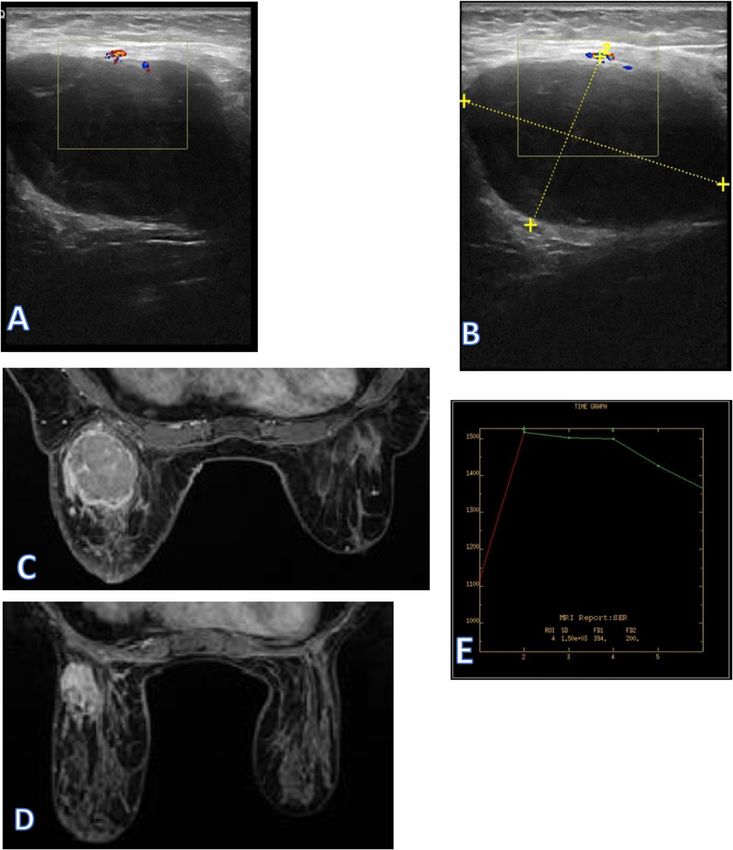
Alawi et al. Egyptian Journal of Radiology and Nuclear Medicine   (2020) 51:96                                                    Page 2 of 13

a shift to pathological examination [1]. Breast lesions in             Table 1 BI-RADS US assessment categories
females younger 25 years are managed differently com-                  Assessment                           Management
pared with those older. The first examination done is                  Category 0: incomplete—need          Recall for additional imaging
ultrasonography, whereas mammography for selected                      additional imaging evaluation
cases. Ultrasonography has more accuracy in the diagno-                Category 1: negative                 Routine screening
sis of dense fibroglandular breasts in young females [2].              Category 2: benign                   Routine screening
Mammography is important in the diagnosis of micro-
                                                                       Category 3: probably benign          Short-interval (6 months) follow-up
calcifications and suspected masses. MR imaging may be                                                      or continued surveillance
useful for those patients with deeper breast lumps or                  Category 4: suspicious—biopsy        Tissue diagnosis
chest wall lesions [3]. The risk of intervention proce-
                                                                          Category 4A: low suspicion for
dures to the developing breast is much greater than that                  malignancy
of the mature breast. So the conservative approach of
                                                                          Category 4B: moderate suspicion
clinical and ultrasonographic follow-up is more com-                      for malignancy
monly done in young females [4].                                          Category 4C: high suspicion for
                                                                          malignancy
Methods                                                                Category 5: highly suggestive of     Tissue diagnosis
This was a retrospective study, a total number of 250                  malignancy
cases with breast lumps under 25 years of age were regis-              Category 6: known biopsy-proven      Surgical excision when clinically
tered in the PACKS of our institution in the period from               malignancy                           appropriate
January 01, 2017 to December 31, 2018. Two hundred
three cases coped with our inclusion criteria that include             irregular, and the orientation of a mass can be described
available histopathological results either by biopsy or                as parallel or not parallel (often described as “taller-
after surgery based on their referring physicians decision.            than-wide” or “vertical”) [7]. When describing mass mar-
Our exclusion criteria were those cases (47) with definite             gins, the most important distinction is between circum-
BIRADS 2 lesions with no available pathology reports.                  scribed and non-circumscribed margins, a feature that
Ultrasonography was done to all patients (203 cases) and               distinguishes masses that require a biopsy from masses
MRI was performed to 26 cases. All cases were histologi-               that can be followed. Circumscribed margins are well
cally verified; their findings were reviewed and compared              defined or sharp. Non-circumscribed margins include
to radiological findings.                                              micro-lobulated, indistinct, angular, and spiculated. Le-
                                                                       sion boundary should be described as an abrupt inter-
Ultrasound assessment                                                  face or an echogenic halo which is a suspicious US
The ultrasound examination was performed with the GE                   finding. When defining the internal echo pattern of a le-
Logiq E9 Medical System, using a 7.5 MHz linear array                  sion, subcutaneous fat within the breast should be used
transducer. Real-time gray-scale and doppler images                    as a reference. Echo pattern descriptors include an-
were obtained. The patients were in a supine position                  echoic, hyperechoic, hypoechoic, isoechoic, and com-
and turned slightly to the contralateral side with the ipsi-           plex. Posterior acoustic features may or may not be seen
lateral upper limb extended cephalad and a pillow placed               when imaging breast masses. Posterior acoustic shadow-
under the ipsilateral shoulder. The palpable mass was                  ing is a suspicious finding and may be seen in cases of
scanned in longitudinal, transverse, and radial planes.                invasive carcinoma, postoperative scar, or macrocalcifi-
The clockface was used to indicate the site of the mass.               cations [8]. Although BI-RADS 0 is used frequently at
The masses were evaluated according to their borders,                  screening mammography, it is less relevant for the US, a
echogenicity, posterior echoes, and depth-width ratio.                 modality that commonly completes the diagnostic work-
Each mass was categorized as either benign or malig-                   up. However, in some instances, additional imaging,
nant. In our study, we classified the breast lesions ac-               such as MR imaging, may be necessary before a final as-
cording to the American College of Radiology (ACR) BI-                 sessment is rendered. BI-RADS 1 (negative) is used if an
RADS. ACR BI-RADS was to address a lack of                             abnormality is not seen in the US. BI-RADS 2 (benign)
standardization and uniformity in practice reporting to                is used when the results of the evaluation are negative
improve communication among radiologists, and sur-                     for malignancy. BI-RADS 3 (probably benign) is a breast
geons (Table 1) [5]. BI-RADS classification for the US is              mass with circumscribed margins, an oval shape, and
based on an analysis of descriptors from several feature               parallel orientation that can be classified as category 3.
categories (Table 2) [6]. It includes six morphologic fea-             This mass should have a risk of malignancy of less than
tures of breast masses: shape, orientation, margin,                    2%. Interval imaging follow-up for lesions categorized as
boundary, internal echo pattern, and posterior acoustic                BI-RADS 3 is recommended by Stavros et al. [9]. The
features. The shape is described as oval, round, or                    BI-RADS 4 is assigned to suspicious lesions for which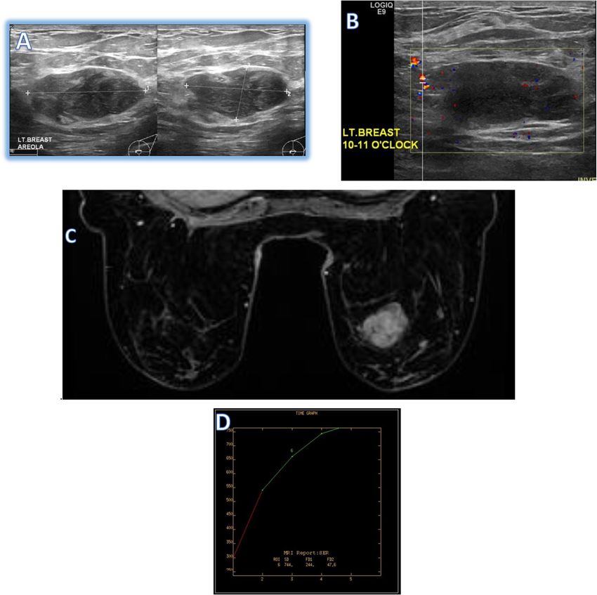
Alawi et al. Egyptian Journal of Radiology and Nuclear Medicine       (2020) 51:96                                                      Page 3 of 13

Table 2 BI-RADS US descriptors
US descriptor             Features favoring         Features favoring malignant                      Indeterminate features
                          benign
Shape of mass             Oval                      Irregular
Orientation of mass       Parallel to skin          Not parallel to skin
Margin of mass            Circumscribed             Micro-lobulated, indistinct, angular, or
                                                    spiculated
Lesion boundary           Abrupt interface          Echogenic halo
Echo pattern              Anechoic or               Complex                                          Isoechoic or hypoechoic
                          hyperechoic
Posterior acoustic                                  Shadowing or combined pattern                    Enhancement or no posterior acoustic
features                                                                                             features

biopsy is recommended. This category is with lesions                        mis- or overdiagnosis. By canceling out the fat signal
having a probability of malignancy of approximately 3–                      within the breast and then using subtraction images, the
94%. Therefore, in the ACR BI-RADS atlas, the sugges-                       ability to identify enhancing lesions becomes more ac-
tion now is to subdivide category 4 into three subgroups                    curate. The axial T2 images are reviewed, as these are
(4A, 4B, and 4C) to better inform the referring clinicians                  fluid sensitive and are excellent for finding cysts, fluid,
and pathologist of the degree of concern. Category 4A                       and inflammation. The T2 images also provide the best
lesions have a low suspicion for malignancy. Category                       evaluation of breast anatomy and skin. The T1 images
4B is considered to have an intermediate suspicion for                      are also excellent for evaluating intraductal blood and
malignancy [10]. Category 4C is used for lesions with                       lymph node.
moderate suspicion but not classic findings of malig-                          In our study, we classify the breast masses by MRI ac-
nancy. BI-RADS 5 (highly suggestive of malignancy) is                       cording to American College of Radiology (ACR) BI-
reserved for findings that almost represent breast cancer                   RADS lexicon (Table 3). All final impressions should be
more than 95%. BI-RADS 6 (known cancer) is for                              complete with each lesion fully categorized [12]. It in-
biopsy-proved malignancy [11].                                              cludes describing the mass; shape: lobulated, round, or
                                                                            oval; margin: circumscribed, not circumscribed, irregular,
MRI assessment                                                              or spiculated; and the internal enhancement characteris-
MRI Breast was performed using 1.5 T (Optima GE,                            tics homogeneous, heterogeneous, or rim enhancement.
USA). The patient was placed prone using a breast coil.                     The non-mass enhancement (NME), distribution: focal,
The MRI protocol included axial T1, T2, diffusion, STIR,                    linear, segmental, regional, multiple regions, or diffuse;
and sagittal STIR sequences contrast was intravenously                      internal     enhancement        patterns:   homogeneous,
injected according to the patient’s weight using the for-
mula of 0.1 mmol/kg with a power injector at a rate of 3                    Table 3 BI-RADS MRI assessment categories and management
mL per second and flushed with 10 cc of saline and dy-
                                                                            Assessment                       Management
namic T1-weighted fat-saturated sequences obtained in
                                                                            Category 0: incomplete—need      Recommend additional imaging:
pre-contrast and post-contrast (60, 120, 180, 240, 300,                     additional imaging evaluation    mammogram or targeted US
and 360 s after contrast administration) phases for bilat-
                                                                            Category 1: negative             Routine breast MRI screening if
eral breasts. Post-processing allows the generation of the                                                   cumulative lifetime risk ≥ 20%
maximum intensity projection (MIP), digital subtraction                     Category 2: benign               Routine breast MRI screening if
sequences, and the kinetic curves. Scanning slice thick-                                                     cumulative lifetime risk ≥ 20%
ness should be 3 mm or less and resolution should be 1                      Category 3: probably benign      Short-interval (6 months) follow-up
mm or less in order to minimize volume averaging. MRI                                                        If the finding is visible on e.g., US, the
offers the advantage of 3D imaging, which is better at                                                       most widely available method should
                                                                                                             be used for follow-up (should be ap-
detecting smaller lesions than 2D imaging because 2D                                                         plied only to lesions not fitting cat-
imaging produces gaps between slices in which a small                                                        egories II and IV, probably benign
lesion may be missed. To optimize the contrast between                                                       findings in high-risk screening should
                                                                                                             rather be biopsied than followed up)
a tumor and surrounding breast parenchyma, the tech-
nique must be excellent. Fat suppression is important in                    Category 4: suspicious           Tissue diagnosis
contrast-enhanced imaging of the breast because lesion                      Category 5: highly suggestive of Tissue diagnosis
                                                                            malignancy
identification in breast MRI relies upon the subtraction
of pre- and post-contrast images. In pre-contrast T1 im-                    Category 6: known biopsy-        Surgical excision when clinically
                                                                            proven malignancy                appropriate
ages, the bright, fatty signal from the breast can lead to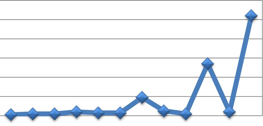
Alawi et al. Egyptian Journal of Radiology and Nuclear Medicine        (2020) 51:96                                          Page 4 of 13

Table 4 Types, frequency, and percentage of the breast lesion in the examined young female
Breast lesions classification                                                                   Frequency                     Percentage
Cystic lesions                             Fibrocystic change                                   54                            26.6%
                                           Abscess and mastitis                                 19                            9.35%
                                           Mammary duct ectasia                                 5                             2.46%
                                           Galactoceles                                         3                             1.47%
                                           Hematomas                                            2                             0.98%
Total cystic lesion                                                                             85                            41.9%
Benign masses                              Fibroadenoma                                         104                           51.2%
                                           Intraductal papilloma                                4                             1.97%
                                           Intramammary lymph node                              4                             1.97%
                                           Lactating adenoma                                    2                             0.98%
                                           Adenomyoepithelioma                                  1                             0.49%
                                           Fibroadenomatoid hyperplasia                         2                             0.98%
Total benign masses                                                                             115                           56.7%
Malignant masses                           Carcinoma IDC                                        3                             1.47%
Total malignant masses                                                                          3                             1.47%

heterogeneous, clumped, or clustered ring. Associated fea-                  the scan. Category 1 is as follows: negative, this is a nor-
tures are as follows: nipple retraction, nipple invasion, skin              mal examination. No abnormal enhancement was found.
retraction, skin thickening, skin invasion, axillary adenopa-               Category 2 includes the following: benign-like category
thy, pectoralis muscle invasion, and chest wall invasion.                   such as intra-mammary lymph node, enhancing and non-
The kinetic curve is as follows: type I (progressive en-                    enhancing fibroadenomas, cysts, old non-enhancing scars
hancement pattern usually considered benign), type II                       or recent scars, postoperative collections, fat-containing
(plateau pattern, considered concerning for malignancy),                    lesions (such as oil cysts, lipomas, galactoceles, and
and type III (washout pattern, considered strongly sug-                     hamartomas). Category 3 is a follows: probably benign, a
gestive of malignancy). Category 0 is as follows: incom-                    finding assessed using this category should have a ≤ 2%
plete—use this for a finding that needs additional imaging                  likelihood of malignancy, but greater than the essentially
evaluation. This may be used for a technically unsatisfac-                  0% likelihood of malignancy of a characteristically benign
tory scan or when more information is needed to interpret                   finding. A probably benign finding is not expected to

  Fig. 1 Frequency of the breast lesion in the examined young female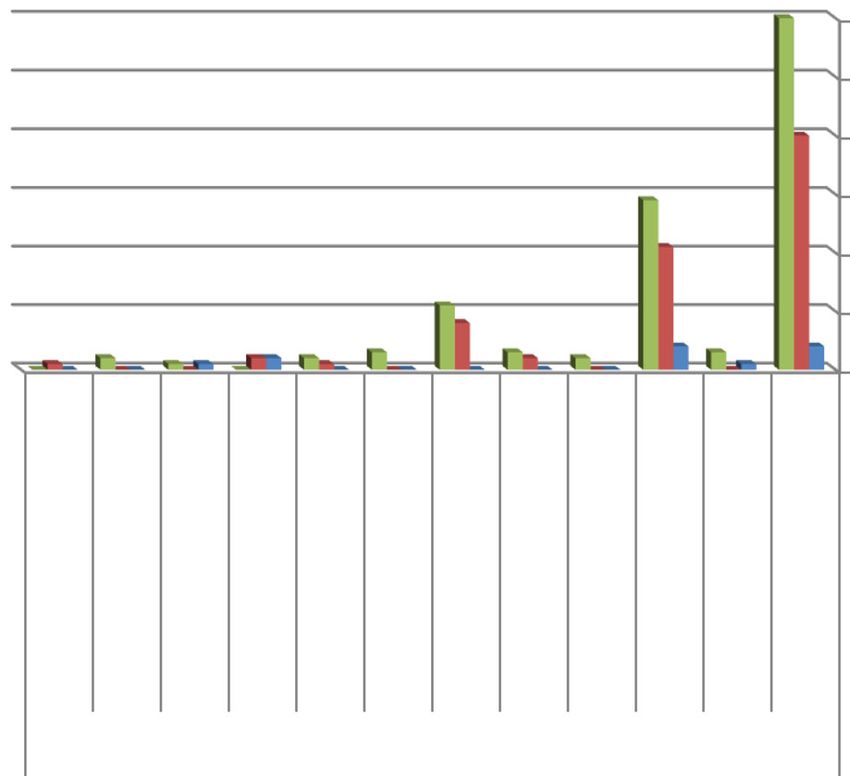
Table 5 Frequency analysis of examined patients regarding to age
Age groups      The breast lesions                                                                                                                                         Total
                                                                                                                                                                                   Alawi et al. Egyptian Journal of Radiology and Nuclear Medicine

                Fibroadenoma Papilloma Fibrocystic Lactating   Mammary        Abscess and   Malignant   Galactoceles LN Hematomas Fibroadenomatoid   Adenomyoepithelioma
                                                   adenoma     duct ectasia   mastitis      IDC                                   hyperplasia
From 11 to 15   4               1       4          0           0              0             0           0          2   1          0                  0                     12
years old
From 16 to 20   40              0       21         0           2              8             0           1          2   0          0                  1                     75
years old
                                                                                                                                                                                   (2020) 51:96

From 21 to 25   60              3       29         2           3              11            3           2          0   1          2                  0                     116
years old
Total           104             4       54         2           5              19            3           3          4   2          2                  1                     203
                                                                                                                                                                                   Page 5 of 13
Alawi et al. Egyptian Journal of Radiology and Nuclear Medicine (2020) 51:96 Page 6 of 13 Fig. 2 Breast lesion in the examined young female according to age group change over the suggested period of imaging. Category 4, malignancy in between. In breast MRI, assessment cat- suspicious of this category is reserved for findings that do egory 4 is not currently divided into subcategories 4A, 4B, not have the classic appearance of malignancy but are suf- and 4C. Category 4 is used for the majority of findings ficiently suspicious to justify a recommendation for bi- prompting breast intervention, which can be performed opsy. For a category 3 assessment is a 2% likelihood of by percutaneous biopsy, by US or stereotactic guidance, or malignancy and for a category 5 assessment is 95%, so cat- by MRI guidance for lesions not visible at either US or egory 4 assessments cover the wide range of likelihood of mammography. Category 5 is highly suggestive of Fig. 3 The histopathology result according to age groups
Alawi et al. Egyptian Journal of Radiology and Nuclear Medicine        (2020) 51:96                                                 Page 7 of 13

Table 6 Frequency analysis of examined patients regarding to age, BIRADS classification, and pathological results
Age group                   Type of examination
                            The ultrasonographic BI-RADS          The MRI BI-RADS                    Histopathology result (benign vs malignant)
              No. of        203                                   26 of 203                          203
              exam. Pat.
                            BIRADS 2    BIRADS3     BI-RADS 5     BIRADS2       BIRADS3   BIRADS 5   Benign                Malignant
From 11 to 15 years old     11          1           0             0             1         0          12                    0
From 16 to 20 years old     72          3           0             0             8         0          75                    0
From 21 to 25 years old     103         10          3             0             14        3          113                   3
Total                       186         14          3             0             23        3          200                   3
Total percent ~             92%         6.7%        1.5%          0%            88%       12%        98.5%                 1.5%

malignancy; these assessments carry a very high probabil-                     (9.35%), mammary duct ectasia 5 cases (2.46%), intraduc-
ity (≥ 95%) of malignancy. Category 6 is known biopsy-                        tal papilloma 4 cases (1.97%), intramammary lymph node
proven malignancy [13].                                                       4 case (1.97%), galactocele 3 cases (1.47%), hematomas 2
                                                                              cases (0.98%), fibroadenomatoid hyperplasia 2 cases
                                                                              (0.98%), lactating adenoma 2 cases (0.98%), and adeno-
Result                                                                        myoepithelioma 1 case (0.49%); however, the malignant
A total of 203 of symptomatic breast lesions were exam-                       group represents 3 cases (1.47%) and were intraductal car-
ined at the radiology department from January 1, 2017 to                      cinoma (Fig. 1). According to age group (Table 5) (Figs. 2
December 31, 2018 in women under 25 years. The cases                          and 3), women from 11 to 15 years were 12 cases includ-
were stratified by age into those 10–15 years, 12 cases                       ing fibroadenoma 4 cases, fibrocystic disease 4 cases, intra-
(5.9%); between 16 and 20 years, 75 cases (36.9%); and                        mammary lymph nodes 2 cases, papilloma 1 case,
those between 21 and 25 years, 116 cases (58.4%). The le-                     hematoma 1 case. From 16 to 20 years was 75 cases in-
sions were primarily class into cystic, benign, and malig-                    cluding fibroadenoma 40 cases, fibrocystic disease 21
nant lesions. There were 115 cases (56.7%) benign, 85                         cases, abscess 8 cases, mammary duct ectasia 2 cases,
cases (41.9%) cystic, and 3 cases (1.47%) malignant lumps                     intramammary lymph nodes 2 cases, galactocele 1 case,
(Table 4). The largest group comprised fibroadenoma                           adenomyoepithelioma 1 case. From 21 to 25 years was 116
representing 104 cases (51.2%) followed by fibrocystic                        cases including fibro adenoma 60 cases, fibrocystic disease
change representing 54 cases (26.6%), abscess 19 cases                        29 cases, abscess 11 cases, papilloma 3 cases, mammary

 Fig. 4 The sonographic BI-RADS according to age groups
Alawi et al. Egyptian Journal of Radiology and Nuclear Medicine (2020) 51:96 Page 8 of 13
duct ectasia 3 cases, intraductal carcinoma 3 cases, lactat- found in our study that pathologic types of breast dis-
ing adenoma 2 cases, galactocele 2 cases, fibroadenoma- ease in women under 25 years are broad and this agrees
toid hyperplasia 2 cases, and hematoma 1 case. Seventeen with Leong et al. [15]. In our study, we found that a
patients of 203 who classified by the US as 14 cases BI- large number of patients complain of breast lump are
RADS 3 (with a strong family history of cancer breast) between 21 and 25 years 116 cases (58.4%), followed by
and 3 cases BI-RADS 5 were proceeding directly to MRI; 16–20 years 75 cases (36.9%), then 10–15 years 12 cases
the same category in MRI and their pathology results were (5.9%). Similar results were reported by Olu-Eddo and
also the same 14 cases were benign and the 3 cases were Ugiagbe [16]. According to Ellen et al. [1], classification,
intraductal carcinoma. One hundred eighty-six patients of in our study divided the lesions into cystic, benign, and
203 continued the 3–6 month-interval follow-up, 9 pa- malignant lesions. One hundred fifteen (49.2%) lesions
tients of 186 (were US BI-RADS category 2) showed an in- were benign cases, 3 (1.4%) were malignant lumps, and
crease in size and changes of US features and need MRI; 85 cases were cystic. In our study, the majority of breast
their category in MRI was changed to BI-RADS category 3 masses in women under 25 years are benign including
(strong family history of cancer breast) and their path- tumors and inflammatory conditions representing ap-
ology results were benign (Table 6) (Figs. 4 and 5), for all proximately 89.2% and 9.36% respectively. This observa-
radiological findings compared with pathological results. tion is compatible with an Indian study conducted by
Malik and Bharadwaj [14]. In the current study, fibro-
Discussion adenomas are found in 104 cases (51.2%) constituted the
Breast masses that occur in the young females are of a largest group of benign lumps, followed by 54 (26.6%)
benign nature with extremely rare exceptions. The cases of fibrocystic disease, then inflammatory lesions 19
young female generally does not consider themselves to cases (9.36%). Malignant lumps constituted 3 cases
be at risk for breast cancer. However, breast cancer can (1.4%) and all were infiltrating duct cell carcinoma and
occur at any age and women of every age should be this cope with finding reported by Egwuonwu et al. [17].
aware of risk factors and breast self-examination tech- Regarding benign masses, fibroadenomas are found in in
niques. The incidence of breast cancer increases with 104 cases (51.2%) constituted the largest group of benign
age, doubling about every 10 years until menopause. lumps and more common between 21 and 25 years (60
Diagnosis of breast cancer in young females is more dif- cases) and this agrees with Valeur et al. [18] that
ficult because of their denser breast tissue [14]. We have revealed fibroadenomas are the most common benign
Fig. 5 The MRI BI-RADS according to age groupsAlawi et al. Egyptian Journal of Radiology and Nuclear Medicine (2020) 51:96 Page 9 of 13 Fig. 6 A 24-year-old patient complains of right breast lump. a and b US revealed right axillary tail large complex hypo echoic lesion with area of breakdown, slightly micro-lobulated margin, and mild echogenic halo boundary is noted (BIRADS V classification). c–e MRI revealed that the mass shows rim enhancement with rapid washout (type III enhancement pattern in kinetic time intensity curve) (category V according to MRI LEXICON classifications). Biopsy revealed intraductal carcinoma tumors in young female patients, accounting for 54–94% in our study, they were 2 cases and one case respectively. of cases. In our study, intraductal papilloma 4 cases Regarding cystic lesions in our study, fibrocystic disease (1.97%) and this agree with Greydanus et al. [19] that re- is the most common pathology and after fibroadenoma vealed it is an uncommon benign disease that occurs in in general benign lesions, it was 54 cases (26.6%), the young females. In our study, the intramammary lymph majority at 21–25 years about 29 cases and this agrees nodes were 4 case (1.97%). Intramammary lymph nodes with Guray and Sahin [24] that revealed fibrocystic are present in up to 47% of breasts. Although they are change is most often diagnosed between the ages of 20 usually located in the upper outer quadrant, they may and 40. Inflammatory changes and abscess 19 cases appear anywhere in the breast [20]. In our 2 cases of lac- (9.35%) between age group 16 and 25 years, 12 cases tating adenoma, patients were lactating in the age group were lactating and this agrees with Chung et al. [25] that 21–25 years and this agrees with Coffin [21] that re- revealed mastitis and abscess are common in lactating vealed lactating adenomas predominantly consist of lob- women. In our study, we found 5 cases duct ectasia, 3 ules with lactational changes that develop in late cases galactocele, all are lactating in the age group 16– pregnancy. Fibroadenomatoid hyperplasia and adeno- 25 years, and 2 cases hematoma with past history of myoepithelioma are rare benign breast lesions [22, 23]; trauma; duct ectasia is a rare entity that occurs in young
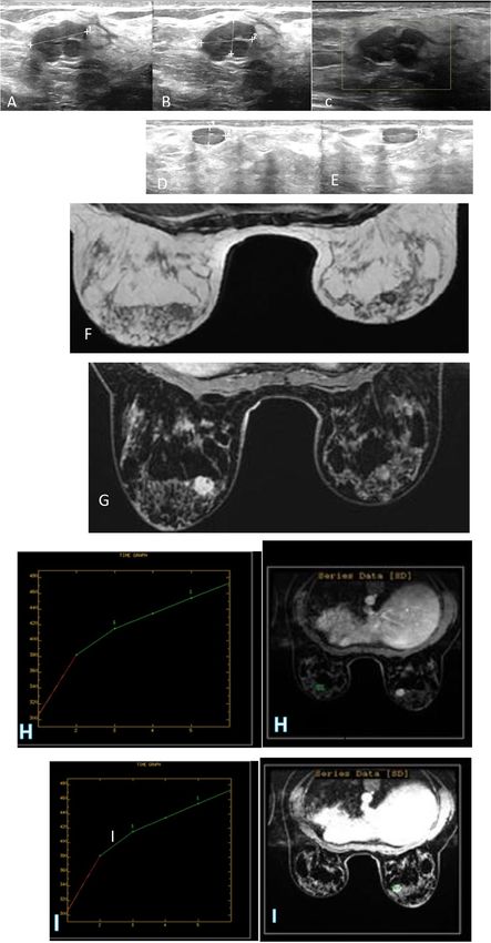
Alawi et al. Egyptian Journal of Radiology and Nuclear Medicine      (2020) 51:96                                                    Page 10 of 13

patients, clinically, patients with duct ectasia present                   advanced stage and more aggressive features. This sug-
with bloody discharge from the nipple or a palpable                        gests that women under 25 years with palpable lumps
mass in the breast [26]. Galactocele is a retention cyst                   should be evaluated adequately to avoid delays in the
filled with milk caused by obstruction of a lactiferous                    diagnosis of palpable breast cancer, regardless of the
duct. It is commonly seen in pregnant or lactating                         very low incidence of breast cancer before 25 years of
women [25]. Hematoma may be associated with a his-                         age. In our study, the 3 malignant breast cases had a
tory of breast trauma or surgery. A recent history of                      positive family history and this agrees with Gordon et al.
trauma or surgery is essential for the diagnosis [26]. Ma-                 [30] that reveal family history is a very important risk
lignancy of breasts is very rare in this age group (Ashi-                  factor that should be considered with breast cancer, for
kori et al. [27]). Malignant neoplasm in our study                         women with a family history of breast cancer in both a
represents 1.44% of lumps. Although this accounts for                      mother and a sister. In our study, the US could detect
the smallest group in the study, however still this fre-                   all the lesions. Because the majority of patients had palp-
quency in young women under 25 years is high and this                      able lumps, targeted US examinations could also im-
agrees with Leong et al. [15]. The malignant lesions are                   prove cancer detection and this agrees with Y.Y. An
present between 21 and 25 years in agreement with                          et al. [31]. In our study, among 26 cases shifting to MRI
Ambreen et al. [28]. The breast cancers in our study ap-                   examination, 3 cases were assigned to US BI-RADS cat-
peared more frequently as mass-type lesions and this                       egory 5 and the same category in MRI which proved to
agrees with Bullier et al. [29] that reveal most of the                    be malignant by pathological results (Fig. 6). Fourteen
breast cancers in women younger than 25 years of age                       cases were assigned to US BI-RADS category 3, the same
were palpable, and palpability was associated with                         category in MRI and their pathology results were benign

 Fig. 7 A 22-year-old patient with history of left breast lump and strong family history of cancer breast. a and b US shows lobulated hypoechoic
 mass with vascularity inside (BIRADS III). c and d MRI revealed that mass appeared lobulated showed homogenous enhancement (type I kinetic
 time intensity curve) (category III MRI lexicon classifications) and biopsy confirmed fibro adenoma
Alawi et al. Egyptian Journal of Radiology and Nuclear Medicine      (2020) 51:96                                                    Page 11 of 13

 Fig. 8 A 25-year-old patient with history of bilateral breast lumps and strong family history of CA breast. Right breast mass (a and b) US shows
 hypoechoic mass (BIRAD II), c 3-month follow-up shows increasing in size and more lobulation (change to BIRADS III). The left breast mass (d) US
 shows smooth outline hypoechoic mass (BIRAD II , e 3-month follow-up shows no significant changes (BIRAD II). f and g MRI, T1, and post-
 contrast study detected the masses in both sides, the right one is lobulated and the left one is smooth margin, h and i) images revealed that the
 right mass shows type I enhancement pattern in kinetic time intensity curve (BIRAD III) and the left mass shows type I enhancement pattern in
 kinetic time intensity curve (BIRADS II). Biopsy confirmed fibro adenoma on both sides
Alawi et al. Egyptian Journal of Radiology and Nuclear Medicine             (2020) 51:96                                                           Page 12 of 13

(Fig. 7), the remaining 9 cases were assigned to US BI-                          Ethics approval and consent to participate
RADS category 2 but shift to MRI examination due to                              This study was approved by the Research Ethics Committee of KAAH (King
                                                                                 Abdul-Aziz Hospital) in Holy Makkah, Saudi Arabia. The patients were less
increasing size, changes in its shape in the US follow-up                        than 16 years old at the time of the study, and written informed consent for
3–6 month, and due to their strong family history of                             their participation was given by their parent or legal guardian. The patients
breast cancer; their category in MRI was changed to BI-                          were more than 16 years old at the time of the study, and written informed
                                                                                 consent for their participation was given. The committee’s reference number
RADS category 3 and their pathology results were be-                             is H-02-K-076-1912-238.
nign (Fig. 8 ), and this agrees with Lee et al. [32] that re-
vealed breast lesions in young females’ biopsy should be
                                                                                 Consent for publication
considered in the event of abnormal imaging findings,                            All patients were less than 16 years old, and written informed consent for
such as non-circumscribed margins, complex solid and                             the publication of this data was given by their parent or legal guardian.
                                                                                 All patients were more than 16 years old at the time of the study, written
cystic components, posterior acoustic shadowing, size
                                                                                 informed consent for their participation was given.
above 3 cm, or an increase in mass size. A clinical his-
tory that includes a risk factor for malignancy, such as a
                                                                                 Competing interests
family history of breast cancer, should prompt consider-                         The authors declare that they have no competing interests.
ation of biopsy even if the lesion has a probably benign
appearance. In our study, there is no significant differ-                        Author details
                                                                                 1
                                                                                  KAAH (King Abdul-Aziz Hospital), Holy Makkah, Kingdom of Saudi Arabia.
ence between ultrasonography and MRI for diagnosis,                              2
                                                                                  Radiology Department, KAAH, Holy Makkah, Kingdom of Saudi Arabia.
and p value is more than 0.05 and this agrees with Qiao-                         3
                                                                                  Radiology Department, Mansoura University, Mansoura, Egypt. 4Breast Unit
hong and Jianwu [33]. Any female patients with a palp-                           Surgery Department, KAAH, Holy Makkah, Kingdom of Saudi Arabia.
                                                                                 5
                                                                                  Pathology Department, KAAH, Holy Makkah, Kingdom of Saudi Arabia. 6King
able breast mass who are under the age of 25 years,                              Abdul-Aziz University, Jeddah, Kingdom of Saudi Arabia.
ultrasound should be the initial imaging modality [34].
Our study has a limitation that it was a retrospective                           Received: 14 February 2020 Accepted: 27 May 2020
study with a small sample size of malignant lesions;
however, our patient group size is still considerable be-
cause breast cancer in women under 25 years of age is                            References
                                                                                 1. Ellen MC, Regino C, Gregory JH et al (2009) Breast masses in children and
quite rare. This study shows the importance of ultrason-                             adolescents: radiologic-pathologic correlation. RadioGraphics 29:907–931
ography in the diagnosis of palpable breast mass in                              2. Venta LA, Dudiak CM, Salomon CG et al (1994) Sonographic evaluation of
women under 25 years which were overwhelmingly                                       the breast. RadioGraphics 14:29–50
                                                                                 3. Dehner LP, Hill DA, Deschryver K (1999) Pathology of the breast in children,
benign.                                                                              adolescents, and young adults. Semin Diagn Pathol 16:235–247
                                                                                 4. Weinstein SP, Conant EF, Orel SG et al (2000) Spectrum of US findings in
                                                                                     pediatric and adolescent patients with palpable breast masses.
Conclusion                                                                           RadioGraphics 20:1613–1621
Most breast lesions in young women are benign. Ultra-                            5. A. American College of Radiology. BI-RADS®—Ultrasound. 1st ed. In: Breast
sonography is an essential first imaging modality in the                             Imaging Reporting and Data System (BI-RADS) atlas. 5th ed. Reston, Va:
                                                                                     American College of Radiology, 2010.
diagnosis of women under 25 years with breast lesions.
                                                                                 6. Hong AS, Rosen EL, Soo MS et al (2005) BI-RADS for sonography: positive
                                                                                     and negative predictive values of sonographic features. AJR Am J
Abbreviations
                                                                                     Roentgenol 184(4):1260–1265
CT: Computed tomography; MRI: Magnetic resonance imaging;
                                                                                 7. Soo MS, Kornguth PJ, Hertzberg BS (1998) Fat necrosis in the breast:
ACR: American College of Radiology; BI-RADS: Breast Imaging-Reporting and
                                                                                     sonographic features. Radiology 206(1):261–269
Data System; US: Ultrasonography; MIP: Maximum intensity projection; 2D: 2
                                                                                 8. Sughra R, Allison LG, Sona AC et al (2010) US of breast masses categorized
dimension; 3D: 3 dimension; NME: Non-mass enhancement
                                                                                     as BI-RADS 3, 4, and 5: pictorial review of factors influencing clinical
                                                                                     management. RadioGraphics 30:1199–1213
Acknowledgements                                                                 9. Stavros AT, Thickman D, Rapp CL et al (1995) Solid breast nodules: use of
Not applicable                                                                       sonography to distinguish between benign and malignant lesions.
                                                                                     Radiology 196:12334
Authors’ contributions                                                           10. Raza S, Chikarmane SA, Neilsen SS et al (2008) BI-RADS 3, 4, and 5 lesions:
MMH carried out the radiological studies, participated in design of the study,       value of US in management—follow-up and outcome. Radiology 248(3):
AS and HQ collected the patients’ data, MMH performed the statistical                773–781
analysis. AA participated in the sequence alignment and drafted the              11. American College of Radiology (2003) BI-RADS®—mammography. In: Breast
manuscript. MH participated in the acquisition of data. WH participated in           Imaging Reporting and Data System (BI-RADS) atlas, 4th edn. American
the sequence alignment. SA participated in the design of the study and               College of Radiology, Reston, Va
performed the statistical analysis. HM conceived of the study, participated in   12. Mahoney MC, Gatsonis C, Hanna L et al (2012) Positive predictive value of
its design and coordination, and helped to draft the manuscript. MMH wrote           BI-RADS MR imaging. Radiology 264:51–58
the paper with revision. All authors read and approved the final manuscript.     13. American College of Radiology. BI-RADS®—MRI. 1st ed. In: Breast Imaging
                                                                                     Reporting and Data System (BI-RADS) atlas. 5th ed. Reston, Va: American
Funding                                                                              College of Radiology, 2010.
This study had no funding from any resource.                                     14. Malik R, Bharadwaj VK (2003) Breast lesions in young females-a 20-year
                                                                                     study for significance of early recognition. Indian J Pathol Microbiol 46:559–
Availability of data and materials                                                   562
The datasets used and/or analyzed during the current study are available         15. Leong SP, Shen ZZ, Liu TJ et al (2010) Is breast cancer the same disease in
from the corresponding author on reasonable request.                                 Asian and Western countries? World J Surg. 34:2308–2324
Alawi et al. Egyptian Journal of Radiology and Nuclear Medicine (2020) 51:96 Page 13 of 13
16. Olu-Eddo AN& Ugiagbe EE. (2011) Benign breast lesions in an African
population: a 25-year histopathological review of 1864 cases. Niger Med J.
52(4):211–216
17. Egwuonwu OA, Anyanwu S, Chianakwana GU et al (2016) Fibroadenoma:
accuracy of clinical diagnosis in females aged 25 years or less. Niger J Clin
Pract 19:336–338
18. Valeur NS, Rahbar H, Chapman T (2015) Ultrasound of pediatric breast
masses: what to do with lumps and bumps. Pediatr Radiol 45:1584–1599
19. Greydanus DE, Matytsina L, Gains M (2006) Breast disorders in children and
adolescents. Prim Care 33:455–502
20. Millet I, Pages E, Hoa D et al (2012) Pearls and pitfalls in breast MRI. Br J
Radiol. 85(1011):197–207
21. Coffin CM (2002) The breast. In: Stocker JT, Dehner LP (eds) Pediatric pathology,
2nd edn. Lippincott Williams & Wilkins, Philadelphia, Pa, pp 993–1015
22. Chen Y, Bekhash A, Kovatich AJ et al (2015) Positive association of
fibroadenomatoid change with HER2-negative invasive breast cancer: a co-
occurrence study. PLOS ONE 10(6):e0129500
23. Yoon Y, Dhananjay C (2013) Adenomyoepithelioma of the breast: a brief
diagnostic review. Arch Pathol Lab Med 137(5):725–729
24. Guray M, Sahin AA (2006) Benign breast diseases: classification, diagnosis,
and management. Oncologist. 11(5):435–449
25. Chung EM, Cube R, Hall GJ, González C et al (2009) From the archives of
the AFIP: breast masses in children and adolescents: radiologic-pathologic
correlation. Radiographics 29:907–931
26. Gao Y, Saksena MA, Brachtel EF et al (2015) How to approach breast lesions
in children and adolescents. Eur J Radiol 84:1350–1364
27. Ashikari H, Jun MY, Farrow JH et al (1977) Breast carcinoma in children and
adolescents. Clin Bull. 7:55–62
28. Ambreen M, Tahir SM, Gulshan AS et al (2011) Breast lumps in adolescent
and young female: are all benign. Journal of the Liaquat University of
Medical and Health Sciences 10:3
29. Bullier G, MacGrogan H, Bonnefoi G et al (2013) Imaging features of
sporadic breast cancer in women under 40 years old: 97 cases. Eur. Radiol.
23:3237–3245
30. Gordon PB (1995) US problem solving in breast imaging: tricks of the trade.
RSNA categorical course in breast imaging.:121–131
31. An YY, Kim SH, Kang BJ et al (2014) Characteristic features and usefulness of
MRI in breast cancer in patients under 40 years old: correlations with
conventional imaging and prognostic factors. Breast Cancer 21:302–315
32. Lee EJ, Chang Y-W, Oh JH et al (2018) Breast lesions in children and
adolescents: diagnosis and management. Korean J Radiol 19(5):978
33. Qiaohong P, Jianwu JI (2019) Diagnostic value of ultrasound combined with
magnetic resonance imaging in different stages of breast cancer. Oncol Lett
17:209–214
34. Jackson VP (1995) The current role of ultrasonography in breast imaging.
Radiol Clin North Am 83:1161–1170
Publisher’s Note
Springer Nature remains neutral with regard to jurisdictional claims in
published maps and institutional affiliations.You can also read