Role of Helicobacter pylori in the triggering and evolution of hemorrhagic gastro duodenal lesions
←
→
Page content transcription
If your browser does not render page correctly, please read the page content below
EXPERIMENTAL AND THERAPEUTIC MEDICINE 22: 1147, 2021

                     Role of Helicobacter pylori in the triggering and
                    evolution of hemorrhagic gastro‑duodenal lesions
          DRAGOS GEORGE POPA1, COSMIN VASILE OBLEAGĂ2*, BOGDAN SOCEA3,4, DRAGOS SERBAN3,5,
         MARIUS EUGEN CIUREA1, MARIAN DIACONESCU2, IONICĂ DANIEL VÎLCEA2, CRISTIAN MEȘINĂ2,
          CECIL MIREA2, DAN NICOLAE FLORESCU6, VLAD DUMITRU BALEANU6, MEDA COMANDASU5,
                 MIHAI SILVIU TUDOSIE3, LAURA CARINA TRIBUS3,7* and BOGDAN NICULESCU8

          Departments of 1Plastic and Reconstructive Surgery, and 2Surgery, Craiova University of Medicine and Pharmacy,
              200349 Craiova; 3Faculty of Medicine, ‘Carol Davila’ University of Medicine and Pharmacy Bucharest,
                 020021 Bucharest; 4Department of General Surgery, ‘Sf. Pantelimon’ Clinical Emergency Hospital,
              021659 Bucharest; 5Fourth Department of General Surgery, Emergency University Hospital Bucharest,
                050098 Bucharest; 6Department of Gastroenterology, Craiova University of Medicine and Pharmacy,
                     200349 Craiova; 7Gastroenterology Department, Emergency University Hospital Bucharest;
                  8
                    Department of Sports and Health, ‘Constantin Brancusi’ University, 210152 Targu Jiu, Romania

                                            Received June 8, 2021; Accepted July 8, 2021

                                                     DOI: 10.3892/etm.2021.10582

Abstract. The majority of studies concerning Helicobacter pylori    NSAIDs was replaced by therapies with oral antiplatelet or
(H. pylori) are oriented towards the implication of infec‑          anticoagulant agents. The need for hemostasis surgery was
tion with H. pylori in processes that end in the formation of       more common in patients who exhibited H. pylori‑positive
neoplasia, without assessing the impact of the bacterium in         UGIB compared to H. pylori‑negative (16 vs. 9.7%). In patients
triggering acute gastroduodenal hemorrhagic episodes. The           with H. pylori‑positive hemorrhagic lesions, gastric resection
present study includes 166 patients with upper digestive hemor‑     was frequently required to obtain hemostasis. Persistence of
rhage, admitted to the ATI Clinic, the Gastroenterology Clinic      H. pylori infection in patients with a history of gastric resec‑
or to the Surgery II Clinic of the County Emergency Clinical        tion (4.1%) still predisposes to a hemorrhagic or neoplastic
Hospital in Craiova, Romania between 2017 and 2019. All             complication.
patients were monitored for evolution and received treatment
according to current guidelines, and hemorrhagic lesions were       Introduction
biopsied. In the study group, 56.8% of the patients with upper
gastrointestinal bleeding (UGIB) were positive for H. pylori        Marked progress has been made in the management of
and 43.2% were negative. In patients less than 50 years of          non‑varicose upper gastrointestinal bleeding (UGIB) due to
age, non‑steroidal anti‑inflammatory drug (NSAID) use and           the introduction of drugs that decrease gastric acid secretion,
H. pylori infection had a cumulative effect in causing bleeding     Helicobacter pylori (H. pylori) eradication therapies, and
lesions, but in patients older than 50 years of age, the use of     improved endoscopic hemostasis, yet, the mortality rate has
                                                                    remained relatively constant (1). H. pylori induces chronic
                                                                    superficial gastritis with neutrophil infiltration into the
                                                                    mucosa, therefore, it has been speculated that H. pylori infec‑
Correspondence to: Dr Dragos Serban, Faculty of Medicine,           tion underlies the bleeding lesion (2). Gastric and duodenal
‘Carol Davila’ University of Medicine and Pharmacy Bucharest,       ulcers are strongly related to H. pylori (2). In initial reports
37 Dionisie Lupu Street, 020021 Bucharest, Romania                  around the world, in the first decade after the discovery of
E‑mail: dragos.serban@umfcd.ro                                      H. pylori, ~95% of duodenal ulcers and 85% of gastric ulcers
                                                                    were associated with H. pylori infection (3), and eradication
*
    Contributed equally
                                                                    of H. pylori changed the natural course of ulcer disease and
                                                                    almost completely prevented the recurrence of the ulcer (4).
Abbreviations: UGIB, upper gastrointestinal bleeding; H. pylori,
Helicobacter pylori; NSAIDs, non‑steroidal anti‑inflammatory        Hemorrhage is the most common complication of ulcer
drugs; IHC, immunohistochemical examination; MALT, gastric          disease and is estimated to be present in 15‑20% of ulcers.
mucosa‑associated lymphoma                                          Approximately 40% of patients with UGIB have a hemor‑
                                                                    rhagic ulcer, and ulcerative disease is therefore the leading
Key words: Helicobacter pylori, upper gastrointestinal bleeding,    cause of upper gastrointestinal hemorrhage (1). Eradication of
endoscopic hemostasis, surgery, anticoagulant agents, neoplasia     H. pylori greatly reduces the risk of ulcer and also bleeding
                                                                    in those patients whose hemorrhage was due to H. pylori
                                                                    infection (4). Mortality varies between 3 and 14% and has not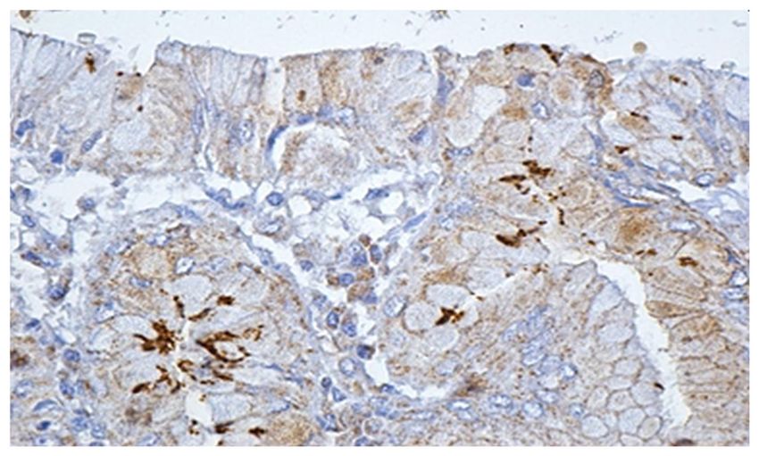
2                           POPA et al: ROLE OF H. pylori IN HEMORRHAGIC GASTRO-DUODENAL LESIONS

changed in the last 10 years (1,4). Mortality increases with         paraffin). The final results were obtained after an interval
age and is significantly higher in patients who are already          of 20‑25 days from the sampling of the biopsy fragments.
hospitalized for associated comorbidities. Risk factors for          At 30 days after discharge, patients were informed of the histo‑
bleeding in peptic ulcer are the administration of non‑steroidal     logical and IHC results performed from biopsies collected at
anti‑inflammatory drugs (NSAIDs), oral anticoagulants and            endoscopic evaluation or by open surgery (in cases where
H. pylori infection. H. pylori infection increases the risk of       hemostasis was obtained surgically). Another purpose for the
UGIB by 1.7 times, but the exact influence of H. pylori in the       introduction of routine IHC was related to the evaluation of the
evolution of UGIB (spontaneous hemostasis or to surgery) is          value of this protocol in the diagnosis of subsequent or asso‑
not known (5,6).                                                     ciated lesions: bleeding: acute/chronic gastritis, metaplasia,
    The present study aimed to analyze the factors of care           cancer or gastric lymphoma. Although histological examina‑
influencing the evolution of non‑variceal UGIB dating some           tion is accurate in providing data on the degree of atrophy,
lesions caused by H. pylori infection, as well as to assess          metaphase, or carcinoma, the addition of IHC examination
the relationship between bacteria and alcohol consumption,           provides additional information and establishes the correct
NSAIDs and oral antiplatelet/anticoagulated agents in trig‑          diagnosis in all cases. The use of IHC was available: the
gering lesion hemorrhage.                                            LSAB (HRP) method [labeled streptavidin biotin (LSAB) and
                                                                     horseradish peroxidase (HRP)] and anti‑H. pylori antibodies
Patients and methods                                                 were used.
                                                                         The evolution of patients was analyzed in terms of: bleeding
The study included 166 patients with clinical signs of UGIB,         rate, the presence of other risk factors, the need for surgical
hospitalized in the ATI Clinic, Gastroenterology Clinic or           treatment, and the length of hospitalization (influenced by
in the Surgery II Clinic of the County Emergency Clinical            surgery). Eradication of H. pylori infection was defined as
Hospital (SCJU) in Craiova, Romania between 2017                     negative results for the fecal antigen testing after specific
and 2019 (3 years). The diagnosis of non‑variceal UGIB was           therapy. We decided to retest for H. pylori for all patients
established by objective clinical examination and endoscopic         tested, regardless of whether the initial result was positive or
examination, and the diagnosis of H. pylori infection was            negative. Retesting was performed after discontinuation of
made by noninvasive tests and by histological and immuno‑            proton pump inhibitors. Testing for H. pylori was performed
histochemical examination (IHC). Testing for H. pylori was           in patients with clinical signs of non‑variceal UGIB without a
conducted in most patients with signs of UGIB after stop‑            previous history of bleeding.
ping the bleeding episode. Patients were tested for H. pylori
using fecal antigen detection (after melena termination,             Results
normal‑looking stool) and by highlighting this bacterium
by specific histological staining (performed from gastric            Testing for H. pylori was consistently positive in both the
endobiopsies or surgery). Biopsy was collected during the            noninvasive test and the histological evaluation, respec‑
first endoscopy, after hemostasis therapy, in patients without       tively IHC. After histological evaluation and IHC, of the
massive bleeding and without the diagnosis of H. pylori              166 patients tested for H. pylori and studied, group A
infection prior to the hemorrhagic episode. The biopsy was           consisted of 96 H. pylori‑positive patients and group B of 70
obtained from the gastric mucosa from 5 different sites,             H. pylori‑negative patients. At 30 days after discharge, all
according to the Updated Sydney System (7): 2 from the               patients were retested. Of the 76 initially negative patients,
antrum, 2 from the mucosa of the gastric body and one from           70 (92.1%) remained negative, and 6 patients (7.9%) became
the gastric incision. In the case of suspicious or certain lesions   positive (histological examination and IHC confirmed their
for malignant lesions, several biopsies were taken from this         positivity for H. pylori). Of the 96 initially positive patients
level. Two types of PENTAX EG‑290 and EVIS EXERA III                 who received specific treatment, 20 remained positive and
Olympus endoscopy devices were used for endoscopic therapy           70 patients became negative.
and biopsy collection. The bioptic samples were processed in             Treatment of H. pylori infection was initiated during
the Pathological Anatomy Laboratory of the SCJU in Craiova           hospitalization, immediately after the bleeding stopped and
and in the Center for Studies of Microscopic Morphology              continued in the outpatient setting. All patients received treat‑
and Immunology within the University of Medicine and                 ment with omeprazole 20 mg twice daily, amoxicillin 1 g twice
Pharmacy in Craiova, Romania. The stains used to highlight           daily and clarithromycin 500 mg twice daily. We decided on
the bacterium were hematoxylin and eosin and Giemsa. IHC             this treatment scheme for several reasons: i) clarithromycin
has superior accuracy in highlighting H. pylori from gastric         and amoxicillin have a low rate of prescription in this area and
biopsy collected from patients with non‑variceal UGIB who            thus there is a low rate of resistance; ii) it is an easy treatment
were both positive and negative on the noninvasive test.             scheme with minimal side effects; and iv) is easy to administer.
    Histological evaluation of endobiopsies using common             In order to prevent possible treatment discontinuation, patients
stains is considered the ‘gold standard’ in identifying this         were asked about the side effects of the therapy and we were
bacterium. The samples obtained were collected in containers         assured of the correct follow‑up of this treatment. Evaluation
with formalin solution and were processed according to               of resistance to first‑line therapy of H. pylori was performed
the standard technique of paraffin inclusion, following the          using the non‑invasive test used at diagnosis (detection of
following steps: fixation in 10% buffered formalin, washing          specific antigen in feces). All patients resistant to first‑line
with water or 80% alcohol, dehydration (by successive alcohol        therapy (22% of total) were re‑evaluated endoscopically and
baths), clarification (by baths of benzene, toluene, xylene and      received second‑line therapy.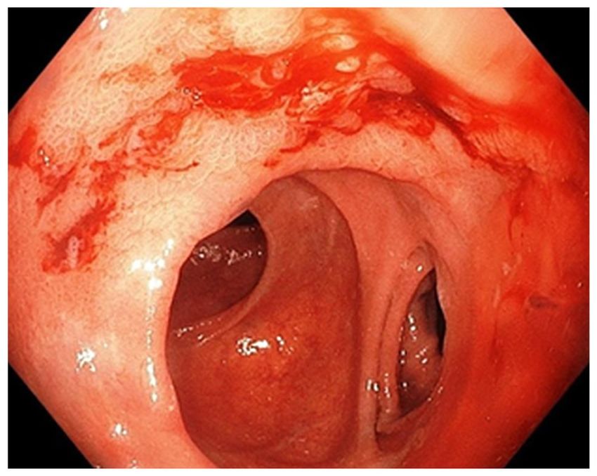
EXPERIMENTAL AND THERAPEUTIC MEDICINE 22: 1147, 2021                                                                3

      Figure 1. Prevalence of H. pylori‑positive hemorrhagic lesions in the studied group according to age (in years). H. pylori, Helicobacter pylori.

    The second non‑invasive test provided information on iden‑                  Table I. Relationship between NSAID use and age‑related anti‑
tifying 6 more patients who at the first test were false‑negative               coagulants/antiplatelet agents in H. pylori‑positive patients.
as well as data on the resistance of the bacterium in the cases
initially treated. We also included in the study 4 patients with                Age (years)                          50 (%)
a history of gastric resection for gastric ulcer. Endoscopic
                                                                                NSAIDs                                 56.52                        16.67
evaluation revealed acute anastomotic ulcer and endoscopic
hemostasis was effective.                                                       Anticoagulants                          4.35                         45
    There was an increased frequency of hemorrhagic lesions                     No treatment                           39.13                        38.33
in H. pylori‑positive men compared to women with the same
                                                                                H. pylori, Helicobacter pylori; NSAIDs, non‑steroidal anti‑inflam‑
lesions (84.04 vs. 15.96%) In the H. pylori‑negative group,
                                                                                matory drugs.
there was also a male prevalence, but with a lower M:F sex
ratio (71.83 vs. 29.17%).
    The presence of H. pylori was increased in the studied
group directly proportional to age ≤50 years, and then                          class IA and IB) were treated with NSAIDs and consumed
decreased slightly in elder age. The most frequent hemor‑                       alcohol chronically compared to 12.50% of H. pylori‑negative
rhagic lesions caused by H. pylori were in the age segment                      patients (Table II).
between 40 and 50 years. In this age segment, >23% of                               Endoscopic evaluation (early or late) was performed in all
non‑variceal UGIB were directly related to the presence of                      patients tested for H. pylori who had hemorrhagic lesions and
H. pylori (Fig. 1).                                                             we classified the gastric lesions found on endoscopy as follows:
    The relationship between NSAID use and H. pylori                            active bleeding requiring endoscopic or surgical hemostasis
infection in patients 50 years of age. Therapies with                    the ability to predict these characteristics can vary greatly (8)
antiplatelet agents or oral anticoagulants were the direct                      (Table III).
causes of UGIB in 45% of patients with H. pylori‑positive                           Acute hemorrhagic lesions (gastric, duodenal ulcer or
gastro‑duodenal lesions >50 years of age. The evolution of                      hemorrhagic gastroduodenitis) were detected more frequently
a non‑variceal UGIB can be significantly influenced by the                      in the group of positive than negative H. pylori patients
value of coagulation times (modifications of these thera‑                       (69 vs. 43 cases). At the same time, the rate of acute lesions in
pies), at the same time the transfusion requirement is higher                   the stomach and duodenum was higher in H. pylori‑positive
in these patients, regardless of the presence of H. pylori                      patients. Chronic hemorrhagic or non‑hemorrhagic gastric
(Table I).                                                                      ulcer was found in similar proportions in both the negative and
    In elderly patients with associated comorbidities, an                       positive patients. Hemorrhagic gastroduodenitis lesions were
episode of UGIB can be fatal. We analyzed the effect of                         found in equal proportions in the studied groups. Evaluation
NSAID use associated with alcohol consumption in patients                       of patients with active bleeding on endoscopic examination
with hemorrhagic lesions, depending on the presence of                          showed that the bleeding rate was high in the H. pylori‑positive
H. pylori and found the following: 28.75% of H. pylori‑positive                 patients who were negative after specific therapy (18.18%)
patients with active bleeding at endoscopic evaluation (Forrest                 compared to those with H. pylori‑negative lesions (15.79%)
4 POPA et al: ROLE OF H. pylori IN HEMORRHAGIC GASTRO-DUODENAL LESIONS
Table II. Determinants of bleeding depending on the presence of H. pylori.
H. pylori‑negative, H. pylori‑positive, H. pylori‑positive,
with NSAID + alcohol (%) without NSAID + alcohol (%) with NSAID + alcohol (%)
Active bleeding lesions 12.5 14.29 28.75
No active bleeding lesions 87.5 85.71 71.25
H. pylori, Helicobacter pylori; NSAID, non‑steroidal anti‑inflammatory drugs.
Table III. Endoscopic evaluation of H. pylori‑positive vs. H. pylori‑negative patients.
H. pylori‑negative H. pylori‑positive
Hemorrhagic acute duodenal ulcer 6 13
Non‑hemorrhagic acute duodenal ulcer 9 9
Hemorrhagic chronic duodenal ulcer 11 6
Hemorrhagic chronic duodenal ulcer 2 6
Hemorrhagic acute gastric ulcer 9 17
Non‑hemorrhagic acute gastric ulcer 3 10
Hemorrhagic chronic gastric ulcer 2 2
Non‑hemorrhagic chronic gastric ulcer 4 3
Hemobilia 1 1
Gastroduodenitis 20 20
Gastric tumor 1 7
Undetectable source 2 2
H. pylori, Helicobacter pylori.
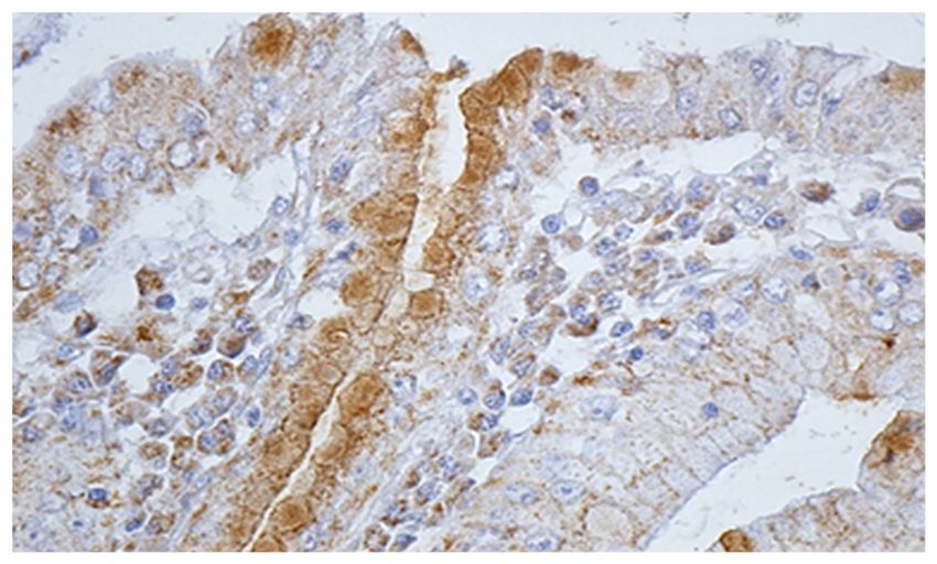
methods showed the presence of H. pylori. Moreover, IHC
detected 6 more cases in which the initial noninvasive tests
were negative, and the histological examination detected only
2 cases out of 6 (Figs. 3 and 4).
In 2 of the 6 cases, the IHC assessment consisted of
metaplasia‑associated lesions. In one case, the diagnosis of
gastric carcinoma was established by IHC, and in another, the
diagnosis of gastric mucosa‑associated lymphoma (MALT)
was established. The patient with gastric carcinoma underwent
surgical operation and underwent a subtotal gastrectomy, and
histological reassessment confirmed the diagnosis of carci‑
noma. In the case of MALT lymphoma, a total gastrectomy
was performed, and the histological evaluation and IHC were
performed on specimens directly from the resected piece.
Surgery for hemostasis was needed more frequently in
patients with H. pylori‑positive UGIB than in H. pylori‑nega‑
tive patients. Approximately 16% of patients with non‑variceal
Figure 2. Upper digestive endoscopy. Ulcer (Forrest IB) of the anastomosis
UGIB and positive H. pylori results required surgery to stop
(gastrojejunal) mouth with ‘belly bleeding’ in an H. pylori‑positive patient.
H. pylori, Helicobacter pylori. the bleeding. In the group of H. pylori‑negative patients, the
need for surgery was lower; respectively 9.7% of patients
underwent surgery (suturing or resection). However, distant
mortality is influenced by the occurrence of bleeding,
and much higher in the treatment‑resistant patients (40%) and in our study, in patients with non‑variceal UGIB due
(Fig. 2). to an H. pylori‑positive lesion, it was lower compared to
Histological and IHC methods represented the most specific H. pylori‑negative patients. We considered this difference
way of detecting this bacterium. In regards to all patients for due to the strict supervision of the therapy in the case of
whom histological and noninvasive tests were positive, IHC H. pylori‑positive patients and due to the information of theEXPERIMENTAL AND THERAPEUTIC MEDICINE 22: 1147, 2021 5
in the development of gastric cancer (12‑14) even though the
prevalence of H. pylori in the remaining stomach appears to be
lower than that in the intact organ (11).
In the present study, the prevalence of H. pylori infec‑
tion increased in direct proportion to age ≤50 years, and
then a slight decrease was found in elderly patients. The
most common hemorrhagic lesions caused by H. pylori were
in the age segment between 40 and 50 years. Thus, in this
age segment, >23% of the patients with non‑variceal UGIB
had gastroduodenal lesions that were directly influenced by
the presence of H. pylori. The prevalence of hemorrhagic
complications caused by H. pylori in the patients studied,
increased sharply in the young age segment (30‑50 years),
Figure 3. Immunohistochemistry exam: intense positive reaction for
H. pylori. H. pylori, Helicobacter pylori.
then gradually decreased, reaching another peak in the age
segment 70‑79 years. Data from the literature report similar
results (15,16). In the study group, for patients 65 years
(mean age: 81 years), reported that 67% of gastric ulcers and
69% of duodenal ulcers were H. pylori‑positive. In addition,
the use of NSAIDs or aspirin, alone or in combination with
H. pylori infection, was reported in 39% of gastric ulcers
and in 25% of patients with duodenal ulcers (17). Studies
do not take into account the difference between the use of
Figure 4. Immunohistochemistry exam: positive reaction for H. pylori. NSAIDs and the use of low‑dose aspirin (antiplatelet effect).
H. pylori, Helicobacter pylori. H. pylori is associated with an increased risk of bleeding in
patients under chronic treatment with AINS or antiplatelet
medication (18‑20). In the present study, therapies with anti‑
patients about their disease and its evolution without treat‑ platelet agents or oral anticoagulants and H. pylori infection
ment. were the most frequent associations encountered in cases
with active bleeding in the study group. At the same time, in
Discussion elderly patients with H. pylori‑positive gastritis who started
long‑term treatment with NSAIDs, oral antiplatelet agents
In the present study, we found an increased frequency of hemor‑ or anticoagulants, treatment of the infection significantly
rhagic lesions among men in the group of H. pylori‑positive reduced the risk of peptic ulcer (21‑23). Patients >50 who
patients compared to the group of H. pylori‑negative patients are administered antiplatelet agents or oral anticoagulants,
(85.04 vs. 70.83%). At the same time, the frequency of hemor‑ in addition to performing an upper digestive endoscopy,
rhagic lesions among H. pylori‑negative women was higher should be considered for a non‑invasive test for the diag‑
than among the H. pylori‑positive women (29.17 vs. 15.96%). nosis of H. pylori (21). In our study, the transformation of
Consistent with these data, Slăvescu et al conducted a study non‑hemorrhagic gastroduodenal H. pylori‑positive lesions
regarding the prevalence of H. pylori infection in children into hemorrhagic lesions was strongly related to alcohol
in Romania, and found a higher prevalence among girls (9). consumption. We analyzed the effect of NSAID consump‑
H. pylori infection seems to act convergently with other tion associated with alcohol consumption depending on
factors, such as alcohol consumption, NSAIDs, and smoking, the presence of H. pylori and found that chronic alcohol
in favor of triggering digestive bleeding. Another hypothesis consumption appeared to have a similar NSAID effect,
that supports these studies showed an increased frequency of so that alcohol consumption without the association with
antibiotic resistance of H. pylori in men compared to women, NSAIDs was considered to cause bleeding in patients
but dependent on the therapeutic regimen followed (10). with H. pylori‑positive lesions in ~14.29% of patients;
Antibiotic resistance of H. pylori and poor medical education 28.75% of H. pylori‑positive patients with active bleeding
may also be factors considered in H. pylori‑positive patients at endoscopic evaluation (class IA and IB) were treated with
who have a history of gastric ulcer for which surgery has been NSAIDs and were chronic consumers of alcohol compared
performed (11) The persistence of H. pylori infection, despite to patients with H. pylori‑negative hemorrhagic lesions
gastric resections, still predisposes to a hemorrhagic compli‑ (12.50% of patients with NSAIDs and alcohol). Studies that
cation (12) According to several studies, H. pylori infection looked at the relationship between H. pylori infection and
in the remaining stomach appears to play an important role alcohol consumption had conflicting results. A multiple6 POPA et al: ROLE OF H. pylori IN HEMORRHAGIC GASTRO-DUODENAL LESIONS
logistical study found that alcohol consumption and active in the usual staining was easier in patients with acute
gastritis pathology were associated with H. pylori infection, lesions of the gastric mucosa (acute gastritis), compared
and active gastritis (hemorrhagic or non‑hemorrhagic) was to chronic (premalignant lesions: atrophic gastritis and
associated with chronic alcohol consumption (24). Recent metaplasia). Specialist studies indicate that when changes
studies found an increased prevalence of H. pylori infection in atrophy occur in the gastric mucosa, a high percentage
in type 2 diabetes (25,26), one plausible explanation being of endobioptic samples become negative on histological
related to the changes in microbiota and low grade chronic evaluation (35), and in areas with metaplasia, H. pylori is
inflammation at the level of the gastroduodenal mucosa (27). undetectable by conventional histological staining in most
There is growing evidence of a bilateral relationship between cases, despite serological evidence (36). In addition, the
H. pylori infection and chronic hepatic diseases (28,29). On low prevalence of H. pylori in antral biopsy specimens with
the one hand, the liver plays significant roles in multiple atrophic mucosa can be explained by an uneven distribution
metabolic pathways, being involved in coagulation and the of bacterial infection resulting from pH‑increasing condi‑
trophicity of gastroduodenal mucosa (30). On the other tions (37,38). IHC evaluation may be useful in conditions
hand, chronic inflammation caused by H. pylori induces where no bacteria is visible in the usual H&E or Giemsa
chronic liver fibrosis (28). staining, but there is histological evidence of inflammation;
Literature data found evidence that H. pylori and NSAIDs in patients with MALT from post‑treatment biopsy speci‑
are major causes of gastroduodenal ulcer, and an in‑depth mens (to ensure that eradication therapy was successful)
analysis of the interaction data revealed that the induction and in forms in which H. pylori cannot be conclusively
effects of ulcers for both factors were cumulative (31). identified (32). Routine IHC evaluation of this pathology is
Eradication of H. pylori in chronic NSAID users was found questionable due to the high cost and need for specialized
to decrease the incidence of ulcer disease (2). Chronic alcohol personnel; however, it can be considered due to the accu‑
consumption appears to have a similar effect as NSAIDs, racy of the diagnosis and the accuracy of identification of
thus chronic alcohol consumption without an association associated lesions (atrophic gastritis, metaplasia, dysplasia,
with NSAIDs was considered to cause bleeding in patients carcinoma, lymphoma) (39).
with H. pylori‑positive lesions in ~14.29% of cases included Aggression and resistance of H. pylori to antibiotic
in group A. In our study, 18.18% of alcohol consuming therapy is correlated positively with an increased rate of active
H. pylori‑positive patients with active bleeding lesions (diag‑ bleeding. Evaluation of patients with active bleeding on endo‑
nosed endoscopically or surgically) became negative after scopic examination showed that the bleeding rate was high in
specific treatment, while 40% of all these patients remained treatment‑resistant H. pylori‑positive patients (40%), compared
positive after therapy. to those who were negative after specific therapy (18.18%).
In the present study, >50% of patients had H. pylori‑posi‑ In contrast, we found that the frequency of hemorrhagic
tive bleeding lesions. The 30‑day retest of the hemorrhagic lesions was much lower in those with H. pylori‑negative
episode in all 166 patients showed that out of the 76 initially lesions (15.79%) compared to H. pylori‑positive patients.
negative patients, 6 patients became positive. These results Literature data show that before the eradication of H. pylori,
are due to the fact that when patients have acute gastroduo‑ 20‑25% of patients with peptic ulcer disease developed
denal bleeding, most diagnostic tests for H. pylori infection complications such as hemorrhage, perforation or stenosis (40)
may show false‑negative results (32) and the sensitivity and and it is estimated that ~1/3 of H. pylori‑positive patients
specificity of the fecal antigen test is 90% (2). Non‑invasive with hemorrhagic ulcer will develop recurrent bleeding in
testing by detecting H. pylori‑specific antigen in feces had the next 1‑2 years in the absence of testing and eradication of
increased sensitivity in the diagnosis of infection in our H. pylori (41). The increased resistance of H. pylori to anti‑
study, so that out of 166 patients tested, a positive diagnosis biotics is also correlated with the higher rate of bleeding at
was established with increased accuracy in 70 of the 76 the distance of the hemorrhagic episode, despite PPI therapy.
H. pylori‑positive patients. Although histological evaluation Endoscopic evaluation should be performed in all patients with
and IHC established the presence of H. pylori in another hemorrhagic lesions for diagnostic or therapeutic purposes,
6 patients, this non‑invasive test had a sensitivity and speci‑ but also to obtain biopsies for histological or IHC diagnosis of
ficity of over 90% in assessing the correct diagnosis. This H. pylori (2). In our study, acute hemorrhagic lesions (gastric,
indirect method of diagnosis is cheaper than other methods duodenal ulcer or hemorrhagic gastroduodenitis) were found
and has the advantage that it can be performed in most more frequently in the group of H. pylori‑positive patients
medical centers (1,2). We found that histological examina‑ than in H. pylori‑negative ones (69 vs. 43 cases). Acute
tion of H. pylori lesions is very accurate in providing data hemorrhagic lesions are more common in H. pylori‑positive
on the degree of atrophy, metaphase or carcinoma, and the patients, so acute hemorrhagic duodenal ulcer and acute
addition of IHC examination brings more information and hemorrhagic gastric ulcer are more common in these patients
establishes the correct diagnosis in all cases. Data from the than in H. pylori‑negative patients (41,42). Chronic hemor‑
literature show that non‑invasive methods and histological rhagic endoscopic lesions (chronic duodenal ulcer, chronic
evaluation of endobiopsies together establish the diagnosis gastric ulcer), as well as non‑hemorrhagic ones, seem to be
of H. pylori infection in >95‑97% of cases (33,34). In the more common in H. pylori‑negative patients. Hemorrhage
patients in the study, the diagnosis of H. pylori infection was may occur more frequently in the colonization phase associ‑
established after performing non‑invasive tests and after ated with acute H. pylori gastritis compared to chronic lesions.
histological evaluation in 94% of cases; this share of the At the same time, tumor lesions were much more common
diagnosis is similar to other studies. Highlighting H. pylori in H. pylori‑positive patients (7 vs. 1 patient). Data from theEXPERIMENTAL AND THERAPEUTIC MEDICINE 22: 1147, 2021 7
literature have shown that endoscopic features have been Patient consent for publication
reported to be useful tools for diagnosing H. pylori infection
in the gastric mucosa. However, the ability to predict these Not applicable.
characteristics can vary greatly (8).
In the present study, the need for surgery for hemostasis Competing interests
was more common in patients with UGIB with H. pylori‑posi‑
tive gastroduodenal lesions, compared to H. pylori‑negative The authors declare that they have no competing interests.
patients. Approximately 16% of patients with non‑variceal
and H. pylori‑positive UGIB required surgery to stop the References
bleeding (16 vs. 9.7%). Regarding the type of surgery, in the
case of H. pylori‑positive patients with UGIB, resection was 1. Obleagă CV, Vere CC, Vîlcea ID, Ciorbagiu MC, Moraru E and
needed in several cases, compared to the group of patients Mirea CS: Helicobacter pylori: Types of diseases, diagnosis,
treatment and causes of therapeutic failure. J Mind Med Sci 3:
with H. pylori‑negative hemorrhagic lesions. At the same time, 156‑161, 2016.
according to other studies, postoperative complications appear 2. Kuipers EJ, Thijs JC and Festen HP: The prevalence of
more frequently in patients who undergo resection (gastrec‑ Helicobacter pylori in peptic ulcer disease. Aliment Pharmacol
Ther 9 (Suppl 20): S59‑S69, 1995.
tomies, duodenopancreatectomies), compared to patients in 3. Hentschell E, Brandstätter G, Dragosics B, Hirschl AM,
whom suturing is performed (16,43‑46). Postoperative adher‑ Nemec H, Schütze K, Taufer M and Wurzer H: Effect of raniti‑
ences are significant causes of long term morbidity, causing dine and amoxycillin plus metronidazole on the eradication of
Helicobacter pylori and the recurrence of duodenal ulcer. N Engl
episodes of abdominal pain and bowel obstruction (47). J Med 328: 308‑312, 1993.
The outcome of patients with UGIB depends very much on 4. Gisbert JP, Khorrami S, Carballo F, Calvet X, Gene E and
the need for surgery for hemostasis. The need for hemostasis Dominguez‑Munoz E: Meta‑analysis: Helicobacter pylori
eradication therapy vs. antisecretory non‑eradication therapy for
surgery in the case of non‑variceal UGIB is the most constant the prevention of recurrent bleeding from peptic ulcer. Aliment
indicator of prolonged hospitalization and high financial Pharmacol Ther 19: 617‑629, 2004.
cost, and the prophylaxis of these episodes of bleeding is the 5. Guo CG, Cheung KS, Zhang F, Chan EW, Chen L, Wong ICK
and Leung WK: Delay in retreatment of Helicobacter pylori
simplest modality of treatment. Thus, the presence of H. pylori infection increases risk of upper gastrointestinal bleeding. Clin
significantly contributes to the increase in hospital stay and Gastroenterol Hepatol 19: 314‑322.e2, 2021.
treatment costs in these patients. 6. Gisbert JP and Abraira V: Accuracy of Helicobacter pylori diag‑
nostic tests in patients with bleeding peptic ulcer: A systematic
review and meta‑analysis. Am J Gastroenterol 101: 848‑863, 2006.
Acknowledgements 7. Stolte M and Meining A: The updated Sydney system:
Classification and grading of gastritis as the basis of diagnosis
and treatment. Can J Gastroenterol 15: 591‑598, 2001.
Not applicable. 8. Mao T, Wang Y, Yin F, Zhao Q, Yang L, Ding X and Tian Z:
Association of endoscopic features of gastric mucosa with
Funding Helicobacter pylori infection in Chinese patients. Gastroenterol
Res Pract 2016: 6539639, 2016.
9. Slăvescu KC, Șarban C, Pîrvan A, Gheban D, Mărgescu C
No funding was received. and Miu N: Prevalence of Helicobacter pylori infection in
children with gastritis and peptic ulcer disease in north‑
western and central Romania. Med Pharmacy Rep 85: 457‑462,
Availability of data and materials 2012.
10. Megraud F: H. pylori antibiotic resistance: Prevalence, impor‑
All data generated or analyzed during this study are included tance and advances in testing. Gut 53: 1374‑1384, 2004.
in this published article. 11. Park S and Chun HJ: Helicobacter pylori infection following
partial gastrectomy for gastric cancer. World J Gastroenterol 20:
2765‑2770, 2014.
Authors' contributions 12. Testerman TL and Morris J: Beyond the stomach: An updated
view of Helicobacter pylori pathogenesis, diagnosis, and treat‑
ment. World J Gastroenterol 20: 12781‑12808, 2014.
DGP, CVO, MEC, DS, CMe, MC and BN led the conception 13. Wroblewski LE, Peek RM Jr and Wilson KT: Helicobacter pylori
and design of this study. BS, DNF, VDB, MST, CMe MEC, and gastric cancer: Factors that modulate disease risk. Clin
MD, LCT, IDV, and BN were responsible for the data collec‑ Microbiol Rev 23: 713‑739, 2010.
14. Conteduca V, Sansonno D, Lauletta G, Russi S, Ingravallo G and
tion and analysis. CMi, DNF, VDB, MC, LCT and DS were Dammacco F: H. pylori infection and gastric cancer: State of the
in charge of reviewing the data and drafting the manuscript. art (Review). Int J Oncol 42: 5‑18, 2013.
DGP, BS, DS, CMi, MST and BN revised critical perspectives 15. Alberto P and Franceschi M: Helicobacter pylori infection in
older people. World J Gastroenterol 20: 6364‑6373, 2014.
for important intellectual content. The final version was read 16. Constantin VD, Paun S, Ciofoaia VV, Budu V and Socea B:
and approved by all the authors. Multimodal management of upper gastrointestinal bleeding
caused by stress gastropathy. J Gastrointestin Liver Dis 18:
279‑284, 2009.
Ethics approval and consent to participate 17. Pilotto A: Aging and upper gastrointestinal disorders. Best Pract
Res Clin Gastroenterol 18 (Suppl): S73‑S81, 2004.
The study was conducted according to the World Medical 18. Venerito M, Schneider C, Costanzo R, Breja R, Röhl FW and
Malfertheiner P: Contribution of Helicobacter pylori infection
Association Declaration of Helsinki, using a protocol approved to the risk of peptic ulcer bleeding in patients on nonsteroidal
by the local Bioethics Committee from County Emergency anti‑inflammatory drugs, antiplatelet agents, anticoagulants,
Clinical Hospital (Craiova, Romania). All patients previously corticosteroids and selective serotonin reuptake inhibitors.
Aliment Pharmacol Ther 47: 1464‑1471, 2018.
signed an informed written consent concerning the hospital‑ 19. Pilotto A and Franceschi M: Helicobacter pylori infection in
ization and research investigation. older people. World J Gastroenterol 20: 6364‑6373, 2014.8 POPA et al: ROLE OF H. pylori IN HEMORRHAGIC GASTRO-DUODENAL LESIONS
20. Lanas A, Dumonceau JM, Hunt RH, Fujishiro M, Scheiman JM, 35. Kokkola A, Rautelin H, Puolakkainen P, Sipponen P,
Gralnek IM, Campbell HE, Rostom A, Villanueva C and Färkkilä M, Haapiainen R and Kosunen TU: Diagnosis of
Sung JJY: Non‑variceal upper gastrointestinal bleeding. Nat Rev Helicobacter pylori infection in patients with atrophic gastritis:
Dis Primers 4: 18020, 2018. Comparison of histology, 13C‑urea breath test, and serology.
21. Pilotto A, Franceschi M, Leandro G, Paris F, Cascavilla L, Scand J Gastroenterol 35: 138‑141, 2000.
Longo MG, Niro V, Andriulli A, Scarcelli C and Di Mario F: 36. Loffeld RJ, Stobberingh E, Flendrig JA and Arends JW:
Proton‑pump inhibitors reduce the risk of uncomplicated peptic Helicobacter pylori in gastric biopsy specimens. Comparison
ulcer in elderly either acute or chronic users of aspirin/non‑steroidal of culture, modified giemsa stain, and immunohistochemistry. A
anti‑inflammatory drugs. Aliment Pharmacol Ther 20: 1091‑1097, retrospective study. J Pathol 165: 69‑73, 1991.
2004. 37. Testoni PA, Colombo E, Cattani L, Longhi M, Bagnolo F,
22. Narayanan M, Reddy KM and Marsicano E: Peptic ulcer disease Lella F, Buizza M and Scelsi R: Helicobacter pylori serology in
and Helicobacter pylori infection. Mo Med 115: 219‑224, 2018. chronic gastritis with antral atrophy and negative histology for
23. Zapata‑Colindres JC, Zepeda‑Gómez S, Montaño‑Loza A, Helicobacter‑like organisms. J Clin Gastroenterol 22: 182‑185,
Vá zquez‑Ba l lesteros E, De Jesús Vi l la lobos J a nd 1996.
Valdovinos‑Andraca F: The association of Helicobacter pylori 38. Kang HY, Kim N, Park YS, Hwang JH, Kim JW, Jeong SH,
infection and nonsteroidal anti‑inflammatory drugs in peptic Lee DH, Jung HC and Song IS: Progression of atrophic gastritis
ulcer disease. Can J Gastroenterol 20: 277‑280, 2006. and intestinal metaplasia drives Helicobacter pylori out of the
24. Zhang L, Eslick GD, Xia HH, Wu C, Phung N and Talley NJ: gastric mucosa. Dig Dis Sci 51: 2310‑2315, 2006.
Relationship between alcohol consumption and active 39. Park YH and Kim N: Review of atrophic gastritis and intestinal
Helicobacter pylori infection. Alcohol Alcohol 45: 89‑94, 2010. metaplasia as a premalignant lesion of gastric cancer. J Cancer
25. Hosseininasab Nodoushan SA and Nabavi A: The interaction of Prev 20: 25‑40, 2015.
Helicobacter pylori infection and type 2 diabetes mellitus. Adv 40. Penston JG: A decade of experience with long‑term continuous
Biomed Res 8: 15, 2019. treatment of peptic ulcers with H2‑receptor antagonists. Aliment
26. He C, Yang Z and Lu NH: Helicobacter pylori infection Pharmacol Ther 7 (Suppl 2): S27‑S33, 1993.
and diabetes: Is it a myth or fact? World J Gastroenterol 20: 41. Laine LA: Helicobacter pylori and complicated ulcer disease.
4607‑4617, 2018. Am J Med 100 (Suppl): S52‑S59, 1996.
27. Suceveanu AI, Stoian AP, Parepa I, Voinea C, Hainarosie R, 42. Campuzano‑Maya G: Hematologic manifestations of
Manuc D, Nitipir C, Mazilu L and Suceveanu AP: Gut micro‑ Helicobacter pylori infection. World J Gastroenterol 20:
biota patterns in obese and type 2 diabetes (T2D) patients from 12818‑12838, 2014.
romanian black sea coast region. Rev Chim 69: 2260‑2267, 2018. 43. Obleagă CV, Vere CC, Pătraşcu AM, Moraru E, Crafciuc AV,
28. Okushin K, Tsutsumi T, Ikeuchi K, Kado A, Enooku K, Fujinaga H, Foarfă MC, Mogoantă SŞ, Streba CT, Bondari S, Paitici Ş et al:
Moriya K, Yotsuyanagi H and Koike K: Helicobacter pylori Severe upper gastrointestinal bleeding determined by a gastric
infection and liver diseases: Epidemiology and insights into lymphoma associated with Helicobacter pylori‑positive atrophic
pathogenesis. World J Gastroenterol 24: 3617‑3625, 2018. gastritis. Rom J Morphol Embryol 58: 611‑617, 2017.
29. Tsai CE, Liang CM, Lee CH, Kuo YH, Wu KL, Chiu YC, Tai WC 44. Obleagă CV, Vere CC, Mogoanţa S, Firuț C, Meșina C,
and Chuah SK: First‑line Helicobacter pylori eradication among Ciorbagiu MC, Mirea CS and Vîlcea ID: Upper gastrointestinal
patients with chronic liver diseases in Taiwan. Kaohsiung J Med bleeding‑initial manifestation of pancreatic head carcinoma.
Sci 32: 397‑402, 2016. Curr Health Sci J 43: 236‑240, 2017.
30. Gheorghe G, Stoian AP, Gaman M, Socea B, Neagu TP, 45. Serban D, Vancea G, Balasescu SA, Socea B, Tudor C and
Stanescu AMA, Bratu OG, Mischianu DLD, Suceveanu AI and Dascalu AM: Informed consent in all surgical specialties: From
Diaconu CC: The Benefits and Risks of Antioxidant Treatment in legal obligation to patient satisfaction. Rom J Leg Med 28:
Liver Diseases. Rev Chim 70: 651, 2019. 317‑321, 2020.
31. Huan JQ, Sridhar S and Hunt RH: Role of Helicobacter pylori 46. Constantin VD, Socea B, Sireţeanu G and Popa F: Epidemiological
infection and nonsteroidal anti‑inf lammatory drugs in aspects and risk factors in the outcome of variceal eso‑gastric
peptic‑ulcer disease: A meta‑analysis. Lancet 359: 14‑22, 2002. bleeding at cirrhosis patients. J Appl Quant Meth 3: 316‑324,
32. Braden B: Diagnosis of Helicobacter pylori infection. BMJ 344: 2008.
828, 2012. 47. Fometescu SG, Costache M, Coveney A, Oprescu SM, Serban D
33. Lee JY and Kim N: Diagnosis of Helicobacter pylori by invasive and Savlovschi C: Peritoneal fibrinolytic activity and adhesio‑
test: Histology. Ann Transl Med 3: 10, 2015. genesis. Chirurgia (Bucur) 108: 331‑340, 2013.
34. Alius C, Tudor C, Badiu CD, Dascalu AM, Smarandache CG,
Sabau AD, Tanasescu C, Balasescu SA and Serban D: This work is licensed under a Creative Commons
Indocyanine green‑enhanced colorectal surgery‑between being Attribution-NonCommercial-NoDerivatives 4.0
superfluous and being a game‑changer. Diagnostics (Basel) 10: International (CC BY-NC-ND 4.0) License.
742, 2020.You can also read