INFREQUENT PLACENTAL AND FETAL INVOLVEMENT IN SARS-COV-2 INFECTION: PATHOLOGY DATA FROM A LARGE MEDICAL CENTER
←
→
Page content transcription
If your browser does not render page correctly, please read the page content below
Journal of
              Developmental
              Biology

Article
Infrequent Placental and Fetal Involvement in SARS-CoV-2
Infection: Pathology Data from a Large Medical Center
Jeffrey Thomas, Yu Sun and Larisa Debelenko *

                                          Department of Pathology and Cell Biology, Columbia University—Irving Medical Center, New York, NY 10032,
                                          USA; jt3236@cumc.columbia.edu (J.T.); ys3271@cumc.columbia.edu (Y.S.)
                                          * Correspondence: ld2863@cumc.columbia.edu

                                          Abstract: In order to determine the frequency of SARS-CoV-2 placental and fetal involvements,
                                          we analyzed placentas of 197 women positive for infection at delivery and fetal tissues in cases of
                                          pregnancy loss in women positive by SARS-CoV-2 PCR (N = 2) and COVID-19 serology (N = 4), using
                                          in situ hybridization (ISH), immunohistochemistry (IHC) and, in selected cases, RT-PCR of tissue
                                          homogenates. The virus was identified in situ, accompanied by intervillositis, in 2 of 197 placentas
                                          (1.02%). In three more cases, SARS-CoV-2 was detected by tissue PCR without in situ localization
                                          and placental inflammation. There were no maternal mortality or association of placental infection
                                          with the clinical severity of COVID-19. All tested neonates born to SARS-CoV-2-positive women
                                          (N = 172) were negative for the virus. There were three pregnancy losses among 197 infected women
                                          and in two cases available fetal tissues were negative for SARS-CoV-2. In one of four fetal autopsies
                                performed in women with positive COVID-19 serology, the mother-to-child transmission (MTCT)

                                          could be inferred based on positive SARS-CoV-2 nucleocapsid IHC in fetal pulmonary endothelium.
Citation: Thomas, J.; Sun, Y.;            Placental involvement by SARS-CoV-2 is rare, but may be underestimated due to its transient nature.
Debelenko, L. Infrequent Placental        MTCT is even rarer, supporting the protective role of placenta in SARS-CoV-2 infection.
and Fetal Involvement in
SARS-CoV-2 Infection: Pathology
                                          Keywords: SARS-CoV-2; IHC; ISH; pregnancy; placenta; intervillositis; autopsy; perinatal; fetal demise
Data from a Large Medical Center. J.
Dev. Biol. 2021, 9, 45. https://
doi.org/10.3390/jdb9040045

                                          1. Introduction
Academic Editors: Simon J. Conway,
Rwik Sen, Carmen Ledesma-Feliciano              According to the recent international retrospective cohort study (PregOuTCOV),
and Navneet Dogra                         3.6% of pregnant women have been infected by SARS-CoV-2 during pregnancy and the
                                          viral exposure was associated with composite adverse obstetric and neonatal outcomes,
Received: 28 August 2021                  which prompted the authors to advocate for vaccination before or early in pregnancy [1].
Accepted: 14 October 2021                 Furthermore, a multinational cohort study established increased risks of maternal and
Published: 16 October 2021                neonatal morbidity and mortality indices in women with COVID-19 diagnosis compared
                                          to women without COVID-19 diagnosis [2].
Publisher’s Note: MDPI stays neutral            Placental involvement by SARS-CoV-2 has been documented by numerous case re-
with regard to jurisdictional claims in   ports [3–10] with the earlier cases recently summarized [11,12] to outline characteristic
published maps and institutional affil-   histomorphologic features of SARS-CoV-2 placental infection. The NIH placenta workshop
iations.                                  established a classification scheme and criteria for the placental involvement depending on
                                          technologies used for the viral detection [13]; however, the frequency of placental infection
                                          in COVID-19 remains unknown.
                                                The question of transplacental or mother-to-child transmission (MTCT) of SARS-CoV-
Copyright: © 2021 by the authors.         2 is complicated with several proposed classification schemes and different sets of data
Licensee MDPI, Basel, Switzerland.        analyzed by different methodologies; however, according to recent reviews, MTCT of
This article is an open access article    SARS-CoV-2 is possible, although, similar to the placental involvement, its incidence and
distributed under the terms and           effects on the newborn have not been elucidated [14,15].
conditions of the Creative Commons              The role of COVID-19 infection in pregnancy loss is even less clear; however, a
Attribution (CC BY) license (https://
                                          retrospective cohort study using a database of pregnant people giving birth in Ontario,
creativecommons.org/licenses/by/
                                          Canada, between July 2002 and December 2020 did not find any cause variation (unusual
4.0/).

J. Dev. Biol. 2021, 9, 45. https://doi.org/10.3390/jdb9040045                                                 https://www.mdpi.com/journal/jdb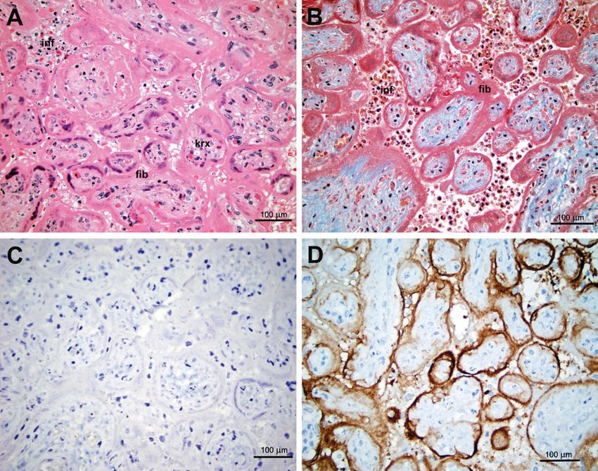
J. Dev. Biol. 2021, 9, 45                                                                                           2 of 10

                            pattern) in preterm birth or stillbirth rates during the first 12 months of COVID-19 pandemic
                            compared with the previous 17.5 years [16].
                                 We analyzed placentas of women infected with SARS-CoV-2 using ISH and ICH in
                            order to determine the frequency of placental/transplacental infection and its association
                            with the disease severity. Fetal tissues from perinatal autopsies in infected women were
                            also analyzed in order to identify cases of MTCT and its possible role in perinatal loss. This
                            analysis summarizes all our cases during the 1st year of the COVID-19 epidemic starting
                            from 15 March 2020, including subsets of cases published by us previously [7,17–19].

                            2. Materials and Methods
                                  The study was approved by the Institutional Review Board (Protocol IRB-AAAT0272).
                            We performed a retrospective review of placental pathology and neonatal autopsy reports
                            and pathology samples from the files of our Pathology Department. Pertinent clinical
                            information was extracted from the medical records. The waver of the patients’ consent
                            was granted based on the criteria which governed the study design, including an adequate
                            plan to protect subjects’ personal information (coding, secure handling) and destroy the
                            patients’ identifiers after the study completion, preventing reuse or disclosure of the
                            personal information. We analyzed clinical information of 1950 women who gave birth
                            during one year since the start of COVID-19 epidemic in March of 2020 and whose placental
                            specimens were available. We included placentas of 197 women who tested positive for
                            SARS-CoV-2 by the nasopharyngeal swab RT-PCR test on admission for labor and delivery
                            during the period from 15 March 2020 to 14 March 2021. Additionally, we analyzed
                            placental pathology of 84 women who had negative PCR, but positive COVID-19 serology
                            tests during the first quarter of 2021. We also studied fetal tissues in 6 cases of perinatal
                            autopsies performed during the same period of 2020/21 if mothers had positive RT-PCR
                            (N = 2) or COVID-19 serology (N = 4) results.
                                  SARS-CoV-2 IHC and ISH were performed on paracentral full length sections of pla-
                            cental disc using probes, antibodies, instruments, and reagents as previously described [7].
                            Placental tissue was refrigerated after delivery and fixed in 10% buffered formaldehyde no
                            later than 24 h after delivery, as recommended by the NIH workshop [13]. One hundred
                            fifty three of 197 (77.7%) placentas of SARS-CoV-2 positive women were studied with
                            combinations of ISH with a sense probe complementary to viral sequences encoding for the
                            spike protein and IHC with anti-nucleocapsid and anti-spike antiviral antibodies. Tissue
                            sections of fetal/neonatal lungs in cases of positive maternal testing (N = 6) were also
                            studied with SARS-CoV-2 ISH and IHC and in 3 cases the viral RT-PCRs of homogenates
                            of the lung tissue were performed as previously described [7].
                                  Placental involvement was defined as definitive in the presence of (1) intervillositis
                            (placentitis) with villous trophoblast necrosis, (2) syncytiotrophoblast positivity for SARS-
                            CoV-2 by ISH and ICH [11–13]. Placental involvement was defined as possible in the
                            presence of positive SARS-CoV-2 RT-PCR of tissue homogenates but negative SARS-CoV-2
                            ISH and IHC and lack of the inflammation in the placental tissue.

                            3. Results
                            3.1. Maternal and Placental Infection
                                  One hundred ninety seven of 1950 (10.1%) women who gave birth between
                            15 March 2020 and 14 March 2021 tested positive for SARS-CoV-2 by RT-PCR of nasopha-
                            ryngeal swab within 24 h of delivery, including 126 of 505 (24.9%) in the second, 29 of 495
                            (5.9%) in the third, 16 of 550 (2.9%) in the fourth quarters of 2020, and 26 of 400 (6.5%)
                            in the first quarter of 2021 (Figure 1). In the first quarter of 2021, the data on COVID-19
                            serology became available and showed that 84 of 284 (29.5%) women were positive for this
                            test, while their RT-PCRs were negative, both taken within 24 h of delivery (Figure 1).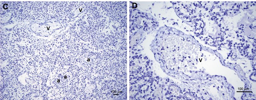
J. Dev. Biol. 2021, 9, x FOR PEER REVIEW 3 of 11
became available and showed that 84 of 284 (29.5%) women were positive for this test,
J. Dev. Biol. 2021, 9, 45 3 of 10
while their RT-PCRs were negative, both taken within 24 h of delivery (Figure 1).
Figure
Figure 1. Dynamics
1. Dynamics of SARS-CoV-2
of SARS-CoV-2 infection
infection in parturient
in parturient womenwomen
in ourinmedical
our medical
center.center.
The infection was detected by nasopharyngeal swab RT-PCR tests from the second
The infection was detected by nasopharyngeal swab RT-PCR tests from the second
quarter of 2020 (Q2/2020) to the first quarter of 2021 (Q1/2021). COVID-19 serology tests
quarter of 2020 (Q2/2020) to the first quarter of 2021 (Q1/2021). COVID-19 serology tests
results available forfor
results available Q1/2021
Q1/2021 are also
are also presented.
presented. N—numbers
N—numbers of studied
of studied placentas
placentas with with
available SARS-CoV-2 testing results in each quarter. The majority of infected women were
available SARS-CoV-2 testing results in each quarter. The majority of infected women
either
were asymptomatic
either asymptomaticororpost postsymptomatic
symptomatic at at the
thetime
timeofofdelivery;
delivery; however,
however, 21%21%(42 (42 of
197) reported symptoms of COVID-19, including six women
of 197) reported symptoms of COVID-19, including six women who had a severe disease, who had a severe disease,
requiring mechanical ventilation in five and extracorporeal oxygenation (ECMO) in one.in one.
requiring mechanical ventilation in five and extracorporeal oxygenation (ECMO)
There
There was,was, however,
however, no no maternal
maternal mortality
mortality in thein studied
the studied
group.group.
The The definitive
definitive placental
placental involvement
involvement by SARS-CoV-2
by SARS-CoV-2 was in
was seen seen
2 ofin197
2 of 197 (1.02%)
(1.02%)
placentas
placentas including
including oneone previously
previously reported
reported casecase
[7]. [7].
The The second
second casecase presented
presented in November
in November 2020 and2020involved
and involved
a 46 yeara 46
oldyear old Gravida
Gravida 2
2 Para1011
Para1011 who who
had an had an induction
induction of laborofatlabor
39 4/7 at weeks
39 4/7and weeks and forceps-assisted
forceps-assisted vaginal de- vaginal
delivery.
livery. She SARS-CoV-2
She was was SARS-CoV-2 RT-PCR-positive
RT-PCR-positive withsymptoms
with mild mild symptoms at the
at the day ofday of delivery
delivery
with
with negative
negative COVID-10
COVID-10 serology
serology and and negative
negative RT-PCR
RT-PCR 10 days
10 days prior.
prior. TheThe antenatal
antenatal his-history
was notable for travel-induced deep vein thrombosis. The newborn girl had Apgar scores 9
tory was notable for travel-induced deep vein thrombosis. The newborn girl had Apgar
scores
and 9 and
at 1 9and
at 15and
min,5 min, respectively,
respectively, and and
birthbirth weight
weight of 3125
of 3125 g (25–50th
g (25–50th percentile).
percentile). The girl
Thewasgirlhealthy-appearing,
was healthy-appearing, vigorous,
vigorous, and passed
and passed all newborn
all newborn screens
screens except
except forfor
an an
elevated
elevated transcutaneous
transcutaneous bilirubin.
bilirubin. Her SARS-CoV-2
Her SARS-CoV-2 RT-PCR RT-PCR testnegative,
test was was negative,
and the and the and
mother
mother andwere
the child the child were discharged
discharged on hospitalonday hospital day health.
2 in good 2 in goodThehealth.
followThe followwere
up visits up unre-
visits were unremarkable.
markable. Placentalincluded
Placental pathology pathology included
patchy patchy intervillous
intervillous inflammation inflammation
with histiocytes
with histiocytes (macrophages)
(macrophages) as a predominantas a predominant
cell type; thiscell type; this intervillositis
intervillositis was accompaniedwas accom-
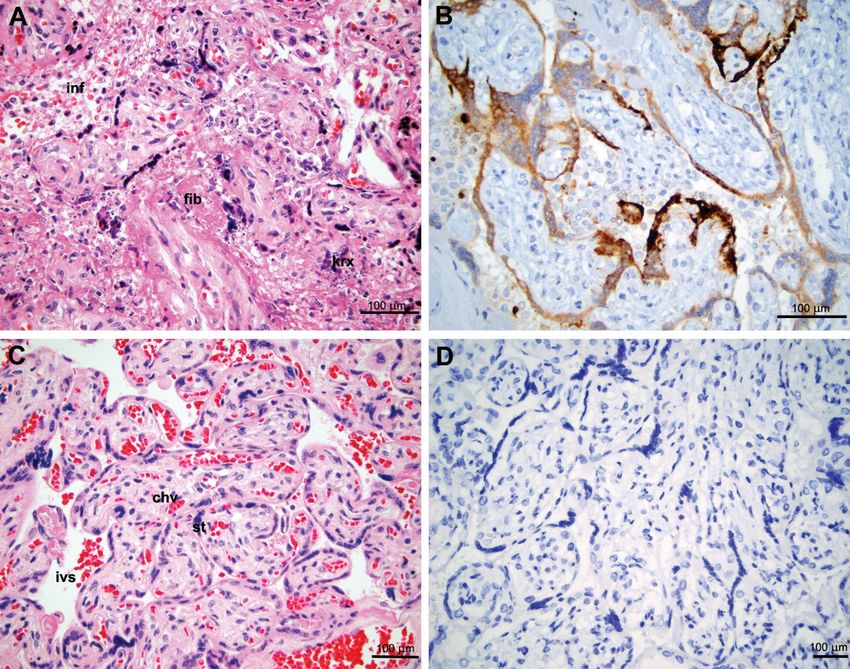
by necrosis
of villous trophoblast with marked karyorrhexis and perivillous accumulation of fibrinoid
proteins (Figure 2A). IHC for SARS-CoV-2 was positive in the syncytiotrophoblast lining
the chorionic villi (Figure 2B).
None of the remaining 195 placentas of infected women showed a constellation of
findings diagnostic of a definitive SARS-CoV-2 involvement; however, three cases that
were reported by us previously [18,19] were consistent with a possible involvement based
on positive RT-PCR of homogenized placental tissue not accompanied by the placental
inflammation and staining with antiviral IHC and ISH.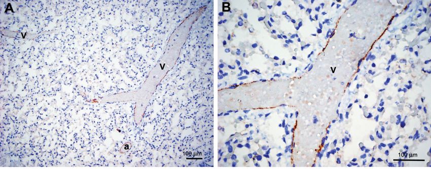
J. Dev. Biol. 2021, 9, x FOR PEER REVIEW 4 of 11
panied by necrosis of villous trophoblast with marked karyorrhexis and perivillous accu-
J. Dev. Biol. 2021, 9, 45 mulation of fibrinoid proteins (Figure 2A). IHC for SARS-CoV-2 was positive in the syn-
4 of 10
cytiotrophoblast lining the chorionic villi (Figure 2B).
Figure2.2.Histopathology
Figure Histopathology andand immunohistochemistry
immunohistochemistry of aof a definitive
definitive placental
placental involvement
involvement by SARS-CoV-2
by SARS-CoV-2 and age-
and age-matched
matched placental control (gestational age: 39 weeks). (A) Intervillositis with mild inflammatory infiltrate
placental control (gestational age: 39 weeks). (A) Intervillositis with mild inflammatory infiltrate in intervillous spacesin intervillous
spaces (inf), massive necrosis of syncytiotrophoblast with karyorrhexis (krx) and perivillous deposition of eosinophilic
(inf), massive necrosis of syncytiotrophoblast with karyorrhexis (krx) and perivillous deposition of eosinophilic fibrinoid
fibrinoid proteins (fib). Hematoxilin and Eosin (H&E). (B) IHC with the antibody against the SARS-CoV-2 nucleocapsid
proteins
protein (fib). Hematoxilin
positive and Eosin (H&E).
in syncytiotrophoblast covering(B)chorionic
IHC with theand
villi antibody against
monocytes the SARS-CoV-2
scattered nucleocapsid
in intervillous protein
spaces. (C) Histol-
positive in syncytiotrophoblast covering chorionic villi and monocytes scattered in intervillous spaces. (C)
ogy of an unaffected placenta at the same gestational age (39 weeks), showing chorionic villi (chv) covered with syncyti-Histology of an
unaffected placenta at the same gestational age (39 weeks), showing chorionic villi (chv) covered with syncytiotrophoblast
otrophoblast (st) and intact intervillous spaces (ivs) with red blood cells. H&E. (D) Negative IHC with antibody against
theand
(st) SARS-CoV-2 nucleocapsid
intact intervillous spacesprotein in the
(ivs) with redunaffected
blood cells.placenta.
H&E. (D) Negative IHC with antibody against the SARS-CoV-2
nucleocapsid protein in the unaffected placenta.
None of the remaining 195 placentas of infected women showed a constellation of
Intervillositis,
findings diagnostic an of unspecific
a definitiveinflammatory placental lesion
SARS-CoV-2 involvement; associated
however, threewith
casesSARS-
that
CoV-2 as well as
were reported bywith some other
us previously infectious
[18,19] and non-infectious
were consistent etiologies,
with a possible was observed
involvement based
in
on10 additional
positive casesof(5.1%)
RT-PCR in the group
homogenized of positive
placental women
tissue not without concurrent
accompanied tissue
by the placental
positivity
inflammationfor SARS-CoV-2 by ICH
and staining with or ISH.
antiviral IHCOne
andofISH.
these 10 cases involved a previously
healthy 36 year old Gravida
Intervillositis, 2 Para inflammatory
an unspecific 1001 woman inplacental
a critical condition due to severe
lesion associated with COVID-
SARS-
19 complicated
CoV-2 as well asbywith
cardiomyopathy and acuteand
some other infectious kidney and respiratory
non-infectious failure,
etiologies, wasnecessitating
observed
ECMO placement.
in 10 additional Because
cases (5.1%) of
in the
the non-reassuring
group of positive fetal heartwithout
women rate tracing, the decision
concurrent tissue
was made to deliver at 24 4/7 weeks by Cesarean section. The baby
positivity for SARS-CoV-2 by ICH or ISH. One of these 10 cases involved a previouslyboy was born with
Apgar
healthyscores 3 and
36 year old 7Gravida
at 1 and2 5Para
min,1001
respectively,
woman in and a low birthweight
a critical condition due of 990 g. He
to severe
developed
COVID-19 severe conditions
complicated associated with
by cardiomyopathy andsystemic hypoxia
acute kidney andand prematurity
respiratory (grade
failure, ne-
1cessitating
germinal matrix
ECMO hemorrhage and moderate
placement. Because respiratory distress,
of the non-reassuring requiring
fetal heart intubation);
rate tracing, the
however, his SARS-CoV-2 RT-PCR studies were negative. The mother fully recovered
within a course of several weeks; the child was on ventilation support for 2 months,
but eventually was weaned off and discharged with persistent pulmonary hypertension.
Placental histopathology showed patchy areas of a mild intervillositis with marked necrosis
of syncytiotrophoblast, highlighted by the complement component C4d and a dense layer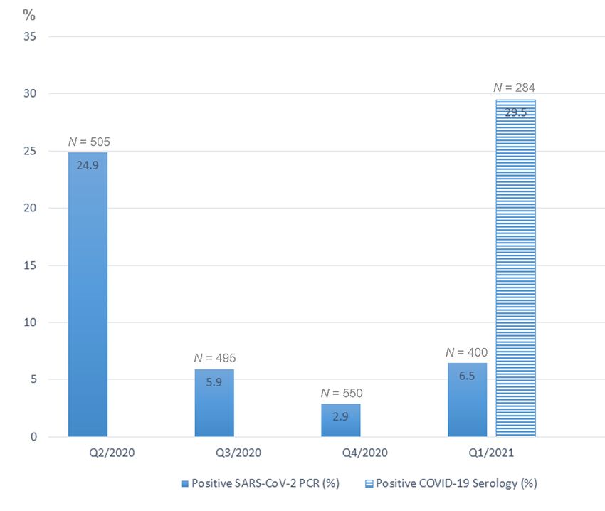
with Apgar scores 3 and 7 at 1 and 5 min, respectively, and a low birthweight of 990 g. He
developed severe conditions associated with systemic hypoxia and prematurity (grade 1
germinal matrix hemorrhage and moderate respiratory distress, requiring intubation);
however, his SARS-CoV-2 RT-PCR studies were negative. The mother fully recovered
J. Dev. Biol. 2021, 9, 45 within a course of several weeks; the child was on ventilation support for 2 months, 5but of 10
eventually was weaned off and discharged with persistent pulmonary hypertension. Pla-
cental histopathology showed patchy areas of a mild intervillositis with marked necrosis
of syncytiotrophoblast, highlighted by the complement component C4d and a dense layer
of
ofperivillous
perivillousfibrin
fibrinenveloping
envelopingaffected
affectedchorionic
chorionicvilli
villiwith
withfocally ischemic
focally cores;
ischemic however,
cores; how-
antiviral ISH and IHC were negative (Figure 3).
ever, antiviral ISH and IHC were negative (Figure 3).
Figure
Figure 3. 3. Histopathology
Histopathology and
and immunohistochemistry
immunohistochemistry of of placenta
placenta in in a parturient
a parturient woman
woman with
with severe
severe COVID-19
COVID-19 (gesta-
(gestational
tional age: 24 4/7 weeks). (A) Intervillositis with scant inflammatory infiltrate (inf) with karyorrhexis, trophoblast necrosis
age: 24 4/7 weeks). (A) Intervillositis with scant inflammatory infiltrate (inf) with karyorrhexis, trophoblast necrosis and
and dense eosinophilic fibrinoid (fib) material enveloping chorionic villi. Villous stromal karyorrhexis (krx), consistent
dense eosinophilic fibrinoid (fib) material enveloping chorionic villi. Villous stromal karyorrhexis (krx), consistent with
with hypoxia, is also seen. H&E. (B) Intervillositis (inf) with trophoblast necrosis, fibrinoid material (fib), enveloping cho-
hypoxia, is also
rionic villi. seen. H&E.
Trichrome stain.(B)
(C)Intervillositis
Negative IHC(inf) with
with trophoblast
antibody againstnecrosis, fibrinoidnucleocapsid
the SARS-CoV-2 material (fib),protein.
enveloping chorionic
(D) IHC with
villi. Trichrome
antibody stain.
against (C) Negative
complement IHC with
complex C4dantibody against
highlighting thethe SARS-CoV-2
interface nucleocapsid
complement protein.and
accumulation (D)necrotic
IHC with antibody
syncytio-
against complement complex C4d highlighting the interface complement accumulation and necrotic syncytiotrophoblast.
trophoblast.
Placentas
Placentas of
of five
five remaining women with
remaining women with severe
severeCOVID-19
COVID-19atatdelivery
deliverywhich
whichwere
were
reported
reportedpreviously
previously [19]
[19] did not show
show acute
acutechanges
changesattributable
attributabletotoinfection;
infection;however,
however,inin
one
oneofofthese
thesecases
casesthetheviral
viralRNA
RNA was
was amplified
amplifiedbyby
RT-PCR
RT-PCR of placental
of placentaltissue homogenates,
tissue homoge-
consistent with a with
nates, consistent possible placental
a possible involvement
placental [13]. [13].
involvement
In the group of 84 women presented with negative viral RT-PCR but positive serology
tests, intervillositis was diagnosed in four cases (4.8%), all negative for SARS-CoV-2 by IHC.
3.2. Fetal and Neonatal Involvement
One hundred seventy-two neonates born to the one hundred and ninety-seven moth-
ers positive for the virus (87.3%) were tested for SARS-CoV-2 by nasopharyngeal swab
RT-PCRs within the first 48 h of life and all test results were negative. There were three
cases of fetal/neonatal loss in the group of 197 SARS-CoV-2 positive women, including
one early second trimester miscarriage, one intrauterine fetal demise (IUFD), and one early
neonatal death. In two of these three cases reported by us previously [18], fetal tissues were
negative for the virus by RT-PCR, IHC, and ISH and the causes of IUFD were defined as
not related to COVID-19 (chronic deciduitis in one case and acute ascending intrauterineJ. Dev. Biol. 2021, 9, 45 6 of 10
infection with group B strep in the second). The third case involved an IUFD at 28 weeks in
a 34 year old Gravida 4 Para 3103 women with the history of systemic lupus erythematosus.
Fetal ultrasound showed corpus callosum agenesis with ventriculomegaly and grey matter
heterotopia. The autopsy was not requested.
During the 1st year of the COVID-19 epidemic, we performed 35 autopsies of stillborn
and live born babies whose maternal SARS-CoV-2 PCR status was known and, besides the
two cases outlined in the previous paragraph, there were no additional perinatal autopsies
from mothers infected with the virus at the time of delivery.
Maternal COVID-19 serology results available in 17 of 35 perinatal autopsies were
positive in four cases (23.5%). In three cases the causes of death included a cord accident
(true umbilical cord knot), immune hydrops in Rh- mother, and non-immune hydrops with
marked pulmonary hypoplasia. In the fourth case of a near term (38 weeks) pregnancy
loss to a 27 year old multigravida with a high body/mass index (35.4), the cause of IUFD
was not apparent. Placental and fetal tissues in the first three cases showed no signs
of inflammation or immunoreactivity with anti-spike or anti-nucleocapsid SARS-CoV-
2 antibodies. In the fourth case, the placenta was negative for the virus; however, an
endothelial staining with anti-nucleocapsid, but not anti-spike, antibody was present
in the fetal lungs (Figure 4A,B). SARS-CoV-2 ISH and RT-PCR from pulmonary tissue
J. Dev. Biol. 2021, 9, x FOR PEER REVIEW 7 of 11
homogenates were, however, negative for replicating viruses. Interestingly, the neonatal
blood was also positive for COVID-19 serology.
Figure4.4.Immunohistochemistry
Figure Immunohistochemistrywith withantibody
antibodyagainst
againstSARS-CoV-2
SARS-CoV-2nucleocapsid
nucleocapsidprotein
proteinininlungs
lungsof
of33stillborn
stillbornfetuses
fetusesof
of mothers with positive COVID-19 serology testing. (A,B) Positive punctate and granular staining of the endothelium of
mothers with positive COVID-19 serology testing. (A,B) Positive punctate and granular staining of the endothelium of
distended veins (v) and small arteries (a) in a 38-week fetus. (C) Negative staining results in a 26-week fetus. (D) Negative
distended veins (v)
staining results in aand small fetus.
30-week arteries (a) in a 38-week fetus. (C) Negative staining results in a 26-week fetus. (D) Negative
staining results in a 30-week fetus.
4. Discussion
The data on the prevalence of COVID-19 infection among parturient women in our
medical center followed the temporal dynamics of the epidemic in New York City with
its peak in the second quarter of 2020, a subsequent drop in the second half of 2020, and a
mild gradual increase in the first quarter of 2021. Interestingly, that near one third (29.5%)J. Dev. Biol. 2021, 9, 45 7 of 10
4. Discussion
The data on the prevalence of COVID-19 infection among parturient women in our
medical center followed the temporal dynamics of the epidemic in New York City with
its peak in the second quarter of 2020, a subsequent drop in the second half of 2020, and a
mild gradual increase in the first quarter of 2021. Interestingly, that near one third (29.5%)
of placentas submitted for pathology evaluation in the first quarter of 2021 came from
women with positive COVID-19 serology tests. The COVID-19 serology test employed by
our medical center uses the ROCHE immunoassay intended for a qualitative detection
of antibodies to SARS-CoV-2 under the Emergency Use Authorization and should be
interpreted as an evidence of prior infection, according to the Center for Disease Control
(CDC) recommendations. Thus, the number 29.5% reflects the prevalence of prior COVID-
19 in the group of pregnant women with high risk and complicated pregnancies whose
placentas have been submitted for pathological evaluation, according to the institutional
policies, and should not be extrapolated to the general population.
Our data also indicate that although 3% of women infected with SARS-CoV-2 had
severe disease at the time of delivery there were no cases of maternal mortality among
symptomatic (as well as asymptomatic) patients and no fetal mortality in the group of
severely ill. In one case, however, the severe COVID-19 disease prompted the delivery at
24+ weeks, leading to a significant fetal morbidity related to prematurity.
To assess the frequency of definitive placental involvement, we used recently proposed
criteria [11–13] which include: (1) placental inflammation (intervillositis); (2) necrosis of
villous trophoblast with perivillous accumulation of fibrillary proteins; (3) in situ identifi-
cation of replicating virus in villous trophoblast. Thus, definitive placental involvement
was identified only in two patients (about 1% in our cohort).
Although in the group of six severely ill patients there were no definitive placental
infection, in one case, reported by us previously [19], positive RT-PCR results from placental
homogenates were consistent with a possible involvement. In this case, an elevated
expression of interferon-induced antiviral transmembrane transcripts was also detected
by molecular techniques while no tissue reaction (inflammation, necrosis) was observed
microscopically. This demonstrates that before the morphological changes become apparent
the infection may develop on the molecular level and if delivery occurs in early stages of
the disease placental lesions may not be detectable on the microscopic level.
Similarly, in the late stages of placental involvement the tissue viral loads likely de-
crease beyond the threshold of detection by in situ techniques, while morphologic features,
including inflammation, necrosis, and fibrinoid deposition, still persist. Thus, intervillositis
observed by us in the severely ill patient on ECMO (Figure 3) was highly suspicious for the
late stage of the infection with features of “burned out” inflammation with marked necrosis
of syncytiotrophoblast and encasement of chorionic villi by dense fibrinoid material caus-
ing ischemic changes in the villous cores. Interestingly, the immunohistochemical staining
for C4d was similar to that described by Libbrecht et al. [9] in their case of SARS-CoV-2
intervillositis; however, as the authors noted, the C4d staining is not specific and had also
been described in the so-called idiopathic intervillositis [20]. Thus, placental involvement
could not be definitively confirmed in the case of the most severe COVID-19 in our cohort.
In sum, our study showed a very low level (about 1%) of definitive placental involve-
ment in SARS-CoV-2 infected parturient women and this involvement did not correlate
with the disease severity, as it was observed in one asymptomatic and one mildly symp-
tomatic cases, while in the group of six patients with severe COVID-19 there was only one
case of possible/early placental involvement and one case demonstrating an organizing
stage of intervillositis, suspicious for a late stage in the evolution of the infectious lesion.
These findings suggest that placental involvement is transient in the course of the infection
and our ability to detect the virus in the placental tissue may be influenced by the time
between the peak of tissue viral load and delivery.
Our data confirmed previous reports [14,15,21,22] on the rarity of perinatal involve-
ment in SARS-CoV-2 as all tested (87.3%) neonates born to the SARS-CoV-2 positive womenJ. Dev. Biol. 2021, 9, 45 8 of 10
were negative for the virus and no perinatal complications or hospital stays have been
attributed to the MTCT of the infection. Three identified cases of the fetal loss among
197 infected parturient women were likely unrelated to SARS-CoV-2 since available fetal
tissues were negative for the virus and pathology other than SARS-CoV-2 infection was
present to explain the demise in each of the three cases.
Our analysis of four perinatal autopsies in women negative by SARS-CoV-2 RT-PCR
but positive for COVID-19 serology showed that in three cases the causes of fetal demise
(cord accident, immune hydrops, and pulmonary hypoplasia) were unlikely related to the
prior maternal infection during pregnancy. In one case, however, the direct cause of the near
term IUFD was not apparent with only one known risk factor (maternal obesity). Positive
COVID-19 serology, which was also detected in the fetal serum, raised a question of a fetal
infection. Unfortunately, the Elecsys® Roche assay which we used does not differentiate
between IgG and IgM antibodies; thus, it is not possible to tell whether the test positivity
was entirely due to the passage of IgG antibodies from the mother or whether a fraction
of IgM antibodies was also present in the fetal serum. The latter would have indicated a
fetal infection, since IgMs do not cross placenta. However, due the test design, the fetal
infection remained indeterminate. In this case the endothelium of fetal lungs showed
immune reaction with the antibody against SARS-CoV-2 nucleocapsid protein; however,
an active SARS-CoV-2 infection due to a replicating virus was ruled out by the negative
ISH and RT-PCR in pulmonary tissue homogenates. The findings were unusual prompting
us to question the observed staining as an unspecific reaction or artefact; however, we have
not observed an artificial staining with this antibody in numerous other cases of placentas
and neonatal lungs (Figure 4C,D).
Interestingly, staining of the endothelium of villous capillaries was reported by
Schwartz et al. [12] in two placentas, including one case of a COVID-19 infection remote
from delivery when no syncytiotrophoblastic staining was observed. A positive staining of
placental Hoffbauer cells was also seen in their cases and the authors hypothesized that the
antibody may have reacted with persistent viral particles engulfed by macrophages and
sequestered in the endothelium.
If this hypothesis is accepted and fetal endothelial cells can internalize the virus or its
debris, the anti-nucleocapsid staining of the pulmonary endothelium in our case of the fetus
positive for COVID-19 serology would be consistent with the history of MTCT of SARS-
CoV-2 sometime during pregnancy. Thus, with all the limitations, this single case is the best
candidate for the MTCT of SARS-CoV-2 in our one-year experience. Interestingly, the virus
has been recently demonstrated in the fetal tissues by RT-PCR and immunofluorecence
IHC in a case of IUFD at 34 weeks to a woman with a mild infection [23]. More data are
needed to develop consensus criteria for the definitive, probable and possible diagnoses of
SARS-CoV-2 infection and MTCT on autopsy material.
Recent study demonstrated the expression of the main SARS-CoV-2 cellular entry
factors in villous trophoblast in term placentas, explaining why this cellular layer gets
attacked by the virus [24]. The individual variability of these expression levels may be
responsible for the different susceptibility to placental infection among women. Because
the MTCT and fetal involvement are even rarer than the placental infection, we agree that
the mechanisms underlying placental defense against SARS-CoV-2 likely involve post entry
viral processing, potentially including phagocytosis by Hofbauer cells and engulfment
by fetal endothelium. Further studies are necessary to understand the role of different
components of the placenta in defending the offspring from SARS-CoV-2.
Author Contributions: J.T. performed data collection, analysis, and manuscript revision; Y.S. per-
formed data collection, analysis, and manuscript revision; L.D. performed conceptualization, data
curation, investigation, project administration, supervision, validation, visualization, and writing of
the manuscript. All authors have read and agreed to the published version of the manuscript.
Funding: This research received no external funding.J. Dev. Biol. 2021, 9, 45 9 of 10
Institutional Review Board Statement: The study was conducted according to the guidelines of the
Declaration of Helsinki, and approved by the Institutional Review Board of Columbia University in
the City of New York (IRB-AAAT0272 approved on 6 June 2021).
Informed Consent Statement: Patient consent was waived due the following reasons: the research
involved no more than minimal risk to the subjects; the waiver or alteration would not adversely
affect the rights and welfare of the subjects; the research could not practicably be carried out without
the waiver or alterations; whenever appropriate, the subjects will be provided with additional
pertinent information after participation.
Data Availability Statement: Not applicable.
Conflicts of Interest: The authors declare no conflict of interest.
References
1. Badr, D.A.; Picone, O.; Bevilacqua, E.; Carlin, A.; Meli, F.; Sibiude, J.; Mattern, J.; Fils, J.-F.; Mandelbrot, L.; Lanzone, A.; et al.
Severe Acute respiratory syndrome coronavirus 2 and pregnancy outcomes according to gestational age at time of infection: The
INTERCOVID Multinational Cohort Study. Emerg. Infect. Dis. 2021, 27, 2535–2543. [CrossRef] [PubMed]
2. Villar, J.; Ariff, S.; Gunier, R.B.; Thiruvengadam, R.; Rauch, S.; Kholin, A.; Roggero, P.; Prefumo, F.; Vale, M.S.D.; Cardona-Perez,
J.A.; et al. Maternal and neonatal morbidity and mortality among pregnant women with and without COVID-19 infection. JAMA
Pediatr. 2021, 175, 817–826. [CrossRef]
3. Patanè, L.; Morotti, D.; Giunta, M.R.; Sigismondi, C.; Piccoli, M.G.; Frigerio, L.; Mangili, G.; Arosio, M.; Cornolti, G. Vertical
transmission of coronavirus disease 2019: Severe acute respiratory syndrome coronavirus 2 RNA on the fetal side of the placenta
in pregnancies with coronavirus disease 2019–positive mothers and neonates at birth. Am. J. Obstet. Gynecol. MFM 2020, 2, 100145.
[CrossRef]
4. Hosier, H.; Farhadian, S.F.; Morotti, R.A.; Deshmukh, U.; Lu-Culligan, A.; Campbell, K.H.; Yasumoto, Y.; Vogels, C.B.; Casanovas-
Massana, A.; Vijayakumar, P.; et al. SARS–CoV-2 infection of the placenta. J. Clin. Investig. 2020, 130, 4947–4953. [CrossRef]
[PubMed]
5. Sisman, J.; Jaleel, M.A.; Moreno, W.; Rajaram, V.; Collins, R.; Savani, R.C.; Rakheja, D.; Evans, A.S. Intrauterine transmission of
SARS-COV-2 infection in a preterm infant. Pediatr. Infect. Dis. J. 2020, 39, e265–e267. [CrossRef]
6. Vivanti, A.J.; Vauloup-Fellous, C.; Prevot, S.; Zupan, V.; Suffee, C.; Do Cao, J.; Benachi, A.; De Luca, D. Transplacental transmission
of SARS-CoV-2 infection. Nat. Commun. 2020, 11, 1–7. [CrossRef] [PubMed]
7. Debelenko, L.; Katsyv, I.; Chong, A.M.; Peruyero, L.; Szabolcs, M.; Uhlemann, A.-C. Trophoblast damage with acute and chronic
intervillositis: Disruption of the placental barrier by severe acute respiratory syndrome coronavirus 2. Hum. Pathol. 2021, 109,
69–79. [CrossRef] [PubMed]
8. Linehan, L.; O’Donoghue, K.; Dineen, S.; White, J.; Higgins, J.R.; Fitzgerald, B. SARS-CoV-2 placentitis: An uncommon
complication of maternal COVID-19. Placenta 2021, 104, 261–266. [CrossRef]
9. Libbrecht, S.; Van Cleemput, J.; Vandekerckhove, L.; Colman, S.; Padalko, E.; Verhasselt, B.; Van de Vijver, K.; Dendooven, A.;
Dehaene, I.; Van Dorpe, J. A rare but devastating cause of twin loss in a near-term pregnancy highlighting the features of severe
SARS-CoV-2 placentitis. Histopathology 2021, 79, 674–676. [CrossRef]
10. Garrido-Pontnou, M.; Navarro, A.; Camacho, J.; Crispi, F.; Alguacil-Guillén, M.; Moreno-Baró, A.; Hernandez-Losa, J.; Sesé, M.;
Cajal, S.R.Y.; Ruíz, I.G.; et al. Diffuse trophoblast damage is the hallmark of SARS-CoV-2-associated fetal demise. Mod. Pathol.
2021, 34, 1704–1709. [CrossRef]
11. Schwartz, D.A.; Baldewijns, M.; Benachi, A.; Bugatti, M.; Collins, R.R.J.; De Luca, D.; Facchetti, F.; Linn, R.L.; Marcelis, L.;
Morotti, D.; et al. Chronic histiocytic intervillositis with trophoblast necrosis is a risk factor associated with placental infection
from coronavirus disease 2019 (COVID-19) and Intrauterine maternal-fetal severe acute respiratory syndrome coronavirus 2
(SARS-CoV-2) Transmission in live-born and stillborn infants. Arch. Pathol. Lab. Med. 2021, 145, 517–528. [CrossRef]
12. Schwartz, D.A.; Baldewijns, M.; Benachi, A.; Bugatti, M.; Bulfamante, G.; Cheng, K.; Collins, R.R.; Debelenko, L.; De Luca, D.;
Facchetti, F.; et al. Hofbauer cells and coronavirus disease 2019 (COVID-19) in pregnancy: Molecular pathology analysis of
villous macrophages, endothelial cells, and placental findings from 22 placentas infected by severe acute respiratory syndrome
coronavirus 2 (SARS-CoV-2) with and without fetal transmission. Arch. Pathol. Lab. Med. 2021. [CrossRef]
13. Roberts, D.J.; Edlow, A.G.; Romero, R.J.; Coyne, C.B.; Ting, D.T.; Hornick, J.L.; Zaki, S.R.; Das Adhikari, U.; Serghides, L.; Gaw,
S.L.; et al. SPECIAL REPORT: A standardized definition of placental infection by severe acute respiratory syndrome coronavirus
2 (SARS-CoV-2), a consensus statement from the National Institutes of Health/Eunice Kennedy Shriver National Institute of
Child Health and Human Development (NIH/NICHD) SARS-CoV-2 placental infection workshop. Am. J. Obstet. Gynecol. 2021.
[CrossRef]
14. Ansari, K.; Kew, T.; Allotey, J.; Thangaratinam, S. Mother-to-child transmission of severe acute respiratory syndrome coronavirus
2. Curr. Opin. Obstet. Gynecol. 2021, 33, 391–399. [CrossRef]
15. Parums, D.V. Editorial: Maternal SARS-CoV-2 Infection and Pregnancy Outcomes from Current Global Study. Data. Med. Sci.
Monit. 2021, 27, e933831. [CrossRef] [PubMed]J. Dev. Biol. 2021, 9, 45 10 of 10
16. Shah, P.S.; Ye, X.Y.; Yang, J.; Campitelli, M.A. Preterm birth and stillbirth rates during the COVID-19 pandemic: A population-
based cohort study. Can. Med. Assoc. J. 2021, 193, E1164–E1172. [CrossRef]
17. Smithgall, M.C.; Liu-Jarin, X.; Hamele-Bena, D.; Cimic, A.; Mourad, M.; Debelenko, L.; Chen, X. Third-trimester placentas of
severe acute respiratory syndrome coronavirus 2 (SARS-CoV-2)-positive women: Histomorphology, including viral immunohis-
tochemistry and in-situ hybridization. Histopathology 2020, 77, 994–999. [CrossRef]
18. Valk, J.E.; Chong, A.M.; Uhlemann, A.-C.; Debelenko, L. Detection of SARS-CoV-2 in placental but not fetal tissues in the second
trimester. J. Perinatol. 2021, 41, 1184–1186. [CrossRef]
19. Mourad, M.; Jacob, T.; Sadovsky, E.; Bejerano, S.; Simone, G.S.-D.; Bagalkot, T.R.; Zucker, J.; Yin, M.T.; Chang, J.Y.; Liu, L.; et al.
Placental response to maternal SARS-CoV-2 infection. Sci. Rep. 2021, 11, 1–12. [CrossRef]
20. Bendon, R.W.; Coventry, S.; Thompson, M.; Rudzinski, E.R.; Williams, E.M.; Oron, A.P. Significance of C4d immunostaining in
placental chronic intervillositis. Pediatr. Dev. Pathol. 2015, 18, 362–368. [CrossRef]
21. Peng, Z.; Wang, J.; Mo, Y.; Duan, W.; Xiang, G.; Yi, M.; Bao, L.; Shi, Y. Unlikely SARS-CoV-2 vertical transmission from mother to
child: A case report. J. Infect. Public Health 2020, 13, 818–820. [CrossRef] [PubMed]
22. Fenizia, C.; Biasin, M.; Cetin, I.; Vergani, P.; Mileto, D.; Spinillo, A.; Gismondo, M.R.; Perotti, F.; Callegari, C.; Mancon, A.; et al.
Analysis of SARS-CoV-2 vertical transmission during pregnancy. Nat. Commun. 2020, 11, 1–10. [CrossRef] [PubMed]
23. Marinho, P.S.; da Cunha, A.J.L.A.; Chimelli, L.; Avvad-Portari, E.; Andreiuolo, F.D.M.; de Oliveira-Szejnfeld, P.S.; Mendes, M.A.;
Gomes, I.C.; Souza, L.R.Q.; Guimarães, M.Z.; et al. Case Report: SARS-CoV-2 Mother-to-child transmission and fetal death
associated with severe placental thromboembolism. Front. Med. 2021, 8, 1315. [CrossRef] [PubMed]
24. Ouyang, Y.; Bagalkot, T.; Fitzgerald, W.; Sadovsky, E.; Chu, T.; Martínez-Marchal, A.; Brieño-Enríquez, M.; Su, E.J.; Margolis, L.;
Sorkin, A.; et al. Term Human Placental Trophoblasts Express SARS-CoV-2 Entry Factors ACE2, TMPRSS2, and Furin. mSphere
2021, 6, e00250-21. [CrossRef]You can also read


