Diagnosis of secondary amyloidosis in alkaptonuria
←
→
Page content transcription
If your browser does not render page correctly, please read the page content below
Millucci et al. Diagnostic Pathology 2014, 9:185
http://www.diagnosticpathology.org/content/9/1/185

 RESEARCH                                                                                                                                          Open Access

Diagnosis of secondary amyloidosis in alkaptonuria
Lia Millucci1†, Lorenzo Ghezzi1†, Giulia Bernardini1†, Daniela Braconi1, Pietro Lupetti2, Federico Perfetto3,
Maurizio Orlandini1 and Annalisa Santucci1*

  Abstract
  Background: Alkaptonuria (AKU) is an inborn error of catabolism due to a deficient activity of homogentisate
  1,2-dioxygenase. Patients suffer from a severe arthropathy, cardiovascular and kidney disease but other organs
  are affected, too. We found secondary amyloidosis as a life-threatening complication in AKU, thus opening
  new perspectives for its treatment. We proved that methotrexate and anti-oxidants have an excellent efficacy
  to inhibit the production of amyloid in AKU model chondrocytes. Owing to the progressive and intractable
  condition, it seems important to detect amyloid deposits at an early phase in AKU and the choice of specimens for a
  correct diagnosis is crucial.
  Methods: Ten AKU subjects were examined for amyloidosis; abdominal fat pad aspirates, labial salivary gland, cartilage
  and synovia specimens were analysed by CR, Th-T, IF, TEM.
  Results: Amyloid was detected in only one abdominal fat pad specimen. However, all subjects demonstrated amyloid
  deposition in salivary glands and in other organ biopsies, indicating salivary gland as the ideal specimen for early
  amyloid detection in AKU.
  Conclusions: This is, at the best of our knowledge, the first report providing correct indications on the diagnosis of
  amyloidosis in AKU, thus offering the possibility of treatment of such co-morbidity to AKU patients.
  Virtual slides: The virtual slide(s) for this article can be found here: http://www.diagnosticpathology.diagnomx.eu/vs/
  13000_2014_185
  Keywords: Serum amyloid A, Ochronosis, Secondary amyloidosis, Salivary gland, Periumbelical fat, Congo Red

Background                                                                              the most common clinical presentation of AKU, but all
Amyloidosis is the deposition of insoluble protein aggregates                           the organs can be affected, too. AKU can be treated symp-
either systemically or in a specific organ. Secondary (AA)                              tomatically during the early stages, whereas for end stages
amyloidosis is caused by a chronic infection or chronic in-                             total joint and heart valve replacements may be required.
flammatory disease where deposits are made up of serum                                  Currently, there is no therapy for AKU, although a clinical
amyloid A (SAA) produced during inflammation.                                           trial with nitisinone is in progress.
   We found SAA amyloidosis as a life-threatening compli-                                  AA amyloidosis is one of the most severe complica-
cation in alkaptonuria (AKU) [1], an ultra-rare (1:250.000-                             tions of several chronic rheumatic disorders [2,3] and is
1.000.000 incidence) autosomal recessive genetic disease                                a secondary complication of AKU due to a chronic in-
due to a deficient activity of homogentisate 1,2-dioxygen-                              flammatory status derived from HGA/BQA-induced oxi-
ase (HGD) leading to the accumulation of homogenti-                                     dative stress [4-12]. SAA-amyloid in AKU co-localizes
sic acid (HGA). HGA-oxidized derivative benzoquinone                                    with ochronotic pigment [1], AKU is a complicated in-
acetic acid (BQA) forms deposits in the connective tissue,                              flammatory multisystemic disease, and any body district
causing a pigmentation known as “ochronosis”, leading to                                expressing HGD may be affected [13].
dramatic organ damage. A severe form of arthropathy is                                     Our findings on AKU as a novel AA amyloidosis opened
                                                                                        new perspectives for its treatment. In fact, the suppression
* Correspondence: annalisa.santucci@unisi.it                                            of SAA below 10 mg/L halts the progression of the disease
†
 Equal contributors
1
 Dipartimento di Biotecnologie, Chimica e Farmacia, Università degli Studi di           and is associated with prolonged survival, reversal of
Siena, via Aldo Moro 2, 53100 Siena, Italy                                              amyloid deposition and recovery of organ function [14].
Full list of author information is available at the end of the article

                                         © 2014 Millucci et al.; licensee BioMed Central Ltd. This is an Open Access article distributed under the terms of the Creative
                                         Commons Attribution License (http://creativecommons.org/licenses/by/4.0), which permits unrestricted use, distribution, and
                                         reproduction in any medium, provided the original work is properly credited. The Creative Commons Public Domain
                                         Dedication waiver (http://creativecommons.org/publicdomain/zero/1.0/) applies to the data made available in this article,
                                         unless otherwise stated.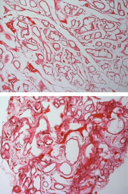
Millucci et al. Diagnostic Pathology 2014, 9:185                                                                   Page 2 of 9
http://www.diagnosticpathology.org/content/9/1/185

Methotrexate [1] and anti-oxidants [9] proved to have an         around the umbilicus of the patients were anesthetized
excellent efficacy to inhibit the production of amyloid in       with 1% lidocaine. The fat was aspirated with an 18-
AKU model chondrocytes, suggesting their introduction            gauge needle connected to a 20 ml syringe. At least two
in AKU therapy.                                                  visible fragments of fat were placed on a glass slide and
   Owing to AKU progressive and intractable condition,           crushed into a single layer by pressing a second slide
it is crucial to detect amyloid deposits at an early phase.      against the first. After the smears were dried in air at 50°C
As ascertained in other secondary amyloidosis, if SAA            for at least 30 minutes and fixed with 20% formalin neu-
deposition cannot be stopped or delayed, serious life-           tral buffer solution (Wako Pure Chemical, Osaka, Japan)
threatening problems can arise [14]. Therefore, SAA              for 5 minutes, these specimens were processed for the
amyloidosis should be diagnosed as early as possible and         Congo Red staining.
the clinicians treating AKU patients need an easy rou-
tine reproducible fast procedure that can be repeated at         Cartilage and synovia
intervals.                                                       Articular cartilage fragments, from AKU subjects who
   In this work, ten AKU subjects were examined for              underwent surgery for joint replacement, were placed in
amyloidosis in abdominal fat pad aspirates and labial sal-       5% formaldehyde. Samples were alcohol dehydrated and
ivary gland (LSG) biopsies. Amyloid was detected in only         paraffin embedded.
one abdominal fat pad specimen. However, all subjects
demonstrated amyloid deposition in LSGs and in other
                                                                 Aortic valve
organ biopsies. These results further support the con-
                                                                 Valve was obtained from an AKU subject who under-
cept that LSG may be the optimal specimen for early de-
                                                                 went biologic aortic valve replacement.
tection of amyloid in AKU and that LSG offers
improved sensitivity in respect to fat pad aspirates in
these patients.                                                  Prostatic calculus
   This is, at the best of our knowledge, the first report       The stone was from the prostate gland of an AKU subject
providing correct indications on the diagnosis of amyl-          who underwent surgical exploration for calculus removal.
oidosis in AKU, thus offering the possibility of treatment
of such co-morbidity to AKU patients.                            Hematoxylin and Eosin (H&E) staining
                                                                 Two sections (5 μm thickness) were obtained from each
Methods                                                          biopsy. Sections were stained with traditional hematoxylin/
Procedures were approved by Siena University Hospital and        eosin (H/E) staining.
national Ethics (Comitato Etico Policlinico Universitario di
Siena, number GGP10058, date 21/07/2010) in accordance
                                                                 Congo Red (CR) staining
with 1975 Helsinki Declaration, revised in 2000 (52nd
                                                                 Romhányi’s CR staining method modified according to
WMA General Assembly, Edinburgh, Scotland, October
                                                                 Bély and Apáthy (2000) was adopted. 5 μm sections of
2000). Informed consent was obtained from all patients.
                                                                 fresh specimen were fixed in cooled 96% ethanol
   All reagents were from Sigma-Aldrich (St. Louis, MO),
                                                                 10 min, rinsed in water, incubated in 1% CR for 40 min,
if not differently specified.
                                                                 washed in water, incubated 10 s in 1 mL 1% sodium hy-
                                                                 droxide in 100 mL of 50% ethanol, incubated 30 s in
LSG
                                                                 Mayer's hematoxylin, sequentially washed in 50%, 75%,
After reviewing AKU patients’ medical history and la-
                                                                 95% ethanol, mounted and observed under a polarized
boratory values (Table 1 and 2), a clinical examination
                                                                 light microscope (Zeiss Axio Lab.A1, Arese, Milano).
was completed and each subject underwent two biopsies
of clinically normal-appearing tissue in the oral cavity
under local anesthesia (2% lidocaine with 1:100,000 epi-         Thioflavin T (Th-T) staining
nephrine). Specimen 1 was taken from the left lateral sur-       AKU LSG samples incubated in 1% Th-T [15,16] were
face of the tongue, containing mucosal and muscle tissue.        mounted and observed under a fluorescence microscope
Specimen 2 was taken from the labial mucosa on the left          (excitation 450 nm, emission 482 nm).
side of the lower lip, containing at least 2 to 3 minor saliv-
ary glands. Both specimens were formalin fixed and ana-          Fluorescence microscopy
lyzed for histopathologic evaluation of amyloid.                 AKU samples in paraffin were cut in 3–5 μm slices and
                                                                 double immunofluorescence stained with anti-SAA and
Abdominal fat                                                    anti-serum amyloid P (SAP) antibodies (Santa Cruz Bio-
Patients underwent a subcutaneous abdominal fat biopsy           technology, CA). Nuclei were counter-stained with blue
via fine-needle aspiration. Skin and subcutaneous tissue         DAPI DNA stain (Abcam, Cambrige, UK).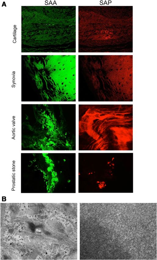
Millucci et al. Diagnostic Pathology 2014, 9:185                                                                                                          Page 3 of 9
http://www.diagnosticpathology.org/content/9/1/185

Table 1 Alkaptonuria patients enrolled for the study and their characteristics
Features                           Patient 1 Patient 2 Patient 3 Patient 4 Patient 5 Patient 6 Patient 7 Patient 8 Patient 9 Patient 10
Age (years)                        71           60            63           42           60            58           42           38            65           55
Sex                                F            F             F            F            M             M            M            F             F            F
Serology
SAA mg/L                           117.70       87.14         97.44        18.16        25.35         25.44        25.13        16.13         28.46        69.90
HGA plasma level μmol/L            31.10        72.90         20.56        15.80        17.02         22.43        18.95        15.21         31.76        13.42
HGA urine level mmol/L             23.70        16.90         22.64        21.70        22.30         18.64        24.10        12.68         22.76        16.60
Congo Red birefringence score
Abdominal fat                      n.d.         -             -            -            -             -            -            -             n.d.         +
Salivary gland                     +++          ++            ++           ++           ++            +++          ++           +++           ++           ++
Synovia                                                       +++
Cartilage                                                     +++                       +++
Aortic valve                                                  +++                       +++
Prostatic stone                                                                         ++
ThT fluorescence (+/−)
Salivary gland                     positive     positive      positive     positive     positive      positive     positive     positive      positive     positive
Synovia                                                       positive                  positive
Cartilage                                                     positive                  positive
Aortic valve                                                  positive
Prostatic stone                                                                         n.d.
SAA immunohistology (+/−)
Salivary gland                     positive     positive      positive     positive     positive      positive     positive     positive      positive     positive
Synovia                                         positive                   positive                   positive     positive
Cartilage                                       positive      positive                  positive      positive     positive
Prostatic stone                                                                         positive
Aortic valve                                                  positive
SAP immunohistology (+/−)
Salivary gland                     positive     positive      positive     positive     positive      positive     positive     positive      positive     positive
Synovia                                                       positive                  positive
Cartilage                                                     positive                  positive
Aortic valve                                                  positive
Prostatic stone                                                                         positive
TEM Amyloid abundance score
                                   +++          +++           ++           +++          +++           +++          ++           +++           ++           +
F: female, M: male, n.d.: not determined. The patients were classified on the basis of Congo red intensity of staining (grades: +++, very intensive; ++, intensive; +,
weak; −, no birefringence). The presence of amyloid deposits in LSG observed at TEM was evaluated as: +++ = very abundant, ++ = medium; + = faint. Positivity
for ThT fluorescence and SAA and SAP immunohistology is also reported.

Transmission electron microscopy (TEM)                                                Results
AKU specimens were fixed in 2.5% glutaraldehyde in                                    Structural features of AKU salivary glands were first
0.1 M cacodylate buffer (CB) pH 7.2 for 3 h at 4°C, post-                             evaluated by means of H&E staining (representative im-
fixed in 1% osmium tetroxide in CB for 2 hours at 4°C,                                ages are presented in Figure 1A-B). Parenchyma was
ethanol dehydrated and embedded in a mixture of Epon–                                 found to be compact only in some limited areas; by con-
Araldite resins. Thin sections, obtained with a Reichert                              trast, a number of vacuoles and a disrupted acinar struc-
ultramicrotome, were stained with uranyl acetate and                                  ture were present in the vast majority of the specimen.
lead citrate and observed with a TEM FeiTecnai G2                                     All of the AKU salivary glands that were examined did
spirit at 80 Kv.                                                                      not contain any trace of lymphocytic infiltration.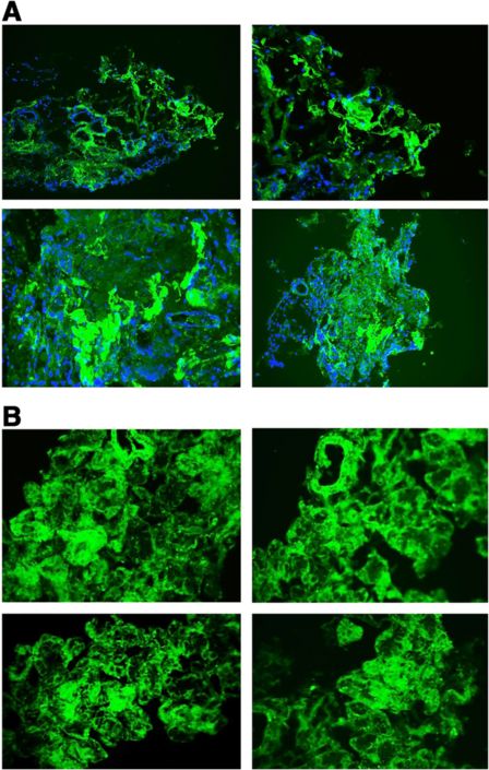
http://www.diagnosticpathology.org/content/9/1/185
                                                                                                                                                                                     Millucci et al. Diagnostic Pathology 2014, 9:185
Table 2 Clinical features of AKU patients
Patient # Date of selection Peripheral neuropathy Orthostatic Ventricular and chronic bacterial Spondylarthropathy Glossomegaly Surgery
          (month/year)                            hypotension atrial dilatation infection
1            08/2011               y                            y                y                    n       y   y             Knee (r), shoulder (r) and hip (l) arthroplasty
2            10/2009               y                            y                n                    y       y   n             Knee (r, l), shoulder (r) and hip (r) arthroplasty
3            03/2012               n                            y                n                    n       y   n             Meniscus (r) surgical repair
4            04/2012               y                            y                n                    n       n   n             None
5            11/2012               y                            y                n                    y       y   n             Knee (l), and hip (l) arthroplasty
6            01/2013               y                            n                n                    y       y   n             Knee (r) arthroplasty
7            05/2012               n                            y                n                    n       n   n             None
8            02/2012               y                            y                y                    n       y   n             Knee (l) arthroplasty
9            03/2013               y                            n                y                    y       y   y             Knee (r) and tendon ankle fracture (l)
10           11/2011               y                            y                n                    n       y   y             Knee (r), and hip (l) arthroplasty
No patient was receiving immunosuppressive or chemotherapy treatment. y = yes; n = no; r = right; l = left.

                                                                                                                                                                                                              Page 4 of 9
Millucci et al. Diagnostic Pathology 2014, 9:185 Page 5 of 9
http://www.diagnosticpathology.org/content/9/1/185
A
B
Figure 1 H/E staining of AKU salivary gland. Some striated ducts
(arrowheads), secretory acini (white arrows) and red blood cells
(black arrows) are indicated. Only representative images are shown.
Magnification: A: 20×; B: 40 × .
In order to diagnose amyloidosis in AKU patients,
diagnostic laboratories adopted periumbelical fat aspirate
CR staining [17] that unfortunately proved to be unsuit-
able for the detection of amyloid deposits in AKU pa-
tients. Indeed, we studied abdominal fat pad aspirates
and labial salivary gland biopsies from 10 AKU patients
in whom subsequent other tissue biopsies for confirm-
ation of AA amyloidosis were also available. All cases
were suspected to have SAA deposits, but nine of them
had negative results on abdominal fat pad aspirates
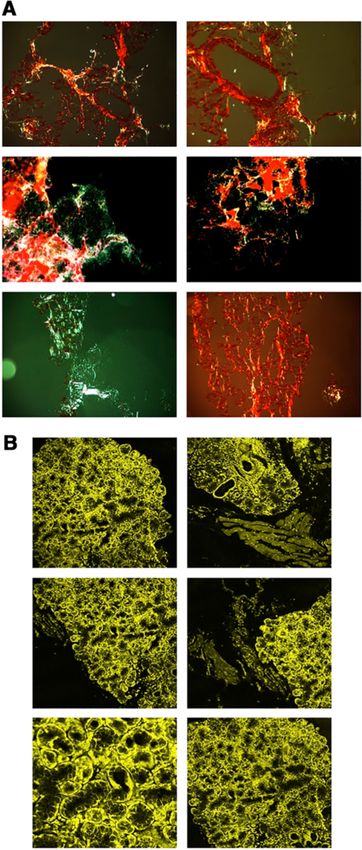
evaluated by polarizing microscopy of CR stained sec- Figure 2 Evidence of amyloid deposits in AKU salivary glands.
tions (not shown). CR staining of fat aspirates from A) Congo Red staining of AKU labial salivary glands amyloid deposits.
three different hospital laboratories were compared in CR stain viewed under polarized light detected the presence of
diffuse amyloid deposits within AKU salivary gland tissue.
all samples for studying inter observer reproducibility Magnification 20×. Representative images from a triplicate set are
and gave negative diagnosis of amyloidosis in nine shown. B) Th-T fluorescence of AKU labial salivary glands amyloid
cases out of ten. deposits shown by confocal microscopy. Analogous results were
Once CR staining was adopted on AKU LSGs, the results obtained from specimens of other patients. Bar: 22 μm.
were unequivocally positive for all ten patients (Figure 2A). Representative images from a triplicate set are shown.
To confirm the presence of amyloid aggregates in LSG
from AKU patients, we performed the Th-T assay. Th-T
fluorescence was evident in all examined samples (10/10)Millucci et al. Diagnostic Pathology 2014, 9:185 Page 6 of 9
http://www.diagnosticpathology.org/content/9/1/185
(Figure 2B). Moreover, by means of immunofluorescence
techniques, we assessed the presence of SAA deposition
in LSG (Figure 3).
Ultrastructural analysis by TEM of AKU LSGs (the
current ‘gold standard’ for detecting amyloid deposits
also in ambiguous cases), confirmed the massive pres-
ence of amyloid fibrils in all specimens. Amyloid de-
posits in LSG were abundant in six patients, moderate
in three, and mild in one (Table 1). Figure 4 shows fine
fibrils, approximately 10 nm in diameter, located in the
secretory stroma of AKU salivary glands of patient #1,
adjacent to the basement membrane. The fibrils were
also present in the interstitial connective tissue, inter-
spersed with collagen fibrils (Figure 4A) and around
interlobular ducts of salivary glands of all patients. In all
observed samples, the glandular stroma contained nu-
merous broken collagen fibrils strictly interconnected
Figure 4 Electron micrographs of AKU salivary glands. A)
Electron micrograph showing fine amyloid fibrils (A) approximately
10 nm in diameter located in close relation to the lamina (LP) of the
secretory end-pieces as well as in the interstitial connective tissue
stroma of AKU labial salivary gland. Pigment granules are visible
amongst collagen fibrils in transverse and longitudinal section (black
arrows). B-C) Electron micrographs of another AKU labial salivary
gland showing a region of the interstitial glandular stroma that
contains fine amyloid fibrils (A), approximately 10 nm in diameter,
interspersed with bundles of collagen fibrils (C). Pigment deposits
are present on broken collagen fibers (black arrows) and scattered
between amyloid fibrils. To be note that amyloid fibrils appear
superimposed to collagen fibers in different area of the tissue.
D-F) Electron micrograph of a third AKU labial salivary gland showing
the glandular stroma containing finely fibrillar amyloid material (A).
In photo E) collagen fibers in transverse section are shown (arrowheads),
many of which presenting electron dense ochronotic deposits located
amongst fibers. In photos E-F), dark ochronotic pigment granules are
indicated (arrowheads) scattered amongst the collagen fiber as well as
amongst amyloid fibrils. Some deposits can be observed located within
amyloid bundles of fibrils and bridging between collagen fibers (white
arrows). A: amyloid; C: collagen; LP lamina propria. Photo A is from
patient #2, photos B-C are from patient #5 and photos D-F are from
Figure 3 SAA and SAP in AKU salivary glands. A) patient #7. Bars, A: 5 μm, B, C and E: 2.5 μm, D and F: 1 μm.
Immunofluorescence staining of SAA deposition in AKU labial
salivary glands. Blue indicates DAPI stained nuclei. Magnification
20×. Representative images from a triplicate set are shown. B) with amyloid fibrils arranged in a scattered manner. More-
Immunofluorescence staining of SAP deposition in AKU labial over, thanks to TEM analysis, it was possible to see evi-
salivary glands. Magnification 20×. Representative images from a
dent pigment traces finely sprinkled over the whole tissue
triplicate set are shown.
(Figure 4B-F). In particular, pigment deposits could beMillucci et al. Diagnostic Pathology 2014, 9:185 Page 7 of 9 http://www.diagnosticpathology.org/content/9/1/185 seen as individual “drop-like” deposits between the colla- examined areas of human body, such as glandular tissues, gen fibers and also scattered over amyloid bundles of may provide insights into the mechanisms of pigment for- fibrils. mation and deposition. Here, we presented evidence to Our study showed, for the first time, that, analogously support the probable association of HGA oxidised prod- to other AKU tissues [1,9], also in LSG amyloid and pig- ucts with collagen and the existence of extracellular mech- ment were strictly interconnected and that collagen fibrils anisms, like as amyloid deposition, mediating ochronotic seemed to feel the effect of destructive action of both pig- pigmentation that overlaps in tissues. ment and amyloid, appearing constantly broken and dam- Confirmatory evaluation of SAA- and SAP-amyloid co- aged when sprinkled and invaded by these two elements. presence (Figure 5A) and TEM observation of amyloid fi- The presence of ochronotic pigmentation in less commonly brils (Figure 5B) in other AKU specimens was possible. Figure 5 Confirmation of amyloidosis in other AKU tissues. A) Positive staining for SAA-and SAP-amyloid in cartilage, synovia, aortic valve and prostatic stone. B) TEM observation of amyloid deposits in cartilage (left image: bar 500 nm) and aortic valve (right image: bar 50 nm).
Millucci et al. Diagnostic Pathology 2014, 9:185                                                                                     Page 8 of 9
http://www.diagnosticpathology.org/content/9/1/185

Discussion                                                       deposition in minor LSG in all AKU patients. This sug-
The tissue source impacts the likelihood of discovering          gests that subclinical amyloid deposition may be more
amyloid deposits. Bone marrow biopsy sensitivity has             easily detected on oral biopsies, and the oral cavity may
been estimated at 63% [18]. Kidney, liver, or cardiac bi-        be the preferred biopsy site for detecting amyloid depos-
opsies have sensitivity as high as 87–98% but are more           ition in AKU patients with no symptoms of systemic
invasive [18]. Rectal biopsy sensitivity ranges from 69%         amyloidosis.
to 97% depending upon the quantity of tissue sampled.               Our study suggests that minor LSG, especially when
Tissue source also impacts amyloid typing. The chemical          the biopsy of affected are lacking, may be the gold stand-
type of amyloid that deposits in AKU is amyloid A pro-           ard for the diagnosis of SAA amyloidosis in AKU.
tein and since this type of amyloid involves diffusely the
                                                                 Abbreviations
mucosa and the submucosa [19], it may be better to pre-          AKU: Alkaptonuria; BQA: Benzoquinone acetate; CR: Congo red;
fer location as the oral cavity or gastrointestinal tract to     HGA: Homogentisic acid; HGD: Homogentisate 1,2-dioxygenase; LSG: Labial
detect them. In particular, LSG biopsy is less invasive          salivary gland; SAA: Serum amyloid A; SAP: Serum amyloid P; Th-T: Thioflavin T.
and cheaper than examination of rectal mucosa. In the            Competing interests
last years, various studies have demonstrated the utility        The authors declare that they have no competing interests.
of LSG biopsy for the detection of amyloid with correl-
                                                                 Authors’ contributions
ation to secondary systemic amyloidosis [20,21].                 LM: conception and design of the study, acquisition of data, collection and
   Amyloid deposition is a dynamic process that can pro-         assembly of data, analysis and interpretation of data, drafting the article, final
gress and stabilize. Quantification of SAA concentration         approval of the version to be submitted. LG: acquisition of data, drafting the
                                                                 article, final approval of the version to be submitted. GB: acquisition of data,
in tissue on regular occasions will similarly reflect the        drafting the article, final approval of the version to be submitted. DB:
accumulation, stabilization or even regression of depos-         acquisition of data, drafting the article, final approval of the version to be
ited amyloid. Abdominal subcutaneous fat tissue seems            submitted. PL: acquisition of data, drafting the article, final approval of the
                                                                 version to be submitted. FP: provision of study materials, analysis and
to be very suitable for this purpose [17,22], because it is      interpretation of data, revising critically for important intellectual content,
easy to obtain by aspiration, but, at least in some cases, it    final approval of the version to be submitted. MO: acquisition of data,
has limited sensitivity and turned out inadequate [23-25].       drafting the article, final approval of the version to be submitted. AS:
                                                                 conception and design of the study, acquisition of data, analysis and
In fact, subcutaneous abdominal fat CR staining is positive      interpretation of data, drafting the article, revising it critically for important
in approximately 80% of patients with AL amyloidosis and         intellectual content, obtaining of funding, final approval of the version to be
less than 65% of patients with AA amyloidosis [24]. Thus,        submitted, responsible for the integrity of the work as a whole. All authors
                                                                 read and approved the final manuscript.
the negative result on staining of the abdominal-fat aspir-
ate in AKU cases does not necessarily indicate negativity        Acknowldegments
of amyloidosis at the diagnosis. Moreover, Tribe [26] did        This work has been supported by Telethon Italy grant GGP10058.
                                                                 The authors thank alkaptonuric patients who generously donated samples
not recommend the use of subcutaneous fat aspiration be-         for the present study, AimAKU (Associazione Italiana Malati di Alcaptonuria,
cause he reported that “fat is rarely involved”. Libbey et al.   ORPHA263402), Toscana Life Sciences Orphan_1 project and Fondazione
[17] reported a 20% false negative result rate. It thus          Monte dei Paschi di Siena 2008–2010, Drs. M. Benucci, A. Mannoni, E. Selvi.
                                                                 The authors also thank Dr. Elisa Vannuccini for TEM analysis.
appears that abdominal fat aspiration is a method not
enough sensitive to detect AA in rheumatic diseases.             Author details
                                                                 1
   In all ten AKU samples, amyloid deposits were identi-          Dipartimento di Biotecnologie, Chimica e Farmacia, Università degli Studi di
                                                                 Siena, via Aldo Moro 2, 53100 Siena, Italy. 2Dipartimento di Scienze della Vita,
fied so underlining the high sensitivity of LSG biopsy in        Università degli Studi di Siena, via A. Moro 2, 53100 Siena, Italy. 3Centro di
the diagnosis of amyloidosis, even in the absence of             Riferimento Regionale per lo Studio dell’Amiloidosi, Dipartimento di
other symptoms.                                                  Medicina Sperimentale e Clinica, viale Pieraccini 18, Università degli Studi di
                                                                 Firenze, 50139 Firenze, Italy.

Conclusions                                                      Received: 12 June 2014 Accepted: 7 September 2014
LSG biopsy appears as a simple and safe method to de-
tect generalized amyloidosis in patients with a chronic          References
inflammatory disease such as AKU. The results of our             1. Millucci L, Spreafico A, Tinti L, Braconi D, Ghezzi L, Paccagnini E, Bernardini
pilot study demonstrate that the prevalence of occult                G, Amato L, Laschi M, Selvi E, Galeazzi M, Mannoni A, Benucci M, Lupetti P,
                                                                     Chellini F, Orlandini M, Santucci A: Alkaptonuria is a novel human
amyloid deposition in patients with AKU may be very                  secondary amyloidogenic disease. Biochim Biophys Acta 1822,
high. Since a prompt detection of amyloid may have sig-              2012:1682–1691.
nificant clinical and economic implications in AKU, it is        2. Lachmann HJ, Goodman HJ, Gilbertson JA, Gallimore JR, Sabin CA, Gillmore
                                                                     JD, Hawkins PN: Natural history and outcome in systemic AA amyloidosis.
fundamental to establish accurately the association of               N Engl J Med 2007, 356:2361–2371.
synchronous amyloidosis. In our limited size sample,             3. Obici L, Raimondi S, Lavatelli F, Bellotti V, Merlini G: Susceptibility to AA
due to ultra-rarity of the disease, the majority of AKU              amyloidosis in rheumatic diseases: a critical overview. Arthritis Rheum
                                                                     2009, 61:1435–1440.
patients had no detectable amyloid deposition on fat pad         4. Bernardini G, Braconi D, Spreafico A, Santucci A: Post-genomics of bone
aspirate. However, we were able to detect amyloid                    metabolic dysfunctions and neoplasias. Proteomics 2012, 12:708–721.
Millucci et al. Diagnostic Pathology 2014, 9:185 Page 9 of 9
http://www.diagnosticpathology.org/content/9/1/185
5. Braconi D, Bernardini G, Bianchini C, Laschi M, Millucci L, Amato L, Tinti L, 24. Buxbaum J: The Amyloidoses. In Rheumatology. 2nd edition. Edited by
Serchi T, Chellini F, Spreafico A, Santucci A: Biochemical and proteomic Klippel JH Dieppe DA. London: Mosby; 1–10.
characterization of alkaptonuric chondrocytes. J Cell Physiol 2012, 25. Halloush RA, Lavrovskaya E, Mody DR, Lager D, Truong L: Diagnosis and
227:3333–3343. typing of systemic amyloidosis: The role of abdominal fat pad fine
6. Braconi D, Bianchini C, Bernardini G, Laschi M, Millucci L, Spreafico A, needle aspiration biopsy. Cytojournal 2010, 6:24.
Santucci A: Redox-proteomics of the effects of homogentisic acid in an 26. Tribe CR: Diagnosis of amyloidosis. Br Med J 1976, 2:943.
in vitro human serum model of alkaptonuric ochronosis. J Inherit Metab
Dis 2011, 34:1163–1176. doi:10.1186/s13000-014-0185-9
7. Braconi D, Laschi M, Amato L, Bernardini G, Millucci L, Marcolongo R, Cite this article as: Millucci et al.: Diagnosis of secondary amyloidosis in
Cavallo G, Spreafico A, Santucci A: Evaluation of anti-oxidant treatments in alkaptonuria. Diagnostic Pathology 2014 9:185.
an in vitro model of alkaptonuric ochronosis. Rheumatology (Oxford) 2010,
49:1975–1983.
8. Braconi D, Laschi M, Taylor AM, Bernardini G, Spreafico A, Tinti L, Gallagher
JA, Santucci A: Proteomic and redox-proteomic evaluation of homogentisic
acid and ascorbic acid effects on human articular chondrocytes. J Cell
Biochem 2010, 111:922–932.
9. Spreafico A, Millucci L, Ghezzi L, Geminiani M, Braconi D, Amato L, Chellini
F, Frediani B, Moretti E, Collodel G, Bernardini G, Santucci A: Antioxidants
inhibit SAA formation and pro-inflammatory cytokine release in a human
cell model of alkaptonuria. Rheumatology (Oxford) 2013, 52:1667–1673.
10. Tinti L, Spreafico A, Braconi D, Millucci L, Bernardini G, Chellini F, Cavallo G,
Selvi E, Galeazzi M, Marcolongo R, Gallagher J, Santucci A: Evaluation of
antioxidant drugs for the treatment of ochronotic alkaptonuria in an
in vitro human cell model. J Cell Physiol 2010, 225:84–91.
11. Tinti L, Spreafico A, Chellini F, Galeazzi M, Santucci A: A novel ex vivo
organotypic culture model of alkaptonuria-ochronosis. Clin Exp Rheumatol
2011, 29:693–696.
12. Tinti L, Taylor AM, Santucci A, Wlodarski B, Wilson PJ, Jarvis JC, Fraser WD,
Davidson JS, Ranganath LR, Gallagher JA: Development of an in vitro model
to investigate joint ochronosis in alkaptonuria. Rheumatology (Oxford) 2011,
50:271–277.
13. Laschi M, Tinti L, Braconi D, Millucci L, Ghezzi L, Amato L, Selvi E, Spreafico
A, Bernardini G, Santucci A: Homogentisate 1,2 dioxygenase is expressed
in human osteoarticular cells: implications in alkaptonuria. J Cell Physiol
2012, 227:3254–3257.
14. Nakamura T: Clinical strategies for amyloid A amyloidosis secondary to
rheumatoid arthritis. Mod Rheumatol 2008, 18:109–118.
15. Picken MM, Herrera GA: Thioflavin T Stain: An Easier and More Sensitive
Method for Amyloid Detection. In Amyloid and Related Disorders. Current
Clinical Pathology. Edited by Picken MM, Dogan A, Herrera GA. Totowa, NJ:
Springer - Humana Press; 2012:187–189.
16. Saeed SM, Fine G: Thioflavin-T for amyloid detection. Am J Clin Pathol
1967, 47:588–593.
17. Libbey CA, Skinner M, Cohen AS: Use of abdominal fat tissue aspirate
in the diagnosis of systemic amyloidosis. Arch Intern Med 1983,
143:1549–1552.
18. van Gameren I, Hazenberg BP, Bijzet J, van Rijswijk MH: Diagnostic accuracy
of subcutaneous abdominal fat tissue aspiration for detecting systemic
amyloidosis and its utility in clinical practice. Arthritis Rheum 2006,
54:2015–2021.
19. Blancas-Mejia LM, Ramirez-Alvarado M: Systemic amyloidoses. Annu Rev
Biochem 2013, 82:745–774.
20. Hachulla E, Janin A, Flipo RM, Saile R, Facon T, Bataille D, Vanhille P, Hatron
PY, Devulder B, Duquesnoy B: Labial salivary gland biopsy is a reliable test
for the diagnosis of primary and secondary amyloidosis. A prospective
clinical and immunohistologic study in 59 patients. Arthritis Rheum 1993,
36:691–697.
21. Sacsaquispe SJ, Antunez-de Mayolo EA, Vicetti R, Delgado WA: Detection of Submit your next manuscript to BioMed Central
AA-type amyloid protein in labial salivary glands. Med Oral Patol Oral Cir
and take full advantage of:
Bucal 2011, 16:e149–e152.
22. Masouye I: Diagnostic screening of systemic amyloidosis by abdominal
fat aspiration: an analysis of 100 cases. Am J Dermatopathol 1997, • Convenient online submission
19:41–45. • Thorough peer review
23. Ansari-Lari MA, Ali SZ: Fine-needle aspiration of abdominal fat pad for
• No space constraints or color figure charges
amyloid detection: a clinically useful test? Diagn Cytopathol 2004,
30:178–181. • Immediate publication on acceptance
• Inclusion in PubMed, CAS, Scopus and Google Scholar
• Research which is freely available for redistribution
Submit your manuscript at
www.biomedcentral.com/submitYou can also read