TEMPLE SYNDROME IN A PATIENT WITH VARIABLY METHYLATED CPGS AT THE PRIMARY MEG3/ DLK1:IG-DMR AND SEVERELY HYPOMETHYLATED CPGS AT THE SECONDARY ...
←
→
Page content transcription
If your browser does not render page correctly, please read the page content below
Kagami et al. Clinical Epigenetics   (2019) 11:42
https://doi.org/10.1186/s13148-019-0640-2

 SHORT REPORT                                                                                                                                   Open Access

Temple syndrome in a patient with variably
methylated CpGs at the primary MEG3/
DLK1:IG-DMR and severely hypomethylated
CpGs at the secondary MEG3:TSS-DMR
Masayo Kagami1* , Atsuhiro Yanagisawa2,3, Miyuki Ota2, Kentaro Matsuoka4, Akie Nakamura1,5, Keiko Matsubara1,
Kazuhiko Nakabayashi6, Shuji Takada7, Maki Fukami1 and Tsutomu Ogata1,8*

  Abstract
  Background: The human chromosome 14q32.2 imprinted region harbors the primary MEG3/DLK1:IG-differentially
  methylated region (DMR) and secondary MEG3:TSS-DMR. The MEG3:TSS-DMR can remain unmethylated only in the
  presence of unmethylated MEG3/DLK1:IG-DMR in somatic tissues, but not in the placenta, because of a hierarchical
  regulation of the methylation pattern between the two DMRs.
  Methods: We performed molecular studies in a 4-year-old Japanese girl with Temple syndrome (TS14).
  Results: Pyrosequencing analysis showed extremely low methylation levels of five CpGs at the MEG3:TSS-DMR and
  grossly normal methylation levels of four CpGs at the MEG3/DLK1:IG-DMR in leukocytes. HumanMethylation450
  BeadChip confirmed marked hypomethylation of the MEG3:TSS-DMR and revealed multilocus imprinting
  disturbance (MLID) including mild hypomethylation of the H19/IGF2:IG-DMR and mild hypermethylation of the
  GNAS A/B:TSS-DMR in leukocytes. Bisulfite sequencing showed markedly hypomethylated CpGs at the MEG3:TSS-
  DMR and irregularly and non-differentially methylated CpGs at the MEG3/DLK1:IG-DMR in leukocytes and apparently
  normal methylation patterns of the two DMRs in the placenta. Maternal uniparental disomy 14 and a deletion
  involving this imprinted region were excluded.
  Conclusions: Such a methylation pattern of the MEG3/DLK1:IG-DMR has not been reported in patients with TS14. It
  may be possible that a certain degree of irregular hypomethylation at the MEG3/DLK1:IG-DMR has prevented
  methylation of the MEG3:TSS-DMR in somatic tissues and that a hypermethylation type MLID has occurred at the
  MEG3/DLK1:IG-DMR to yield the apparently normal methylation pattern in the placenta.
  Keywords: Temple syndrome, Multilocus imprinting disturbance, Primary DMR, Secondary DMR

Background                                                                           differentially methylated regions (DMRs), i.e., the
The human chromosome 14q32.2 imprinted region harbors                                germline-derived primary MEG3/DLK1:IG-DMR and the
a cluster of imprinted genes, including paternally expressed                         postfertilization-derived secondary MEG3:TSS-DMR [3].
DLK1 and RTL1 and maternally expressed MEG3, RTL1as,                                 Both DMRs are methylated on the paternally inherited al-
MEG8, snoRNAs, and microRNAs [1, 2]. The parental ori-                               lele and unmethylated on the maternally transmitted allele
gin dependent expression patterns of these imprinted genes                           in somatic tissues such as leukocytes and skin fibroblasts.
are regulated by the methylation patterns of two                                     In the placenta, the MEG3/DLK1:IG-DMR alone remains
                                                                                     as a DMR with the same methylation pattern, and the
                                                                                     MEG3:TSS-DMR is rather hypomethylated regardless of
* Correspondence: kagami-ms@ncchd.go.jp; tomogata@hama-med.ac.jp
1
 Department of Molecular Endocrinology, National Research Institute for              the parental origin [1, 4]. Consistent with such methylation
Child Health and Development, 2-10-1 Okura, Setagaya-ku, Tokyo 157-8535,             patterns, the unmethylated MEG3/DLK1:IG-DMR and
Japan                                                                                MEG3:TSS-DMR of maternal origin function as imprinting
Full list of author information is available at the end of the article

                                       © The Author(s). 2019 Open Access This article is distributed under the terms of the Creative Commons Attribution 4.0
                                       International License (http://creativecommons.org/licenses/by/4.0/), which permits unrestricted use, distribution, and
                                       reproduction in any medium, provided you give appropriate credit to the original author(s) and the source, provide a link to
                                       the Creative Commons license, and indicate if changes were made. The Creative Commons Public Domain Dedication waiver
                                       (http://creativecommons.org/publicdomain/zero/1.0/) applies to the data made available in this article, unless otherwise stated.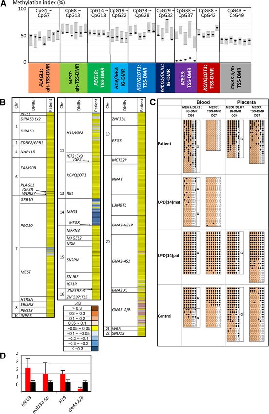
Kagami et al. Clinical Epigenetics   (2019) 11:42                                                                  Page 2 of 9

control centers in the placenta and somatic tissues, respect-      Here, we report a TS14 patient with severely hypo-
ively. Furthermore, the MEG3/DLK1:IG-DMR acts hier-              methylated MEG3:TSS-DMR and considerably methyl-
archically as an upstream regulator for the methylation          ated MEG3/DLK1:IG-DMR, together with partial MLIDs
pattern of the MEG3:TSS-DMR in somatic tissues, but not          at several DMRs, and discuss the atypical methylation
in the placenta [4]. Thus, the MEG3:TSS-DMR can stay             pattern of the MEG3/DLK1:IG-DMR.
unmethylated only in the presence of unmethylated MEG3/
DLK1:IG-DMR in somatic tissues.                                  Patient and methods
   Maternal uniparental disomy 14 (UPD(14)mat), micro-           Case report
deletions involving paternally expressed DLK1 (and               Clinical findings of this Japanese girl are summarized in
RTL1), and epimutations (hypomethylations) affecting             Table 1. She was conceived naturally to a 35-year-old
both DMRs of paternal origin cause a constellation of            father and a 38-year-old mother and was delivered by
clinical features including pre- and post-natal growth           cesarean section at 34 weeks of gestation because of
failure, muscular hypotonia, feeding difficulties, small         intrauterine growth retardation. The placenta was small
hands and feet, and precocious puberty [5]. The name             and histologically characterized by hypoplastic and
“Temple syndrome” (TS14) has been approved for such              edematous villi and chorioamnionitis (Fig. 1a). At birth,
conditions affecting the 14q32.2 imprinted region [5].           her length was 36.8 cm (− 2.9 SD), weight 1.18 kg (− 3.8
Thus, the diagnosis of TS14 is primarily based on gen-           SD), and occipitofrontal circumference (OFC) 30.7 cm
etic rather than clinical findings, although the precise         (− 0.4 SD). Apgar score was seven at 1 min and nine at
definition of TS14 has not yet been established. Import-         5 min. She was admitted to a neonatal intensive care
antly, there is no single report of an isolated epimutation      unit for 95 days, to receive tube feeding for feeding diffi-
of the MEG3/DLK1:IG-DMR or the MEG3:TSS-DMR in                   culty and resulting failure to thrive. Brain magnetic res-
somatic tissues, in agreement with the hierarchical inter-       onance imaging, auditory brainstem response, and
action between the two DMRs.                                     echocardiography showed no abnormal findings. Routine
   TS14 is associated with Silver-Russell syndrome               laboratory tests were normal, and chromosome analysis
(SRS)-compatible phenotype and Prader-Willi syndrome             revealed a 46,XX karyotype. Physical examination re-
(PWS)-like hypotonia with variable expressivity and incom-       vealed all the six Netchine-Harbison scoring features for
plete penetrance, in infancy to early childhood [6, 7]. In-      SRS (Fig. 1b) [9] and a marked hypotonia characteristic
deed, our recent study in 32 patients with molecularly           of PWS [10]. While she received growth hormone treat-
confirmed TS14 has revealed both SRS-compatible pheno-           ment for short stature born small for gestational age
type and PWS-like hypotonia in ~ 50% of patients,                from 3 years of age, she remained small with a relatively
SRS-compatible phenotype alone in ~ 20% of patients,             large OFC (Fig. 1c). On the last examination at 4 years
PWS-like hypotonia alone in ~ 20% of patients, and               and 5/12 months of age, her height was 85.0 cm (− 4.4
non-syndromic growth failure in the remaining ~ 10% of           SD), weight 7.8 kg (− 7.8 SD), and OFC 46.8 cm (− 2.0
patients in infancy to early childhood [8], when their infant-   SD). She still required tube feeding and showed obvious
ile phenotypes were assessed by the presence or absence of       developmental delay with a developmental quotient of
clinical features utilized in the Netchine-Harbison scoring      66.
system developed for the clinical diagnosis of SRS [9] and
by that of clinical features prompting genetic testing for       Ethical approval
PWS [10]. From late childhood, however, TS14 patients fre-       This study was approved by the Institutional Review
quently exhibit truncal obesity inconsistent with SRS and        Board Committees at National Center for Child Health
gonadotropin-dependent precocious puberty contrastive to         and Development and was performed after obtaining
central hypogonadotropism in PWS [8]. Thus, the overall          written informed consent.
phenotype would argue for TS14 being an independent
clinical entity.                                                 Samples and primers
   Recent studies have identified variable degrees of multilo-   Genomic DNA (gDNA) was extracted from fresh leuko-
cus imprinting disturbances (MLIDs) in a subset of patients      cytes with FlexiGene DNA Kit (Qiagen, Hilden, Germany)
with imprinting diseases (IDs) caused by epimutations [11].      and from paraffin-embedded placenta with AllPrep DNA/
Indeed, MLIDs have been detected in several patients with        RNA FFPE Kit (Qiagen). Metaphase spreads were prepared
TS14 caused by epimutations, although their clinical fea-        from lymphocytes. Total RNA was obtained from
tures remain within the phenotypic spectrum of TS14 [12].        Epstein-Barr virus-transformed lymphoblastoid cell lines
The underlying mechanism(s) leading to MLIDs is unknown          with an AllPrep DNA/RNA/miRNA Universal Kit (Qiagen),
in most patients, while mutations of causative or candidate      and cDNA samples were prepared with oligo (dT) primers
genes for MLID have been identified in a certain fraction of     using Superscript III Reverse Transcriptase (Thermo Fisher
patients and/or their mothers (for review, see [13]).            Scientific, Waltham, MA, USA) or TaqMan Advanced
Kagami et al. Clinical Epigenetics        (2019) 11:42                                                                                         Page 3 of 9

Table 1 Clinical features of this patient                                      Table 1 Clinical features of this patient (Continued)
                               This patient        TS14 patients                                               This patient       TS14 patients
                                                   (n = 32) (Ref. [8])                                                            (n = 32) (Ref. [8])
Genetic causes                 Epimutation         UPD(14)mat                  PWS salient features prompting genetic testing < 6 years
                                                   (n = 23)                      Hypotonia (with poor          +                  21/31 (68%)
                                                   Epimutation (n = 6)           suck)
                                                   Paternal deletion (n = 3)
                                                                                 Global developmental          +                  5/26 (19%)
Sex (male to female)           Female              18:14                         delay (≥ 2 years)
Karyotype                      46,XX               …                           Developmental status
Pregnancy and delivery                                                           Age at head control           18                 6.5 (3–10) (n = 25)
  Gestational age (weeks)      34                  39 (30–41) (n = 31)           (months)

  Placental weight g (%)*      195 (47)            74 (56–120) (n = 7)           Age at sitting without        24                 10 (6–15) (n = 25)
                                                                                 support (months)
  Medically assisted           –                   2/30 (7%)
  reproduction                                                                   Age at standing with          4 5/12             …
                                                                                 support (years)
  Paternal age at              35                  33 (22–48) (n = 29)
  childbirth (years)                                                             Intellectual disability       +                  2/12 (17%)

  Maternal age at              38                  30 (22–42) (n = 31)           Speech delay                  +                  …
  childbirth (years)                                                             IQ/DQ                         DQ = 66 (at 4 5/ 90 (53–114) (n = 12)
Growth                                                                                                         12 years)

  Birth length-SDS             − 2.9               − 2.1 (− 4.0 to + 1.4)        Neurological and/or           –                  5/32 (16%)
                                                   (n = 29)                      emotional problems

  Birth weight-SDS             − 3.8               − 2.7 (− 4.6 to + 3.8)      Other findings
                                                   (n = 31)                      Joint hypermobility           +                  10/30 (33%)
  Birth OFC-SDS                − 0.4               −1.2 (− 3.9 to + 1.4)         Scoliosis                     –                  6/32 (19%)
                                                   (n = 27)
                                                                                 Recurrence otitis media       –                  …
  Present age (years to        4:5                 9.3 (0.7–62) (n = 32)
  months)                                                                        Clinodactyly                  –                  11/28 (39%)
  Present height-SDS           − 4.4               − 2.3 (− 8.0 to + 0.2)      The data of the previously reported 32 patients with TS 14 are shown as the
                                                   (n = 32)                    median (range) or frequency. For frequency, the denominators indicate the
                                                                               number of patients examined for the presence or absence of each feature,
  Present weight-SDS           − 7.8               − 1.5 (− 5.7 to + 4.3)      and the numerators represent the number of patients assessed to be positive
                                                   (n = 32)                    for that feature; thus, the differences between the denominators and the
                                                                               numerators denote the number of patients evaluated to be negative for
  Present OFC-SDS              − 2.0               − 1.8 (− 4.9 to − 0.7)      that feature
                                                   (n = 13)                    OFC occipitofrontal circumference
TS14 clinical features                                                         *Assessed by the gestational age-matched placental weights [23]
                                                                               †Birth OFC-SDS ≥ 1.5 above birth length-SDS and/or birth weight-SDS
  Pre- and/or post-natal       +                   31/32 (97%)                 ‡Postnatal relative macrocephaly is found in 38% of patients
  growth failure
  Obesity                      –                   …
                                                                               miRNA Assays (Thermo Fisher Scientific). Primers
  Muscular hypotonia           +                   21/31 (66%)                 utilized in this study are listed in Additional file 1:
  Small hands                  +                   29/32 (91%)                 Table S1.
  Feeding difficulty           +                   19/30 (63%)
  Early onset of puberty       …                   13/17 (76%)                 Methylation analysis
                                                                               Methylation analysis was performed for 49 CpG sites at
SRS Netchine-Harbison          6/6                 4 (0–6) (n = 21)
scoring system criteria                                                        nine DMRs, including four CpGs at the MEG3/
  Birth length and/or          +                   26/31 (84%)
                                                                               DLK1:IG-DMR and five CpGs at the MEG3:TSS-DMR,
  weight ≤ – 2 SDS                                                             involved in the development of known IDs, by pyrose-
  Height at ~ 2 years ≤ − 2    +                   24/26 (93%)                 quencing with PyroMark Q24 (Qiagen), and for 753
  SDS                                                                          CpG sites at multiple DMRs widely distributed on the
  Relative macrocephaly        +                   14/27 (52%)‡                genome, including the MEG3:TSS-DMR but not the
  at birth†                                                                    MEG3/DLK1:IG-DMR, with HumanMethylation450
  Prominent forehead (1–       +                   19/30 (63%)                 BeadChip (Illumina, San Diego, CA, USA), using
  3 years)                                                                     bisulfite-treated leukocyte gDNA samples. The data
  Body asymmetry               +                   7/30 (23%)                  from 50 and 11 healthy subjects were utilized as refer-
  Feeding difficulties         +                   19/30 (63%)                 ences for pyrosequencing and HumanMethylation450
                                                                               BeadChip, respectively. In HumanMethylation450
Kagami et al. Clinical Epigenetics (2019) 11:42 Page 4 of 9
BeadChip analysis, each CpG site was interpreted as ab- 1.7.1 and were mapped to the hs37d5 (GRCh37) refer-
normally methylated when the |Δβ| was > 3 SD and > ence sequence using the BWA 0.7.12. PCR duplicates
0.05, and each DMR was assessed as abnormally methyl- were removed by Picard 1.83. Multi-sample calling for
ated when > 20% of CpGs within the DMR showed ab- single-nucleotide and short indel variations was per-
normal methylation levels, as employed previously [12]. formed by GATK 2.8. Common variants were excluded
These analyses were not performed for placental gDNA on the basis of 1000 genomes project data [15] and
because of a lack of reference data. Furthermore, bisul- Human Genetic Variation Database [16].
fite sequencing was carried out for six CpGs at the
MEG3/DLK1:IG-DMR and seven CpGs at the Results
MEG3:TSS-DMR using leukocyte and placental gDNA Methylation analysis
samples. Of the 49 CpGs examined by pyrosequencing, Pyrosequencing analysis showed extremely low methyla-
only four CpGs (CpG14, CpG15, and CpG18 at the tion levels of five CpGs at the MEG3:TSS-DMR and
PEG10:TSS-DMR; and CpG45 at the GNAS A/ variable but grossly normal methylation levels of four
B:TSS-DMR) were included in the list of HumanMethy- CpGs at the MEG3/DLK1:IG-DMR with a relatively
lation450 BeadChip, whereas all nine CpGs at the hypermethylated CpG and a relatively hypomethylated
MEG3/DLK1:IG-DMR and MEG3:TSS-DMR were ana- CpG, as well as normal methylation levels of most CpGs
lyzed by bisulfite sequencing. The detailed methods have at the remaining seven DMRs (Fig. 2a). The methylation
been described previously [4, 6, 14]. level of each CpG was well reproduced by four times of
independent analyses with a variation in the methylation
UPD and deletion analyses index within 2%. HumanMethylation450 BeadChip ana-
We performed microsatellite analysis for seven loci on lysis also revealed marked hypomethylation of nearly all
chromosome 14, genome-wide array comparative genomic CpGs at the MEG3:TSS-DMR accompanied by a hyper-
hybridization (aCGH) and single-nucleotide polymorphism methylated CpG at the MEG8-DMR [12], together with
(SNP) array using SurePrint G3 ISCA CGH + SNP Micro- mild hypomethylation of 30% of CpGs at the H19/
array Kit (catalog number G4890, 4 × 180 K format) (Agi- IGF2:IG-DMR and mild hypermethylation of 64% of
lent Technologies, Santa Clara, CA, USA), aCGH using a CpGs at the GNAS A/B:TSS-DMR, in addition to a few
custom-build dense oligo-microarray for chromosome of hypo- or hypermethylated CpGs at several DMRs
14q32.2–q32.3 (Design ID 032112, Agilent Technologies), (Fig. 2b) (for details, see Additional file 1: Table S2 and
and fluorescence in situ hybridization (FISH) for the its footnotes). The Beadchip analysis was performed once.
MEG3/DLK1:IG-DMR and MEG3:TSS-DMR, as reported For the H19/IGF2:IG-DMR, the CpGs around CpG19–22
previously [1, 14]. examined by pyrosequencing were shown to be normally
methylated by HumanMethylation450 BeadChip analysis;
Expression analysis for the GNAS A/B:TSS-DMR, the CpGs around CpG43–
We performed quantitative PCR analysis using TaqMan 49 examined by pyrosequencing were found to be hyper-
real-time PCR Assay. cDNA samples were subjected to methylated by HumanMethylation450 BeadChip analysis.
an ABI PRISM 7000 (Thermo Fisher Scientific) with (Additional file 1: Figure S1). Bisulfite sequencing dis-
probe-primer mixtures (catalog assay No: Hs00262142 closed markedly hypomethylated CpGs at the
for H19, Hs04188276 for IGF2, Hs00171584 for DLK1, MEG3:TSS-DMR and rather irregularly (non-differen-
Hs00292028 for MEG3, and Hs00419701 for MEG8; tially) methylated CpGs at the MEG3/DLK1:IG-DMR in
assay ID: 477901_mir for miR134-5p; and custom assay leukocytes and apparently similar methylation patterns of
ID: AJ70MG9 for GNAS-A/B) (Thermo Fisher Scien- the MEG3/DLK1:IG-DMR and MEG3:TSS-DMR between
tific). Data were normalized against GAPDH (catalog placentas of this patient and a control subject (Fig. 2c).
No: 4326317E), except for miR134-5p which were nor-
malized against miR361-5p (assay ID: 478056_mir)
(Thermo Fisher Scientific). UPD and deletion analyses
UPD (14) mat and a deletion involving the 14q32.2
Whole exome sequencing imprinted region were excluded (Additional file 1: Table
Whole-exome sequencing was carried out for the patient S3 and Additional file 1: Figure S2).
and her parents with SureSelect Human All Exon V5
(Agilent Technologies). Captured libraries were se- Expression analysis
quenced with a HiSeq 1500 (Illumina) with 101-base Quantitative PCR analyses showed increased expression of
pair (bp) paired-end reads, as reported previously [12]. MEG3 and miR134-5p regulated by the MEG3:TSS-DMR,
Reads from each sample were trimmed by removing increased expression of H19 controlled by the H19/
adapters and low-quality bases at ends using cutadapt IGF2:IG-DMR, and decreased expression of GNAS A/BKagami et al. Clinical Epigenetics (2019) 11:42 Page 5 of 9
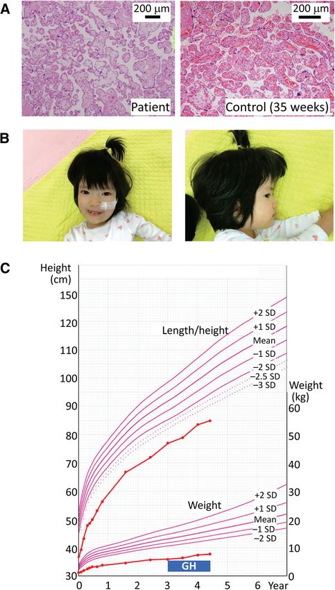
Fig. 1 Clinical findings. a Histological finding of the placenta. b Facial photos at 4 years of age. c Growth chart of this patient. She receives
growth hormone (GH) therapy (0.23 mg/kg/week) for short stature born small-for-date from 3 years of age
regulated by the GNAS A/B-DMR (Fig. 2d). IGF2, DLK1, because no cSNP was identified in MEG3, miR134-5p,
and MEG8 expression was not detected. and H19, biparental expression of these genes was not
demonstrated.
Whole exome sequencing
No pathogenic variant was identified in causative or can- Discussion
didate genes for MLID, such as ZFP57, NLRP2, NLRP7, We identified an epimutated (hypomethylated)
KHDC3L, NLRP5, TRIM28, PADI6, OOEP, UHRF1, and MEG3:TSS-DMR in leukocyte gDNA of a 4-year-old girl
ZAR1 [13], of this patient and the parents. Additionally, with typical SRS features and PWS-like marked hypotonia.Kagami et al. Clinical Epigenetics (2019) 11:42 Page 6 of 9 Fig. 2 The results of methylation and expression analyses. a Methylation indices (MIs, the ratios of methylated CpGs at each CpG site) obtained by pyrosequencing for 49 CpGs. Black circles represent the mean MIs calculated after four times of analyses in this patient, and gray vertical bars indicate the reference ranges (minimum–maximum) obtained from 50 control subjects. b Heatmap indicating the Δβ values for 753 CpG sites examined by the HumanMethylation450 BeadChip. A single row indicates a single CpG site. The methylation levels of CpG sites are classified into nine categories based on Δβ values. For the formal nomenclature of examined DMRs/loci, see Monk et al. [22]. c Bisulfite sequencing analysis for the MEG3/DLK1:IG-DMR (CG4) and MEG3:TSS-DMR (CG7). Each line indicates each clone, and filled and open circles represent methylated and unmethylated cytosines at the CpG dinucleotides, respectively. The four CpGs at the MEG3/DLK1:IG-DMR and five CpGs at the MEG3:TSS-DMR highlighted in orange have also been examined by pyrosequencing. Since the CG4 region contains a G/A SNP (rs12437020), genotyping data for this SNP are also shown; the leukocytes and placental samples are derived from different control subjects as indicated by different genotyping data. d Quantitative real-time PCR analysis using immortalized lymphocytes. Shown are relative mRNA expression levels of MEG3, H19, GNAS-A/B, and miR134-5p (mean ± SE). The expression studies were performed three times for each sample Notably, the MEG3:TSS-DMR alone was found to be se- presence of both SRS-compatible phenotype and PWS-like verely hypomethylated by the different methylation analysis phenotype, we made the diagnosis of TS14 in this patient, methods, while mildly hypo- or hypermethylated CpGs although the diagnosis of SRS with an aberrant 14q32.2 were also detected in other DMRs by HumanMethyla- imprinted region would also be acceptable at this age [8]. tion450 BeadChip analysis. On the basis of the definitive The MEG3/DLK1:IG-DMR showed confounding genetic aberration at the 14q32.2 imprinted region and the methylation patterns which were inconsistent with the
Kagami et al. Clinical Epigenetics   (2019) 11:42                                                           Page 7 of 9

markedly hypomethylated MEG3:TSS-DMR in leukocyte               MLID was identified in several DMRs of this patient
gDNA and obvious placental hypoplasia. Indeed, pyrose-       by HumanMethylation450 BeadChip analysis. The MLID
quencing indicated variable but grossly normal methyla-      may be involved in the phenotypic development of this
tion levels for the four CpGs at the MEG3/                   patient. Indeed, hypomethylation of the H19/
DLK1:IG-DMR in leukocytes, and bisulfite sequencing          IGF2:IG-DMR and hypermethylation of the GNAS A/
showed a considerable degree of irregular (non-differen-     B:TSS-DMR are frequently associated with SRS somatic
tial) methylation pattern in leukocytes and apparently       features including compromised body and placental
normal methylation pattern in the placenta. Although         growth [9, 19]. Since quantitative PCR analysis revealed
the methylation levels obtained by pyrosequencing are        not only increased expression of MEG3 and miR134-5p
known to be higher for the MEG3/DLK1:IG-DMR than             but also elevated expression of H19 and decreased ex-
for the MEG3:TSS-DMR in patients with TS14 [6, 12],          pression of GNAS A/B, it is likely that the MLID re-
such methylation patterns with grossly normal methyla-       sulted in altered expression of relevant imprinted genes,
tion levels at the MEG3/DLK1:IG-DMR have not been            contributing to the development of SRS phenotype with
reported in patients with TS14. Furthermore, variable        persistent severe growth failure unresponsive to growth
methylation levels ranging from an apparently normal         hormone treatment and postnatal relative macrocephaly
level to a severely skewed level at different DMRs of a      which is infrequent in TS14 [8]. In addition, since pa-
single imprinted region have been described only for         tients with MLIDs are frequently associated with devel-
GNAS-DMRs in patients with sporadic pseudohypopar-           opmental delay [13], MLID may also be relevant to the
athyroidism type Ib [17]. Since the grossly normal           obvious developmental delay of this patient.
methylation levels indicated by pyrosequencing were             Several matters should also be pointed out with regard
found to be due to irregular methylation rather than to      to the MLID. First, the MLID remained relatively mild
differential methylation by bisulfite sequencing, this im-   and occurred at several CpGs at each affected DMR in
plies that normal methylation levels do not necessarily      the absence of a mutation in causative or candidate
represent a differential methylation pattern.                genes for MLID. This implies that the MLID has taken
   The underlying factor(s) leading to the atypical          place as an incidental posy-zygotic event. Second, the
methylation patterns of the MEG3/DLK1:IG-DMR                 mild hypomethylation of the H19/IGF2:IG-DMR and
remains to be clarified. However, the MEG3/                  hypermethylation of the GNAS A/B:TSS-DMR were in-
DLK1:IG-DMR are more methylated than the                     dicated by HumanMethylation450 BeadChip analysis,
MEG3:TSS-DMR in leukocytes of patients with TS14,            but not by pyrosequencing analysis, as reported previ-
as described above [6, 12]. Furthermore, Kota et al.         ously [12], although HumanMethylation450 BeadChip
have reported that the unmethylated Meg3/                    analysis was performed just once. In this regard, the
Dlk1:IG-DMR of maternal origin harbors bidirection-          number of examined CpGs is much larger in Human-
ally expressed cis-acting relatively short (mostly < 500     Methylation450 BeadChip analysis than in pyrosequenc-
bp and up to 750 bp) non-coding RNAs (IG-DMR                 ing analysis, and the methylation level of each CpG is
RNA) that exerts enhancer-like functions on the Meg3         evaluated by the Δβ value in HumanMethylation450
promoter and protects the Meg3:TSS-DMR from de               BeadChip analysis and by the comparison with the nor-
novo methylation in mice [18]. Thus, it may be possible      mal range (within the normal range or not) in pyrose-
that a certain degree of irregular hypomethylation at        quencing analysis. These factors would explain why
the MEG3/DLK1:IG-DMR, as observed in this patient,           HumanMethylation450 BeadChip analysis is more
can prevent methylation of the MEG3/DLK1:IG-DMR              powerful for the detection of abnormal methylation
in leukocytes (somatic tissues) by producing a reduced       levels than pyrosequencing. Third, MLID occurred not
but functionally sufficient amount of IG-DMR RNAs.           only as a hypomethylation type but also as a hyperme-
In this case, the methylation pattern of the MEG3/           thylation type. Such MLIDs with both hypomethylated
DLK1:IG-DMR in the placenta may be explained by as-          and hypermethylated DMRs in the absence of a gene
suming that the MEG3/DLK1:IG-DMR was once hypo-              mutation have been identified in multiple patients (Add-
methylated to produce placental hypoplasia and,              itional file 1: Figure S3). Although the examined CpGs
subsequently, subjected to hypermethylation type             and the utilized methylation analysis methods are vari-
MLID in a relatively late developmental stage. However,      able among patients, the data imply that hypomethyla-
it may also be possible that methylated clones were          tion is more prevalent than hypermethylation and that
preferentially amplified because of the poor quality of      hypomethylation primarily occurs at various primary
gDNA samples extracted from the paraffin-embedded            DMRs whereas hypermethylation primarily takes place
placenta. In addition, this hypermethylation type MLID       at several specific secondary DMRs such as the ZDBF2/
might have taken place to a lesser degree in leukocytes      GPR1:IG-DMR, ZNF597:TSS-DMR, MEG8:Int2-DMR,
(somatic tissues) than in the placenta.                      GNAS-NESP:TSS-DMR, and GNAS A/B:TSS-DMR.
Kagami et al. Clinical Epigenetics        (2019) 11:42                                                                                           Page 8 of 9

Thus, it might be possible that defective methylation                          Consent for publication
maintenance of primary DMRs occurs incidentally in the                         We obtained written informed consent from the patient or the patient’s
                                                                               parents to publish patient’s clinical and molecular information as well as
post-zygotic period, followed by hypermethylation of                           facial photographs.
specific DMRs that are hypermethylated when adjacent
primary DMRs are hypomethylated, as has been re-                               Competing interests
ported for the above secondary DMRs [11, 12, 20, 21].                          The authors declare that they have no competing interests.

However, this notion remains purely speculative and
awaits further investigations.                                                 Publisher’s Note
                                                                               Springer Nature remains neutral with regard to jurisdictional claims in
                                                                               published maps and institutional affiliations.
Conclusion
                                                                               Author details
We identified a considerably methylated MEG3/                                  1
                                                                                Department of Molecular Endocrinology, National Research Institute for
DLK1:IG-DMR        and     severely    hypomethylated                          Child Health and Development, 2-10-1 Okura, Setagaya-ku, Tokyo 157-8535,
MEG3:TSS-DMR in a patient with typical TS14                                    Japan. 2Department of Pediatrics, Yaizu City Hospital, 1000 Doubara, Yaizu,
                                                                               Shizuoka 425-8505, Japan. 3Department of Pediatrics, JR Tokyo General
somatic and placental phenotype. These data will
                                                                               Hospital, 2-1-3 Yoyogi, Shibuya-ku, Tokyo 151-8528, Japan. 4Department of
help clarify the hierarchical interaction between the                          Pathology, Dokkyo Medical University, Saitama Medical Center, 2-1-50
two DMRs in somatic tissues and the biological                                 Minami-Koshigaya, Koshigaya, Saitama 343-8555, Japan. 5Department of
                                                                               Pediatrics, Hokkaido University Graduate School of Medicine, Kita 15, Nishi 7,
function of the primary DMR in the placenta.
                                                                               Kita-ku, Sapporo 060-8638, Japan. 6Department of Maternal-Fetal Biology,
                                                                               National Research Institute for Child Health and Development, 2-10-1 Okura,
                                                                               Setagaya-ku, Tokyo 157-8535, Japan. 7Department of Systems BioMedicine,
Additional file                                                                National Research Institute for Child Health and Development, 2-10-1 Okura,
                                                                               Setagaya-ku, Tokyo 157-8535, Japan. 8Department of Pediatrics, Hamamatsu
 Additional file 1: Table S1. Primers utilized in this study. Table S2.        University School of Medicine, 1-20-1 Handayama, Higashi-ku, Hamamatsu,
 Methylation levels (β-values) of each CpG site in leukocyte DNA samples.      Shizuoka 431-3192, Japan.
 Table S3. The results of micosatellite analysis. Figure S1. Methylation
 analyses of the H19/IGF2:IG-DMR, MEG3:TSS-DMR, and GNAS A/B:TSS-DMR,          Received: 27 December 2018 Accepted: 28 February 2019
 using leukocyte gDNA samples. Figure S2. Lack of UPD (14) mat and
 microdeletion in this patient. Figure S3. Representative data in patients
 with both hypomethylation-type and hypermethylation-type of MLID in           References
 the absence of a mutation in causative or candidate genes for MLID.           1. Kagami M, Sekita Y, Nishimura G, Irie M, Kato F, Okada M, et al. Deletions
 (PDF 1007 kb)                                                                     and epimutations affecting the human 14q32.2 imprinted region in
                                                                                   individuals with paternal and maternal upd (14)-like phenotypes. Nat Genet.
                                                                                   2008;40:237–42.
Abbreviations                                                                  2. Ogata T, Kagami M. Kagami-Ogata syndrome: a clinically recognizable upd
aCGH: Array comparative genomic hybridization; DMR: Differentially                 (14) pat and related disorder affecting the chromosome 14q32.2 imprinted
methylated region; FISH: Fluorescence in situ hybridization; gDNA: Genomic         region. J Hum Genet. 2016;61:87–94.
DNA; ID: Imprinting disorder; MLID: Multilocus imprinting disturbance;         3. Okae H, Chiba H, Hiura H, Hamada H, Sato A, Utsunomiya T, et al. Genome-
OFC: Occipitofrontal circumference; PWS: Prader-Willi syndrome; SNP: Single-       wide analysis of DNA methylation dynamics during early human
nucleotide polymorphism; SRS: Silver-Russell syndrome; TS14: Temple                development. PLoS Genet. 2014;10:e1004868.
syndrome; UPD(14)mat: Maternal uniparental disomy chromosome 14                4. Kagami M, O'Sullivan MJ, Green AJ, Watabe Y, Arisaka O, Masawa N, et al.
                                                                                   The IG-DMR and the MEG3-DMR at human chromosome 14q32.2:
Acknowledgements                                                                   hierarchical interaction and distinct functional properties as imprinting
We are grateful to the patient and her parents for their cooperation.              control centers. PLoS Genet. 2010;17:e1000992.
                                                                               5. Ioannides Y, Lokulo-Sodipe K, Mackay DJ, Davies JH, Temple IK. Temple
                                                                                   syndrome: improving the recognition of an underdiagnosed chromosome
Funding                                                                            14 imprinting disorder: an analysis of 51 published cases. J Med Genet.
The work was supported by grants from the Japan Agency for Medical                 2014;51:495–501.
Research and Development (JP17ek0109141, JP18ek0109373, and                    6. Kagami M, Mizuno S, Matsubara K, Nakabayashi K, Sano S, Fuke T, et al.
JP18ek0109301), National Center for Child Health and Development (28-6),           Epimutations of the IG-DMR and the MEG3-DMR at the 14q32.2 imprinted
Takeda Science Foundation, and the Japan Society for the Promotion of              region in two patients with Silver-Russell syndrome-compatible phenotype.
Science (15K15096).                                                                Eur J Hum Genet. 2015;23:1062–7.
                                                                               7. Hosoki K, Kagami M, Tanaka T, Kubota M, Kurosawa K, Kato M, et al.
Availability of data and materials                                                 Maternal uniparental disomy 14 syndrome demonstrates Prader-Willi
Not applicable.                                                                    syndrome-like phenotype. J Pediatr. 2009;155:900–3.
                                                                               8. Kagami M, Nagasaki K, Kosaki R, Horikawa R, Naiki Y, Saitoh S, et al. Temple
                                                                                   syndrome: comprehensive molecular and clinical findings in 32 Japanese
Authors’ contributions
                                                                                   patients. Genet Med. 2017;19:1356–66.
Molecular analysis was performed by MK, AN, KMtsubara, and KN. Clinical
                                                                               9. Wakeling EL, Brioude F, Lokulo-Sodipe O, O'Connell SM, Salem J, Bliek J,
follow-up was carried out by AY, MO, and TO. Placental histology was exam-
                                                                                   et al. Diagnosis and management of Silver-Russell syndrome: first
ined by KMatsuoka. The paper was written by MK and TO and reviewed and
                                                                                   international consensus statement. Nat Rev Endocrinol. 2017;13:105–24.
edited by ST and MF. All authors read and approved the final manuscript.
                                                                               10. Gunay-Aygun M, Schwartz S, Heeger S, O'Riordan MA, Cassidy SB. The
                                                                                   changing purpose of Prader-Willi syndrome clinical diagnostic criteria and
Ethics approval and consent to participate                                         proposed revised criteria. Pediatrics. 2001;108:E92.
Approval to conduct this study was obtained from the Ethical Committee of      11. Bens S, Kolarova J, Beygo J, Buiting K, Caliebe A, Eggermann T, et al.
the Institutional Review Board Committee at the National Center for Child          Phenotypic spectrum and extent of DNA methylation defects associated
Health and Development (518).                                                      with multilocus imprinting disturbances. Epigenomics. 2016;8:801–16.
Kagami et al. Clinical Epigenetics (2019) 11:42 Page 9 of 9
12. Kagami M, Matsubara K, Nakabayashi K, Nakamura A, Sano S, Okamura K,
et al. Genome-wide multilocus imprinting disturbance analysis in Temple
syndrome and Kagami-Ogata syndrome. Genet Med. 2017;19:476–82.
13. Begemann M, Rezwan FI, Beygo J, Docherty LE, Kolarova J, Schroeder C,
et al. Maternal variants in NLRP and other maternal effect proteins are
associated with multilocus imprinting disturbance in offspring. J Med Genet.
2018;55:497–504.
14. Kagami M, Kato F, Matsubara K, Sato T, Nishimura G, Ogata T. Relative
frequency of underlying genetic causes for the development of UPD (14)
pat-like phenotype. Eur J Hum Genet. 2012;20:928–32.
15. 1000 Genomes Project Consortium, Abecasis GR, Auton A, Brooks LD, MA
DP, Durbin RM, et al. An integrated map of genetic variation from 1,092
human genomes. Nature. 2012;491:56–65.
16. Higasa K, Miyake N, Yoshimura J, Okamura K, Niihori T, Saitsu H, et al.
Human genetic variation database, a reference database of genetic
variations in the Japanese population. J Hum Genet. 2016;61:547–53.
17. Maupetit-Méhouas S, Mariot V, Reynès C, Bertrand G, Feillet F, Carel JC, et al.
Quantification of the methylation at the GNAS locus identifies subtypes of
sporadic pseudohypoparathyroidism type Ib. J Med Genet. 2011;48:55–63.
18. Kota SK, Llères D, Bouschet T, Hirasawa R, Marchand A, Begon-Pescia C,
et al. ICR noncoding RNA expression controls imprinting and DNA
replication at the Dlk1-Dio3 domain. Dev Cell. 2014;31:19–33.
19. Kawashima S, Nakamura A, Inoue T, Matsubara K, Horikawa R, Wakui K, et al.
Maternal uniparental disomy for chromosome 20: physical and
endocrinological characteristics of five patients. J Clin Endocrinol Metab.
2018;103:2083–8.
20. Maeda T, Higashimoto K, Jozaki K, Yatsuki H, Nakabayashi K, Makita Y, et al.
Comprehensive and quantitative multilocus methylation analysis reveals the
susceptibility of specific imprinted differentially methylated regions to
aberrant methylation in Beckwith-Wiedemann syndrome with epimutations.
Genet Med. 2014;16:903–12.
21. Court F, Martin-Trujillo A, Romanelli V, Garin I, Iglesias-Platas I, Salafsky I,
et al. Genome-wide allelic methylation analysis reveals disease-specific
susceptibility to multiple methylation defects in imprinting syndromes. Hum
Mutat. 2013;34:595–602.
22. Monk D, Morales J, den Dunnen JT, Russo S, Court F, Prawitt D, et al.
Nomenclature group of the European Network for Human Congenital
Imprinting Disorders. Recommendations for a nomenclature system for
reporting methylation aberrations in imprinted domains. Epigenetics. 2018;
13:117–21.
23. Nakayama M. Placental pathology. Tokyo: Igaku Shoin; 2002. p. 106. (in
Japanese)You can also read