Peripheral blood neurotrophic factor levels in children with autism spectrum disorder: a meta analysis - Nature
←
→
Page content transcription
If your browser does not render page correctly, please read the page content below
www.nature.com/scientificreports
OPEN Peripheral blood neurotrophic
factor levels in children with autism
spectrum disorder: a meta‑analysis
Shu‑Han Liu, Xiao‑Jie Shi, Fang‑Cheng Fan & Yong Cheng*
Increasing evidence suggests that abnormal regulation of neurotrophic factors is involved in the
etiology and pathogenesis of Autism Spectrum Disorder (ASD). However, clinical data on neurotrophic
factor levels in children with ASD were inconsistent. Therefore, we performed a systematic review
of peripheral blood neurotrophic factors levels in children with ASD, and quantitatively summarized
the clinical data of peripheral blood neurotrophic factors in ASD children and healthy controls. A
systematic search of PubMed and Web of Science identified 31 studies with 2627 ASD children and
4418 healthy controls to be included in the meta-analysis. The results of random effect meta-analysis
showed that the peripheral blood levels of brain-derived neurotrophic factor (Hedges’ g = 0.302;
95% CI = 0.014 to 0.591; P = 0.040) , nerve growth factor (Hedges’ g = 0.395; 95% CI = 0.104 to 0.686;
P = 0.008) and vascular endothelial growth factor (VEGF) (Hedges’ g = 0.097; 95% CI = 0.018 to 0.175;
P = 0.016) in children with ASD were significantly higher than that of healthy controls, whereas blood
neurotrophin-3 (Hedges’ g = − 0.795; 95% CI = − 1.723 to 0.134; P = 0.093) and neurotrophin-4 (Hedges’
g = 0.182; 95% CI = − 0.285 to 0.650; P = 0.445) levels did not show significant differences between
cases and controls. Taken together, these results clarified circulating neurotrophic factor profile in
children with ASD, strengthening clinical evidence of neurotrophic factor aberrations in children with
ASD.
Autism Spectrum Disorder (ASD) refer to a group of neurodevelopmental disorders characterized primarily by
restrictive, repetitive patterns of behaviors, loof of interest or activity, and social communication impairment.
The disease includes autistic disorder, Asperger’s syndrome and pervasive developmental disorder-not otherwise
specified1,2. According to the Autism and Developmental Disabilities Monitoring Network, the number of chil-
dren diagnosed with ASD has increased by 150% since 2000, with the overall prevalence of ASD estimated at 16.8
per 1000 children aged 8 years in 20143. Twin studies have shown that the concordance of ASD in monozygotic
twins is higher than that in dizygotic twins, suggesting that ASD is highly h eritable4. However, the coincidence
rate between identical twins and autism and related diseases was less than 100%, indicating that environmental
factors also play a role in A SD5.
The neurotrophins are a family of proteins that have been shown to play an important role in the central and
peripheral nervous system, which control a number of aspects of survival, development, and function of n eurons6.
The so-called “classic” neurotrophin family includes nerve growth factor (NGF), brain-derived neurotrophic
factor (BDNF), neurotrophin-3 (NT-3) and neurotrophin-4 (NT-4)7. Recently, research findings from analyses
of postmortem and peripheral tissue and molecular genetic studies led to the hypothesis that neurotrophins—
as crucial regulators of neuroplasticity—impacting on the pathophysiologic features of A SD8. In a trios-based
association study, Nishimura et al. showed that the BDNF SNP haplotype combinations significantly associated
with ASD9. Moreover, Postmortem studies found elevated BDNF protein levels in the fusiform gyrus t issue10 and
elevated NT-3 protein levels in the cerebellum11 from patients with ASD. However, molecular genetic studies and
postmortem studies on neurotrophic factors were scarce, and therefore it is difficult to evaluate the robustness
of the associations of neurotrophins with ASD.
Increasing number of clinical studies measured peripheral blood levels of neurotrophic factors in children
with ASD, due to the easy accessibility of blood and “periphery as a window to the brain” hypothesis12. However,
clinical data on neurotrophic factor levels in children with ASD have yielded inconsistent results. One study
showed that the serum BDNF levels of children with ASD were significantly higher than that of the control
subjects13, whereas Makkonen et al.14 suggested that was no significant difference in serum BDNF concentrations
Center On Translational Neuroscience, College of Life and Environmental Sciences, Minzu University of China,
27 South Zhongguancun Avenue, Zhongguancun South St, Haidian District, Beijing 100081, China. * email:
yongcheng@muc.edu.cn
Scientific Reports | (2021) 11:15 | https://doi.org/10.1038/s41598-020-79080-w 1
Vol.:(0123456789)
www.nature.com/scientificreports/
Figure 1. PRISMA flowchart of the literature search.
between cases and controls, and another study showed that BDNF serum levels were significantly decreased in
ASD children when compared with controls15. For VEGF, results from Emanuele et al. showed that VEGF levels
in patients with ASD were lower than that of healthy controls16. In contrast, one study indicated that VEGF levels
did not show significantly difference between children with ASD and healthy controls17. Moreover, studies have
demonstrated inconsistent data for IGF-1 and IGF-2 levels comparing ASD children and healthy controls18,19.
Given the inconsistent findings on neurotrophic factors in children with ASD, a meta-analysis on this subject
is necessary.
To clarify neurotrophic factor profile in children with ASD, here we undertook a meta-analysis of studies
measuring neurotrophic factor levels in blood of children with ASD and healthy controls.
Results
The initial search generated a total of 1217 records: 545 were searched from PubMed database, 670 from Web
of Science and 2 additional records identified from the reference lists of relevant studies. After scanning the
titles and abstracts, 62 articles relevant to present subject were identified for full-text scrutiny. Several studies
were excluded as they did not have necessary data (11 studies)20–30; lack of healthy controls (6 studies)31–36; sam-
ple source is not peripheral blood (5 studies)18,37–40; samples derived from postmortem brain (3 studies)10,11,41;
participants were adults (3 study)16,42,43; had patient samples that overlapped with another studies (1 study)44;
neurotrophic factors were studied in less than 3 articles (1 study)19 and non-English publication (1 studies)45.
Therefore, a total of 31 studies met the criteria were included for this meta-analysis (Fig. 1)8,12,13,15,17,46–71. Demo-
graphic and clinical profile of the included studies were presented in Supplementary Table S1.
Main association of peripheral blood neurotrophic factor levels with ASD in children. We first
compared the peripheral blood BDNF levels between 2380 children with ASD and 4191 healthy controls extracted
from 27 studies. Random-effects meta-analysis showed that ASD children had significantly increased levels of
Scientific Reports | (2021) 11:15 | https://doi.org/10.1038/s41598-020-79080-w 2
Vol:.(1234567890)www.nature.com/scientificreports/
Figure 2. Forest plot for random-effects meta-analysis for BDNF. Forest plot for random-effects meta-analysis
on differences in blood BDNF concentrations between children with autism spectrum disorder (ASD) and
healthy controls. The sizes of the squares are proportional to study weight. Diamond marker indicates pooled
effect size. CI, confidence interval.
BDNF compared with healthy controls (Fig. 2, Hedges’ g = 0.302; 95% CI = 0.014 to 0.591; P = 0.040). Then, we
compared blood NGF levels between 100 children with ASD and 84 healthy controls extracted from 3 studies,
and the results showed that compared with healthy controls, children with ASD had significantly increased NGF
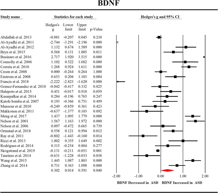
levels (Fig. 3A, Hedges’ g = 0.395; 95% CI = 0.104 to 0.686; P = 0.008). In addition, we compared the blood VEGF
levels of 844 children with ASD and 2460 healthy controls extracted from 3 studies, which showed significantly
increased VEGF levels in ASD children when compared with healthy controls (Fig. 3B, Hedges’ g = 0.097; 95%
CI = 0.018 to 0.175; P = 0.016). However, we did not observe significant differences between children with ASD
and healthy controls for peripheral blood NT-3 (Hedges’ g = − 0.795; 95% CI = − 1.723 to 0.134; P = 0.093) and
NT-4 (Hedges’ g = 0.182; 95% CI = − 0.285 to 0.650; P = 0.445) levels (Fig. 3C,D).
Investigation of heterogeneity. Of the three neurotrophic factors that were significantly associated with
ASD, NGF and VEGF did show no significant between-study heterogeneity, whereas BDNF showed high levels
of heterogeneity. Therefore, we next performed subgroup analyses to investigate potential source of the high
levels of heterogeneity for studies measuring BDNF concentrations.
The sources of heterogeneity include category variables (sample source, assay type and medication status)
and continuous variables (sample size, age, sex, publication year, course of disease and disease severity). Due
to a substantial lack of information regarding medication status, course of disease and disease severity, we only
Scientific Reports | (2021) 11:15 | https://doi.org/10.1038/s41598-020-79080-w 3
Vol.:(0123456789)www.nature.com/scientificreports/
Figure 3. Forest plot for random-effects meta-analysis for NGF, VEGF, NT-3 and NT-4. Forest plot for random-
effects meta-analysis on differences in blood NGF (A), VEGF (B), NT-3 (C) and NT-4 (D) concentrations
between children with autism spectrum disorder (ASD) and healthy controls. The sizes of the squares are
proportional to study weight. Diamond marker indicates pooled effect size. CI, confidence interval.
Main effect Heterogeneity Publication bias
No. with ASD/
TF No. of studies controls Hedges g (95% CI) Z score P value Q statistic df P value I2 Statistic Egger intercept P value
BDNF 27 2380/4191 0.302 (0.014 to 0.591) 2.054 0.040 537.149 26 0.000 95.160 2.13562 0.11851
NGF 3 100/84 0.395 (0.104 to 0.686) 2.662 0.008 0.909 2 0.635 0.000 1.13092 0.71027
NT-3 4 140/114 − 0.795 (− 1.723 to 0.134) − 1.678 0.093 33.086 3 0.000 90.933 − 7.11871 0.26344
NT-4 4 485/838 0.182 (− 0.285 to 0.650) 0.764 0.445 18.275 3 0.000 83.585 1.37770 0.61376
VEGF 3 844/2460 0.097 (0.018 to 0.175) 2.414 0.016 0.611 2 0.737 0.000 − 0.59890 0.23760
Table 1. Summary of comparative outcomes for measurements of neurotrophic factor levels. df, degrees of
freedom; ASD, Autism Spectrum Disorder; BDNF, Brain-Derived Neurotrophic Factor; NGF, Nerve Growth
Factor; NT-3, Neurotrophin-3; NT-4, Neurotrophin-4; VEGF, Vascular Endothelial Growth Factor; NTF,
Neurotrophic Factor.
performed subgroup analyses on sample sources (serum and plasma) and assay types (ELISA and no ELISA), as
well as meta-regression analyses based on sample size, age, sex and publication year.
Subgroup analyses suggested that sampling source did not address the between-study heterogeneity, and
we still observed significant heterogeneity for studies plasma (Q = 43.966; I 2 = 88.628; P < 0.001) and serum
(Q = 344.797; I2 = 95.650; P < 0.001) BDNF levels (see Supplementary Fig. S1). Additionally, subgroup analyses
stratified by assay type showed increased BDNF levels in ASD children when compared with controls in ELISA
group (20 studies, Hedges’ g = 0.404; 95% CI = 0.024 to 0.785; P = 0.037), but not in non-ELISA group (7 studies,
Hedges’ g = 0.015; 95% CI = − 0.618 to 0.647; P = 0.964). Again, we observed high levels of heterogeneity among
studies for ELISA group (Q = 413.794; I2 = 95.408; P < 0.001) and non-ELISA group (Q = 119.886; I2 = 94.995;
P < 0.001) (see Supplementary Fig. S2).
Meta-regression analyses showed that sample size, age, gender and publication year had no moderating effects
on the outcome of the meta-analysis (P > 0.05 in all analyses) (see Supplementary Fig. S3).
Visual inspection of funnel plots suggested no risk of publication bias for studies analyzing BDNF, NGF and
VEGF levels, and these were confirmed by the results of the Egger’s test (see Supplementary Fig. S4 and Table 1).
Discussion
In this study, we performed a comprehensive investigation on the changes of peripheral neurotrophic factors in
children with ASD. We included 31 studies with 2486 ASD children and 4303 healthy controls measuring five
neurotrophic factors, and reported the levels of BDNF, NGF and VEGF were elevated in children with ASD.
However, no significant associations were found between NT-3 or NT-4 and ASD. In addition, we found high
levels of between-study heterogeneity for BDNF, whereas NGF and VEGF did not show between-study heteroge-
neities. We further performed subgroup analysis based on the sample source and assay type, and meta-regression
Scientific Reports | (2021) 11:15 | https://doi.org/10.1038/s41598-020-79080-w 4
Vol:.(1234567890)www.nature.com/scientificreports/
analysis based on sample size, age, gender and publication year for studies measuring BDNF levels. However, we
did not find potential sources of heterogeneity. This is consistent with a previous meta-analysis on blood BDNF
levels in ASD children, which included a relatively small number of studies published in 201672. Although studies
from the literature in the past decade has produced inconsistent results, and the role of neurotrophic factors in
children with ASD is still unclear, this study provides strong clinical evidence that the levels of BDNF, NGF and
VEGF in peripheral blood of children with ASD are higher than those in healthy controls, strengthening the
clinical evidence that neurotrophic factor plays a critical role in ASD onset and/or development.
Despite the largely unknown etiology and pathogenesis of ASD, researches have consistently demonstrated
that children with ASD are accompanied by abnormal brain development73,74. Courchesne et al. investigated
the neural basis of brain overgrowth in ASD children at the cellular level and found abnormal increases in the
number of neurons in the prefrontal cortex of ASD boys75. Additionally, it has been suggested that that the
brain volume overgrowth in early ASD children is mainly due to the increased proliferation of neural progeni-
tor cells76. Neurotrophic factors play a positive role on the proliferation of embryonic neural progenitor c ells27,
and an in vivo study showed that BDNF increased neurogenesis in the granule cell layer of hippocampus in
rats77. Importantly, BDNF levels are temporally regulated during development, which has been suggested to be
required for proper neuronal development and f unctions78. Therefore, it is very likely that in the early stage of
ASD children, abnormal regulation of BDNF leads to the subsequent long-term changes in the brain structure
and function. Furthermore, it has been reported the excessive brain growth in ASD children occur prior to
most clinical manifestations of the d isease79, raising the possibility that the observed BDNF aberrations in ASD
children found in the meta-analysis contributed to the excessive growth in brains of early ASD children.
We noted that levels of two other neurotrophic factors, NGF and VEGF, also increased compared with
healthy controls. VEGF is a key signaling molecule of the central nervous system which is involved in neu-
roprotection, neuronal survival and axonal g rowth80. Similarly, NGF is also involved in important aspects of
nerve cell growth, differentiation, survival, and regeneration, and thought may represent a serological marker
for autistic c hildren23,81. It is possible that that the structural changes in the brains of children with ASD may
also be contributed by the abnormalities of NGF and VEGF. Therefore, targeting the neurotrophic factor system
may provide a novel strategy for the potential treatment of ASD, and future studies are needed to validate the
hypothesis. However, since the studies included in this meta-analysis analyzed NGF levels by ELISA method,
and the commercially available ELISA kits could not differentiate between pro and mature forms of NGF, it is
unclear whether the levels of the mature form of NGF (the form with neurotrophic activity) were up-regulated in
ASD children. Thus, another explanation for the observed NGF aberrations in ASD children is the compromise
of proNGF to mature NGF conversion in the disease, which requires further investigations.
Abnormalities of neurotrophic factors were also thought to be associated with other neurological diseases,
such as Alzheimer’s disease and schizophrenia. Previous meta-analyses have demonstrated that blood BDNF
levels were significantly decreased in patients with Alzheimer’s disease, whereas blood NGF and VEGF levels did
not show significant differences between patients with Alzheimer’s disease and control subjects82. Additionally,
meta-analyses showed that blood BDNF and NGF levels were significantly decreased in schizophrenia patients
when compared with controls83,84. In contrast, blood VEGF levels were found not to be significantly different
between first-episode schizophrenia patients and controls, whereas medicated multiple-episode schizophrenia
had higher levels of VEGF than that of control subjects85. It is considered that ASD shares many molecular
pathways with other neuropsychiatric diseases including schizophrenia. However, the present meta-analysis
revealed heightened blood BDNF, NGF and VEGF levels in ASD children, it is very likely that patients with
ASD have a unique neurotrophic factor profile comparing with other neuropsychiatric diseases, and this may
partially explain the pathogenesis of ASD.
Although no significant between-study heterogeneity was found in the analysis of blood NGF and VEGF
levels, one explanation for the low heterogeneity is that relatively few studies have been included. Additionally,
we found high levels of heterogeneity among studies analyzing BDNF levels. Here we used subgroup and meta-
regression analyses to adjust confounders that could explain the between-study heterogeneity. The potential
moderators that we have analyzed including sampling source, assay type, sample size, publication year, age and
gender did not address the heterogeneity. Obviously, the unexplained heterogeneity may due to other variables
that we have not analyzed, such as medication status, disease severity and life style. However, the limited infor-
mation on these variables in the included studies prevented us from analyzing whether the potential confound-
ers had moderating effects on the outcome of the meta-analysis, therefore highlighting the need to control the
variables in future studies.
Despite this work provides strong clinical evidence of the increased blood neurotrophic factor profile in
children with ASD, there are still some limitations in this study. Firstly, the meta-analysis of peripheral blood
neurotrophic factor levels in children with ASD and healthy controls produced a summary of results mainly from
cross-sectional studies. Therefore, it is unclear whether the abnormal levels of neurotrophic factors are the cause
or consequence for the development of ASD. Secondly, this meta-analysis analyzed blood neurotrophic factors
levels in ASD children, but the neurotrophic factor profile in the brains of ASD children is largely unknown.
The third limitation of the meta-analysis is that the effect size is very small, and the number of participants is
very small except for BDNF and VEGF. In addition, a small number of studies evaluated NT-3 and NT-4, which
may make it difficult to observe significant associations between the two neurotrophins and ASD. It should be
noted that our meta-analysis showed a trend of decreased blood NT-3 levels in ASD children when compared
with controls (P = 0.093). It is likely that we would observe a significant association between NT-3 and ASD with
increased number of studies and sample size from future studies. Lastly, our study included only English articles,
which may lead to publication bias. However, considering that we have excluded only one non-English articles,
this is unlikely to have a significantly impact on the outcome of our meta-analysis.
Scientific Reports | (2021) 11:15 | https://doi.org/10.1038/s41598-020-79080-w 5
Vol.:(0123456789)www.nature.com/scientificreports/
In conclusion, the results of our meta-analysis showed the elevated peripheral blood BDNF, NGF and VEGF
concentrations as a manifestation of children with ASD, strengthening the clinical evidence of an abnormal
neurotrophic factor profile in children with ASD. Thus, future investigators into neurotrophic factors as potential
therapeutic targets for the treatment of ASD are warranted.
Method
This was an exploratory meta-analysis, and adhered to the guidelines that are recommended by the PRISMA
statement (Preferred Reporting Items for Systematic reviews and Meta-Analysis)86.
Search strategy and study selection. We have conducted a systematic search of peer-reviewed
English articles using PubMed and Web of Science databases up to December 23, 2019. Our search strategy
was:(neurotrophin OR neurotrophic factor OR brain-derived neurotrophic factor OR BDNF OR nerve growth
factor OR NGF OR neurotrophin-3 OR NT-3 OR neurotrophin-4 OR NT-4 OR glial cell-derived neurotrophic
factor OR GDNF OR insulin-like growth factor OR IGF OR vascular endothelial growth factor OR VEGF) AND
(Autism), without year limitation. Additionally, we checked the reference list of relevant studies.
Original articles were screened according to the title and abstract, and then were scrutinized based on the
following criteria: (1) measured peripheral blood neurotrophic factors; (2) neurotrophic factor that were avail-
able in three or more studies; (3) studies which provide the neurotrophic factor concentrations and standard
deviation, or sample size and P value; (4) compared with matched healthy controls; (5) studies were excluded
if the samples were from adult ASD patients, this is because neurotrophic factor levels were altered in adult68,
which could have a confounding effect.
Data extraction. Data from each included study was extracted by one investigator, and was verified by
another investigator. Any inconsistencies were settled by discussions. Sample sizes, mean neurotrophic factor
concentrations, standard deviation and P values were extracted as primary outcomes to generate effective size
(ES). For studies that did not report neurotrophic factor concentrations, the sample size and P values were used
to calculate the effect size87. Data on author last name, publication year, country of region, age, gender, sample
source (serum or plasma sample or dried blood spot), diagnosis and assay type were also extracted.
Statistical analysis. All statistical analyses were performed by comprehensive Meta-Analysis version 2
software. The ES was produced by sample size, mean concentration and standard deviation (SD), or by sample
size and P value if the data of mean concentration were not available. The standardized mean difference of
neurotrophic factor levels between children with ASD and healthy controls was calculated as ES, and converted
into Hedge’s g statistic, which provides an unbiased adjusted ES for sample size88. We calculated ES estimates
by evaluating each neurotrophic factor. And we used a random effects model in this meta-analysis, because if
between-study heterogeneity is significant, the random effects model can produce a wider 95% confidence inter-
val than the fixed effect model89.
We used the Cochrane Q test and I 2 statistics to assess the heterogeneity among studies. P < 0.10 was con-
sidered statistically significant. The inconsistent levels among studies was decided by the I2 index to reflect the
impact of heterogeneity, and an I 2 index of 0.25, 0.50, 0.75 indicated low, moderate and high levels of heteroge-
neity, respectively90. We used subgroup analysis and unrestricted maximum-likelihood random-effects meta-
regressions of ES to evaluate whether theoretically related covariates influence the outcome of meta-analysis.
Publication bias was assessed via visual inspection of funnel plots, and Egger’s test was used to estimate the
statistical significance.
We set all the statistical significances at P < 0.05 in this study except for where noted.
Received: 6 April 2020; Accepted: 25 November 2020
References
1. Li, D., Karnath, H. O. & Xu, X. Candidate biomarkers in children with autism spectrum disorder: a review of MRI studies. Neurosci.
Bull. 33, 219–237. https://doi.org/10.1007/s12264-017-0118-1 (2017).
2. Sharma, S. R., Gonda, X. & Tarazi, F. I. Autism Spectrum Disorder: Classification, diagnosis and therapy. Pharmacol. Ther. 190,
91–104. https://doi.org/10.1016/j.pharmthera.2018.05.007 (2018).
3. Baio, J. et al. Prevalence of Autism Spectrum Disorder among children aged 8 years—autism and developmental disabilities moni-
toring network, 11 sites, United States, 2014. MMWR Surveill. Summ. 67, 1–23. https://doi.org/10.15585/mmwr.ss6706a1 (2018).
4. Sahin, M. & Sur, M. Genes, circuits, and precision therapies for autism and related neurodevelopmental disorders. Science https
://doi.org/10.1126/science.aab3897 (2015).
5. Matelski, L. & Van de Water, J. Risk factors in autism: thinking outside the brain. J. Autoimmun. 67, 1–7. https://doi.org/10.1016/j.
jaut.2015.11.003 (2016).
6. Skaper, S. D. Neurotrophic factors: an overview. Methods Mol. Biol. 1727, 1–17. https://doi.org/10.1007/978-1-4939-7571-6_1
(2018).
7. Keefe, K. M., Sheikh, I. S. & Smith, G. M. Targeting neurotrophins to specific populations of neurons: NGF, BDNF, and NT-3 and
their relevance for treatment of spinal cord injury. Int. J. Mol. Sci. https://doi.org/10.3390/ijms18030548 (2017).
8. Wang, M. et al. Increased serum levels of brain-derived neurotrophic factor in autism spectrum disorder. NeuroReport 26, 638–641.
https://doi.org/10.1097/wnr.0000000000000404 (2015).
9. Nishimura, K. et al. Genetic analyses of the brain-derived neurotrophic factor (BDNF) gene in autism. Biochem. Biophys. Res.
Commun. 356, 200–206. https://doi.org/10.1016/j.bbrc.2007.02.135 (2007).
Scientific Reports | (2021) 11:15 | https://doi.org/10.1038/s41598-020-79080-w 6
Vol:.(1234567890)www.nature.com/scientificreports/
10. Garcia, K. L. et al. Altered balance of proteolytic isoforms of pro-brain-derived neurotrophic factor in autism. J. Neuropathol. Exp.
Neurol. 71, 289–297. https://doi.org/10.1097/NEN.0b013e31824b27e4 (2012).
11. Sajdel-Sulkowska, E. M., Xu, M. & Koibuchi, N. Increase in cerebellar neurotrophin-3 and oxidative stress markers in autism.
Cerebellum 8, 366–372. https://doi.org/10.1007/s12311-009-0105-9 (2009).
12. Zhang, Q. B., Jiang, L. F., Kong, L. Y. & Lu, Y. J. Serum Brain-derived neurotrophic factor levels in Chinese children with autism
spectrum disorders: a pilot study. Int. J. Dev. Neurosci. 37, 65–68. https://doi.org/10.1016/j.ijdevneu.2014.06.013 (2014).
13. Meng, W. D. et al. Elevated serum brain-derived neurotrophic factor (BDNF) but not BDNF gene val66met polymorphism is
associated with autism spectrum disorders. Mol. Neurobiol. 54, 1167–1172. https://doi.org/10.1007/s12035-016-9721-9 (2017).
14. Makkonen, I. et al. Brain derived neurotrophic factor and serotonin transporter binding as markers of clinical response to fluoxetine
therapy in children with autism. J. Pediatr. Neurol. 9, 1–8. https://doi.org/10.3233/JPN-2010-0446 (2011).
15. Francis, K. et al. Brain-derived neurotrophic factor (BDNF) in children with ASD and their parents: a 3-year follow-up. Acta
Psychiatr. Scand. 137, 433–441. https://doi.org/10.1111/acps.12872 (2018).
16. Emanuele, E. et al. Serum levels of vascular endothelial growth factor and its receptors in patients with severe autism. Clin. Biochem.
43, 317–319. https://doi.org/10.1016/j.clinbiochem.2009.10.005 (2010).
17. Kajizuka, M. et al. Serum levels of platelet-derived growth factor BB homodimers are increased in male children with autism. Prog.
Neuropsychopharmacol. Biol. Psychiatry 34, 154–158. https://doi.org/10.1016/j.pnpbp.2009.10.017 (2010).
18. Riikonen, R. et al. Cerebrospinal fluid insulin-like growth factors IGF-1 and IGF-2 in infantile autism. Dev. Med. Child Neurol.
48, 751–755. https://doi.org/10.1017/S0012162206001605 (2006).
19. Mills, J. L. et al. Elevated levels of growth-related hormones in autism and autism spectrum disorder. Clin. Endocrinol. (Oxf.) 67,
230–237. https://doi.org/10.1111/j.1365-2265.2007.02868.x (2007).
20. Battaglia, A. Sensory impairment in mental retardation: a potential role for NGF. Arch. Ital. Biol. 149, 193–203. https://doi.
org/10.4449/aib.v149i2.1362 (2011).
21. Bou Khalil, R. Is insulin growth factor-1 the future for treating autism spectrum disorder and/or schizophrenia?. Med. Hypotheses
99, 23–25. https://doi.org/10.1016/j.mehy.2016.12.004 (2017).
22. Das, U. N. Nutritional factors in the pathobiology of autism. Nutrition 29, 1066–1069. https://doi.org/10.1016/j.nut.2012.11.013
(2013).
23. Galvez-Contreras, A. Y., Campos-Ordonez, T., Gonzalez-Castaneda, R. E. & Gonzalez-Perez, O. Alterations of growth factors in
autism and attention-deficit/hyperactivity disorder. Front. Psychiatry 8, 126. https://doi.org/10.3389/fpsyt.2017.00126 (2017).
24. Koh, J. Y., Lim, J. S., Byun, H. R. & Yoo, M. H. Abnormalities in the zinc–metalloprotease–BDNF axis may contribute to mega-
lencephaly and cortical hyperconnectivity in young autism spectrum disorder patients. Mol. Brain 7, 64. https://doi.org/10.1186/
s13041-014-0064-z (2014).
25. Korzeniewski, S. J. et al. Elevated protein concentrations in newborn blood and the risks of autism spectrum disorder, and of
social impairment, at age 10 years among infants born before the 28th week of gestation. Transl. Psychiatry 8, 115. https://doi.
org/10.1038/s41398-018-0156-0 (2018).
26. Li, L. Y., Jiang, N. & Zhao, Y. Could acupuncture have a role in the treatment of autism spectrum disorder via modulation of BDNF
expression and activation?. Acupunct. Med. 32, 503–505. https://doi.org/10.1136/acupmed-2014-010602 (2014).
27. Numakawa, T., Odaka, H. & Adachi, N. Actions of Brain-derived neurotrophic factor and glucocorticoid stress in neurogenesis.
Int. J. Mol. Sci. https://doi.org/10.3390/ijms18112312 (2017).
28. Steinman, G. IGF—autism prevention/amelioration. Med. Hypotheses 122, 45–47. https://doi.org/10.1016/j.mehy.2018.10.015
(2019).
29. Tsai, S. J. Is autism caused by early hyperactivity of brain-derived neurotrophic factor?. Med. Hypotheses 65, 79–82. https://doi.
org/10.1016/j.mehy.2005.01.034 (2005).
30. Steinman, G. & Mankuta, D. Insulin-like growth factor and the etiology of autism. Med. Hypotheses 80, 475–480. https://doi.
org/10.1016/j.mehy.2013.01.010 (2013).
31. Al-Ayadhi, L., El-Ansary, A., Bjorklund, G., Chirumbolo, S. & Mostafa, G. A. Impact of Auditory Integration Therapy (AIT) on
the plasma levels of human glial cell line-derived neurotrophic factor (GDNF) in Autism Spectrum Disorder. J. Mol. Neurosci. 68,
688–695. https://doi.org/10.1007/s12031-019-01332-w (2019).
32. Moradi, H., Sohrabi, M., Taheri, H., Khodashenas, E. & Movahedi, A. The effects of different combinations of perceptual-motor
exercises, music, and vitamin D supplementation on the nerve growth factor in children with high-functioning autism. Comple-
ment. Ther. Clin. Pract. 31, 139–145. https://doi.org/10.1016/j.ctcp.2018.02.005 (2018).
33. Riikonen, R. & Vanhala, R. Levels of cerebrospinal fluid nerve-growth factor differ in infantile autism and Rett syndrome. Dev.
Med. Child. Neurol. 41, 148–152. https://doi.org/10.1017/s0012162299000328 (1999).
34. Skogstrand, K. et al. Simultaneous measurement of 25 inflammatory markers and neurotrophins in neonatal dried blood spots by
immunoassay with xMAP technology. Clin. Chem. 51, 1854–1866. https://doi.org/10.1373/clinchem.2005.052241 (2005).
35. Spratt, E. G. et al. Pilot study and review: physiological differences in BDNF, a potential biomarker in males and females with
autistic disorder. Int. Neuropsychiatr. Dis. J. 3, 19–26. https://doi.org/10.9734/INDJ/2015/12118 (2015).
36. Sukasem, C. et al. Pharmacogenetics of risperidone-induced insulin resistance in children and adolescents with autism spectrum
disorder. Basic Clin. Pharmacol. Toxicol. 123, 42–50. https://doi.org/10.1111/bcpt.12970 (2018).
37. Anlar, B. et al. Urinary epidermal and insulin-like growth factor excretion in autistic children. Neuropediatrics 38, 151–153. https
://doi.org/10.1055/s-2007-990282 (2007).
38. Li, Q. et al. Transplantation of umbilical cord blood mononuclear cells increases levels of nerve growth factor in the cerebrospinal
fluid of patients with autism. Genet. Mol. Res. 14, 8725–8732. https://doi.org/10.4238/2015.July.31.21 (2015).
39. Makkonen, I., Kokki, H., Kuikka, J., Turpeinen, U. & Riikonen, R. Effects of fluoxetine treatment on striatal dopamine transporter
binding and cerebrospinal fluid insulin-like growth factor-1 in children with autism. Neuropediatrics 42, 207–209. https://doi.
org/10.1055/s-0031-1291242 (2011).
40. Vanhala, R., Turpeinen, U. & Riikonen, R. Low levels of insulin-like growth factor-I in cerebrospinal fluid in children with autism.
Dev. Med. Child. Neurol. 43, 614–616. https://doi.org/10.1017/s0012162201001116 (2001).
41. Sheikh, A. M. et al. BDNF-Akt-Bcl2 antiapoptotic signaling pathway is compromised in the brain of autistic subjects. J. Neurosci.
Res. 88, 2641–2647. https://doi.org/10.1002/jnr.22416 (2010).
42. Hashimoto, K. et al. Reduced serum levels of brain-derived neurotrophic factor in adult male patients with autism. Prog. Neu-
ropsychopharmacol. Biol. Psychiatry 30, 1529–1531. https://doi.org/10.1016/j.pnpbp.2006.06.018 (2006).
43. Miyazaki, K. et al. Serum neurotrophin concentrations in autism and mental retardation: a pilot study. Brain Dev. 26, 292–295.
https://doi.org/10.1016/S0387-7604(03)00168-2 (2004).
44. Abdallah, M. W. et al. Amniotic fluid MMP-9 and neurotrophins in autism spectrum disorders: an exploratory study. Autism Res.
5, 428–433. https://doi.org/10.1002/aur.1254 (2012).
45. Tsukurova, L. A. A neuroprotective approach to optimizing treatment and correction activities in children with autism spectrum
disorders. Zh. Nevrol. Psikhiatr. Im. S S Korsakova 118, 51–56. https://doi.org/10.17116/jnevro20181185251 (2018).
46. Skogstrand, K. et al. Reduced neonatal brain-derived neurotrophic factor is associated with autism spectrum disorders. Transl.
Psychiatry 9, 252. https://doi.org/10.1038/s41398-019-0587-2 (2019).
Scientific Reports | (2021) 11:15 | https://doi.org/10.1038/s41598-020-79080-w 7
Vol.:(0123456789)www.nature.com/scientificreports/
47. Ormstad, H. et al. Serum tryptophan, tryptophan catabolites and brain-derived neurotrophic factor in subgroups of youngsters
with autism spectrum disorders. CNS Neurol. Disord. Drug Targets 17, 626–639. https://doi.org/10.2174/18715273176661807201
63221(2018).
48. Gomez-Fernandez, A. et al. Children with autism spectrum disorder with regression exhibit a different profile in plasma
cytokines and adhesion molecules compared to children without such regression. Front. Pediatr. 6, 264. https://doi.org/10.3389/
fped.2018.00264(2018).
49. Businaro, R. et al. Interleukin-18 modulation in autism spectrum disorders. J. Neuroinflam. 13, 2. https://doi.org/10.1186/s1297
4-015-0466-6 (2016).
50. Pecorelli, A. et al. Cytokines profile and peripheral blood mononuclear cells morphology in Rett and autistic patients. Cytokine
77, 180–188. https://doi.org/10.1016/j.cyto.2015.10.002 (2016).
51. Bryn, V. et al. Brain derived neurotrophic factor (BDNF) and autism spectrum disorders (ASD) in childhood. Eur. J. Paediatr.
Neurol. 19, 411–414. https://doi.org/10.1016/j.ejpn.2015.03.005 (2015).
52. Halepoto, D. M., Bashir, S., Zeina, R. & Al-Ayadhi, L. Y. Correlation Between Hedgehog (Hh) protein family and brain-derived
neurotrophic factor (BDNF) in Autism Spectrum Disorder (ASD). J. Coll. Phys. Surg. Pak. 25, 882–885. https://doi.org/10.2015/
JCPSP.882885 (2015).
53. Taurines, R. et al. Altered peripheral BDNF mRNA expression and BDNF protein concentrations in blood of children and ado-
lescents with autism spectrum disorder. J. Neural Transm. (Vienna) 121, 1117–1128. https://doi.org/10.1007/s00702-014-1162-x
(2014).
54. Kasarpalkar, N. J., Kothari, S. T. & Dave, U. P. Brain-derived neurotrophic factor in children with Autism Spectrum Disorder. Ann
Neurosci 21, 129–133. https://doi.org/10.5214/ans.0972.7531.210403 (2014).
55. Rodrigues, D. H. et al. Circulating levels of neurotrophic factors in autism spectrum disorders. Neuro Endocrinol. Lett. 35, 380–384
(2014).
56. Chandley, M. et al. Gene expression deficits in pontine locus coeruleus astrocytes in men with major depressive disorder. J. Psy-
chiatry Neurosci. JPN 38, 276–284. https://doi.org/10.1503/jpn.120110 (2013).
57. Dincel, N. et al. Serum nerve growth factor levels in autistic children in Turkish population: a preliminary study. Indian J. Med.
Res. 138, 900–903 (2013).
58. Abdallah, M. W. et al. Neonatal levels of neurotrophic factors and risk of autism spectrum disorders. Acta Psychiatr. Scand. 128,
61–69. https://doi.org/10.1111/acps.12020 (2013).
59. Tostes, M. H., Teixeira, H. C., Gattaz, W. F., Brandao, M. A. & Raposo, N. R. Altered neurotrophin, neuropeptide, cytokines and
nitric oxide levels in autism. Pharmacopsychiatry 45, 241–243. https://doi.org/10.1055/s-0032-1301914 (2012).
60. Al-Ayadhi, L. Y. Relationship between Sonic hedgehog protein, brain-derived neurotrophic factor and oxidative stress in autism
spectrum disorders. Neurochem. Res. 37, 394–400. https://doi.org/10.1007/s11064-011-0624-x (2012).
61. Khundakar, A., Morris, C., Oakley, A. & Thomas, A. Cellular pathology within the anterior cingulate cortex of patients with late-
life depression: a morphometric study. Psychiatry Res. 194, 184–189. https://doi.org/10.1016/j.pscychresns.2011.04.008 (2011).
62. Ray, B., Long, J. M., Sokol, D. K. & Lahiri, D. K. Increased secreted amyloid precursor protein-alpha (sAPPalpha) in severe autism:
proposal of a specific, anabolic pathway and putative biomarker. PLoS ONE 6, e20405. https://doi.org/10.1371/journal.pone.00204
05 (2011).
63. AL-Ayadhi, L. Serum levels of brain-derived neurotrophic factor (BDNF) in autistic children in central Saudi Arabia. The Open
Conference Proceedings Journal 2, 36–40. https://doi.org/10.2174/2210289201102010036 (2011).
64. Mona Mansour, A. M., Azam, H. & Henedy, M. Brain derived neurotrophic factor in autism. Curr. Psychiatr. 17, 23–29 (2010).
65. Correia, C. T. et al. Increased BDNF levels and NTRK2 gene association suggest a disruption of BDNF/TrkB signaling in autism.
Genes Brain Behav. 9, 841–848. https://doi.org/10.1111/j.1601-183X.2010.00627.x (2010).
66. Croen, L. A. et al. Brain-derived neurotrophic factor and autism: maternal and infant peripheral blood levels in the Early Markers
for Autism (EMA) Study. Autism Res. 1, 130–137. https://doi.org/10.1002/aur.14 (2008).
67. Enstrom, A. et al. Peripheral blood leukocyte production of BDNF following mitogen stimulation in early onset and regressive
autism. Am. J. Biochem. Biotechnol. 4, 121–129. https://doi.org/10.3844/ajbbsp.2008.121.129 (2008).
68. Katoh-Semba, R. et al. Age-related changes in BDNF protein levels in human serum: differences between autism cases and normal
controls. Int. J. Dev. Neurosci. 25, 367–372. https://doi.org/10.1016/j.ijdevneu.2007.07.002 (2007).
69. Nelson, P. et al. Selected neurotrophins, neuropeptides, and cytokines: developmental trajectory and concentrations in neonatal
blood of children with autism or Down syndrome. Int. J. Dev. Neurosci. 24, 73–80. https://doi.org/10.1016/j.ijdevneu.2005.10.003
(2006).
70. Connolly, A. M. et al. Brain-derived neurotrophic factor and autoantibodies to neural antigens in sera of children with autis-
tic spectrum disorders, Landau-Kleffner syndrome, and epilepsy. Biol. Psychiatry 59, 354–363. https://doi.org/10.1016/j.biops
ych.2005.07.004 (2006).
71. Nelson, K. B. et al. Neuropeptides and neurotrophins in neonatal blood of children with autism or mental retardation. Ann. Neurol.
49, 597–606 (2001).
72. Qin, X. Y. et al. Association of peripheral blood levels of brain-derived neurotrophic factor with autism spectrum disorder in
children: a systematic review and meta-analysis. JAMA Pediatr. 170, 1079–1086. https: //doi.org/10.1001/jamape diatr ics.2016.1626
(2016).
73. Sparks, B. et al. Brain structural abnormalities in young children with autism spectrum disorder. Neurology 59, 184–192. https://
doi.org/10.1212/wnl.59.2.184 (2002).
74. Dementieva, Y. et al. Accelerated head growth in early development of individuals with autism. Pediatr. Neurol. 32, 102–108. https
://doi.org/10.1016/j.pediatrneurol.2004.08.005 (2005).
75. Courchesne, E. et al. Neuron number and size in prefrontal cortex of children with autism. JAMA 306, 2001–2010. https://doi.
org/10.1001/jama.2011.1638 (2011).
76. Piven, J., Elison, J. T. & Zylka, M. J. Toward a conceptual framework for early brain and behavior development in autism. Mol.
Psychiatry 22, 1385–1394. https://doi.org/10.1038/mp.2017.131 (2017).
77. Scharfman, H. et al. Increased neurogenesis and the ectopic granule cells after intrahippocampal BDNF infusion in adult rats. Exp.
Neurol. 192, 348–356. https://doi.org/10.1016/j.expneurol.2004.11.016 (2005).
78. Mori, T., Shimizu, K. & Hayashi, M. Differential expression patterns of TrkB ligands in the macaque monkey brain. NeuroReport
15, 2507–2511. https://doi.org/10.1097/00001756-200411150-00015 (2004).
79. Lainhart, J. & Lange, N. Increased neuron number and head size in autism. JAMA 306, 2031–2032. https://doi.org/10.1001/
jama.2011.1633 (2011).
80. Yasuhara, T., Shingo, T. & Date, I. The potential role of vascular endothelial growth factor in the central nervous system. Rev.
Neurosci. 15, 293–307. https://doi.org/10.1515/revneuro.2004.15.4.293 (2004).
81. Cortesi, M., Alfei, E., Barale, F. & Fusar-Poli, P. Linking autism, regression and Landau-Kleffner syndrome: integrative role of nerve
growth factor. Med. Hypotheses 68, 1178–1179. https://doi.org/10.1016/j.mehy.2006.10.029 (2007).
82. Du, Y. et al. Postmortem brain, cerebrospinal fluid, and blood neurotrophic factor levels in Alzheimer’s disease: a systematic review
and meta-analysis. J. Mol. Neurosci. 65, 289–300. https://doi.org/10.1007/s12031-018-1100-8 (2018).
83. Fernandes, B. S. et al. Peripheral brain-derived neurotrophic factor in schizophrenia and the role of antipsychotics: meta-analysis
and implications. Mol. Psychiatry 20, 1108–1119. https://doi.org/10.1038/mp.2014.117 (2015).
Scientific Reports | (2021) 11:15 | https://doi.org/10.1038/s41598-020-79080-w 8
Vol:.(1234567890)www.nature.com/scientificreports/
84. Qin, X. Y., Wu, H. T., Cao, C., Loh, Y. P. & Cheng, Y. A meta-analysis of peripheral blood nerve growth factor levels in patients
with schizophrenia. Mol. Psychiatry 22, 1306–1312. https://doi.org/10.1038/mp.2016.235 (2017).
85. Misiak, B., Stramecki, F., Stanczykiewicz, B., Frydecka, D. & Lubeiro, A. Vascular endothelial growth factor in patients with schizo-
phrenia: a systematic review and meta-analysis. Prog. Neuropsychopharmacol. Biol. Psychiatry 86, 24–29. https://doi.org/10.1016/j.
pnpbp.2018.05.005 (2018).
86. Liberati, A. et al. The PRISMA statement for reporting systematic reviews and meta-analyses of studies that evaluate health care
interventions: explanation and elaboration. J. Clin. Epidemiol. 62, e1-34. https://doi.org/10.1016/j.jclinepi.2009.06.006 (2009).
87. Cohen, J. A power primer. Psychol. Bull. 112, 155–159. https://doi.org/10.1037//0033-2909.112.1.155 (1992).
88. Qin, X. Y. et al. Decreased peripheral brain-derived neurotrophic factor levels in Alzheimer’s disease: a meta-analysis study
(N=7277). Mol. Psychiatry 22, 312–320. https://doi.org/10.1038/mp.2016.62 (2017).
89. Wei, Z., Li, X., Li, X., Liu, Q. & Cheng, Y. Oxidative stress in Parkinson’s disease: a systematic review and meta-analysis. Front. Mol.
Neurosci. 11, 236. https://doi.org/10.3389/fnmol.2018.00236 (2018).
90. Higgins, J. P. T. & Thompson, S. G. Quantifying heterogeneity in a meta-analysis. Stat. Med. 11, 1539–1558 (2002).
Acknowledgements
This study was supported by the National Science Foundation of China (81703492), Beijing Natural Science
Foundation (7182092), the Minzu University Research Fund Q8 (2018CXTD03), and the MUC 111 project.
Authors Contributions
Y.C conceived and designed this study; S-H.L and X-J.S extracted the data; S-H.L and F-C.F performed statisti-
cal analyses; All authors analyzed and interpreted the data; S-H.L drafted the manuscript with critical revisions
from Y.C.
Competing interests
The authors declare no competing interests.
Additional information
Supplementary Information The online version contains supplementary material available at https://doi.
org/10.1038/s41598-020-79080-w.
Correspondence and requests for materials should be addressed to Y.C.
Reprints and permissions information is available at www.nature.com/reprints.
Publisher’s note Springer Nature remains neutral with regard to jurisdictional claims in published maps and
institutional affiliations.
Open Access This article is licensed under a Creative Commons Attribution 4.0 International
License, which permits use, sharing, adaptation, distribution and reproduction in any medium or
format, as long as you give appropriate credit to the original author(s) and the source, provide a link to the
Creative Commons licence, and indicate if changes were made. The images or other third party material in this
article are included in the article’s Creative Commons licence, unless indicated otherwise in a credit line to the
material. If material is not included in the article’s Creative Commons licence and your intended use is not
permitted by statutory regulation or exceeds the permitted use, you will need to obtain permission directly from
the copyright holder. To view a copy of this licence, visit http://creativecommons.org/licenses/by/4.0/.
© The Author(s) 2021
Scientific Reports | (2021) 11:15 | https://doi.org/10.1038/s41598-020-79080-w 9
Vol.:(0123456789)You can also read