DigitalCommons@USU Utah State University
←
→
Page content transcription
If your browser does not render page correctly, please read the page content below
Utah State University
DigitalCommons@USU

Undergraduate Honors Capstone Projects                                                Honors Program

5-2021

Effects of Sex and Autism on Oxytocin Receptors in the
Substantia Nigra of the Human Brain
Kip Dooley
Utah State University

Follow this and additional works at: https://digitalcommons.usu.edu/honors

   Part of the Biology Commons

Recommended Citation
Dooley, Kip, "Effects of Sex and Autism on Oxytocin Receptors in the Substantia Nigra of the Human Brain"
(2021). Undergraduate Honors Capstone Projects. 692.
https://digitalcommons.usu.edu/honors/692

This Thesis is brought to you for free and open access by
the Honors Program at DigitalCommons@USU. It has
been accepted for inclusion in Undergraduate Honors
Capstone Projects by an authorized administrator of
DigitalCommons@USU. For more information, please
contact digitalcommons@usu.edu.
EFFECTS OF SEX AND AUTISM ON OXYTOCIN RECEPTORS
   IN THE SUBSTANTIA NIGRA OF THE HUMAN BRAIN

                                        By

                                   Kip Dooley

                     Capstone submitted in partial fulfillment
                     of the requirements for graduation with

                      UNIVERSITY HONORS

                                With a major in
                                Human Biology
                         in the department of Biology

Approved:

______________________________               _______________________________
Capstone Mentor                              Departmental Honors Advisor
Dr. Sara Freeman                             Dr. Brett Adams

                     ___________________________________
                    Director of University Honors Program
                               Dr. Kristine Miller

                       UTAH STATE UNIVERSITY
                              Logan, UT

                                  Spring 2021
© 2021 Kip Dooley All Rights Reserved
i

                                             Abstract

       Oxytocin, a hormone present in the mammalian brain, has been shown to be a vital

component of social function in animals and may have a role in the social deficits associated

with Autism Spectrum Disorder in humans. Based on previous studies from our lab, there are

oxytocin receptors in the human substantia nigra, a basal ganglia structure in the midbrain that is

important in both movement and reward pathways. The substantia nigra contains two subsections

that are defined by the neurotransmitters they contain: the pars compacta, which is dopaminergic,

and the pars reticulata, which is GABAergic. By localizing oxytocin receptors in either the pars

compacta or pars reticulata, we can infer the role of that region as it relates to social function. We

previously attempted to identify the pars compacta using immunohistochemistry for tyrosine

hydroxylase, but the background signal was too high to reliably be used to delineate the

boundaries, so we are trying a new approach. We used Nissl staining, which has been shown to

reveal dopaminergic neurons in the substantia nigra and has been used to distinguish the pars

compacta from the pars reticulata. Once identified, we used the borders of the pars compacta to

quantify oxytocin receptors within the substantia nigra in a neuroanatomically informed way by

overlaying microscope images of tissue (with the pars compacta outlined) with the receptor

autoradiographs, which visualize oxytocin receptors in the substantia nigra. The tissue was

acquired from four distinct groups: eight typically developing (TD) males, seven TD females,

eight males with Autism Spectrum Disorder, and seven females with Autism Spectrum Disorder.

We analyzed the oxytocin receptor binding to determine the effect of sex and autism on oxytocin

receptor density in the pars compacta. Females with ASD exhibited significantly reduced OXTR

density when compared to both males with ASD and TD females, which may be related to

differences in expression of symptoms between males with ASD and females with ASD. Future
ii directions of this research are aimed at defining the role of the oxytocin system in individuals with Autism Spectrum Disorder and how it relates to the social deficits present in those individuals.
iii

                                      Acknowledgements

        First, I would like to thank my research and capstone mentor, Dr. Sara Freeman for the

support and guidance she has offered throughout both the capstone process and my time in her

lab. I am very grateful to have had the opportunity to be a part of her lab and contribute to the

amazing work she does. Her abundant knowledge and willingness to guide me through the

writing process has been invaluable to my capstone project as well as my academic writing

abilities, and for that I can not thank her enough.

        I would also like to acknowledge Michelle Palumbo, who sliced and mounted the brain

tissue used in this project.

        Lastly, I want to thank my incredible support system in my friends and especially my

family. My family, particularly my mom, has been incredibly supportive throughout this process

and was very willing to listen and offer support in conversations about my capstone project.
Table of Contents Introduction ................................................................................................................................... 1 Materials and Methods ................................................................................................................. 3 Results ............................................................................................................................................ 7 Discussion..................................................................................................................................... 10 Reflective Writing ....................................................................................................................... 14 References .................................................................................................................................... 17 Professional Author Bio ............................................................................................................. 19
List of Figures

Figure 1: Total, nonspecific, and specific OXTR autoradiograms ....................................... 6
Figure 2: Nissl-stained section and specific binding autoradiogram .................................... 6
Figure 3: Effect of sex and ASD on OXTR density in the pars compacta of the SN .......... 8
Figure 4: Association between age and OXTR density in the SN ......................................... 8
Figure 5: Association between postmortem interval and OXTR density in the SN ............ 9
Figure 6: Association between ASD symptom severity and OXTR density ...................... 10
1

Introduction
       Oxytocin (OT) has been shown to play an important role in social function in animals and

is known to modulate social behavior (Carter et al., 2020). Because of its relationship with social

functions, OT is implicated in the biology of Autism Spectrum Disorder (ASD), which is a

developmental neuropsychiatric condition that is characterized in part by social deficits. OT is a

neurotransmitter that acts in the brain by binding to the OT receptor (OXTR). Examining the site

of action of OT can provide insight into its function as it relates to ASD in both males and

females. Analyses of OXTR density differences between TD and ASD specimens may reveal

some of the mechanisms of ASD and its associated social deficits.

       ASD is a complicated condition that is a major area of ongoing research and is

characterized by three classes of symptoms. These include difficulty interacting socially, deficits

in both verbal and non-verbal communication, and repetitive or harmful behavior patterns

(Beuker et al., 2012). ASD predominantly affects males, who are four times as likely to be

diagnosed with ASD than females, which may be due to neuroanatomical differences or

underdiagnosis in females (Fombonne, 2009). In order to gain a better understanding of ASD,

many studies, including this one, focus on differences in brain chemistry and anatomy between

typically developing brain tissue and brain tissue from individuals with ASD.

       The substantia nigra (SN) is a small structure in the midbrain that contains two

anatomically interwoven yet functionally distinct subunits, the dopaminergic pars compacta and

the GABAergic pars reticulata. The pars compacta is involved in reward pathways and fine

motor control while the pars reticulata has an inhibitory effect on downstream neural targets

when stimulated (Caputi et al., 2013; see also Hodge & Butcher, 1980). Previously, Paval (2017)

proposed that autistic behavior is partially a result of abnormalities in the dopaminergic system
2
housed in the midbrain. Previous work from our lab has identified dense OXTR binding in the
SN of the human brain but has not been able to identify which of these two subunits the signal
resides in. Knowing the specific location of the OXTR within the SN and whether they are
present in one or both subdivisions influences how we interpret the function of OT in that area.
Other studies, such as Jacob et al. (2007), provide support for a connection between the
OXTR gene and ASD in certain populations. Ribeiro et al. (2018) identified two polymorphisms
at the OXTR gene that may play a role in the diagnosis of ASD and its associated behaviors.
Over a decade of clinical research has provided evidence for symptom improvement in ASD
after treatment with intranasal OT, although some studies have reported no changes (Guastella et
al., 2015 see also; Anagnostou et al., 2012). However, for an OT treatment to be effective, the
OXTR system must be working properly. Freeman et al. (2018) found evidence for dysregulated
OXTR in the ASD brain. However, like the other studies mentioned previously, this study did
not evaluate sex differences or study the SN. While these studies have suggested that
dysfunctions and differences in both dopamine and OT circuits are potential contributing factors
to ASD, neuroanatomical support for these hypotheses is lacking. Our study expands on these
results with a targeted design that explores OXTR density as it relates to both the neuroanatomy
and sex of the individual based on ASD diagnosis.
The most common technique used to investigate neural OXTR is receptor
autoradiography. In this technique, brain tissue sections are incubated in a radioactively-labeled
ligand that binds to the receptor of interest. Once the radioligand binds to the receptors in the
tissue, it emits radiation, which is detected by radiosensitive film that darkens in the pattern and
density of receptor binding. The OXTR radioligand binds selectively in rodents, but has been
shown to exhibit cross-reactivity with AVPR1a receptors in primate brain tissue. To address this3
cross-reactivity, a pharmacologically-informed, competitive binding protocol for receptor
autoradiography that selectively distinguishes OXTR and AVPR1A distributions in primate brain
tissue was used that yields macroscopic grayscale images of receptor locations and densities
(Freeman et al., 2018).
This study focuses on the pars compacta and potential differences in OXTR density that
may be associated with ASD. Experimental groups include: males with ASD, females with ASD,
and matched neurotypical male and female control specimens. This experimental design allows
us the ability to directly assess the effects of ASD and sex on OXTR binding in the SN. We
expect that ASD individuals will have lower amounts of OXTR in the SN than the TD controls.
Despite the male-bias in ASD diagnosis, a significant difference does not exist in the overall
severity of symptoms between males and females diagnosed with ASD (Werling & Geschwind,
2014). However, differences exist in how these symptoms are presented. For example, females
with ASD tend to internalize their symptoms resulting in anxiety, depression, and other
emotional symptoms while males externalize behavior problems like aggressive behavior,
hyperactivity, and increased restrictive behaviors (Werling & Geschwind, 2014). These sex
differences in presentation of symptoms may be related to differences in OXTR density between
males and females with ASD and their matched TD controls.
Materials and Methods
Specimens
Unfixed, frozen blocks of postmortem human brain tissue were provided by the
University of Maryland Brain and Tissue Bank, which is a Brain and Tissue Repository of the
NIH NeuroBioBank. The provided tissue includes 30 specimens and contains tissue from four4
distinct groups: TD males (n=8), TD females (n=7), males with ASD (n=8), and females with
ASD (n=7).
Tissue Preparations
The unfixed, frozen blocks of de-identified human brain tissue containing the SN were
stored at -80℃, brought to -20℃, sectioned at 20 µm on a cryostat, and mounted to Fisher
Superfrost-Plus slides. Slides were sealed in a box with the addition of a desiccant and were
stored at -80℃ until they were removed for receptor autoradiography.
Competitive-binding receptor autoradiography was performed to selectively reveal
OXTR as described in Freeman et al., 2018. Until recently, locations of OXTR and AVPR1a, a
structurally-related receptor, were not able to be dependably mapped with the use of the
commercially-available radioligands alone, specifically 125I-ornithine vasotocin analog for
OXTR (125I-OVTA) and 125I-linearized vasopressin antagonist for AVPR1a (125I-LVA) (Perkin
Elmer, Waltham, MA). Because of the structural similarities between OXTR and AVPR1a, there
is pharmacological cross-reactivity in this system (Song & Albers, 2017). When used in primate
brains, these radioligands are now known to bind to both receptors (Freeman et al., 2014). In
order to avoid the receptor cross-reactivity, our lab developed the first dependable method for
visualizing OXTR and AVPR1a in the primate brain through the use of a modified form of
receptor autoradiography, where the brain tissue is co-incubated with the radioligand and a
selective competitor compound that blocks one of the receptor subtypes to reveal binding only to
the receptor of interest. This approach has proven to be valid in the use of postmortem brain
tissue from monkeys (Freeman et al., 2014a; see also Freeman et al., 2014b) and humans
(Freeman et al., 2017) to show, selectively, either OXTR or AVPR1a. In the current study, we
used this method of competitive-binding receptor autoradiography to specifically locate OXTR5
binding in the SN of the human brain. After the assay, the slides were exposed to Carestream
BioMax MR film (Kodak, Rochester, NY, USA) for 10 days and then developed.
Visualization of Dopaminergic Neurons
In order to identify the pars compacta, we initially attempted staining the tissue for
tyrosine hydroxylase (TH) using immunohistochemistry (protocol adapted from Lonstein 2007)
to visualize dopaminergic neurons. However, the background signal was too high to reliably use
the TH stained sections in our analysis. Based on Domesick, Stinus, & Paskevich (1983), we
know that dopaminergic neurons in the SN can be distinguished from GABAergic neurons when
stained for Nissl substance using thionin. Fresh frozen 20 µm sections mounted to Fisher
SuperFrost Plus slides were kept in 4% paraformaldehyde at 4℃ for 1 week to fix the tissue.
Slides were then dipped in deionized water and soaked twice in 50% chloroform and ethyl
alcohol for 1.5 hours before being hydrated in descending concentrations of ethyl alcohol. The
slides were dipped in 0.25% thionin then water before being dehydrated in ascending
concentrations of ethyl alcohol and xylenes. Slides were coverslipped using CytoSeal 60
(Radnor, Wayne, PA, USA). The stained slides were examined using brightfield microscopy
using a Keyence BZ-X800 (Keyence Corporation of America, Itasca, IL, USA) microscope.
High resolution images of the Nissl-stained sections were used to anatomically determine the
boundaries of the pars compacta and thus inform the analysis of the OXTR autoradiograms.
Quantification
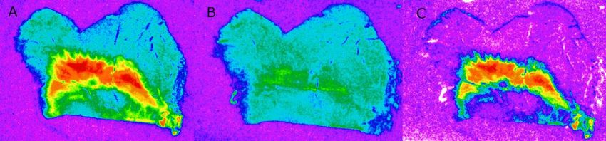
Three representative sections of the SN from each specimen were quantified. For each
section, images of radioligand binding remaining in the presence of the competitor were digitally
subtracted from the corresponding image of total binding to yield an image that represented
specific binding (Figure 1 A-C).6
Figure 1: Total, nonspecific, and specific OXTR autoradiograms
A. Autoradiogram showing total binding. B. Autoradiogram showing nonspecific binding. C.
Autoradiogram showing OXTR specific binding (subtraction of B from A), which was used to
measure OXTR binding densities.
Digital densitometry was performed on the specific OXTR autoradiograms using MCID
Core to quantify the density of OXTR in the pars compacta. Images of Nissl stained tissue
sections were placed side by side with the corresponding OXTR specific binding autoradiogram
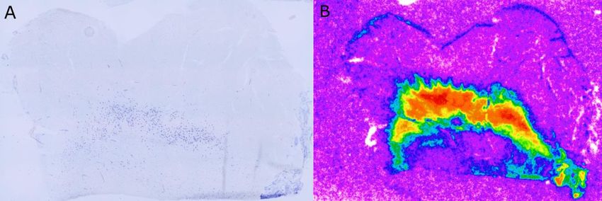
in order to accurately outline the pars compacta on the autoradiogram (Figure 2 A & B).
Figure 2: Nissl-stained section and specific binding autoradiogram
A. A Nissl-stained tissue section showing dark blue dopaminergic neurons of the pars compacta.
B. The corresponding autoradiogram showing specific OXTR binding against background.7
Statistical Analysis
Statistical analyses and data visualization were performed in Graphpad Prism. A two-way
ANOVA was used to determine whether there was a main effect of ASD or sex on OXTR
density in the pars compacta and to identify a potential interaction effect between these two
factors. Linear regressions were used to evaluate whether there was a correlation between OXTR
binding and age and between OXTR binding and postmortem interval (PMI). An exploratory
linear regression analysis on a subset of specimens was used to evaluate the potential relationship
between OXTR density and ASD symptom severity by correlating OXTR binding with scores
from the Autism Diagnostic Inventory-Revised (ADI-R) (Rutter, Lord, & LeCouteur, 2003).
This psychiatric questionnaire is composed of three sections: Section A-Qualitative
Abnormalities in Reciprocal Social Interaction, Section B (Verbal or Nonverbal)-Qualitative
Abnormalities in Communication, and Section C-Restrictive, Repetitive, and Stereotyped
Patterns of Behaviors. For each section, higher scores are indicative of greater impairment
(Rutter & LeCouteur, 2003). For all tests, alpha was set to p8
p=0.0012; Figure 3) and between females with ASD and TD control females (adjusted p=0.0093;
Figure 3).
Figure 3: Effect of sex and ASD on OXTR density in the pars compacta of the SN
Because age has been found to impact OXTR binding in previous studies in human brain
tissue (Freeman et al., 2018), we looked for an association between age and OXTR density in the
pars compacta of the SN. We found no effect of age on OXTR density (Figure 4; R2 = 0.0098, p
= 0.6238).
Figure 4: Association between age and OXTR density in the SN9
Because the brains of the donors in this study were removed at varying intervals after
death, we looked for an association between postmortem interval (time between death and
collection; PMI) and OXTR density. Although protein degradation after death is a concern that
could reduce radioligand binding to our receptors of interest, we found no significant effect of
PMI on OXTR density (Figure 5; R2=0.0564; p=0.2328).
Figure 5: Association between postmortem interval and OXTR density in the SN
Expression of ASD symptoms can be evaluated using the ADI-R methods previously
mentioned, which consist of three sections: A, B, and C. We looked for an association between
severity of ASD symptoms, expressed in ADI-R score, and OXTR density. We found no
significant effect of ADI-R score on sections A (Reciprocal Social Interaction) or C (stereotyped
behavior) and OXTR density in the SN. Section B of the ADI-R test, which is not included in our
study, evaluates verbal and nonverbal communication. Because OT has not been implicated in
the early transition from nonverbal to verbal communication, we did not include it in this study.10
Figure 6: Association between ASD symptom severity and OXTR density
Discussion
The current study is the first to evaluate whether OXTR differs in the SN of individuals
with ASD compared to matched TD controls and whether sex differences exist in OXTR density.
We found a main effect of sex and a significant interaction between sex and diagnosis. Females
with ASD exhibited significantly reduced OXTR density when compared to both males with
ASD and TD females, which may be related to difference in expression of symptoms between
males with ASD and females with ASD.
Along with a male-bias, autism has been shown to produce unique symptoms in females
compared to their male counterparts (Werling & Geschwind, 2014). As discussed previously, the
severity of symptoms in males and females with ASD is not significantly different. However,
females with ASD are more likely to internalize their symptoms, which is a stark contrast to the
hyperactive and aggressive behavior typically seen in males with ASD (Werling & Geschwind,
2014). A recent study highlighting sex differences in ASD symptoms found that girls with ASD
exhibit greater deviance from a sex-specific mean of standardized social scores when compared
to boys with ASD (Lundström et al., 2019). These findings support the evaluation of ASD11
symptoms and how those symptoms are presented from a sex-specific viewpoint that takes these
differences into account. Our results provide a possible neurobiological explanation for the
differences in presentation of symptoms between males and females, and may contribute to
potential treatments of ASD in both males and females.
One method of treatment that has been explored is administration of intranasal OT to
individuals with ASD. Bernaerts et al. (2020) found no significant improvements in 40 subjects
that were administered nasal OT and that submitted follow-up questionnaires 24 hours, 4 weeks,
and 1 year post-treatment. However, each of the 40 adult subjects involved in the study was
male. Parker et al. (2017) found that intranasal oxytocin treatment led to improved social
functions in children with ASD. 84% of the subjects in this study, though, were male, and the
authors noted that their sample was not able to identify sex differences in response to treatment
(Parker et al., 2017). Sex differences are emerging as important factors to consider in both the
biology and treatment of ASD. Intranasal OT treatment, although promising in male children
with ASD, should be further explored in females with ASD. Because of the reduced density of
OXTR we found in females with ASD, potential treatment methods may differ in males and
females with ASD.
Previous studies have found that age impacts OXTR binding in the human brain
(Freeman et al., 2018). Among five brain regions investigated, Freeman et al. (2018) found that
OXTR density was negatively correlated with age across all specimens, both TD and ASD. TD
specimens also exhibited a peak of OXTR density in early life that was absent in ASD specimens
(Freeman et al., 2018). Although an effect of age on OXTR density was found, that study did not
include the SN. We looked for an association between age and OXTR density in the SN and
found no significant association across all specimens. Our results support and expand on those12
initial findings and indicate that the effect of age may be specific to certain regions of the brain,
like the ventral pallidum.
In addition to a main effect of sex and an interaction between sex and diagnosis, we
found multiple negative results worth noting. We found no association between PMI and OXTR
density in the SN, as we hypothesized based on previous studies examining PMI and OXTR
density. Our study supports the findings of Freeman et al. (2017) where effects of PMI were
evaluated up to 33 hours post-death with no significant association. However, our specimens
extend those findings and include PMIs above 60 hours in a novel region of interest in the SN
while still exhibiting no significant association. We also found no effect of symptom severity on
OXTR density in the SN.
Despite our results, it is important to acknowledge the limitations present in a subset of
our statistical analysis. Although the sample size of our study is in line with other studies using
postmortem brain tissue from clinical populations, the sample size of specimens that included
Section A and Section C social scores (Figure 6) is limited. Because of this reduced sample size,
further studies may be necessary to confirm a lack of correlation between ASD symptom severity
and OXTR density in the SN.
Few studies examine sex-differences in ASD, and even fewer explore neuroanatomical
differences between TD and ASD specimens. Our findings give insight into the known sex-
differences in ASD symptom presentation and may provide a neurobiological basis for future
exploration of ASD treatments involving OT and for future studies of ASD and how it relates to
OT in the human brain. Single nucleotide polymorphisms (SNPs), or variations at a single
nucleotide in a DNA sequence, present in the OXTR gene have been implicated in ASD (LoParo
& Waldman, 2015). These DNA differences, in conjunction with our findings, support the need13 for exploration of genetic mechanisms underlying ASD, which may be involved in the sex- differences in OXTR density between ASD females and ASD males, as well as differences between ASD females and TD females. Our lab plans to follow up this study by examining mRNA in the SN to determine whether the dopaminergic neurons of the pars compacta express OXTR.
14

                                      Reflective Writing
       Of my many experiences during my four years at Utah State University, tackling a

research project in the lab of Dr. Freeman has been one of the most challenging and also one of

the most rewarding. Despite experiencing multiple setbacks and readjustments, I was able to

complete my capstone project and obtain exciting results. My capstone project is titled “Effects

of Sex and Autism on Oxytocin Receptors in the Human Substantia Nigra” and is an intersection

of my experiences in the classroom and lab with my career goal of becoming a physician.

       I joined the Freeman lab at the beginning of my junior year at USU. Before joining Dr.

Freeman’s lab, I spent two years learning and practicing laboratory techniques as well as

learning the importance of the research that takes place on campus and the impacts it has on life

outside of the university. My work in the Freeman lab is a culmination of my classroom and

course-associated lab experiences and is an appropriate capstone to my undergraduate education.

Working on my capstone project has been a rare opportunity that has allowed me to apply a

range of different skills including analytical thinking, hands-on laboratory techniques, written

and verbal communication, design, and problem solving.

       With my passion for medicine in mind, the topic of my capstone project is very relevant

and timely. Because of Dr. Freeman’s previous work, I was fortunate enough to be able to work

with postmortem human brain tissue from clinical populations including those that had autism

and those that were typically developing. Autism is a very prevalent yet mysterious condition

that is an area of focus in many clinical studies. Although a reliable treatment for autism is not

likely in the immediate future, our work is directly contributing to a growing field of knowledge

surrounding autism. This study is the first to explore anatomical differences in the midbrain, and

specifically the substantia nigra, of individuals with autism, and our results contribute to the

understanding of the mechanisms underlying autism.
15
This project has helped me build a very strong relationship with my research mentor, Dr.
Freeman, who is incredibly supportive of my education and future goals. One of the reasons this
project has created such a meaningful relationship is the challenges and setbacks that we have
faced throughout. Our initial plan was to automate the process in a way where we could write a
macro, or script, in the program we used to produce our results and run images through the
macro in batches, letting the computer do the work. However, that proved to be much more
difficult than anticipated. After many brainstorming sessions and conference calls with software
representatives, we decided that we were running out of time and that the only logical way
forward was to do the analysis by hand, one image at a time. Before deciding to do the analysis
manually, we had gotten so close so many times before finding a new problem that would halt
the entire process. However, every time we hit a roadblock we were able to regroup and come up
with another solution. Through this experience, Dr. Freeman also helped me understand that
science is often very fluid, and the first approach is very unlikely to be the last as circumstances,
methods, and ideas change.
Throughout my time in the Freeman lab I have presented research three times at USU at
both the fall and spring research symposiums. When I arrived at the library for my first poster
presentation, I was shocked by the magnitude of the event and by how many presenters there
were. Every college in the university was represented and the research projects ranged from
CRISPR-Cas9 gene editing to whether or not cats are friendlier than dogs. While I was aware of
the research-oriented approach of Utah State, I did not understand that research was conducted to
such an extent on campus. Getting to interact with dedicated researchers from so many
disciplines offered a unique experience to engage with other motivated students, each working to
make a difference in their respective field.16
Although my capstone is not a hands-on service-oriented project, I believe that it has the
potential to impact communities throughout the world. Autism is a very prevalent condition that
is likely here to stay and is affecting more people every year. This project contributes to the
understanding of the condition and provides insight into potential mechanisms that underlie the
associated social deficits. Since little is currently understood of the neurobiology behind autism,
every new finding presents opportunities to identify or create treatments that can alleviate some
of the symptoms associated with the condition.
My honors capstone project has presented numerous challenges, but has led to
tremendous growth both academically and personally. My capstone would not have been
possible without the guidance and support of Dr. Freeman or collaboration with both students
and professionals. Science is an interdisciplinary field where one person rarely has all the
answers they need and where progress is not made alone. My research built on previous findings
and I hope that others will look to my project as a starting point to further our collective
knowledge and understanding of a condition that has affected nearly everyone in one way or
another.17
References
Bernaerts, S., Boets, B., Bosmans, G. et al. Behavioral effects of multiple-dose oxytocin
treatment in autism: a randomized, placebo-controlled trial with long-term follow-up.
Molecular Autism 11, 6 (2020). https://doi.org/10.1186/s13229-020-0313-1
Beuker, K. T., Schjølberg, S., Lie, K. K., Donders, R., Lappenschaar, M., Swinkels, S. H.,
& Buitelaar, J. K. (2012). The structure of autism spectrum disorder symptoms in
the general population at 18 months. Journal of Autism and Developmental Disorders,
43(1), 45-56. doi:10.1007/s10803-012-1546-4
Caputi, A., Melzer, S., Michael, M., & Monyer, H. (2013). The long and short of GABAergic
neurons. Current Opinion in Neurobiology, 23(2), 179-186.
doi:10.1016/j.conb.2013.01.021
Carter CS, Kenkel WM, MacLean EL, Wilson SR, Perkeybile AM, Yee JR, Ferris CF, Nazarloo
HP, Porges SW, Davis JM, Connelly JJ, Kingsbury MA. Is Oxytocin "Nature's
Medicine"? Pharmacol Rev. 2020 Oct;72(4):829-861. doi: 10.1124/pr.120.019398.
PMID: 32912963; PMCID: PMC7495339.
Domesick, V., Stinus, L., & Paskevich, P. (1983). The cytology of dopaminergic and
nondopaminergic neurons in the substantia nigra and ventral tegmental area of the rat: A
light- and electron-microscopic study. Neuroscience, 8(4), 743-765. doi:10.1016/0306-
4522(83)90007-6
Fombonne, E. Epidemiology of Pervasive Developmental Disorders. Pediatr Res 65, 591–598
(2009). https://doi.org/10.1203/PDR.0b013e31819e7203
Freeman, S. M. et al. Neuroanatomical distribution of oxytocin and vasopressin 1a receptors in
the socially monogamous coppery titi monkey (Callicebus cupreus). Neuroscience
https://doi.org/10.1016/j.neuroscience.2014.04.055 (2014a).
Freeman, S. M., Inoue, K., Smith, A. L., Goodman, M. M. & Young, L. J. The neuroanatomical
distribution of oxytocin receptor binding and mRNA in the male rhesus macaque
(Macaca mulatta). Psychoneuroendocrinology 45, 128–141 (2014b).
Freeman, S.M., Palumbo, M.C., Lawrence, R.H. et al. Effect of age and autism spectrum
disorder on oxytocin receptor density in the human basal forebrain and midbrain. Transl
Psychiatry 8, 257 (2018). https://doi.org/10.1038/s41398-018-0315-3
Freeman, S. M., Smith, A. L., Goodman M. M. & Bales K. L. Selective localization of oxytocin
receptors and vasopressin 1a receptors in the human brainstem. Soc. Neurosci. 12, 113–
123 (2017).18
Guastella, A. J., Gray, K. M., Rinehart, N. J., Alvares, G. A., Tonge, B. J., Hickie, I. B., Keating,
C. M., Cacciotti-Saija, C., & Einfeld, S. L. (2015). The effects of a course of intranasal
oxytocin on social behaviors in youth diagnosed with autism spectrum disorders: a
randomized controlled trial. Journal of child psychology and psychiatry, and allied
disciplines, 56(4), 444–452. https://doi.org/10.1111/jcpp.12305
Hodge, G.K., Butcher, L.L. Pars compacta of the substantia nigra modulates motor activity but is
not involved importantly in regulating food and water intake. Naunyn-Schmiedeberg's
Arch. Pharmacol. 313, 51–67 (1980). https://doi.org/10.1007/BF00505805
Jacob, S., Brune, C. W., Carter, C., Leventhal, B. L., Lord, C., & Cook, E. H. (april 2007).
Association of the oxytocin receptor gene (OXTR) in Caucasian children and adolescents
with autism. Neuroscience Letters, 417(1), 6-9. doi:10.1016/j.neulet.2007.02.001
LoParo, D., Waldman, I. The oxytocin receptor gene (OXTR) is associated with autism spectrum
disorder: a meta-analysis. Mol Psychiatry 20, 640–646 (2015).
https://doi.org/10.1038/mp.2014.77
Lundström, S., Mårland, C., Kuja-Halkola, R., Anckarsäter, H., Lichtenstein, P., Gillberg, C.,
& Nilsson, T. (2019). Assessing autism in females: The importance of a sex-specific
comparison. Psychiatry Research, 282, 112566. doi:10.1016/j.psychres.2019.112566
Parker, K. J., Oztan, O., Libove, R. A., Sumiyoshi, R. D., Jackson, L. P., Karhson, D. S., . . .
Hardan, A. Y. (2017). Intranasal oxytocin treatment for social deficits and biomarkers of
response in children with autism. PNAS, 114(30), 81119-8124.
doi:https://doi.org/10.1073/pnas.1705521114
Pavăl, D. (september, 2017). A dopamine hypothesis of autism spectrum disorder.
Developmental Neuroscience, 39(5), 355-360. doi:10.1159/000478725
Rutter, M., Lord, C., & LeCouteur, A. (2003). Autism diagnostic interview: Revised (ADI-
R). Los Angeles, CA: Western Psychological Services.
Song, Z. & Albers, H. E. Cross-talk among oxytocin and arginine–vasopressin receptors:
relevance for basic and clinical studies of the brain and periphery. Front.
Neuroendocrinol. https://doi.org/10.1016/j.yfrne.2017.10.004 (2017).
Werling, D. M. & Geschwind, D. H. (2013). Sex differences in autism spectrum disorders.
Current Opinion in Neurology, 26(2), 146-153. doi:10.1097/wco.0b013e32835ee54819
Professional Author Bio
Kip Dooley is a Senior at Utah State University studying Biology with an emphasis in
Human Biology and minors in Chemistry, Business, and Yoga Studies. He has spent two years as
a member of the Freeman lab and is an undergraduate research scholar. In Spring of 2019 he was
awarded the Joseph K.-K. Li Research Scholarship. He has presented research three times at the
2019 Fall Student Research Symposium, 2020 Spring Research Symposium, and the 2021 Spring
Research Symposium where he won Best Poster in the Life Science category. Following
graduation in May of 2021 he will apply to medical school to pursue his dream of becoming a
physician.You can also read