Coming Unbound: Disrupting Automatic Integration of Synesthetic Color and Graphemes by Transcranial Magnetic Stimulation of the Right Parietal Lobe
←
→
Page content transcription
If your browser does not render page correctly, please read the page content below
Coming Unbound: Disrupting Automatic Integration
                         of Synesthetic Color and Graphemes by Transcranial
                            Magnetic Stimulation of the Right Parietal Lobe

                                             Michael Esterman1,2, Timothy Verstynen1, Richard B. Ivry1,
                                                             and Lynn C. Robertson1,2

                                                                                                                                                                                  Downloaded from http://mitprc.silverchair.com/jocn/article-pdf/18/9/1570/1756351/jocn.2006.18.9.1570.pdf by guest on 18 May 2021
                  Abstract
                 & In some individuals, a visually presented letter or number                                 for normal color–shape binding. Using a color-naming task with
                 automatically evokes the perception of a specific color, an ex-                              colored letters that were either congruent or incongruent with
                 perience known as color–grapheme synesthesia. It has been                                    the synesthetic photism, we demonstrate that inhibition of
                 suggested that parietal binding mechanisms play a role in the                                the right posterior parietal lobe with repetitive TMS transiently
                 phenomenon. We used a noninvasive stimulation technique,                                     attenuates synesthetic binding. These findings suggest that syn-
                 transcranial magnetic stimulation (TMS), to determine whether                                esthesia (the induction of color from shape) relies on similar
                 the posterior parietal lobe is critical for the integration of color                         mechanisms as found in normal perception (where the percep-
                 and shape in color–grapheme synesthesia, as it appears to be                                 tion of color is induced by wavelength). &

                  INTRODUCTION
                                                                                                              junction visual search (Donner et al., 2002; Ashbridge,
                  In color–grapheme synesthesia, specific graphemes                                           Walsh, & Cowey, 1997). In addition, patients with bilateral
                  are automatically seen in specific colors. For example,                                     PPC lesions have difficulty in correctly conjoining shape
                  the letter ‘‘A’’ is always perceived in a particular shade                                  and color. For example, R.M., a patient with Balint’s syn-
                  of red. This unusual variety of color–form binding                                          drome, makes frequent illusory conjunctions in free view-
                  has been well characterized behaviorally. However, the                                      ing conditions (Friedman-Hill, Robertson, & Treisman,
                  neural correlates of synesthesia are not well under-                                        1995). Together, these findings suggest that although syn-
                  stood (Rich & Mattingley, 2002). Although functional                                        esthesia is an atypical integration of color and shape, it
                  magnetic resonance imaging (fMRI) has shown that                                            may rely on similar mechanisms to normal perceptual fea-
                  increased activity in extrastriate cortex is associated                                     ture binding (Cohen-Kadosh & Henik, 2006; Sagiv, Heer,
                  with the perception of synesthetic photisms (Sperling,                                      & Robertson, 2006; Robertson, 2003; Grossenbacher &
                  Prvulovic, Linden, Singer, & Stirn, 2006; Hubbard, Arman,                                   Lovelace, 2001).
                  Ramachandran, & Boynton, 2005), other brain imaging                                            Further evidence that parietal mechanisms may be
                  studies of color–grapheme synesthesia have shown neu-                                       involved in synesthesia come from behavioral studies
                  ral correlates outside of the ventral occipital cortex in                                   demonstrating that binding of the synesthetic photism
                  the posterior parietal cortex (PPC). Rich et al. (2003)                                     requires awareness of the inducing grapheme and is
                  found activation in the right PPC (at the junction of                                       modulated by whether the grapheme is within the
                  the occipital cortex) during synesthetic color–grapheme                                     spotlight of attention (Sagiv et al., 2006; Palmeri, Blake,
                  perception, an area implicated in normal binding of                                         Marois, Flanery, & Whetsell, 2002; Mattingley, Rich,
                  color and shape (Donner et al., 2002). Others have                                          Yelland, & Bradshaw, 2001). The idea that synesthesia
                  found activation in the intraparietal sulcus (IPS) associ-                                  arises from operations of an attentional binding mech-
                  ated with color–grapheme synesthesia, both in the left                                      anism is challenged by findings indicating preattentive
                  hemisphere (Weiss, Zilles, & Fink, 2005) and bilaterally                                    activation of synesthetic percepts (Smilek, Dixon, &
                  (Elias, Saucier, Hardie, & Sarty, 2003).                                                    Merikle, 2004; Ramachandran & Hubbard, 2001). These
                     The PPC has already been associated with color–form                                      contradictory results may be explained in part by indi-
                  binding in normal perception using tasks that involve con-                                  vidual differences between synesthetes (Hubbard &
                                                                                                              Ramachandran, 2005).
                                                                                                                 We sought to test the hypothesis that parietal binding
                  1
                    University of California, Berkeley, 2Veterans Administration                              mechanisms that are necessary for normal perception
                  Medical Center, Martinez, CA                                                                also play a role in color–grapheme synesthesia. To this

                                                                                                                    Journal of Cognitive Neuroscience 18:9, pp. 1570–1576
Downloaded from http://www.mitpressjournals.org/doi/pdf/10.1162/jocn.2006.18.9.1570 by guest on 24 October 2021
end, we used a color-naming task (Mattingley et al., TMS (rTMS) was applied over the primary visual cortex
2001; Dixon, Smilek, Cudahy, & Merikle, 2000). On (V1) to test for the effects of generalized brain stimula-
each trial, a colored letter was presented in one of tion on responses in this task.
three colors and the participant pressed a key to indi-
cate the color. The color was either congruent or
incongruent with the synesthetic photism (Figure 1B). METHODS
Synesthetic facilitation occurs when responses to let-
ters presented in their synesthetic color (congruent Participants
condition) are faster than baseline responses (neutral All procedures for this experiment were approved by the
Downloaded from http://mitprc.silverchair.com/jocn/article-pdf/18/9/1570/1756351/jocn.2006.18.9.1570.pdf by guest on 18 May 2021
characters that do not evoke a synesthetic photism). local ethical review board at University of California at
Synesthetic interference occurs when responses to let- Berkeley. We tested two color–grapheme synesthetes
ters presented in colors that are incongruent with recruited from the local population at the university:
their synesthetic photism (incongruent condition) are C.P. (27 years old) and E.F. (22 years old), both right-
slower than baseline. This task was performed follow- handed women. Both report having a unique set of
ing transcranial magnetic stimulation (TMS) of the alphanumeric–color associations (see Figure 1A) that are
left or right parietal cortex. This method allowed us to stable over time. Both reported their colors were pro-
investigate whether transient inhibition of the parietal jected in the external world and appeared as a ‘‘property’’
cortex influenced synesthetic induction of color by of the inducing character, which was also in the ‘‘mind’s
shape. Specifically, would parietal stimulation reduce eye.’’ Both synesthetes would be classified as ‘‘projectors’’
any photism-induced facilitation or interference? Such (Sagiv et al., 2006; Dixon, Smilek, & Merikle, 2004). It has
changes would be consistent with this region contribut- been shown in a prior behavioral study that attention
ing to binding of color and shape in synesthesia. modulates the extent of C.P.’s synesthetic experience
We targeted the angular gyrus at the junction of the (Sagiv et al., 2006). Moreover, her experiences were pre-
posterior IPS and transverse occipital sulcus (IPS/TOS), viously associated with modest activation of lower visual
a region associated with color–form binding in normal areas, leading to her classification as a ‘‘higher synes-
perception (Donner et al., 2002). In addition, activation thete’’ (see participant C.H.P. in Hubbard et al., 2005).
in the right IPS/TOS has been observed during synes- We did not test participants with normal perception
thetic color–grapheme perception (Rich et al., 2003), in this study because they have no synesthetic photism
suggesting a correspondence between normal and syn- to influence color naming (see behavioral procedures
esthetic binding of color and form. In separate sessions, described below) and training controls with color–
stimulation was applied over this parietal region of the grapheme associations would not replicate the experi-
right and left hemisphere. In another session, repetitive ence of synesthesia.
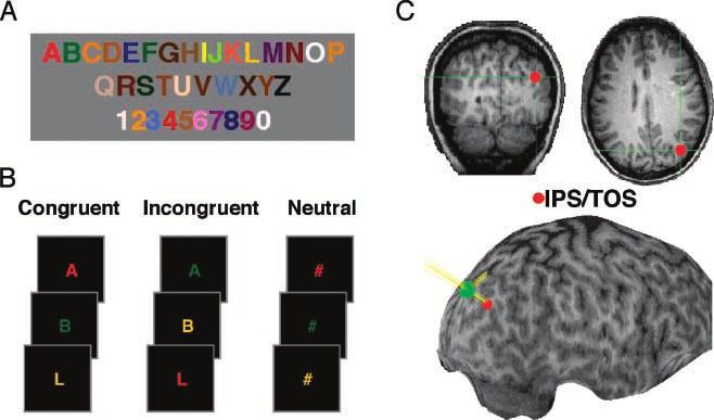
Figure 1. Behavioral and TMS
procedure for participant E.F.
(A) Synesthetic alphabet. (B)
Individualized experimental
conditions. The task was to
name the stimulus color. In
the congruent condition, the
stimulus color matched the
synesthetic color. In the
incongruent condition, the
stimulus color did not match
the synesthetic color. Neutral
characters did not evoke a
synesthetic color. (C) Target
stimulation site (right IPS/
TOS) shown on coronal
and axial slices, plus a 3-D
reconstruction of the
anatomical image for E.F.
Target location is shown
in red, center of magnetic
coil is shown as green
spheres in the 3-D image,
and estimated pulse and
orthogonal trajectories
are shown as yellow lines.
Esterman et al. 1571
Downloaded from http://www.mitpressjournals.org/doi/pdf/10.1162/jocn.2006.18.9.1570 by guest on 24 October 2021Behavioral Procedure TOS and V1). The voxel location was marked on a skull-
stripped reconstructed image.
Before testing, we estimated the specific RGB screen
The scalp location was determined by using a stereo-
values for each letter of each participant’s synesthetic
taxic localization system (Brainsight, Rogue-Research
alphabet. They were seated comfortably 30 cm in front
Inc., Montreal, Canada). Coil position over the target
of a 19-in. CRT monitor (70 Hz refresh rate). An alpha-
regions was monitored online during the stimulation
numeric symbol was presented on the screen and the
epochs. In addition, trajectory estimates of the TMS
participant was instructed to adjust the RGB values until
pulse were estimated intermittently throughout record-
the color on the screen matched their synesthetic
ing (green dots and yellow lines in Figure 1C). Although
photism. For testing, we chose those letters that evoked
Downloaded from http://mitprc.silverchair.com/jocn/article-pdf/18/9/1570/1756351/jocn.2006.18.9.1570.pdf by guest on 18 May 2021
we chose our region of interest (ROI) based on each
the most red, green, and yellow color associations,
individual’s anatomy, we estimated the Talairach coor-
respectively. Each shade of red, green, and yellow was
dinates of the right IPS/TOS site as (30, 74, 32), close
chosen to closely match the synesthetic photism.
to the area associated with feature binding (22, 71, 27)
To quantify each participant’s synesthesia we used a
in Donner et al. (2002). Talairach space is primarily driv-
color-naming task. A series of colored single letters (red,
en by the need to standardize across individuals and
green, or yellow; see above) were presented, each for
assumes a standard sulcal and gyral geometry. By nature,
1000 msec, in a color that was either congruent (con-
it is insensitive to individual differences in brain struc-
gruent trials) or incongruent (incongruent trials) with
ture. The locations in the present study were identified
the synesthetic photism, or a symbol (‘‘#’’) was pre-
by using the pattern of gross anatomical landmarks of
sented that did not evoke a synesthetic photism (neutral
the IPS, angular gyrus, and TOS. Thus, this coordinate
trials; see Figure 1B for examples). Participants pressed a
is only an estimate of our stimulation location.
button with their right hand indicating the true color of
rTMS was performed by using an iron-cored figure-8
the letter as quickly as possible while trying to ignore the
coil (NeoTonus Inc., Marietta, GA; see Epstein & Davey,
synesthetic photism. Reaction time to manually respond
2002). Before each session, the participant’s active mo-
to the screen color was recorded as the difference
tor threshold was determined as the point at which four
between the onset of the letter and the subsequent
to six visible twitches of the thumb were detected fol-
keypress. There was no deadline to respond. Following
lowing 10 pulses over the motor cortex while the thumb
a response, there was a 1000 msec intertrial interval.
and index finger were held together in a pinchlike pos-
Stimuli were presented with PsychLab software (Teren
ture. C.P.’s threshold was between 35% and 38% maxi-
Gum, Boston, VA), and recorded with a CMU button box
mum stimulator output, whereas E.F.’s threshold was
(three buttons pressed with three different fingers).
consistent at 45%. Stimulation was then set to 115% of
Each block consisted of 120 trials (48 congruent, 48
motor threshold for the remainder of the experiment.
incongruent, and 24 neutral, representing all combina-
Each stimulation epoch consisted of 480 consecutive
tions of colors and characters). After each stimulation
pulses that were delivered at a rate of 1 Hz (8 min). This
epoch (sham rTMS or real rTMS; see below), partici-
low-frequency design causes a transient inhibition of the
pants were tested on two blocks of trials: an early block
underlying cortex, with the duration of the effect rough-
(1–5 min poststimulation) and a late block (6–10 min
ly equal to the duration of the stimulation at 1 Hz
poststimulation). Order of sham and real TMS was
(Pascual-Leone et al., 1998). Thus, maximal behavioral
counterbalanced for each participant. Each sham and
effects of TMS were expected during the early block
real TMS epoch was performed twice in each session,
(minutes 1–5 post stimulation) diminishing during the
alternating between stimulation types. Separate sessions
late block (minutes 6–10 post stimulation). During rTMS
were done for each target region (right parietal, left
blocks, the coil was oriented to deliver stimulation
parietal, V1). For each session, 960 trials were collected
directly to the target cortical location. For sham control
per participant.
rTMS blocks, the coil was oriented 908 away from the
scalp so that no pulses perturbed underlying neural
tissue. The subjects were naive to whether they were
Transcranial Magnetic Stimulation
receiving sham or real stimulation.
We first obtained high-resolution anatomical MRIs for
each participant. The images for C.P. were acquired in a
Data Analysis
previous study using a MPRAGE sequence on a 1.5T
Siemens Vision scanner at the University of California, Given the small sample size and the propensity for
San Diego (1 1 1 isotropic voxels, 180 slices). The large between-subjects variability with synesthesia, we
images for E.F. were acquired using a MPFLASH protocol adopted a strong within-subject method to analyze the
on a Varian INNOVA 4T system at the University of stimulation effects. The TMS effects were statistically
California, Berkeley (2 2 2 isotropic voxels, 128 analyzed by using randomization methods on a single-
slices). We identified the target regions for TMS stimu- subject basis (e.g., bootstrapping; see Manly, 1997). All
lation from each participant’s MRI (left and right IPS/ analyses excluded any reaction times greater than four
1572 Journal of Cognitive Neuroscience Volume 18, Number 9
Downloaded from http://www.mitpressjournals.org/doi/pdf/10.1162/jocn.2006.18.9.1570 by guest on 24 October 2021standard deviations from the mean (less than 1% of trials where Xr is the array of values pulled from the
for each participant). We first recorded the interference randomized null distribution.
effect for each of the six conditions (3 regions 2 rTMS/
sham epochs) by subtracting reaction times to incon-
gruent trials from neutral trials. A ‘‘true’’ TMS effect RESULTS AND DISCUSSION
score (Xt) was determined by subtracting the interfer-
ence effect observed following sham stimulation from Following sham rTMS, both participants showed syn-
the effect found after real rTMS. A score of zero indicates esthetic interference in all six blocks (E.F.: mean =
no change in interference following rTMS, a positive 51 msec, range 32–90 msec; C.P.: mean = 31 msec;
Downloaded from http://mitprc.silverchair.com/jocn/article-pdf/18/9/1570/1756351/jocn.2006.18.9.1570.pdf by guest on 18 May 2021
score indicates increased interference following rTMS, range, 19–48 msec). In contrast, rTMS of the right pa-
and a negative score represents a decreased interference rietal ROI significantly attenuated interference for both
effect after rTMS. We then randomly recategorized participants during the early test block (by 54 msec for
reaction times between neutral and incongruent trials E.F. and 48 msec for C.P., p < .01; see Figure 3). For
and recalculated a new interference effect. This was done E.F., interference returned to baseline levels in the late
separately for rTMS and sham trials, following which a block (61 msec); however, C.P.’s scores were still atten-
new TMS effect score was calculated. This scrambling, uated (10 msec), suggesting the effects of rTMS had
recategorization, and subtraction process was repeated not completely worn off. C.P.’s responses were generally
for 10,000 iterations to produce a hypothetical probabil- faster overall following real rTMS, likely due to gen-
ity distribution of interference effects that would occur eralized arousal. More importantly, the attenuation of
simply by chance. Figure 2A shows an example of a interference seen after right IPS/TOS stimulation is the
random distribution of interference effects for C.P. result of a disproportionate decrease in reaction times
following right-hemisphere stimulation. The dashed line on incongruent trials (see Table 1).
represents her true TMS effect. These distributions In contrast to right-hemisphere stimulation, rTMS
closely resembled normal distributions, as verified using over the left PPC had no effect on interference (see
probability plots (Figure 2B; Chambers, Cleveland, Figure 3; C.P.: p = .22, E.F.: p = .09). Thus, the
Kleiner, & Tukey, 1983). disruption of synesthesia following right parietal rTMS
The probability of getting the true observed effects by does not generalize to the left-hemisphere homologue.
chance ( p) can then be directly calculated as: This is consistent with neurophysiological evidence that
the right parietal cortex plays a more critical role in
normal color–form binding than does the left parietal
p ¼ ðXr > Xt Þ cortex (Ashbridge et al., 1997).
Figure 2. (A) A histogram of simulated TMS effects representing a hypothetical null distribution for C.P. after right PPC stimulation. The
observed reaction times were randomly recategorized (with replacement) and new interference effects were calculated from these permutated
data sets. This process was repeated 10,000 times to produce a distribution of values that represent what would be observed purely by chance.
The dashed line represents the true observed TMS effect immediately following stimulation. (B) A normal probability plot, comparing the
data presented in (A) with values from a normal distribution with mean of 0.04 msec and standard deviation of 20 msec. The approximately
straight line indicates that the hypothetical null distribution ref lect a normal gaussian process.
Esterman et al. 1573
Downloaded from http://www.mitpressjournals.org/doi/pdf/10.1162/jocn.2006.18.9.1570 by guest on 24 October 2021disruption of synesthetic color–form integration, thus
reducing the conflict when the synesthetic photism
did not correspond to the real color of the letter.
These findings are consistent with evidence implicating
the IPS/TOS in feature binding in normal perception
(Donner et al., 2002; Friedman-Hill et al., 1995) and
suggests that this region contributes to feature binding
even when the perceived color is evoked by shape
rather than wavelength (Robertson, 2003).
Downloaded from http://mitprc.silverchair.com/jocn/article-pdf/18/9/1570/1756351/jocn.2006.18.9.1570.pdf by guest on 18 May 2021
The present study supports the theory that feedback
from a multimodal association region, like the parietal
cortex, contributes to the perception of a synesthetic
photism (Cohen-Kadosh & Henik, 2006; Sagiv et al.,
2006; Robertson, 2003; Grossenbacher & Lovelace,
2001). Although this does not exclude the possibility
that direct connections between fusiform regions also
play a role in synesthesia (Ramachandran & Hubbard,
2001), it suggests that cross wiring between these re-
Figure 3. Effects of TMS on synesthetic interference. Graph gions is not always sufficient to bind the synesthetic
displays effects of rTMS on synesthetic interference (incongruent percept to the inducing character.
minus neutral) for both participants immediately following An alternative explanation for our findings is that
stimulation (early block; see text). Both participants demonstrated parietal TMS attenuates competition between perceptu-
a strong attenuation of interference following right parietal
compared to sham rTMS, but no change following stimulation ally related codes, similar to competition produced be-
of the left parietal region or V1 (see Methods for discussion of tween the perceptually and conceptually related codes
statistical procedure and Table 1 for reaction times and standard in the traditional Stroop task where the color of the
deviations in all conditions). word may be congruent or incongruent with the word
itself. According to this view, parietal TMS would inter-
fere with competition between the color word and color
rTMS over V1 also had no effect on interference (see (traditional Stroop) as well as competition between two
Figure 3; C.P.: p = .28, E.F.: p = .21). Thus, the colors, one induced by wavelength and the other by the
disruption of synesthesia following right parietal rTMS letter (synesthesia), rather than interfere with feature
is not due to generalized arousal following stimulation binding per se. Although imaging studies have asso-
and suggests that V1 does not play a critical role in ciated parietal activity with Stroop tasks (MacLeod &
color–form binding. MacDonald, 2000), it is unlikely that the parietal lobe
Unlike interference, facilitation was not reliably ob- plays a critical role in the competitive process underlying
served in our participants. Naming colors that were the traditional Stroop effect for several reasons. First,
synesthetically congruent produced consistently faster bilateral stimulation of the posterior parietal lobes does
responses for participant E.F only (36 msec). C.P. did not influence the magnitude of Stroop interference or
not show consistent facilitation in the sham blocks facilitation (Hayward, Goodwin, & Harmer, 2004).
(mean = 14 msec; range, –26 to 44 msec). Facilitation Second, neuropsychological studies of patients with
is not reliably observed in traditional Stroop tasks in parietal lesions have demonstrated normal Stroop inter-
normal observers (Tzelgov, Henik, & Berger, 1992; ference compared to controls. Interestingly, this effect
MacLeod, 1991) as well as tasks similar to ours when has been reported in patients with either left, right, or
tested in other synesthetes (Sagiv et al., 2006; Mattingley bilateral infarcts (Vivas, Humphreys, & Fuentes, 2003;
et al., 2001; Dixon et al., 2000). It has been proposed Robertson, Treisman, Friedman-Hill, & Grabowecky,
that facilitation, when observed in traditional Stroop 1997; Berti, Frassinetti, & Umilta, 1994), and these same
tasks, may be due to inadvertent reading, which would patients may exhibit profound deficits in color–form
explain our weak facilitation effect, given that ‘‘reading’’ binding (see Robertson, 2003). In contrast, alterations
the letter would not lead to correct responses in our in Stroop performance have been associated with dam-
paradigm (MacLeod, 1991). Our results are consistent age to the prefrontal cortex (Kato, 2001; Stuss, Floden,
with studies of normal perceivers showing that facilita- Alexander, Levine, & Katz, 2001).
tion and interference rely on different mechanisms Third, the three studies that have implicated the
(Tzelgov et al., 1992). parietal lobe in color–grapheme synesthesia have not
In sum, immediately following rTMS of the right PPC, involved Stroop tasks, indicating that the parietal lobe’s
both participants showed a significant attenuation of role in synesthesia is not dependent on response in-
interference normally induced by their synesthesia. We terference (Weiss et al., 2005; Elias et al., 2003; Rich
propose that right parietal rTMS produced a transient et al., 2003). Together, these different lines of research
1574 Journal of Cognitive Neuroscience Volume 18, Number 9
Downloaded from http://www.mitpressjournals.org/doi/pdf/10.1162/jocn.2006.18.9.1570 by guest on 24 October 2021Table 1. Single-subject Reaction Times
Participant E.F. Participant C.P.
Neutral Congruent Incongruent Neutral Congruent Incongruent
Right parietal
Early phase
Sham 476 ± 103 424 ± 77 546 ± 114 615 ± 161 574 ± 158 647 ± 186
Downloaded from http://mitprc.silverchair.com/jocn/article-pdf/18/9/1570/1756351/jocn.2006.18.9.1570.pdf by guest on 18 May 2021
rTMS 481 ± 88 423 ± 58 497 ± 96 550 ± 145 511 ± 118 535 ± 94
Late phase
Sham 483 ± 86 453 ± 97 531 ± 92 548 ± 108 575 ± 169 597 ± 185
rTMS 509 ± 117 441 ± 80 570 ± 131 591 ± 166 546 ± 145 601 ± 184
Left parietal
Early phase
Sham 492 ± 120 434 ± 81 523 ± 116 503 ± 99 503 ± 104 544 ± 152
rTMS 469 ± 84 449 ± 93 530 ± 141 499 ± 118 471 ± 96 523 ± 134
Late phase
Sham 496 ± 90 462 ± 94 529 ± 119 533 ± 113 521 ± 111 557 ± 132
rTMS 471 ± 95 462 ± 108 532 ± 133 495 ± 114 490 ± 103 536 ± 145
V1
Early phase
Sham 452 ± 76 419 ± 70 485 ± 91 591 ± 121 547 ± 141 611 ± 156
rTMS 440 ± 99 412 ± 62 488 ± 107 511 ± 106 486 ± 107 544 ± 144
Late phase
Sham 423 ± 75 413 ± 64 514 ± 113 568 ± 117 556 ± 168 587 ± 150
rTMS 443 ± 98 418 ± 74 501 ± 84 524 ± 127 511 ± 132 529 ± 152
Shown are mean reaction times and standard deviations across each condition (neutral, congruent, or incongruent), phase (early, late), participant
(E.F., C.P.) and each stimulation site (right parietal, left parietal, V1).
strongly suggest that our effect is not due to response also be greater in individuals such as C.P., in whom the
interference in general. extent of early visual activation has been shown to be
Behavioral and physiological evidence suggests that lesser (see Hubbard et al., 2005). Parietal participation in
synesthesia is a heterogeneous phenomenon (Dixon & the synesthetic experience may correspond to the ex-
Smilek, 2005; Hubbard et al., 2005). These individual tent that attention is required to elicit the photism. The
differences may be related to the degree of parietal variability in the expression of color–grapheme synes-
involvement. One behavioral factor that may contribute thesia makes the examination of individual differences
to this heterogeneity is the difference between projec- vital to our understanding of this fascinating phenome-
tors, those who see the synesthetic photism in the non (Dixon & Smilek, 2005; Hubbard et al., 2005; Dixon
world, and associators, those who see the photism in et al., 2004). Our results have illuminated one of poten-
the mind’s eye (Dixon et al., 2004). Although this factor tially several mechanisms responsible for synesthesia
has not been explored with respect to the degree of and are consistent with theories of how normal binding
parietal involvement, one hypothesis to consider in occurs within the human brain.
future research is that synesthetes who show greater Future studies must examine the role of parietal bind-
parietal involvement may tend to be ‘‘projectors,’’ par- ing mechanisms in other synesthetes, as well as investi-
ticularly when the synesthetic percept is tightly bound gate other parietal and temporal lobe regions that have
spatially to the inducing grapheme. Physiologically, the been implicated in spatial attention and binding, such as
degree of parietal contribution among synesthetes may the superior parietal cortex and the superior temporal
Esterman et al. 1575
Downloaded from http://www.mitpressjournals.org/doi/pdf/10.1162/jocn.2006.18.9.1570 by guest on 24 October 2021sulcus, using both TMS and fMRI. In addition, single-pulse MacLeod, C. M. (1991). Half a century of research on the
TMS will be able to elaborate on the temporal dynamics Stroop effect: An integrative review. Psychological
Bulletin, 109, 163–203.
of the right PPC’s involvement in synesthesia. MacLeod, C. M., & MacDonald, P. A. (2000). Interdimensional
interference in the Stroop effect: Uncovering the
cognitive and neural anatomy of attention. Trends in
Acknowledgments Cognitive Sciences, 4, 383–391.
We thank Noam Sagiv for discussions and motivation that led Manly, B. (1997). Randomization, bootstrap and Monte
to this project, Noam Sobel for comments on an early version Carlo methods in biology (2nd ed.). New York: Chapman
of this manuscript, and Edward Hubbard for his constructive and Hall/CRC.
comments during the review process. Mattingley, J. B., Rich, A. N., Yelland, G., & Bradshaw, J. L.
Downloaded from http://mitprc.silverchair.com/jocn/article-pdf/18/9/1570/1756351/jocn.2006.18.9.1570.pdf by guest on 18 May 2021
(2001). Unconscious priming eliminates automatic binding
Reprint requests should be sent to Michael Esterman, Depart- of colour and alphanumeric form in synaesthesia. Nature,
ment of Psychology and Neuroscience Institute, University of Cali- 410, 580–582.
fornia, Berkeley, CA, USA, or via e-mail: esterman@berkeley.edu. Palmeri, T. J., Blake, R., Marois, R., Flanery, M. A., & Whetsell,
W. (2002). The perceptual reality of synesthetic colors.
Proceedings of the National Academy of Sciences, U.S.A.,
REFERENCES 99, 4127–4131.
Pascual-Leone, A., Tormos, J. M., Keenan, J., Tarazona, F.,
Ashbridge, E., Walsh, V., & Cowey, A. (1997). Temporal Canete, C., & Catala, M. D. (1998). Study and modulation
aspects of visual search studied by transcranial magnetic of human cortical excitability with transcranial magnetic
stimulation. Neuropsychologia, 35, 1121–1131. stimulation. Journal of Clinical Neurophysiology, 15,
Berti, A., Frassinetti, F., & Umilta, C. (1994). Nonconscious 333–343.
reading? Evidence from neglect dyslexia. Cortex, 30, 181–197. Ramachandran, V. S., & Hubbard, E. M. (2001). Psychophysical
Chambers, J., Cleveland, W., Kleiner, B., & Tukey, P. (1983). investigations into the neural basis of synaesthesia.
Graphical methods for data analysis (Wadsworth). Proceedings of the Royal Society of London, Series B,
Boston: Duxbury Press. Biological Sciences, 268, 979–983.
Cohen-Kadosh, R., & Henik, A. (2006). Color congruity Rich, A. N., & Mattingley, J. B. (2002). Anomalous perception
effect: Where do colors and numbers interact in in synaesthesia: A cognitive neuroscience perspective.
synesthesia? Cortex, 42, 259–263. Nature Reviews, 3, 43–52.
Dixon, M. J., & Smilek, D. (2005). The importance of Rich, A. N., Puce, A., Syngeniotis, A., Williams, M. A.,
individual differences in grapheme–color synesthesia. Howard, M. A., McGlone, F., & Mattingley, J. B. (2003).
Neuron, 45, 821–823. Colour my brain: A functional neuroimaging study of
Dixon, M. J., Smilek, D., Cudahy, C., & Merikle, P. M. color–graphemic synaesthesia. Paper presented at the
(2000). Five plus two equals yellow. Nature, 406, 365. annual meeting of the Cognitive Neuroscience Society,
Dixon, M. J., Smilek, D., & Merikle, P. M. (2004). Not New York, NY.
all synaesthetes are created equal: Projector versus Robertson, L., Treisman, A., Friedman-Hill, S., & Grabowecky,
associator synaesthetes. Cognitive, Affective, and M. (1997). The interaction of spatial and object pathways:
Behavioral Neuroscience, 4, 335–343. Evidence from Balint’s syndrome. Journal of Cognitive
Donner, T. H., Kettermann, A., Diesch, E., Ostendorf, F., Neuroscience, 9, 295–317.
Villringer, A., & Brandt, S. A. (2002). Visual feature and Robertson, L. C. (2003). Binding, spatial attention and
conjunction searches of equal difficulty engage only partially perceptual awareness. Nature Reviews Neuroscience, 4,
overlapping frontoparietal networks. Neuroimage, 15, 16–25. 93–102.
Elias, L. J., Saucier, D. M., Hardie, C., & Sarty, G. E. (2003). Sagiv, N., Heer, J., & Robertson, L. C. (2006). Does binding
Dissociating semantic and perceptual components of of synesthetic color to the evoking grapheme require
synaesthesia: Behavioral and functional neuroanatomical attention? Cortex, 42, 232–242.
investigations. Cognitive Brain Research, 16, 232–237. Smilek, D., Dixon, M. J., & Merikle, P. M. (2004). Binding of
Epstein, C. M., & Davey, K. R. (2002). Iron-core coils for graphemes and synesthetic colors in color–graphemic
transcranial magnetic stimulation. Journal of Clinical synesthesia. In N. Sagiv & L. Robertson (Eds.), Synesthesia:
Neurophysiology, 19, 376–381. Perspectives from cognitive neuroscience. New York:
Friedman-Hill, S., Robertson, L., & Treisman, A. (1995). Parietal Oxford University Press.
contributions to visual feature binding: Evidence from a Sperling, J. M., Prvulovic, D., Linden, D. E. J., Singer, W., &
patient with bilateral lesions. Science, 269, 853–855. Stirn, A. (2006). Neuronal correlates of colour–graphemic
Grossenbacher, P. G., & Lovelace, C. T. (2001). Mechanisms synaesthesia: A fMRI study. Cortex, 42, 295–303.
of synesthesia: Cognitive and physiological constraints. Stuss, D. T., Floden, D., Alexander, M. P., Levine, B., &
Trends in Cognitive Sciences, 5, 36–41. Katz, D. (2001). Stroop performance in focal lesion
Hayward, G., Goodwin, G. M., & Harmer, C. J. (2004). The patients: Dissociation of processes and frontal lobe
role of the anterior congulate cortex in the counting lesion location. Neuropsychologia, 39, 771–786.
Stroop task. Experimental Brain Research, 154, 355–358. Tzelgov, J., Henik, A., & Berger, J. (1992). Controlling Stroop
Hubbard, E. M., Arman, A. C., Ramachandran, V. S., & effects by manipulating expectations for color words.
Boynton, G. M. (2005). Individual differences among Memory & Cognition, 20, 727–735.
grapheme–color synesthetes: Brain–behavior correlations. Vivas, A. B., Humphreys, G. W., & Fuentes, L. J. (2003).
Neuron, 45, 975–985. Inhibitory processing following damage to the parietal
Hubbard, E. M., & Ramachandran, V. S. (2005). Neurocognitive lobe. Neuropsychologia, 41, 1531–1540.
mechanisms of synesthesia. Neuron, 48, 509–520. Weiss, P. H., Zilles, K., & Fink, G. R. (2005). When visual
Kato, M. (2001). Prefrontal lobes and the attentional control: perception causes feeling: Enhanced cross-modal
A neuropsychological study using modified Stroop test. processing in grapheme–color synesthesia. Neuroimage,
Rinsho Shinkeigaku, 41, 1134–1136. 28, 859–868.
1576 Journal of Cognitive Neuroscience Volume 18, Number 9
Downloaded from http://www.mitpressjournals.org/doi/pdf/10.1162/jocn.2006.18.9.1570 by guest on 24 October 2021You can also read