Copper-Doped Ordered Mesoporous Bioactive Glass: A Promising Multifunctional Platform for Bone Tissue Engineering - MDPI
←
→
Page content transcription
If your browser does not render page correctly, please read the page content below
bioengineering

Article
Copper-Doped Ordered Mesoporous Bioactive Glass:
A Promising Multifunctional Platform for Bone
Tissue Engineering †
Francesco Baino
 Institute of Materials Physics and Engineering, Department of Applied Science and Technology,
 Politecnico di Torino, 10129 Turin, Italy; francesco.baino@polito.it
 † Dedicated to the cherished memory of Mariagrazia Cirro, a candid and free spirit.

 Received: 28 April 2020; Accepted: 20 May 2020; Published: 21 May 2020

 Abstract: The design and development of biomaterials with multifunctional properties is highly
 attractive in the context of bone tissue engineering due to the potential of providing multiple
 therapies and, thus, better treatment of diseases. In order to tackle this challenge, copper-doped
 silicate mesoporous bioactive glasses (MBGs) were synthesized via a sol-gel route coupled with an
 evaporation-induced self-assembly process by using a non-ionic block co-polymer as a structure
 directing agent. The structure and textural properties of calcined materials were investigated by X-ray
 powder diffraction, scanning-transmission electron microscopy and nitrogen adsorption-desorption
 measurements. In vitro bioactivity was assessed by immersion tests in simulated body fluid (SBF).
 Preliminary antibacterial tests using Staphylococcus aureus were also carried out. Copper-doped glasses
 revealed an ordered arrangement of mesopores (diameter around 5 nm) and exhibited apatite-forming
 ability in SBF along with promising antibacterial properties. These results suggest the potential
 suitability of copper-doped MBG powder for use as a multifunctional biomaterial to promote bone
 regeneration (bioactivity) and prevent/combat microbial infection at the implantation site, thereby
 promoting tissue healing.

 Keywords: biomaterials; bioglass; porosity; bioactivity; antibacterial; tissue engineering

1. Introduction
      Over the last years, there has been an increasing interest in investigating the biological effects that
can be elicited by ionic dissolution products released by implanted biomaterials. In fact, it is known
that several trace elements are involved in cell metabolic processes and act as enzyme cofactors, thereby
playing key roles in regulating many functions of the body [1]. Hence, the controlled release of dopants
from biomaterials is a valuable approach to modulate the therapeutic response and, ultimately, promote
healing and regeneration in tissue engineering strategies [2,3].
      Bacterial infection is one of the major causes hindering tissue healing and leading to implant
failure; in this regard, a special set of metallic cations with antimicrobial properties (e.g., Ag+ , Ga3+ ,
Cu2+ ) has been suggested for therapeutic purposes. Silver has been well-known to have a bactericidal
activity since ancient times [4]. Silver ions are more effective against Gram-negative bacteria than
Gram-positive species. The antibacterial effect of Ag+ ions is associated to the silver affinity with
disulfide (S–S) and sulfhydryl (–SH) groups available on the proteins of microbial cell walls. As a result
of the binding reaction with silver, normal metabolic processes of bacteria, such as oxidative metabolism
and uptake of nutrients, are disrupted leading to cell death [5].
      The antibacterial properties of gallium are due to the competition that Ga3+ ions establish with
   3+
Fe ions in many biochemical reactions owing to the similarity of their ionic radii (“Trojan horse”

Bioengineering 2020, 7, 45; doi:10.3390/bioengineering7020045             www.mdpi.com/journal/bioengineering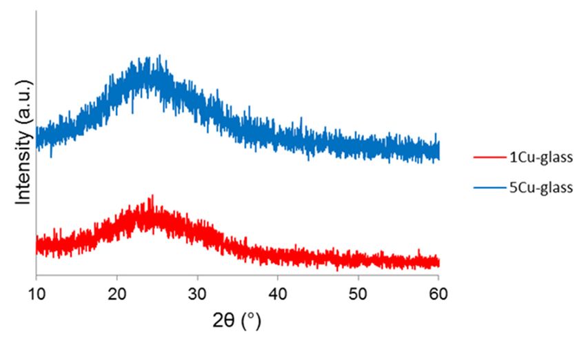
Bioengineering 2020, 7, 45                                                                          2 of 10

effect). Uptake of Ga3+ ions leads to the inhibition of some key biological reactions in bacteria, such as
those involved in DNA and protein synthesis [6].
     Copper ions can kill bacteria due to the generation of reactive oxygen species (ROS), lipid
peroxidation, protein oxidation and DNA degradation [7]. Copper ions exhibit good antibacterial
activity against both Gram-positive and Gram-negative bacteria [8] and, very interestingly,
can stimulate the formation of collagen by bone cells, thereby contributing to osteogenesis
and inhibiting osteoporosis [9]. All these attractive features make copper a valuable dopant to
be incorporated in bioactive ceramics and glasses for making multifunctional biomaterials, which
combine osteoconduction/osteoinduction with new therapeutic extra-functionalities.
     Copper-doped hydroxyapatite microspheres have been prepared by chemical co-precipitation [10],
high-temperature solid-phase synthesis [11], ion-exchange methods [12] and pneumatic extrusion
printing [13].
     Incorporation of copper in bioactive silicate glasses has been reported via melt-quenching
route [14] or sol-gel process [15]. Surface functionalization of sol-gel glasses with copper nanoparticles
was achieved by applying impregnation routes and proper thermal treatments [16]. Resorbable
copper-doped phosphate glass fibers were also fabricated by drawing for potential application in
wound healing and skin tissue engineering [17]. Doping of sol-gel silicate glass compositions with
copper has been recently proposed as an interesting approach for obtaining multifunctional biomaterials
combining tissue regenerative and antibacterial capabilities [18–21]. Specifically, the use of ion-doped
biomedical glasses in the context of antibiotic-free antibacterial applications has been reviewed by
Kaya et al. [22].
     This work reports the synthesis of copper-doped glasses via a modified sol-gel method
incorporating supramolecular chemistry, which allows mesoporous bioactive materials to be obtained.

2. Materials and Methods

2.1. Preparation
      The process used for the synthesis of copper-doped silicate glasses was a sol-gel-type route
commonly known as the evaporation-induced self-assembly (EISA) method, which is applied to
produce mesoporous materials. The parent binary glass belonged to the 80SiO2 -20CaO (mol.%)
system; CuO was introduced to partially substitute CaO in the glass composition, thus obtaining
80SiO2 -19CaO-1CuO (1Cu-glass) and 80SiO2 -15CaO-5CuO (5Cu-glass) formulations (mol.%).
      The glass synthesis procedure was adapted from that reported by Yan et al. [23] for the preparation
of mesoporous silicate glasses, which initially did not contain copper. The non-ionic block copolymer
EO20 -PO70 -EO20 (Pluronic P123, Mw = 5800 g/mol, Sigma-Aldrich, St. Louis, MO, USA) was
used as a structure-directing agent, while tetraethoxysilane (TEOS), calcium nitrate tetrahydrate
(Ca(NO3 )2 ·4H2 O) and copper chloride (CuCl2 ) (all the reagents were purchased from Sigma-Aldrich,
St. Louis, MO, USA) were used to supply SiO2 , CaO and CuO, respectively. Firstly, 4.0 g of Pluronic
P123 were dissolved in 60.0 g of ethanol with 1.0 g of 0.5 M HCl used as a catalyst under constant
stirring at room temperature; then, once Pluronic P123 was completely dissolved, TEOS and salts
were slowly added over 3 h following this order: 6.7 g of TEOS, 1.8 or 1.425 g of Ca(NO3 )2 ·4H2 O (for
1Cu-glass and 5Cu-glass, respectively), and 0.054 or 0.27 g of CuCl2 (for 1Cu-glass and 5Cu-glass,
respectively). The sols were then poured into Petri dishes to allow the EISA process to occur at room
temperature. Aged gels were removed from the dishes and calcined in air at 650 ◦ C for 5 h (heating
and cooling rate of 2 and 5 ◦ C/min); the selection of calcination temperature was also performed
according to the results from thermogravimetric analysis (TGA) on the gels. The calcined materials
were finally ground by ball milling (Pulverisette 0, Fritsch, Idar-Oberstein, Germany) and sieved by
stainless steel sieves with a mesh of 32 µm (Giuliani Technologies, Torino, Italy).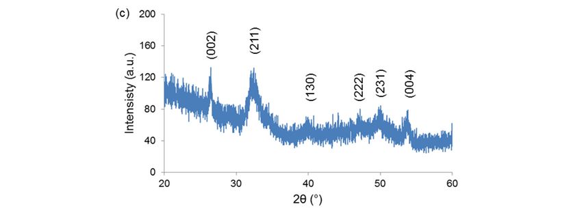
Bioengineering 2020, 7, 45                                                                                3 of 10

2.2. Characterization
     Calcined materials underwent wide-angle (2θ within 10–60◦ ) X-ray powder diffraction (XRPD) by
using a X’Pert Pro PW3040/60 diffractometer (PANalytical, Eindhoven, The Netherlands) operating at
40 kV and 30 mA with Bragg-Brentano camera geometry and Cu Kα incident radiation (wavelength
λ = 0.15405 nm) in order to assess the presence of crystalline phases.
     Small-angle XRPD (2θ within 0.8–4◦ ) was also performed in order to assess the presence of an
ordered pore symmetry in the materials.
     Textural parameters were assessed by nitrogen adsorption-desorption measurements
performed at −196 ◦ C (Quantachrome Autosorb1, Quantachrome, Boynton Beach, FL, USA).
The Brunauer-Emmet-Teller (BET) method [24] and the density functional theory (DFT) isotherm
reconstruction approach [25] were used to determine pore volume, specific surface area (SSA), pore size
distribution and mean pore size.
     The porous structure was also examined by means of scanning-transmission electron microscopy
(STEM) (Merlin, Zeiss, Oberkochen, Germany) operating at 30 kV.
     In vitro bioactivity was assessed in terms of apatite-forming ability by immersion tests in simulated
body fluid (SBF) for 2 weeks. SBF was prepared according to the protocol recommended by Kokubo
and Takadama [26]. Small cylinders of pressed powders (diameter = 10 mm, height = 5 mm) were
maintained in polyethylene bottles filled with SBF at 37 ◦ C in a static incubator; a ratio of sample mass
to solution volume of 1.5 mg/mL was used, as recommended in previous studies [27,28]. At the end of
the experiment, the samples were extracted, rinsed with ethanol to stop reactions, left to dry overnight
at room temperature and analyzed by SEM equipped with energy-dispersive spectroscopy (EDS)
probe (SEM-EDS, Merlin, Zeiss, Oberkochen, Germany) after being sputter-coated with chromium.
The sample surface was also analyzed by X-ray diffraction (XRD) method according to the procedure
described above.
     Antibacterial activity was assessed against a standard Staphylococcus aureus strain by performing
the Kirby-Bauer test according to the Performance Standards for Antimicrobial Disk Susceptibility Test
(Approved Standard, 9th Ed., NCCLS, Villanova, PA, USA, 2006).

3. Results and Discussion
     TGA was useful to select the calcination temperature (Figure 1). An increase of mass loss up
to 650 ◦ C was observed (about 30% of total mass loss); above this temperature, the mass remained
constant, confirming that the surfactant was completely removed from the material. Mass reduction
could be attributed to two major events associated to the removal of ethanol and water from the gel
(70–200 ◦ C) and the thermal decomposition of organics (surfactant) and nitrates (300–500 ◦ C).
      Bioengineering 2020, 7, x FOR PEER REVIEW                                                     4 of 10

      Figure 1. Thermogravimetric analysis (TGA) of the gel corresponding to the 80SiO2 -20CaO (mol.%)
           Figure 1. Thermogravimetric analysis (TGA) of the gel corresponding to the 80SiO2-20CaO (mol.%)
      nominal  composition: mass loss and lass loss derivative.
            nominal composition: mass loss and lass loss derivative.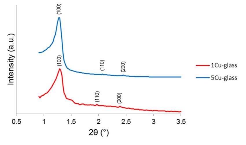
Bioengineering 2020, 7, 45                                                                                 4 of 10

     The wide-angle XRPD patterns of calcined 1Cu-glass and 5Cu-glass are displayed in Figure 2
and confirm
      Figure 1.the amorphous nature
                 Thermogravimetric       of both
                                     analysis      materials,
                                               (TGA)           ascorresponding
                                                       of the gel  proved by thetopresence
                                                                                   the 80SiOof  only broad
                                                                                             2-20CaO       haloes
                                                                                                      (mol.%)
in thenominal
       2θ range              ◦
                  of 15 to 35mass
                               , which  is typical  of derivative.
                                                       silicate glasses.
                composition:       loss and  lass loss

       Figure 1. Thermogravimetric analysis (TGA) of the gel corresponding to the 80SiO2-20CaO (mol.%)
       nominal composition: mass loss and lass loss derivative.

      Figure 2. Wide-angle X-ray powder diffraction (XRPD) patterns of copper-doped materials
         Figure 2. Wide-angle X-ray powder diffraction (XRPD) patterns of copper-doped materials after
      after calcination.
                                                 calcination.
     The small-angle XRPD patterns of both glasses (Figure 3) show three diffraction peaks that can be
attributed to the (100), (110) and (200) reflections of a two-dimensional hexagonal p6mm lattice [23].
The d100Figure
          values
               2. were 6.7 and
                  Wide-angle   6.8 powder
                             X-ray nm 1Cu-glass   and(XRPD)
                                          diffraction  5Cu-glass,  corresponding
                                                              patterns             to cell
                                                                       of copper-doped     parameters
                                                                                        materials after of 7.7
and 7.8 nm (assuming a perfect two-dimensional        hexagonal
                                                 calcination.    symmetry).

                  Figure 3. Small-angle XRPD patterns of copper-doped materials after calcination.

                  Figure 3. Small-angle XRPD patterns of copper-doped materials after calcination.
                  Figure 3. Small-angle XRPD patterns of copper-doped materials after calcination.
    The mesoporous nature of the materials was further confirmed by STEM investigation
along the [100] direction (Figure 4), which allowed revealing an ordered arrangement of parallel
one-dimensional nanopores (nano-channels). Rough measurements of pore dimeter yielded a value of
around 5.7 nm.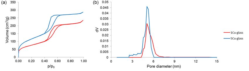
Bioengineering 2020, 7, 45 5 of 10
Bioengineering 2020, 7, x FOR PEER REVIEW 5 of 10
Figure
Figure 4. Scanning-transmission
4. Scanning-transmission (STEM)
(STEM) image
image of 5Cu-glass
of 5Cu-glass recorded
recorded along
along the [100]
the [100] direction,
direction, showing
showing a parallel arrangement of nano-sized channels
a parallel arrangement of nano-sized channels (mesopores). (mesopores).
Nitrogen
Nitrogen adsorption–desorptionmeasurements
adsorption–desorption measurements (Figure
(Figure5)5)further
further confirmed
confirmed the the
existence of of
existence
uniform nanopores in the mesoscale range. Both glasses exhibited a type-IV isotherm, which is
uniform nanopores in the mesoscale range. Both glasses exhibited a type-IV isotherm, which is
associated to pores with size between 2 and 50 nm (i.e., mesopores, according to the International
associated to pores with size between 2 and 50 nm (i.e., mesopores, according to the International
Union for Pure and Applied Chemistry (IUPAC) definition) [29]. The shape of the hysteresis loop
Unioncould
for Pure andinformation
provide Applied Chemistry
about the(IUPAC)
pore shapedefinition) [29].
[30]: in both The shape
materials, the of
looptheshape
hysteresis loop
suggests thecould
provide information
presence about mesopores
of cylindrical the pore shape
with [30]: in both
hexagonal materials,
symmetry, the loop
which shape
is typical of suggests
MCM-41 the presence
ordered
of cylindrical
mesoporousmesopores with
silica [31]. hexagonal
These symmetry,
results are in good which is typical
agreement of MCM-41
with the findings ordered mesoporous
from small-angle
silica XRPD
[31]. These
(Figureresults are in good
3). Quantification ofagreement withisthe
textural features findings from
summarized small-angle
in Table 1; the value XRPD
of pore(Figure
size 3).
(5.1 nm) is of
Quantification close to thatfeatures
textural determined more roughlyin
is summarized byTable
STEM1;measurements
the value of pore(5.7 nm).
size (5.1 nm) is close to
that The data
determined shown
more in Table
roughly by 1STEM
revealmeasurements
that the pore volume
(5.7 and SSA of the mesoporous glasses
nm).
Bioengineering 2020, 7, x FOR PEER REVIEW 6 of 10
decrease as the copper content increases. These results suggest that the incorporation of Cu2+ ions
may have a negative effect on the precursor condensation, disrupting the ordered orientation of
(SiO4)4− units during the self-assembling reaction of the glass. Interestingly, although the total
amount of modifiers (calcium and copper) is equal to 20 mol.% in all materials, the “disturbing”
effect is higher when different types of modifiers are simultaneously introduced. This effect was also
observed elsewhere when other modifiers (e.g., zinc or cerium) were added to the silicate network of
mesoporous silicate glasses [32]. The mechanism behind this effect in mesoporous glasses is still to
be elucidated, but a role could be played by the higher difference of modifier’s ionic radius as
compared to silicon, which is the major forming element of the glass network (ionic radii: 0.210 nm
for Si4+, 0.231 nm for Ca2+ and 0.140 nm for Cu2+, hence the absolute differences |∆Si-Cu| = 0.070 nm >
|∆Si-Ca| = 0.021 nm).
The copper-depending trend of pore volume and SSA displayed in Table 1 is also consistent
Figure
Figure 5. Nitrogen adsorption-desorption measurements performed on calcined
on glasses: (a) isotherms
with that5.recently
Nitrogen adsorption-desorption
observed by Luo et al. [33] formeasurements performed
Cu-doped nanofibrous calcined
mesoporous glassglasses: (a)
scaffolds.
and (b)
Unlike pore
isotherms size
poreand distributions.
(b) pore
volume andsize
SSA,distributions.
the mean pore size is not apparently affected by the increasing content
of copper
Table 1. in the glass composition.
Textural characteristics of calcined mesoporous glasses obtained by nitrogen
Figure
SSA6a and
of all b reveals
mesoporous
adsorption-desorption the formation
glasses
porosimetry. collected inofTable
calcium phosphatehigher
1 is significantly globular
than agglomerates
that assessed foron the
surface
bothofmelt-derived
both Cu-doped materials
(less than 1 m2/g)after
and immersion in glasses
sol-gel silicate SBF, thus demonstrating
produced the apatite-forming
without using a structure
directing agent (few tens of m /g) /g)
2/g) [34];3 this is consistent2 with previous
ability of these glass compositions in vitro. The newly-formed phase exhibit a (nm)
Sample Pore Volume (cm SSA (m Mean findings
Pore on
Size several
“cauliflower”
mesoporous
structure formed glass
0Cu-glass types and compositions
by needle-like nano-sized
0.265 [35,36].
crystals: this450is the typical morphological
5.0 “fingerprint”
of the hydroxyapatite-like
1Cu-glass phases0.232
grown on the surface432 of bioactive glasses upon soaking in SBF.
5.1 nitrogen
Table 1. Textural characteristics of calcined mesoporous glasses obtained by
Semi-quantitative compositional
adsorption-desorption
assessment (EDS) on the agglomerates formed on 1Cu-glass and
porosimetry.
5Cu-glass 0.165 275 5.1
5Cu-glass yielded Ca-to-P atomic ratios of 1.85 and 1.91, respectively. These values are higher than
Sample
the Ca-to-P atomic ratio Pore volume (cm3/g)
of stoichiometric SSA (m2/g)
hydroxyapatite (1.67), but canMean pore size (nm)
be justified considering the
The 0Cu-glass
boundary data shown
effects in Table
due
0.265that the pore volume450
to the1 reveal
finite volume involved in and SSA of the mesoporous
compositional
5.0
assessment glasses
by EDS. decrease
On the
asother 1Cu-glass
the copper content increases. 0.232 results suggest that432
These the incorporation of Cu5.12+ ions may have a
hand, non-stoichiometric hydroxyapatite has been reported to commonly form on silicate
negative 5Cu-glass
effect 0.165 275the ordered orientation 5.1 of (SiO )4− units
glasses in SBFon the precursor
[37,38]. condensation,
XRD analysis disrupting
(Figure 6c) confirmed the formation of hydroxyapatite 4 (PDF
during the self-assembling reaction of the glass. Interestingly,
code: 01-086-0740) on the surface of samples during immersion in SBF. although the total amount of modifiers
(calcium and copper)
At present, is equal to 20community
the biomaterials mol.% in all materials,
assumes thatthe
the“disturbing”
formation ofeffect is higher when
a hydroxyapatite-like
different
layer ontypes of modifiers
the surface are simultaneously
of a given material soaked introduced.
in SBF isThis effectofwas
a proof its also observed
bioactivity elsewhere
and, to some
extent, of its bone bonding ability in vivo [26]. Being bioactive, the Cu-doped mesoporous materials
produced in this work can be included in the versatile class of mesoporous bioactive glasses
(MBGs), which have attracted great interest over the last few years for potential use in tissue
engineering applications [39].
Previous studies showed that MBGs with high silica content (>80 mol.%) are highly versatileBioengineering 2020, 7, 45 6 of 10
when other modifiers (e.g., zinc or cerium) were added to the silicate network of mesoporous silicate
glasses [32]. The mechanism behind this effect in mesoporous glasses is still to be elucidated, but a
role could be played by the higher difference of modifier’s ionic radius as compared to silicon, which
is the major forming element of the glass network (ionic radii: 0.210 nm for Si4+ , 0.231 nm for Ca2+
and 0.140 nm for Cu2+ , hence the absolute differences |∆Si-Cu | = 0.070 nm > |∆Si-Ca | = 0.021 nm).
The copper-depending trend of pore volume and SSA displayed in Table 1 is also consistent
with that recently observed by Luo et al. [33] for Cu-doped nanofibrous mesoporous glass scaffolds.
Unlike pore volume and SSA, the mean pore size is not apparently affected by the increasing content
of copper in the glass composition.
SSA of all mesoporous glasses collected in Table 1 is significantly higher than that assessed for
both melt-derived (less than 1 m2 /g) and sol-gel silicate glasses produced without using a structure
directing agent (few tens of m2 /g) [34]; this is consistent with previous findings on several mesoporous
glass types and compositions [35,36].
Figure 6a,b reveals the formation of calcium phosphate globular agglomerates on the surface of
both Cu-doped materials after immersion in SBF, thus demonstrating the apatite-forming ability of
these glass compositions in vitro. The newly-formed phase exhibit a “cauliflower” structure formed by
needle-like nano-sized crystals: this is the typical morphological “fingerprint” of the hydroxyapatite-like
phases grown on the surface of bioactive glasses upon soaking in SBF. Semi-quantitative compositional
assessment (EDS) on the agglomerates formed on 1Cu-glass and 5Cu-glass yielded Ca-to-P atomic ratios
of 1.85 and 1.91, respectively. These values are higher than the Ca-to-P atomic ratio of stoichiometric
Bioengineering 2020,
hydroxyapatite 7, x FOR
(1.67), butPEER
can REVIEW
be justified considering the boundary effects due to the finite 7 ofvolume
10
involved in compositional assessment by EDS. On the other hand, non-stoichiometric hydroxyapatite
justify the absence of a clear antibacterial halo around the sample doped with the lower amount of
has been reported to commonly form on silicate glasses in SBF [37,38]. XRD analysis (Figure 6c)
copper. In summary, these early results demonstrate that both copper-doped MBG compositions
confirmed
exhibitthean formation
antimicrobial of hydroxyapatite (PDF code:aureus
effect against Staphylococcus 01-086-0740) on thefurther
and motivate surface of sampleson
investigation during
immersion in SBF.
these highly promising bioactive and antibacterial multifunctional biomaterials.
Figure 6. In6.vitro
Figure bioactivity
In vitro tests:
bioactivity tests:SEM
SEMmicrographs showingthe
micrographs showing the“cauliflower”
“cauliflower” calcium-phosphate
calcium-phosphate
agglomerates
agglomeratesformed on on
formed (a)(a)
1Cu-glass
1Cu-glassandand(b)
(b) 5Cu-glass afterimmersion
5Cu-glass after immersion forfor 2 weeks
2 weeks in SBF;
in SBF; (c) XRD
(c) XRD
analysis
analysis on 5Cu-glass
on 5Cu-glass (2 weeks
(2 weeks ininSBF),
SBF),which
which reveals
reveals the
thediffraction
diffractionpeaks of hydroxyapatite
peaks of hydroxyapatiteformed
formed
during
during the test.
the test.Bioengineering 2020, 7, 45 7 of 10
Bioengineering 2020, 7, x FOR PEER REVIEW 7 of 10
justify
Atthe absence
present, the of a clear antibacterial
biomaterials community halo around
assumes thethe
that sample doped
formation of awith the lower amount
hydroxyapatite-like of
layer
copper. In summary,
on the surface of a giventhese early soaked
material results in
demonstrate
SBF is a proofthatofboth copper-doped
its bioactivity and, toMBG
somecompositions
extent, of its
exhibit an antimicrobial
bone bonding effect
ability in vivo against
[26]. BeingStaphylococcus
bioactive, the aureus
Cu-doped andmesoporous
motivate further investigation
materials producedon in
these highly
this work canpromising
be included bioactive and antibacterial
in the versatile multifunctional
class of mesoporous biomaterials.
bioactive glasses (MBGs), which have
attracted great interest over the last few years for potential use in tissue engineering applications [39].
Previous studies showed that MBGs with high silica content (>80 mol.%) are highly versatile
carriers for antibiotics but exhibit negligible [19] or no antibacterial effect (equivalent effect to that
of plastic control [40]) in the short term if used alone. Therefore, incorporation of copper was
thought as a valuable strategy to impart inherent antimicrobial extra-functionalities to these silicate
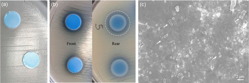
biomaterials. The results of the antibacterial tests performed on 1Cu-glass and 5Cu-glass discs are
shown in Figure 7. As shown in Figure 7a, 1Cu-glass composition is apparently unable to create an
inhibitory halo for bacteria around the sample. On the contrary, an inhibitory halo can be clearly
observed around the 5Cu-glass sample (region of total inhibition around 2 mm around the outer
surface) (Figure 7b). These results suggest that the initial concentration of copper in the MBG
composition is key in dictating the antibacterial behavior, in agreement with previous observations
reported by other authors. However, we should take into account that antibacterial materials can
exert their antiseptic effect via (i) release-killing mode, which is due to the release of antibacterial
ions, and/or (ii) contact-killing mode, if bacteria come in direct contact with the biomaterial. Hence,
the visual inspection of the surface of 1Cu-glass sample is important to clarify whether a contact-killing
antibacterial effect can still be elicited. In this regard, the lack of bacteria on the surface of the sample
brought into contact with the bacteria-inoculated plate during the Kirby-Bauer test can provide an
evidence of contact-mode antibacterial capacity. Figure 7c shows a SEM image of the 1Cu-glass
surface after the Kirby-Bauer test: although this samples showed no inhibition halo, the image
clearly shows that only few clusters of Staphylococcus aureus survived after 24 h of incubation on
this material. Furthermore, it is worth underlining that the antibacterial effect of copper ions was
reported to be more significant against Gram-negative bacteria, such as Escherichia Coli, compared to
Gram-negative strains such as Staphylococcus Aureus [19], which could partially justify the absence of a
clear Figure
antibacterial halobioactivity
6. In vitro around the sample
tests: doped with showing
SEM micrographs the lower amount
the of copper.
“cauliflower” In summary, these
calcium-phosphate
earlyagglomerates
results demonstrate
formed onthat both copper-doped
(a) 1Cu-glass MBG after
and (b) 5Cu-glass compositions
immersion exhibit an antimicrobial
for 2 weeks in SBF; (c) XRDeffect
against Staphylococcus
analysis on 5Cu-glass aureus andin
(2 weeks motivate further
SBF), which investigation
reveals the diffractiononpeaks
theseofhighly promising
hydroxyapatite bioactive
formed
and antibacterial multifunctional biomaterials.
during the test.
Figure
Figure 7.7.In
Invitro
vitroantibacterial
antibacterial experiments
experiments (24inhibition
(24 h): h): inhibition haloontests
halo tests on (a) 1Cu-glass
(a) 1Cu-glass and (b)
and (b) 5Cu-glass;
5Cu-glass;
(c) (c) SEM showing
SEM micrograph micrograph showingofthe
the surface surfaceafter
1Cu-glass of 1Cu-glass after
the test (the whitethearrows
test (the white the
highlight arrows
few
highlight the few clusters of
clusters of bacteria survived). bacteria survived).
4. Conclusions
4. Conclusions
Copper-doped MBGs
Copper-doped MBGs were
wereobtained
obtainedbybya wet (sol-gel)
a wet route
(sol-gel) in which
route a non-ionic
in which surfactant
a non-ionic was
surfactant
incorporated as a mesopore template. After calcination, the glasses exhibited an ordered
was incorporated as a mesopore template. After calcination, the glasses exhibited an ordered structure
of mesopores
structure arranged arranged
of mesopores accordingaccording
to hexagonal symmetry.
to hexagonal The presence
symmetry. of a mesoporous
The presence texture
of a mesoporous
was thewas
texture reason
thebehind
reason the apatite-forming
behind propertyproperty
the apatite-forming of these of
glasses
these despite
glasses the highthe
despite content
high of silica
content
of silica (80 mol.%), as the large SSA (275–450 m2/g) was key to enhance the ion-exchangeBioengineering 2020, 7, 45 8 of 10
(80 mol.%), as the large SSA (275–450 m2 /g) was key to enhance the ion-exchange phenomena between
glass and solution during immersion in SBF. The amount of copper in the MBG composition played
a role in affecting both textural and functional properties: as copper increased, the SSA decreased
but the antibacterial effect against Staphylococcus aureus was more significant. These preliminary
observations show promise for the potential use of copper-doped MBGs in bone tissue engineering
applications and motivate further investigation on these materials.
Funding: This research received no external funding.
Conflicts of Interest: The author declares no conflict of interest.
References
1. Habibovic, P.; Barralet, J.E. Bioinorganics and biomaterials: Bone repair. Acta Biomater. 2011, 7, 3013–3026.
[CrossRef] [PubMed]
2. Hoppe, A.; Güldal, N.S.; Boccaccini, A.R. A review of the biological response to ionic dissolution products
from bioactive glasses and glass-ceramics. Biomaterials 2011, 32, 2757–2774. [CrossRef] [PubMed]
3. Kargozar, S.; Baino, F.; Hamzehlou, S.; Hill, R.; Mozafari, M. Bioactive glasses entering the mainstream.
Drug Discov. Today 2018, 23, 1700–1704. [CrossRef] [PubMed]
4. Mijnendonckx, K.; Leys, N.; Mahillon, J.; Silver, S.; Van Houdt, R. Antimicrobial silver: Uses, toxicity
and potential for resistance. BioMetals 2013, 26, 609–621. [CrossRef]
5. Silvestry-Rodriguez, N.; Sicairos-Ruelas, E.E.; Gerba, C.P.; Bright, K.R. Silver as a Disinfectant. Rev. Environ.
Contam. Toxicol. 2007, 191, 23–45. [CrossRef]
6. Pourshahrestani, S.; Zeimaran, E.; Kadri, N.A.; Gargiulo, N.; Samuel, S.; Naveen, S.V.; Kamarul, T.; Towler, M.
Gallium-containing mesoporous bioactive glass with potent hemostatic activity and antibacterial efficacy.
J. Mater. Chem. B 2015, 4, 71–86. [CrossRef]
7. Chatterjee, A.K.; Chakraborty, R.; Basu, T. Mechanism of antibacterial activity of copper nanoparticles.
Nanotechnology 2014, 25, 135101. [CrossRef]
8. Mehtar, S.; Wiid, I.; Todorov, S.D. The antimicrobial activity of copper and copper alloys against nosocomial
pathogens and Mycobacterium tuberculosis isolated from healthcare facilities in the Western Cape: An in-vitro
study. J. Hosp. Infect. 2008, 68, 45–51. [CrossRef]
9. Liu, C.; Fu, X.; Pan, H.; Wan, P.; Wang, L.; Tan, L.; Wang, K.; Zhao, Y.; Yang, K.; Chu, P.K. Biodegradable
Mg-Cu alloys with enhanced osteogenesis, angiogenesis, and long-lasting antibacterial effects. Sci. Rep. 2016,
6, 27374. [CrossRef]
10. Nam, P.T.; Thom, N.T.; Phuong, N.T.; Xuyen, N.T.; Hai, N.S.; Anh, N.T.; Dung, P.T.; Thanh, D.T.M. Synthesis,
characterization and antimicrobial activity of copper doped hydroxyapatite. Vietnam J. Chem. 2018, 56,
672–678. [CrossRef]
11. Pogosova, M.A.; Kazin, P.E.; Tretyakov, Y. Synthesis and characterisation of copper doped Ca–Li
hydroxyapatite. Nucl. Instrum. Methods Phys. Res. Sect. B Beam Interact. Mater. Atoms 2012, 284,
33–35. [CrossRef]
12. Li, Y.; Ho, J.; Ooi, C.P. Antibacterial efficacy and cytotoxicity studies of copper (II) and titanium (IV)
substituted hydroxyapatite nanoparticles. Mater. Sci. Eng. C 2010, 30, 1137–1144. [CrossRef]
13. Chi, W.; Zou, J.; Ai, F.; Lin, Y.; Li, W.; Cao, C.; Yang, K.; Zhou, K. Research of Cu-Doped Hydroxyapatite
Microbeads Fabricated by Pneumatic Extrusion Printing. Materials 2019, 12, 1769. [CrossRef] [PubMed]
14. Milković, L.; Hoppe, A.; Detsch, R.; Boccaccini, A.R.; Zarkovic, N. Effects of Cu-doped 45S5 bioactive glass
on the lipid peroxidation-associated growth of human osteoblast-like cellsin vitro. J. Biomed. Mater. Res. Part
A 2013, 102, 3556–3561. [CrossRef] [PubMed]
15. Varmette, E.A.; Nowalk, J.R.; Flick, L.M.; Hall, M.M. Abrogation of the inflammatory response in
LPS-stimulated RAW 264.7 murine macrophages by Zn- and Cu-doped bioactive sol-gel glasses. J. Biomed.
Mater. Res. Part A 2009, 90, 317–325. [CrossRef] [PubMed]
16. Bonici, A.; Lusvardi, G.; Malavasi, G.; Menabue, L.; Piva, A. Synthesis and characterization of bioactive
glasses functionalized with Cu nanoparticles and organic molecules. J. Eur. Ceram. Soc. 2012, 32, 2777–2783.
[CrossRef]Bioengineering 2020, 7, 45 9 of 10
17. Neel, E.A.; Ahmed, I.; Pratten, J.; Nazhat, S.; Knowles, J.C. Characterisation of antibacterial copper releasing
degradable phosphate glass fibres. Biomaterials 2005, 26, 2247–2254. [CrossRef]
18. Li, J.; Zhai, D.; Lv, F.; Yu, Q.; Ma, H.; Yin, J.; Yi, Z.; Liu, M.; Chang, J.; Wu, C. Preparation of copper-containing
bioactive glass/eggshell membrane nanocomposites for improving angiogenesis, antibacterial activity
and wound healing. Acta Biomater. 2016, 36, 254–266. [CrossRef]
19. Bari, A.; Bloise, N.; Fiorilli, S.; Novajra, G.; Vallet-Regí, M.; Bruni, G.; Torres-Pardo, A.; González-Calbet, J.M.;
Visai, L.; Vitale-Brovarone, C. Copper-containing mesoporous bioactive glass nanoparticles as multifunctional
agent for bone regeneration. Acta Biomater. 2017, 55, 493–504. [CrossRef]
20. Bains, R.; Sharma, P.; Mir, R.A.; Jeet, S.; Kaur, G.; Pandey, O.P. Influence of CuO/MgO ratio on the gene
expression, cytocompatibilty, and antibacterial/anticancerous/analgesic drug loading kinetics for (15-x)
CuO-xMgO-10P2O5-60SiO2-10CaO-5ZnO (2.5 ≤ x ≤ 12.5) mesoporous bioactive glasses. J. Biomed. Mater.
Res. Part A 2018, 106, 2116–2130. [CrossRef]
21. Moghanian, A.; Ghorbanoghli, A.; Kazem-Rostami, M.; Pazhouheshgar, A.; Salari, E.; Yazdi, M.S.;
Alimardani, T.; Jahani, H.; Jazi, F.S.; Tahriri, M. Novel antibacterial Cu/Mg-substituted 58S-bioglass:
Synthesis, characterization and investigation of in vitro bioactivity. Int. J. Appl. Glas. Sci. 2019, in press.
[CrossRef]
22. Kaya, S.; Cresswell, M.; Boccaccini, A.R. Mesoporous silica-based bioactive glasses for antibiotic-free
antibacterial applications. Mater. Sci. Eng. C 2018, 83, 99–107. [CrossRef] [PubMed]
23. Yan, X.; Yu, C.; Zhou, X.; Tang, J.; Zhao, D. Highly Ordered Mesoporous Bioactive Glasses with Superior
In Vitro Bone-Forming Bioactivities. Angew. Chem. Int. Ed. 2004, 43, 5980–5984. [CrossRef] [PubMed]
24. Brunauer, S.; Emmett, P.H.; Teller, E. Adsorption of Gases in Multimolecular Layers. J. Am. Chem. Soc. 1938,
60, 309–319. [CrossRef]
25. Landers, J.; Gor, G.Y.; Neimark, A.V. Density functional theory methods for characterization of porous
materials. Colloids Surf. A Physicochem. Eng. Asp. 2013, 437, 3–32. [CrossRef]
26. Kokubo, T.; Takadama, H. How useful is SBF in predicting in vivo bone bioactivity? Biomaterials 2006, 27,
2907–2915. [CrossRef]
27. Nommeots-Nomm, A.; Labbaf, S.; Devlin, A.; Todd, N.; Geng, H.; Solanki, A.K.; Tang, H.M.; Perdika, P.;
Pinna, A.; Ejeian, F.; et al. Highly degradable porous melt-derived bioactive glass foam scaffolds for bone
regeneration. Acta Biomater. 2017, 57, 449–461. [CrossRef]
28. Baino, F.; Fiume, E.; Miola, M.; Leone, F.; Onida, B.; Vernè, E. Fe-doped bioactive glass-derived scaffolds
produced by sol-gel foaming. Mater. Lett. 2019, 235, 207–211. [CrossRef]
29. Sing, K.S.W. Reporting physisorption data for gas/solid systems with special reference to the determination
of surface area and porosity (Recommendations 1984). Pure Appl. Chem. 1985, 57, 603–619. [CrossRef]
30. Sing, K.S.; Williams, R.T. Physisorption Hysteresis Loops and the Characterization of Nanoporous Materials.
Adsorpt. Sci. Technol. 2004, 22, 773–782. [CrossRef]
31. Arcos, D.; Vallet-Regí, M. Sol–gel silica-based biomaterials and bone tissue regeneration. Acta Biomater. 2010,
6, 2874–2888. [CrossRef] [PubMed]
32. Salinas, A.; Shruti, S.; Malavasi, G.; Menabue, L.; Vallet-Regí, M. Substitutions of cerium, gallium and zinc in
ordered mesoporous bioactive glasses. Acta Biomater. 2011, 7, 3452–3458. [CrossRef] [PubMed]
33. Luo, H.; Xiao, J.; Peng, M.; Zhang, Q.; Yang, Z.; Si, H.; Wan, Y. One-pot synthesis of copper-doped mesoporous
bioglass towards multifunctional 3D nanofibrous scaffolds for bone regeneration. J. Non Cryst. Solids 2020,
532, 119856. [CrossRef]
34. Sepulveda, P.; Jones, J.; Hench, L.L. Characterization of melt-derived 45S5 and sol-gel-derived 58S bioactive
glasses. J. Biomed. Mater. Res. 2001, 58, 734–740. [CrossRef]
35. Wu, C.; Chang, J. Mesoporous bioactive glasses: Structure characteristics, drug/growth factor delivery
and bone regeneration application. Interface Focus 2012, 2, 292–306. [CrossRef]
36. Izquierdo-Barba, I.; Vallet-Regí, M. Mesoporous bioactive glasses: Relevance of their porous structure
compared to that of classical bioglasses. Biomed. Glas. 2015, 1, 140–150. [CrossRef]
37. López-Noriega, A.; Arcos, D.; Izquierdo-Barba, I.; Sakamoto, Y.; Terasaki, O.; Vallet-Regí, M.
Ordered Mesoporous Bioactive Glasses for Bone Tissue Regeneration. Chem. Mater. 2006, 18, 3137–3144.
[CrossRef]
38. Fiume, E.; Tulyaganov, D.; Ubertalli, G.; Vernè, E.; Baino, F. Dolomite-Foamed Bioactive Silicate Scaffolds for
Bone Tissue Repair. Materials 2020, 13, 628. [CrossRef]Bioengineering 2020, 7, 45 10 of 10
39. Wu, C.; Chang, J. Multifunctional mesoporous bioactive glasses for effective delivery of therapeutic ions
and drug/growth factors. J. Control. Release 2014, 193, 282–295. [CrossRef] [PubMed]
40. Wu, C.; Zhou, Y.; Xu, M.; Han, P.; Chen, L.; Chang, J.; Xiao, Y. Copper-containing mesoporous bioactive glass
scaffolds with multifunctional properties of angiogenesis capacity, osteostimulation and antibacterial activity.
Biomaterials 2013, 34, 422–433. [CrossRef]
© 2020 by the author. Licensee MDPI, Basel, Switzerland. This article is an open access
article distributed under the terms and conditions of the Creative Commons Attribution
(CC BY) license (http://creativecommons.org/licenses/by/4.0/).You can also read