Characterization of POU2F1 Gene and Its Potential Impact on the Expression of Genes Involved in Fur Color Formation in Rex Rabbit - MDPI
←
→
Page content transcription
If your browser does not render page correctly, please read the page content below
G C A T
T A C G
G C A T
           genes
Article
Characterization of POU2F1 Gene and Its Potential
Impact on the Expression of Genes Involved in Fur
Color Formation in Rex Rabbit
Naisu Yang 1 , Bohao Zhao 1 , Shuaishuai Hu 1 , Zhiyuan Bao 1 , Ming Liu 1 , Yang Chen 1,2 and
Xinsheng Wu 1,2, *
 1   College of Animal Science and Technology, Yangzhou University, Yangzhou 225009, China;
     dx120180101@yzu.edu.cn (N.Y.); zhao598841633@163.com (B.Z.); 18852726848@163.com (S.H.);
     18352764997@163.com (Z.B.); mLiu1994@163.com (M.L.); yangc@yzu.edu.cn (Y.C.)
 2   Joint International Research Laboratory of Agriculture & Agri-Product Safety, Yangzhou University,
     Yangzhou 225009, China
 *   Correspondence: xswu@yzu.edu.cn; Tel.: +86-514-8799-7194

 Received: 26 April 2020; Accepted: 18 May 2020; Published: 20 May 2020

 Abstract: The naturally colorful fur of the Rex rabbit is becoming increasingly popular in the modern
 textile market. Our previous study found that POU class 2 homeobox 1 gene (POU2F1) potentially
 affects the expression of genes involved in fur color formation in the Rex rabbit, but the function and
 regulation of POU2F1 has not been reported. In this study, the expression patterns of POU2F1 in Rex
 rabbits of various colors, as well as in different organs, were analyzed by RT-qPCR. Interference and
 overexpression of POU2F1 were used to identify the potential effects of POU2F1 on other genes related
 to fur color formation. The results show that the levels of POU2F1 expression were significantly
 higher in the dorsal skin of the brown and protein yellow Rex rabbits, compared with that of the black
 one. POU2F1 mRNAs were widespread in the tissues examined in this study and showed the highest
 level in the lungs. By transfecting rabbit melanocytes with an POU2F1-overexpression plasmid,
 we found that the POU2F1 protein was located at the nucleus, and the protein showed the classic
 characteristics of a transcription factor. In addition, abnormal expression of POU2F1 significantly
 affected the expression of pigmentation-related genes, including SLC7A11, MITF, SLC24A5, MC1R,
 and ASIP, revealing the regulatory roles of POU2F1 on pigmentation. The results provide the basis
 for further exploration of the role of POU2F1 in fur color formation of the Rex rabbit.

 Keywords: POU2F1; fur color; Rex rabbit; overexpression; RNAi

1. Introduction
     Rex rabbit fur is becoming a fashionable raw material for modern textiles and has outstanding
features, including aesthetic appeal, smoothness, softness, lightness, flexibility, and heat-retaining
properties. The multiple natural colors of the Rex rabbit have made it increasingly popular. In particular,
some special colors, such as chinchilla, brown, protein yellow, and protein chinchilla, have the
advantages of being not only attractive but also nontoxic and ecological, as no artificial dyes are
required [1]. The huge demand in the market for fashionable Rex rabbit fur has promoted research
into the characteristics of its fur color.
     The formation of fur color in mammals is a complex process influenced by diverse factors,
including the environment, management, and the genetic background [2,3]. Briefly, the relative
quantity and distribution of melanocyte products, such as eumelanin and pheomelanin, determine
the fur color [4,5]. It has been reported that gene expression affects the pigmentation of rabbit fur
through various mechanisms; for example, some genes, such as SLC7A11 [6–8], melanocortin receptor

Genes 2020, 11, 575; doi:10.3390/genes11050575                                     www.mdpi.com/journal/genes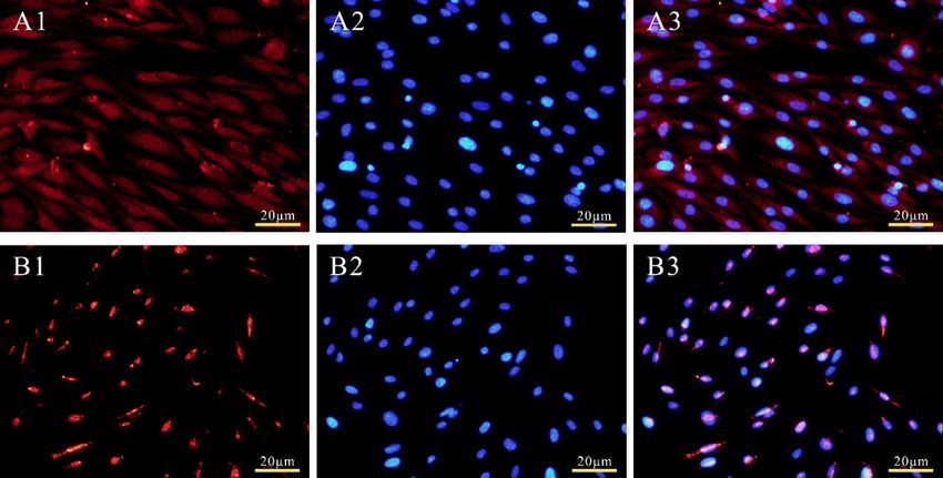
Genes 2020, 11, 575                                                                                 2 of 12

1 (MC1R) [9–11], microphthalmia-associated transcription factor (MITF) [12], and ASIP [13], are likely
to influence the production and deposition of melanin.
     Recently, SLC7A11 was found to be modulated by POU class 2 homeobox 1 gene (POU2F1) in rabbit
pigmentation [14]. POU2F1, also known as OCT1, OTF1, or oct-1B [15], is a potent regulator of stress
responses, metabolism, and tumorigenicity and is itself regulated by phosphorylation, ubiquitination,
O-GlcNAcylation, and other mechanisms [16]. Recent studies into POU2F1 have focused on its impacts
on cancers and tumors [17–20], especially hepatocellular carcinoma [21,22]. Furthermore, POU2F1 was
identified as a transcription factor that binds to the promoter region of SLC7A11 via two binding sites
in the Rex rabbit [14]. However, little is known about the role of POU2F1 in fur color formation in
mammals, and the effects of the gene on the fur color of the Rex rabbit remain unclear.
     Therefore, this study is the next stage of our research to analyze the potential impacts of POU2F1
on the significant genes involved in the formation of Rex rabbit fur color. The expression pattern
of POU2F1 in the dorsal skin of the Rex rabbit with different fur colors, and in different organs,
was analyzed by RT-qPCR. Additionally, a pcDNA3.1(+)-Myc-POU2F1 vector and siRNAs were
constructed to analyze the potential regulatory roles of POU2F1 on SLC7A11, as well as some other
pigmentation-related genes, such as SLC24A5, MITF, MC1R, CREB1, and ASIP, to reveal the regulatory
role of POU2F1 in fur color formation of the Rex rabbit.

2. Materials and Methods

2.1. Animals and Samples
     Eighteen 6-month-old Rex rabbits with 6 different fur colors (n = 3 for each color), including
black (BL), chinchilla (CH), white (WH), brown (BR), protein yellow (PY), and protein chinchilla (PC),
were provided by a rabbit breeding farm in Yuyao, Zhejiang, China. A 1 cm2 sample of dorsal skin
tissue was collected from Rex rabbits of each color (n = 3) after anesthesia by injection of sodium
pentobarbital solution (0.7%) into the ear vein. Tissue samples of different organs (heart, liver, spleen,
lung, kidney, jejunum, colon, ileum, cecum, rectum, dorsal skin, sacculus rotundus, and gizzard) were
collected from other white and black Rex rabbits (n = 3, respectively). White is the most common
color of Rex rabbit and is widely used for fur production around the world because it is easily dyed
and has good plasticity, while black rabbits were chosen for their striking contrast. These rabbits
were euthanized by ear vein injection of 25 mL air after deep anesthesia, and organ samples were
collected about 5 min after confirmation of the absence of heartbeat and death. All tissue samples were
placed in liquid nitrogen immediately after being cut into small pieces and stored at −80 ◦ C until use.
The experimental procedures were approved by the Animal Care and Use Committee of Yangzhou
University (Yangzhou, China, 24 October 2017, No. 201710001).

2.2. Melanocyte Culture and RNA Extraction
     Melanocytes were separated by two-step enzymatic digestion from a 1.5 × 1.5 cm2 section of
dorsal skin from white Rex rabbits according to our previous report [23]. Total RNA of the dorsal
skin and organs was extracted using RNAsimple Total RNA kit (Tiangen, Beijing, China), and
total RNA from melanocytes was extracted using Trizol reagent (Invitrogen, Carlsbad, CA, USA),
according to the manufacturer’s instructions. Electrophoresis with 1% agarose gel was used to
monitor RNA degradation and contamination. RNA purity and concentration were measured using
a NanoPhotometer spectrophotometer (Thermo Fisher Scientific, Wilmington, NC, USA).

2.3. Real-Time qPCR
     The dorsal skin and organs were submitted for quantitative real-time PCR to detect the expression
levels of POU2F1. Briefly, cDNA was synthesized from approximately 1 µg RNA using HiScript II Q
Select RT SuperMix for qPCR (Vazyme, Nanjing, China). Then, the cDNA was used for RT-qPCR with
the AceQ qPCR SYBRR Green Master Mix (Vazyme, Nanjing, China), based on the manufacturer’s
Genes 2020, 11, 575                                                                              3 of 12

instructions. To improve the data reliability, cDNA of each sample was tested three times for technical
replication, and three biological replicates were tested for each group. In order to prevent deviation
due to possible unstable expression of reference genes, glyceraldehyde 3-phosphate dehydrogenase
(GAPDH), β-actin, and hypoxanthine phosphoribosyltransferase 1 (HPRT1) were used as reference
genes [24]. The primer sequences for RT-qPCR are listed in Table 1. The output data were analyzed
using QuantStudioR 5 (Applied Biosystems), and the expression levels were analyzed according to the
2−∆∆Ct method [25].

                                 Table 1. Primer sequences for RT-qPCR.

               Genes      F/R             Sequences of Primers (50 -30 )      Accession No.
                           F          AGCTTGCCATGTCCAAACCAG
               MITF                                                          XM_008260927.1
                           R          TTCATACTTGGGCACTCGCTCT
                           F           CCTCGGCACCCCACTTGCAG
               MC1R                                                          NW_003159591.1
                           R          CAGCACCTCCTTGAGCGTCCTG
                           F         CTGTCCCTCATGAAGACTACCGTT
             SLC24A5                                                         XM_002717863.2
                           R          ACACCACCTATGTTCAACTGGC
                           F        CTGTGCTTCCTCACTGCCTATAGCC
               ASIP                                                          NM_001122939.1
                           R        TTCAGCGCCACAATGGAGACCGAA
                           F          CCTCCCCAGCACTTCCTACACA
              CREB1                                                          XM_008259050.1
                           R          TTCAGCTCCTCAATCAGCGTCT
                           F            TCACCATTGGCTACGTGCT
             SLC7A11                                                           NC_013683.1
                           R          GCCACAAAGATCGGAACTGCT
                           F        GCAGCAGCAGCAGATTCAAGAATG
             POU2F1                                                          XM_017345713.1
                           R          GTGTGCCTGTGTTGCCATCTCC
                           F          CACCAGGGCTGCTTTTAACTCT
             GAPDH                                                           NM_001082253.1
                           R          CTTCCCGTTCTCAGCCTTGACC
                           F             CGGCACCAGGGCGTGAT
              β-actin                                                         NM_001101683
                           R            CGCTTGCTCTGGGCCTCGT
                           F           CGTCGAGGACTTGGAAAGGG
              HPRT1                                                          NM_001105671.1
                           R           TTGAGCACACAGAGGGCTAC
                                   F, forward primers; R, reverse primers.

2.4. Construction of pcDNA3.1(+)-Myc-POU2F1 Vector
     The PrimeScript™ 1st Strand cDNA Synthesis Kit (Takara, Beijing, China) was used to
synthesize Rex rabbit skin cDNA of high quality for the construction of the POU2F1 overexpression
vector. The CDS sequence of the POU2F1 gene was amplified by PCR using Phanta Max
Super-Fidelity DNA Polymerase (Vazyme, Nanjing, China), according to the mRNA sequence of
rabbit POU2F1 (NC_013681.1). The amplified CDS sequence of POU2F1 was subcloned into an NheI-
and EcoRI-digested pcDNA3.1(+)-Myc vector (Invitrogen) (Forward primer: gggagacccaagctggctagc
ATGGCGGACGGAGGAGCA; Reverse primer: gtagtcggatcctttgaattcCTGTGCCTTGGAGGCGGT), and
the recombinant plasmid was named pcDNA3.1(+)-Myc-POU2F1 (Figure 1) for the subsequent steps.
The pcDNA3.1(+)-Myc-POU2F1 was transferred into melanocytes to overexpress the POU2F1 gene,
and the expression levels of the fur-color-related genes (SLC24A5, SLC7A11, MITF, MC1R, CREB1, and
ASIP) were detected by RT-qPCR, according to the above method.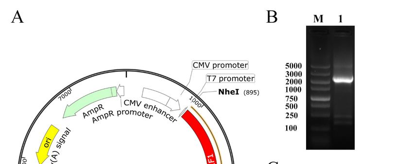
Genes 2020,
   Genes    11, 575
         2020,  11, x FOR PEER REVIEW                                                                             4 of
                                                                                                                4 of 12 12

      Figure 1. Identification and construction of pcDNA3.1(+)-Myc-POU2F1. (A) Plasmid map
      of Figure   1. Identification and construction
         pcDNA3.1(+)-Myc-POU2F1.          The gene of    pcDNA3.1(+)-Myc-POU2F1.
                                                      sequence encoding POU2F1 was(A) Plasmid
                                                                                       inserted map
                                                                                                  into of
                                                                                                        the
         pcDNA3.1(+)-Myc-POU2F1.       The  gene   sequence  encoding POU2F1   was   inserted   into  the
     pcDNA3.1(+)-Myc vector between the corresponding restriction sites NheI and EcoRI. (B) PCR products
         pcDNA3.1(+)-Myc vector between the corresponding restriction sites NheI and EcoRI. (B) PCR
      of POU2F1 gene. M, DL5000 DNA Marker, Lane 1, POU2F1 mRNA sequence. (C) Identification
         products of POU2F1 gene. M, DL5000 DNA Marker, Lane 1, POU2F1 mRNA sequence. (C)
      of pcDNA3.1(+)-Myc-POU2F1 digested by NheI and EcoRI. M, DL10000 DNA Marker. Lane 1,
         Identification of pcDNA3.1(+)-Myc-POU2F1 digested by NheI and EcoRI. M, DL10000 DNA Marker.
     production of pcDNA3.1(+)-Myc-POU2F1 after NheI and EcoRI digestion. Lane 2, production of
         Lane 1, production of pcDNA3.1(+)-Myc-POU2F1 after NheI and EcoRI digestion. Lane 2, production
     pcDNA3.1(+)-Myc-POU2F1 after EcoRI digestion. Lane 3, pcDNA3.1(+)-Myc-POU2F1 before digestion.
         of pcDNA3.1(+)-Myc-POU2F1 after EcoRI digestion. Lane 3, pcDNA3.1(+)-Myc-POU2F1 before
         digestion.
2.5. Subcellular Localization of POU2F1 Protein
      PSORT
    2.5.         (www.psort.org)
         Subcellular                  was used
                     Localization of POU2F1          to predict the localization of the POU2F1 protein.
                                               Protein
The pcDNA3.1(+)-Myc-POU2F1 was transferred into melanocytes using ExFect 2000 (Vazyme, Nanjing,
          PSORT (www.psort.org) was used to predict the localization of the POU2F1 protein. The
China), according to the manufacturer’s instructions with 1 µg pcDNA3.1(+)-Myc-POU2F1 in 1 µL
    pcDNA3.1(+)-Myc-POU2F1 was transferred into melanocytes using ExFect 2000                    (Vazyme, Nanjing,
ExFect 2000 for each well, and cultured in a 24-well plate with CO2 at 37 ◦ C for 24 h. Then, 4%
    China), according to the manufacturer’s instructions with 1μg pcDNA3.1(+)-Myc-POU2F1 in 1 μL
paraformaldehyde      was used to fix the cells at room temperature (RT) for 20 min, followed by penetration
    ExFect 2000 for each well, and cultured in a 24-well plate with CO2 at 37 °C for 24 h. Then, 4%
in 0.3%
    paraformaldehyde(Solarbio,
          Triton  X-100   was usedBeijing,
                                       to fix China)
                                              the cellsat at
                                                          RTroom
                                                              for 10temperature
                                                                     min and blocking     in goat
                                                                                   (RT) for        serum
                                                                                              20 min,      at 37 ◦ Cbyfor
                                                                                                        followed
30 min.    The cells
    penetration       wereTriton
                  in 0.3%   incubated
                                   X-100with    the primary
                                           (Solarbio,  Beijing,antibody
                                                                 China) at(Myc)
                                                                            RT forat104 ◦min
                                                                                         C overnight     and in
                                                                                              and blocking    with
                                                                                                                 goatthe
second   antibody   (Cy3-conjugate)     at RT for 2 h.  After  dying  in DAPI   for 10 min,  the  cells were
    serum at 37 °C for 30 min. The cells were incubated with the primary antibody (Myc) at 4 °C overnight     observed
andand
     photographed      under
         with the second       a fluorescent
                           antibody            invertedatmicroscope.
                                      (Cy3-conjugate)                     The dying
                                                             RT for 2 h. After cells were  washed
                                                                                      in DAPI   for 10with
                                                                                                        min,PBS  (three
                                                                                                             the cells
times  forobserved
    were    5 min/time)
                     andbetween      each of
                          photographed        the above
                                            under          steps. inverted microscope. The cells were washed
                                                   a fluorescent
    with PBS (three times for 5 min/time) between each of the above steps.
2.6. Inhibition of POU2F1 by siRNA
   2.6. Inhibition
     Three  siRNAs of POU2F1
                       (with 50 by
                                FAMsiRNA
                                      modification) and negative control siRNAs were ordered from Suzhou
GenePharma
         Three Co.,   Ltd.(with
                 siRNAs     (Suzhou,
                                  5′ FAM China)   (Table 2). and
                                             modification)   The negative
                                                                  siRNAs were     diluted
                                                                            control  siRNAsinto 20 µM
                                                                                              were     with from
                                                                                                    ordered DEPC
water,  and GenePharma
   Suzhou    then the complexed
                             Co., Ltd. 2(Suzhou,
                                          µL diluted    siRNAs
                                                   China)  (Tableand  1 µLsiRNAs
                                                                  2). The   Lipofectamine    2000into
                                                                                    were diluted   (InvitrogenTM)
                                                                                                      20 μM with
   DEPC
were        water, to
      transferred    andthethen   the complexed
                            melanocytes     when the2 cell
                                                         μL confluence
                                                             diluted siRNAs
                                                                        reached  and  1 μL Lipofectamine
                                                                                   approximately            2000
                                                                                                   70%. After  24 h,
   (InvitrogenTM)
fluorescence          were transferred
               microscopy     was used to  to the melanocytes
                                               detect          when the
                                                      the transfection    cell confluence
                                                                       efficiency.        reached
                                                                                    The total RNAapproximately
                                                                                                    extracted from
Genes 2020, 11, 575 5 of 12
Genes 2020, 11, x FOR PEER REVIEW 5 of 12
70%. After 24 h, fluorescence microscopy was used to detect the transfection efficiency. The total RNA
the cells after transfection was used for RT-qPCR to examine the expression levels of POU2F1 and
extracted from the cells after transfection was used for RT-qPCR to examine the expression levels of
other genes, according to the above methods.
POU2F1 and other genes, according to the above methods.
Table 2. Primer sequences of siRNAs.
Table 2. Primer sequences of siRNAs.
siRNAs S/A Sequences of Primers (50 -30 )
siRNAs S/A Sequences of Primers (5′-3′)
S S GGAAGAGCCCAGUGACCUUTT
GGAAGAGCCCAGUGACCUUTT
siRNA1
siRNA1
A AAAGGUCACUGGGCUCUUCCTT
AAGGUCACUGGGCUCUUCCTT
S CCUUGAACCUCAGCUUUAATT
siRNA2 S CCUUGAACCUCAGCUUUAATT
siRNA2 A UUAAAGCUGAGGUUCAAGGTT
A UUAAAGCUGAGGUUCAAGGTT
S GCCUCCACCUCCGAGACAUTT
siRNA3 S
siRNA3 A GCCUCCACCUCCGAGACAUTT
AUGUCUCGGAGGUGGAGGCTT
A AUGUCUCGGAGGUGGAGGCTT
S, sense strands; A, anti-sense strands.
S, sense strands; A, anti-sense strands.
2.7. 2.7.
Statistical Analysis
Statistical Analysis
All All
values in the
values text
in the and
text andfigures
figuresarearepresented
presentedas asthe means ±±standard
the means standarddeviation
deviation (SD)
(SD) calculated
calculated
by GraphPad Prism software (GraphPad Software, La Jolla, CA, USA). A paired
by GraphPad Prism software (GraphPad Software, La Jolla, CA, USA). A paired T-test from SPSS T-test from SPSS25 25
waswas
performed between
performed between2 groups
2 groups (control and
(control each
and test
each group).
test group).Statistical
Statisticaldifferences
differences are
are presented
presented at
probability
at probability of pGenes 2020, 11, x FOR PEER REVIEW 6 of 12
Genes 2020, 11, 575 6 of 12
for calculating the relative expression level in corresponding Rex rabbits in figure B. * p < 0.05, ** p <
0.01, and *** p < 0.001.
3.2. The Tissue Distribution of POU2F1 Gene Expression in WH- and BL-Rex Rabbits
3.2. The Tissue Distribution of POU2F1 Gene Expression in WH- and BL-Rex Rabbits
POU2F1 expression in different organs of the WH- and BL-Rex rabbits is shown in Figure 2B.
POU2F1
The relative expression
expression in different
levels organs
of POU2F1 of the WH-
in different and BL-Rex
organs rabbits
of BL-Rex is shown
rabbits in Figure
normalized by 2B.
three
The relative expression levels of POU2F1 in different organs of BL-Rex rabbits
reference genes (GAPDH, β-actin and HPRT1) were generally consistent (Supplementary Figure S1). normalized by three
reference
POU2F1 genes (GAPDH,
expression β-actin and
in the jejunum, HPRT1)
which had were generally
a relatively lowconsistent
and stable (Supplementary
expression level Figure
in BL-S1).
and
POU2F1
WH-Rex expression
rabbits, in theasjejunum,
was used a controlwhich had as
(defined a relatively low and
1) to calculate thestable expression
fold change level inexpression
in POU2F1 BL- and
WH- Rex
in other rabbits,
organs. POU2F1was was
usedwidely
as a control
expressed(defined as 1) organs
in different to calculate
of thethe
WH-fold
andchange
BL-Rex inrabbits,
POU2F1 and
expression in other organs. POU2F1 was widely expressed in different organs
almost all other organs (except dorsal skin and ileum) had significantly higher expression levelsof the WH- and BL-than
Rex rabbits,
jejunum and In
(p < 0.05). almost all other
addition, organs
POU2F1 (except
showed thedorsal
highestskin and ileum)
expression hadinsignificantly
levels higher
the lung in both WH-
expression levels than jejunum (p < 0.05). In addition, POU2F1 showed the highest expression levels
and BL-Rex rabbits (29.15 and 7.99 times higher, respectively), while the dorsal skin of BL-Rex rabbit
in the lung in both WH- and BL-Rex rabbits (29.15 and 7.99 times higher, respectively), while the
had significantly lower POU2F1 expression (0.06 times).
dorsal skin of BL- Rex rabbit had significantly lower POU2F1 expression (0.06 times).
3.3. Subcellular Localization of POU2F1 Protein
3.3. Subcellular Localization of POU2F1 Protein
PSORT predicted that 95.7% of the POU2F1 protein would be located at the nucleus. Subcellular
PSORT predicted that 95.7% of the POU2F1 protein would be located at the nucleus. Subcellular
localization analysis was used to determine the localization of the POU2F1 protein. As shown in
localization analysis was used to determine the localization of the POU2F1 protein. As shown in
Figure 3A, the empty pcDNA3.1(+)-Myc vector without the POU2F1 gene was randomly distributed
Figure 3A, the empty pcDNA3.1(+)-Myc vector without the POU2F1 gene was randomly distributed
throughout the whole cell, including the nucleus and cytoplasm. The pcDNA3.1(+)-Myc-POU2F1 was
throughout the whole cell, including the nucleus and cytoplasm. The pcDNA3.1(+)-Myc-POU2F1 was
only found
only found inin
thethenucleus
nucleus(Figure
(Figure3B),
3B),indicating that the
indicating that the localization
localizationofofthe
thePOU2F1
POU2F1 protein
protein in in
thethe
dorsal skin
dorsal cells
skin ofofRex
cells Rexrabbits
rabbitswas
wasnuclear,
nuclear,which
which is
is consistent withthe
consistent with theprediction.
prediction.
Figure
Figure 3. 3. Subcellularlocalization
Subcellular localizationofofPOU2F1
POU2F1 protein
protein in
in rabbit
rabbit melanocytes.
melanocytes. TheThepcDNA3.1(+)-Myc
pcDNA3.1(+)-Myc
(A1–A3) and recombinant plasmids pcDNA3.1(+)-Myc-POU2F1 (B1–B3)
(A1–A3) and recombinant plasmids pcDNA3.1(+)-Myc-POU2F1 (B1–B3) were transientlywere transiently transfected
transfected
into rabbit melanocytes. Cells in groups A and B are melanocytes magnified 40 times by fluorescence
into rabbit melanocytes. Cells in groups A and B are melanocytes magnified 40 times by fluorescence
inverted
inverted microscope.A1
microscope. A1(B1)
(B1) is
is the
the Myc-tag
Myc-tag dyed
dyed byby Cy3,
Cy3,and
andA2A2(B2)
(B2)isisthe
theDAPI-dyed
DAPI-dyed nucleus,
nucleus,
which
which areare merged
merged inin
A3A3(B3).
(B3).
3.4.3.4.
TheThe Effects
Effects ofofPOU2F1
POU2F1Gene
Geneon
onOther
OtherPigmentation-Related
Pigmentation-Related Genes
Genes
POU2F1
POU2F1 siRNAinterference
siRNA interferenceand
andoverexpression
overexpression were
were performed
performedininmelanocytes
melanocytes totoanalyze thethe
analyze
potential functional mechanisms of POU2F1 during melanin deposition in the dorsal skin of
potential functional mechanisms of POU2F1 during melanin deposition in the dorsal skin of Rex rabbits, Rex
rabbits,by
followed followed by detection
detection of the expression
of the expression levels oflevels of pigment-related
pigment-related genesgenes (SLC7A11,
(SLC7A11, SLC24A5,
SLC24A5, MITF,
MITF, MC1R, CREB1 and ASIP) by RT-qPCR. siRNA1, siRNA2, and siRNA3
MC1R, CREB1 and ASIP) by RT-qPCR. siRNA1, siRNA2, and siRNA3 were successfully transferredwere successfully
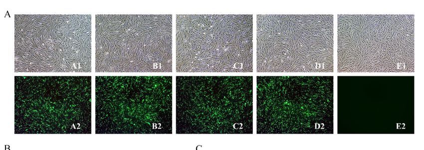
into melanocytes (Figure 4A); siRNA3 had the best interference efficiency (85.09%) (Figure 4B,C) and
was used for POU2F1 interference in the next stage of the experiment.Genes 2020, 11, x FOR PEER REVIEW 7 of 12
transferred into melanocytes (Figure 4A); siRNA3 had the best interference efficiency (85.09%)
Genes 2020, 11, 575
(Figure 4B,C) and was used for POU2F1 interference in the next stage of the experiment. 7 of 12
Figure 4. Interference efficiency of siRNAs on POU2F1 expression. (A) Melanocyte morphology
Figure 4. Interference efficiency of siRNAs on POU2F1 expression. (A) Melanocyte morphology 24 h
24 h after FAM-siRNAs transfection. (A–E) represent siRNA-1, siRNA-2, siRNA-3, NC, and blank,
after FAM-siRNAs transfection. A–E represent siRNA-1, siRNA-2, siRNA-3, NC, and blank,
respectively. (B,C) show interference efficiency of siRNAs on POU2F1 expression. *** p < 0.001.
respectively. (B,C) show interference efficiency of siRNAs on POU2F1 expression. *** p < 0.001.
The POU2F1 gene was overexpressed by 1326.3 times the baseline by pcDNA3.1(+)-Myc-POU2F1
TheSimultaneously,
(Figure 5A). POU2F1 gene was overexpressed
the SLC7A11 gene wasby 1326.3 times the
significantly baseline by pcDNA3.1(+)-Myc-
downregulated 0.55 times (Figure 5B),
POU2F1
indicating the(Figure 5A).regulation
negative Simultaneously,
of thethe SLC7A11
SLC7A11 geneby
gene was significantly
POU2F1. downregulated
In addition, 0.55 SLC24A5
MITF and times
(Figure 5B), indicating the negative regulation of the SLC7A11 gene by POU2F1. In addition,
were downregulated (0.56 and 0.73 times, respectively), but ASIP was significantly upregulated MITF
and SLC24A5 were downregulated (0.56 and 0.73 times, respectively), but ASIP was significantly
3.76 times. However, when POU2F1 expression was inhibited by siRNA3 (Figure 5C), the SLC7A11
upregulated 3.76 times. However, when POU2F1 expression was inhibited by siRNA3 (Figure 5C),
gene showed significant overexpression at 1.58 times the baseline (Figure 5D), emphasizing the
the SLC7A11 gene showed significant overexpression at 1.58 times the baseline (Figure 5D),
inhibitory role of POU2F1 on SLC7A11. In addition, ASIP was significantly downregulated 0.63 times,
emphasizing the inhibitory role of POU2F1 on SLC7A11. In addition, ASIP was significantly
and MC1R was significantly
downregulated 0.63 times,upregulated
and MC1R was2.62 times.
significantly upregulated 2.62 times.
Figure 5. Cont.Genes 2020, 11, 575 8 of 12
Figure 5. Expression levels of SLC7A11 and other pigmentation-related genes after the overexpression
and interference of POU2F1. (A,C) are the relative gene expression levels of POU2F1 in rabbit
melanocytes transfected with pcDNA3.1(+)-Myc-POU2F1 vector and siRNA-3, respectively; (B,D) are
the relative expression levels of other pigmentation-related genes in rabbit melanocytes transfected
with pcDNA3.1(+)-Myc-POU2F1 vector and siRNA-3, respectively. The relative expression levels of the
above genes were measured by RT-qPCR and calculated by the 2−∆∆Ct method. * p < 0.05, ** p < 0.01,
and *** p < 0.001.
4. Discussion
In this study, we compared the expression levels of POU2F1 in Rex rabbits of various colors
to investigate the possible impacts of the gene on fur color. POU2F1 was generally but differently
expressed in the dorsal skin of Rex rabbits, revealing its potential roles in fur color formation. POU2F1
showed the lowest expression levels in BL-Rex rabbits and the highest expression levels in the BR-Rex
rabbit. In addition, the opposite pattern of expression of the SLC7A11 gene was identified in Rex rabbits
with the same color [14]. This is consistent with the negative regulation of the SLC7A11 gene by POU2F1,
which was reported previously. Additionally, SLC7A11 was reported to positively regulate melanin
levels, and higher SLC7A11 gene expression levels led to increased melanin deposition [14]. This may
explain why POU2F1 and SLC7A11 showed the highest and lowest expression levels, respectively,
in BL-Rex rabbits.
Additionally, the tissue distribution of POU2F1 expression was analyzed to explore potential
differential expression. Because the use of GAPDH as a reference gene to correct gene expression
levels in the Rex rabbit was relatively novel, another two reference genes (β-actin and HPRT1) were
employed for the detection of POU2F1 expression levels. The expression levels of POU2F1 normalized
by GAPDH, HPRT1 and β-actin were roughly consistent, which revealed the usability of GAPDH as
a reference gene in the Rex rabbit and improved the reliability of the data in this study. POU2F1 was
widely expressed in different organs of white and black Rex rabbits, with the highest expression levels
in the lungs. POU2F1 has been reported to be involved in carcinogenesis and other disease processes
by promoting cell proliferation and metastasis [26], e.g., in type 2 diabetes [15,27], head and neck
cancer [28], cervical cancer [19], and liver cancer [29]. The wide involvement of POU2F1 in various
kinds of cancer and in the immune response to cancers suggests its general expression in different
organs, which was also found in this study. Interestingly, most recent studies on the POU2F1 gene
have focused on its regulatory impacts on liver cancer [21,22,29]. POU2F1 was found to be highly
expressed in the liver of Rex rabbits, suggesting a potential immune-related role for the POU2F1 gene,
a phenomenon which deserves further research. In addition, POU2F1 showed the highest expressionGenes 2020, 11, 575 9 of 12
levels in the lungs, which may be related to the expression and important roles of SLC7A11 in response
to lung cancer [30,31].
As a transcription factor, POU2F1 was predicted to be expressed and to have some biological
functions in the nucleus of melanocytes of the Rex rabbit, as was reported in human HEK-293T
cells [32]. A subcellular localization study confirmed the prediction that the POU2F1 protein is located
at the nucleus and does not have a transmembrane structure, which are classical characteristics of
transcription factors. POU2F1 played biological roles in the nucleus of melanocytes, suggesting it has
a potential role as a transcription factor regulating the expression of some other genes. Overexpression
and interference of POU2F1 revealed its negative regulatory effect on the SLC7A11 gene, which
was consistent with the findings of our previous study. Abnormal expression of POU2F1 not only
regulated the expression of the SLC7A11 gene but also some other fur-color-related genes. The positive
influence of POU2F1 on the expression of ASIP was also identified in this study, which fits with the
negative relationship between ASIP and SLC7A11 [14]. In addition, expression of the MC1R gene
was upregulated when POU2F1 expression was affected. Both ASIP and MC1R have been reported
to be involved in the melanogenesis pathway and the regulation of fur color formation [9,10,13].
Furthermore, the overexpression of POU2F1 negatively regulated the expression of SLC24A5 and
MITF, which are related to pigmentation. The findings of various studies indicate the important role
SLC24A5 has on human skin pigmentation: it regulates human epidermal melanogenesis and related
diseases, such as nonsyndromic oculocutaneous albinism [33–35]. MITF plays significant roles in
signal transduction and transcription in melanocytes during the color formation of skin and fur [36,37].
In addition, ASIP, MC1R, SLC24A5, and MITF were found to not only affect the formation of human
skin color [38,39] but also affect fur color in different animals, including mice [37], sheep [40,41],
horses [42–44], pigs [45], and rhesus macaques [46]. The significant associations between POU2F1 and
other fur-color-related genes further revealed the potential impacts of POU2F1 on Rex rabbit fur color.
5. Conclusions
The POU2F1 gene was significantly more highly expressed in the dorsal skin of brown and
protein yellow Rex rabbits compared with that of Rex rabbits with black, chinchilla, white, and protein
chinchilla fur. POU2F1 affects the expression of genes, including SLC24A5, SLC7A11, MITF, MC1R,
and ASIP, that are related to the formation of fur color in Rex rabbits. This study highlighted the
potential regulatory role of POU2F1 in the fur color of Rex rabbits.
Supplementary Materials: The following are available online at http://www.mdpi.com/2073-4425/11/5/575/s1,
Figure S1: Expression level of POU2F1 in different organ tissues of Black Rex rabbit.
Author Contributions: Conceptualization, N.Y.; Formal analysis, B.Z.; Funding acquisition, X.W.; Investigation,
N.Y., Z.B. and M.L.; Resources, S.H.; Supervision, X.W.; Writing—original draft, N.Y.; Writing—review & editing,
Y.C. All authors have read and agreed to the published version of the manuscript.
Funding: This research was funded by Modern Agricultural Industrial System Special Funding, grant
number CARS-43-A-1.
Conflicts of Interest: The authors declare no conflict of interest.
References
1. Tang, J.; Zhang, Y. Color Rabbit Hair Property and Market Foreground. Adv. Mater. Res. 2011, 332, 35–40.
[CrossRef]
2. Kulikov, A.V.; Bazhenova, E.Y.; Kulikova, E.A.; Fursenko, D.V.; Trapezova, L.I.; Terenina, E.E.; Mormede, P.;
Popova, N.K.; Trapezov, O.V. Interplay between aggression, brain monoamines and fur color mutation in the
American mink. Genes Brain Behav. 2016, 15, 733–740. [CrossRef] [PubMed]
3. Cieslak, M.; Reissmann, M.; Hofreiter, M.; Ludwig, A. Colours of domestication. Biol. Rev. 2011, 86, 885–899.
[CrossRef] [PubMed]
4. Chang, T.-S. An Updated Review of Tyrosinase Inhibitors. Int. J. Mol. Sci. 2009, 10, 2440–2475. [CrossRef]
[PubMed]Genes 2020, 11, 575 10 of 12
5. Sturm, R.A. Molecular genetics of human pigmentation diversity. Hum. Mol. Genet. 2009, 18, R9–R17.
[CrossRef]
6. Kim, J.Y.; Kanai, Y.; Chairoungdua, A.; Cha, S.H.; Matsuo, H.; Kim, D.K.; Inatomi, J.; Sawa, H.; Ida, Y.;
Endou, H. Human cystine/glutamate transporter: cDNA cloning and upregulation by oxidative stress in
glioma cells. Biochim. Biophys. Acta (BBA) Biomembr. 2001, 1512, 335–344. [CrossRef]
7. Chintala, S.; Li, W.; Lamoreux, M.L.; Ito, S.; Wakamatsu, K.; Sviderskaya, E.V.; Bennett, D.C.; Park, Y.-M.;
Gahl, W.A.; Huizing, M.; et al. Slc7a11 gene controls production of pheomelanin pigment and proliferation
of cultured cells. Proc. Natl. Acad. Sci. USA 2005, 102, 10964–10969. [CrossRef]
8. Li, H.-T.; He, X.; Zhou, Z.; Zhao, S.-H.; Zhang, W.-X.; Liu, G.; Zhao, Z.-S.; Jia, B. Expression levels of Slc7a11
in the skin of Kazakh sheep with different coat colors. Hereditas (Beijing) 2012, 34, 1314–1319. [CrossRef]
9. Luo, H.; Hou, J.N.; Li, Y.-M.; Yan, S.Q. Cloning and Sequence of MC1R Gene 50 -Flanking Sequence in Rabbit.
China Anim. Husb. Vet. Med. 2011, 38, 89–92.
10. Yang, G.-L.; Fu, D.-L.; Lang, X.; Wang, Y.-T.; Cheng, S.-R.; Fang, S.-L.; Luo, Y.-Z. Mutations in MC1R Gene
Determine Black Coat Color Phenotype in Chinese Sheep. Sci. World J. 2013, 2013, 1–8. [CrossRef]
11. Thomas, A.C.; Heux, P.; Santos, C.; Arulvasan, W.; Solanky, N.; Carey, M.E.; Gerrelli, D.; Kinsler, V.A.;
Etchevers, H.C. Widespread dynamic and pleiotropic expression of the melanocortin-1-receptor (MC1R)
system is conserved across chick, mouse and human embryonic development. Birth Defects Res. 2018, 110,
443–455. [CrossRef] [PubMed]
12. Flesher, J.L.; Paterson-Coleman, E.K.; Vasudeva, P.; Ruiz-Vega, R.; Marshall, M.; Pearlman, E.; MacGregor, G.R.;
Neumann, J.; Ganesan, A.K. Delineating the role of MITF isoforms in pigmentation and tissue homeostasis.
Pigment Cell Melanoma Res. 2019, 33, 279–292. [CrossRef] [PubMed]
13. Fontanesi, L.; Forestier, L.; Allain, D.; Scotti, E.; Beretti, F.; Deretz-Picoulet, S.; Pecchioli, E.; Vernesi, C.;
Robinson, T.; Malaney, J.L.; et al. Characterization of the rabbit agouti signaling protein (ASIP) gene:
Transcripts and phylogenetic analyses and identification of the causative mutation of the nonagouti black
coat colour. Genomics 2010, 95, 166–175. [CrossRef]
14. Chen, Y.; Hu, S.; Mu, L.; Zhao, B.; Wang, M.; Yang, N.; Bao, G.; Zhu, C.; Wu, X. Slc7a11 Modulated by POU2F1
is Involved in Pigmentation in Rabbit. Int. J. Mol. Sci. 2019, 20, 2493. [CrossRef] [PubMed]
15. Ng, M.C.Y.; Lam, V.K.L.; Tam, C.H.T.; Chan, A.W.; So, W.-Y.; Ma, R.C.; Zee, B.; Waye, M.M.Y.; Mak, W.W.;
Hu, C.; et al. Association of the POU class 2 homeobox 1 gene (POU2F1) with susceptibility to Type 2
diabetes in Chinese populations. Diabet. Med. 2010, 27, 1443–1449. [CrossRef]
16. Kang, J.; Shen, Z.; Lim, J.-M.; Handa, H.; Wells, L.; Tantin, D. Regulation of Oct1/Pou2f1 transcription activity
by O-GlcNAcylation. FASEB J. 2013, 27, 2807–2817. [CrossRef]
17. Xiao, Y.; Xiang, L.; Dai, W.; Tang, W.; Wang, J.; Zhang, Y.; Wu, X.; Liu, G.; Chen, Y.; Zhu, H.; et al.
The POU2F1/miR-4490/USP22 Axis Regulates Cell Proliferation and Metastasis in Gastric Cancer. Available
online: https://ssrn.com/abstract=3384868 (accessed on 5 May 2019). [CrossRef]
18. Dey, A.; Sen, S.; Maulik, U. Studying POU2F1 to Unveil the Structural Facet for Pan-Cancer Analysis
Considering the Functional Annotations and Sequence-Structure Space Paradigm. bioRxiv 2019, 806489.
19. Zhang, R.; Lu, H.; Lyu, Y.-Y.; Yang, X.-M.; Zhu, L.-Y.; Yang, G.-D.; Jiang, P.-C.; Re, Y.; Song, W.-W.; Wang, J.-H.
E6/E7-P53-POU2F1-CTHRC1 axis promotes cervical cancer metastasis and activates Wnt/PCP pathway.
Sci. Rep. 2017, 7, 44744. [CrossRef]
20. Vázquez-Arreguín, K.; Bensard, C.; Schell, J.C.; Swanson, E.; Chen, X.; Rutter, J.; Tantin, D. Oct1/Pou2f1 is
selectively required for colon regeneration and regulates colon malignancy. PLoS Genet. 2019, 15, e1007687.
[CrossRef]
21. Zhong, Y.; Huang, H.; Chen, M.; Huang, J.; Wu, Q.; Yan, G.-R.; Chen, D. POU2F1 over-expression correlates
with poor prognoses and promotes cell growth and epithelial-to-mesenchymal transition in hepatocellular
carcinoma. Oncotarget 2017, 8, 44082–44095. [CrossRef]
22. Hong, Y.Z.; Guan, Y.C.; Shi, P.W.; Yu, C.; Jian, P.H. POU2F1 promotes growth and metastasis of hepatocellular
carcinoma through the FAT1 signaling pathway. Am. J. Cancer Res. 2017, 7, 1665–1679.
23. Chen, Y.; Hu, S.; Wang, M.; Zhao, B.; Yang, N.; Li, J.; Chen, Q.; Liu, M.; Zhou, J.; Bao, G.; et al. Characterization
and Establishment of an Immortalized Rabbit Melanocyte Cell Line Using the SV40 Large T Antigen. Int. J.
Mol. Sci. 2019, 20, 4874. [CrossRef] [PubMed]Genes 2020, 11, 575 11 of 12
24. Svingen, T.; Letting, H.; Hadrup, N.; Hass, U.; Vinggaard, A.M. Selection of reference genes for quantitative
RT-PCR (RT-qPCR) analysis of rat tissues under physiological and toxicological conditions. PeerJ 2015, 3, 855.
[CrossRef] [PubMed]
25. Schmittgen, T.D.; Livak, K.J. Analyzing real-time PCR data by the comparative CT method. Nat. Protoc. 2008,
3, 1101–1108. [CrossRef] [PubMed]
26. Xie, C.-H.; Cao, Y.-M.; Huang, Y.; Shi, Q.-W.; Guo, J.-H.; Fan, Z.-W.; Li, J.-G.; Chen, B.-W.; Wu, B.-Y. Long
non-coding RNA TUG1 contributes to tumorigenesis of human osteosarcoma by sponging miR-9-5p and
regulating POU2F1 expression. Tumor Biol. 2016, 37, 15031–15041. [CrossRef] [PubMed]
27. Batool, A.; Jahan, N.; Sun, Y.; Hanif, A.; Xue, H. Genetic association of IDE, POU2F1, PON1, IL1 alpha and
IL1 beta with type 2 diabetes in Pakistani population. Mol. Biol. Rep. 2014, 41, 3063–3069. [CrossRef]
28. Sharpe, D.J.; Orr, K.S.; Moran, M.; White, S.J.; McQuaid, S.; Lappin, T.R.; Thompson, A.; James, J. POU2F1
activity regulates HOXD10 and HOXD11 promoting a proliferative and invasive phenotype in Head and
Neck cancer. Oncotarget 2014, 5, 8803–8815. [CrossRef]
29. Liu, Y.; Wang, Y.; Sun, X.; Mei, C.; Zha, X. MiR-449a promotes liver cancer cell apoptosis by down-regulation
of Calpain6 and POU2F1. Oncotarget 2015, 7, 13491–13501. [CrossRef]
30. Moreno, J.A.; Lambros, M.P.; Ouchi, K.; Kuwahara, Y.; Iehara, T.; Konishi, E.; Hosoi, H. Abstract 3209:
The expression of SLC7A11 transporter in lung and pancreatic cancer tissues at different stages of development.
Tumor Biol. 2015, 75, 3209. [CrossRef]
31. Ji, X.; Qian, J.; Rahman, S.M.J.; Siska, P.J.; Zou, Y.; Harris, B.K.; Hoeksema, M.D.; Trenary, I.A.; Heidi, C.;
Eisenberg, R.; et al. xCT (SLC7A11)-mediated metabolic reprogramming promotes non-small cell lung cancer
progression. Oncogene 2018, 37, 5007–5019. [CrossRef]
32. Gao, J.; Chen, Y.; Yang, Y.; Liang, J.; Xie, J.; Liu, J.; Li, S.; Zheng, G.; Xie, L.; Zhang, R. The transcription
factor Pf-POU3F4 regulates expression of the matrix protein genes Aspein and Prismalin-14 in pearl oyster
(Pinctada fucata). FEBS J. 2016, 283, 1962–1978. [CrossRef] [PubMed]
33. Wei, A.; Zang, D.-J.; Zhang, Z.; Liu, X.-Z.; He, X.; Yang, L.; Wang, Y.; Zhou, Z.; Zhang, M.-R.; Dai, L.-L.; et al.
Exome Sequencing Identifies SLC24A5 as a Candidate Gene for Nonsyndromic Oculocutaneous Albinism.
J. Investig. Dermatol. 2013, 133, 1834–1840. [CrossRef] [PubMed]
34. Ginger, R.S.; Askew, S.E.; Ogborne, R.M.; Wilson, S.; Ferdinando, D.; Dadd, T.; Smith, A.M.;
Kazi, S.; Szerencsei, R.T.; Winkfein, R.J.; et al. SLC24A5 Encodes atrans-Golgi Network Protein with
Potassium-dependent Sodium-Calcium Exchange Activity That Regulates Human Epidermal Melanogenesis.
J. Biol. Chem. 2007, 283, 5486–5495. [CrossRef] [PubMed]
35. Lamason, R.; Mohideen, M.-A.P.; Mest, J.R.; Wong, A.C.; Norton, H.L.; Aros, M.C.; Jurynec, M.; Mao, X.;
Humphreville, V.R.; Humbert, J.E.; et al. SLC24A5, a Putative Cation Exchanger, Affects Pigmentation in
Zebrafish and Humans. Science 2005, 310, 1782–1786. [CrossRef] [PubMed]
36. Goding, C.R. Mitf from neural crest to melanoma: Signal transduction and transcription in the melanocyte
lineage. Genes Dev. 2000, 14, 1712–1728. [PubMed]
37. Bauer, G.L.; Praetorius, C.; Bergsteinsdóttir, K.; Hallsson, J.H.; Gísladóttir, B.K.; Schepsky, A.; Swing, D.A.;
O’Sullivan, T.N.; Arnheiter, H.; Bismuth, K.; et al. The Role of MITF Phosphorylation Sites During Coat Color
and Eye Development in Mice Analyzed by Bacterial Artificial Chromosome Transgene Rescue. Genetics
2009, 183, 581–594. [CrossRef]
38. Jacobs, L.C.; Hamer, M.A.; Gunn, D.A.; Deelen, J.; Lall, J.S.; Van Heemst, D.; Uh, H.-W.; Hofman, A.;
Uitterlinden, A.G.; Griffiths, C.E.M.; et al. A Genome-Wide Association Study Identifies the Skin Color
Genes IRF4, MC1R, ASIP, and BNC2 Influencing Facial Pigmented Spots. J. Investig. Dermatol. 2015, 135,
1735–1742. [CrossRef]
39. Landi, M.T.; Kanetsky, P.A.; Tsang, S.; Gold, B.; Munroe, D.; Rebbeck, T.; Swoyer, J.; Ter-Minassian, M.;
Hedayati, M.; Grossman, L.; et al. MC1R, ASIP, and DNA Repair in Sporadic and Familial Melanoma in
a Mediterranean Population. J. Natl. Cancer Inst. 2005, 97, 998–1007. [CrossRef]
40. Fontanesi, L.; Dall’Olio, S.; Beretti, F.; Portolano, B.; Russo, V. Coat colours in the Massese sheep breed are
associated with mutations in the agouti signalling protein (ASIP) and melanocortin 1 receptor (MC1R) genes.
Animal 2010, 5, 8–17. [CrossRef]
41. Han, J.; Yang, M.; Yue, Y.; Guo, T.; Liu, J.; Niu, C.; Yang, B. Analysis of agouti signaling protein (ASIP) gene
polymorphisms and association with coat color in Tibetan sheep (Ovis aries). Genet. Mol. Res. 2015, 14,
1200–1209. [CrossRef]Genes 2020, 11, 575 12 of 12
42. Rieder, S.; Taourit, S.; Mariat, D.; Langlois, B.; Guérin, G. Mutations in the agouti (ASIP), the extension
(MC1R), and the brown (TYRP1) loci and their association to coat color phenotypes in horses (Equus caballus).
Mamm. Genome 2001, 12, 450–455. [CrossRef] [PubMed]
43. Kreuzer, S. A Mutation in the Equine SLC24A5 Gene Is Associated With A Dilution Of Black Horses.
In Proceedings of the World Congress on Genetics Applied to Livestock Production, Leipzig, Germany,
1–6 August 2010.
44. Haase, B.; Signer-Hasler, H.; Binns, M.M.; Obexer-Ruff, G.; Hauswirth, R.; Bellone, R.R.; Burger, D.; Rieder, S.;
Wade, C.M.; Leeb, T. Accumulating Mutations in Series of Haplotypes at the KIT and MITF Loci Are Major
Determinants of White Markings in Franches-Montagnes Horses. PLoS ONE 2013, 8, e75071. [CrossRef]
[PubMed]
45. Mao, H.; Ren, J.; Ding, N.; Xiao, S.; Huang, L. Genetic variation within coat color genes of MC1R and ASIP in
Chinese brownish red Tibetan pigs. Anim. Sci. J. 2010, 81, 630–634. [CrossRef] [PubMed]
46. Bradley, B.J.; Gerald, M.S.; Widdig, A.; Mundy, N.I. Coat Color Variation and Pigmentation Gene Expression
in Rhesus Macaques (Macaca mulatta). J. Mamm. Evol. 2012, 20, 263–270. [CrossRef]
© 2020 by the authors. Licensee MDPI, Basel, Switzerland. This article is an open access
article distributed under the terms and conditions of the Creative Commons Attribution
(CC BY) license (http://creativecommons.org/licenses/by/4.0/).You can also read