In vivo delivery of MMP3-shRNA and Sox9 lentivirus cocktail enhances matrix synthesis to prevent lumbar disc degeneration - X-MOL
←
→
Page content transcription
If your browser does not render page correctly, please read the page content below
Original papers

In vivo delivery of MMP3-shRNA and Sox9 lentivirus cocktail
enhances matrix synthesis to prevent lumbar disc degeneration
Zheng Zhao1,A–F, Siyuan Li2,A–D,F, Hui Huang1,A,B,F, Jing Fang1,B,C,F, Huawei Wei1,B,C,F, Yongming Xi1,A–F
1
    Department of Orthopedics, Affiliated Hospital of Qingdao University, China
2
    Department of Orthopedics, Shandong Provincial Third Hospital, Jinan, China

A – research concept and design; B – collection and/or assembly of data; C – data analysis and interpretation;
D – writing the article; E – critical revision of the article; F – final approval of the article

    Advances in Clinical and Experimental Medicine, ISSN 1899–5276 (print), ISSN 2451–2680 (online)                                         Adv Clin Exp Med. 2020;29(6):639–647

Address for correspondence
Yongming Xi
                                                                 Abstract
E-mail: yongming_xi@yahoo.com                                    Background. Intervertebral disc degeneration (IDD) is characterized by increased proteolytic degradation
                                                                 of the extracellular matrix (ECM), leading to a loss of collagen II and proteoglycan in the nucleus pulposus (NP).
Funding sources
The National Natural Science Foundation of China
                                                                 Although MMP3 has been reported to play a central role in disc degeneration, it is still unknown whether
(NSFC, grant No. 81470104).                                      gene therapy targeting MMP3 can inhibit IDD.

Conflict of interest
                                                                 Objectives. To investigate whether lentivirus-mediated MMP3 knockdown is capable of attenuating IDD.
None declared                                                    More importantly, we also explored whether combined gene therapy that simultaneously antagonizes
                                                                 MMP3 and overexpresses Sox9 can synergistically inhibit IDD and induce augmented matrix reconstitution
                                                                 in the degenerative NP.
Received on September 22, 2019
Reviewed on March 31, 2020
                                                                 Material and methods. We performed direct injection of lentiviral vectors LV-MMP3-shRNA and/or LV-
Accepted on April 29, 2020                                       Sox9 into rabbit lumbar discs. The animals were scanned using magnetic resonance imaging (MRI) at 8,
                                                                 12 and 24 weeks after the operation. We also evaluated the gene expression and synthesis of NP matrix
Published online on June 26, 2020
                                                                 components, including collagen II, aggrecan and proteoglycan.
                                                                 Results. The MRI scans showed remarkable needle-puncture-induced progressive IDD in animals injected
                                                                 with PBS or 10^7 viral particles (VP) of the control virus. In contrast, injection of 10^7 VP of LV-MMP3-shRNA
                                                                 or LV-Sox9 substantially inhibited IDD. MMP3 knockdown or Sox9 overexpression stimulated collagen II and
                                                                 aggrecan expression, as well as proteoglycan synthesis. Notably, the injection of a cocktail of LV-MMP3-shRNA
                                                                 and LV-Sox9 (5 × 10^6 VP each) greatly delayed the development of IDD and induced the highest levels
                                                                 of collagen II and proteoglycan production, indicating a synergistic effect in ECM induction.
                                                                 Conclusions. Our results suggest that gene therapy targeting MMP3 is an efficient way to delay IDD.
                                                                 Combined gene therapy possesses a stronger capacity to induce matrix components in degenerative NP
                                                                 tissue than single-gene delivery.
                                                                 Key words: gene therapy, disc degeneration, SOX9, MMP3
Cite as
Zhao Z, Li S, Huang H, Fang J, Wei H, Xi Y. In vivo delivery
of MMP3-shRNA and Sox9 lentivirus cocktail enhances matrix
synthesis to prevent lumbar disc degeneration. Adv Clin
Exp Med. 2020;29(6):639–647. doi:10.17219/acem/121509

DOI
10.17219/acem/121509

Copyright
© 2020 by Wroclaw Medical University
This is an article distributed under the terms of the
Creative Commons Attribution 3.0 Unported (CC BY 3.0)
(https://creativecommons.org/licenses/by/3.0/)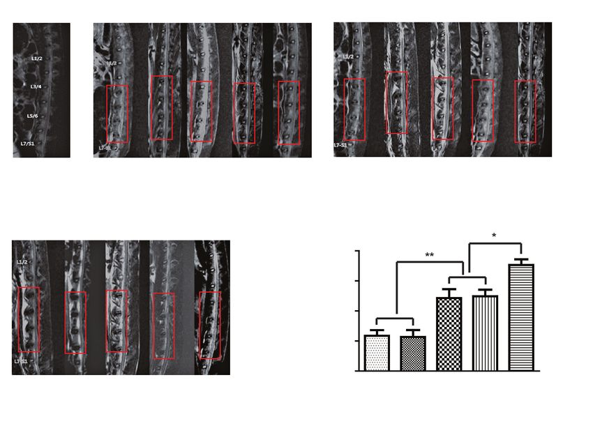
640 Z. Zhao et al. Combined gene therapy for disc degeneration
Introduction to prevent or delay IDD.11 Nishida et al. conducted an initial
study showing that direct injection of adenoviral TGFβ1
Intervertebral disc degeneration (IDD) is a major cause vectors into the rabbit lumbar discs led to a 100% increase
of most musculoskeletal disorders of the spine that re- in proteoglycan synthesis compared with intact control
sults in lower back pain, morbidity and physical disability.1 tissues.12 Since then, gene therapy using several growth fac-
A lthough the precise etiology and pathophysiology of disc tors, including BMP2, GDF5, IGF1, and interleukin (IL)-1
degeneration has not been fully elucidated, IDD is generally receptor antagonist (IL-1Ra) for disc degeneration has been
believed to be a consequence of increased proteolytic deg- tested both in vitro and in vivo, as previously reported.11,13
radation of extracellular matrix (ECM) macromolecules. Co-transduction of adeno-associated virus (AAV)-based
During the disc degenerative process, the nucleus pulposus Sox9 and OP1 vectors was reported to exhibit a stronger
(NP) loses water and large amounts of aggregating pro- effect in the induction of proteoglycan and collagen II than
teoglycans and type II collagen (COL2A1), which leads AAV-Sox9 or AAV-OP1 alone.14
to ECM breakdown and structural failure.2 So far, gene therapy which targets an individual MMP
A variety of studies have focused on the mechanisms molecule (particularly MMP3) against disc degeneration
underlying ECM degradation in IDD pathogenesis. An in- has not yet been reported. In our study, we performed direct
creasing amount of evidence has shown that matrix metal- injection of lentiviral vectors expressing MMP3-shRNA
loproteinases (MMPs) play important roles in the degrada- into NP tissues of lumbar discs in rabbits and evaluated
tion of matrix components during the disc degenerative the reconstitution of matrix components, specifically
process. Several MMP family members, such as MMP1, collagen type II and proteoglycan. We also investigated
MMP2, MMP3, MMP7, MMP8, MMP9, MMP12, and whether a cocktail of both MMP3-shRNA and Sox9 lenti-
MMP26, have been reported to be increasingly expressed viruses could trigger a synergistic effect to further augment
in degenerated disc tissues, suggesting an association matrix synthesis.
between MMP and IDD. 3–6 Notably, Bachmeier et al.
reported that among all MMPs tested (MMP1, MMP2,
MMP3, MMP7, MMP8, MMP9, and MMP13), the expres- Material and methods
sion of MMP3 was consistently and substantially upregu-
lated in degenerated disc samples, and this process was Lentiviral vectors
accompanied with increased enzymatic activity of MMP3,7
implying that MMP3 plays a central role in disc degenera- The lentiviral vectors harboring MMP3-shRNA (LV-
tion. Besides elevated matrix-degrading MMPs, the loss MMP3-shRNA), Sox9 (LV-Sox9), or empty vectors were
of matrix-producing Sox9 also plays a role in ECM deg- provided by Biowit Technologies (Shenzhen, China).
radation in IDD. Being a key transcription factor in chon- All lentivirus vectors were stored at −80°C and diluted
drogenic differentiation, Sox9 can directly stimulate gene with phosphate-buffered saline (PBS) to a concentration
expression of collagen type II and aggrecan,8 the major core of 5 × 10⁵ viral particles (vp)/μL right before use.
protein in proteoglycan. Sive et al. detected a strong signal
of Sox9 in normal NP tissues using in situ hybridization, Animal study
which was accompanied by high-level expression of col-
lagen II and aggrecan.9 However, the expression of Sox9 This is an experimental study in a rabbit model. The pro-
decreases with aging and disc degeneration.10 These stud- tocol for the animal study was reviewed and approved
ies suggest that MMP-induced excessive ECM degrada- by the Animal Care Committee of Qingdao University,
tion or loss of Sox9-reduced ECM synthesis is critically China. A total of 25 New Zealand white rabbits were used
implicated in the pathogenesis of IDD. Hence, molecular in this study, with an average age of 4 months and an av-
therapy which targets MMP or restores Sox9 can be used erage body weight of 2.5 kg. Before surgery, the animals
to reconstitute matrix components in a degenerative NP. underwent magnetic resonance imaging (MRI) to exclude
Although spine disorders resulting from disc degen- congenital spine deformity and disc disease.
eration can be treated with surgery, including discectomy For direct gene transfer, the animals were anesthetized
or spinal fusion, they are not curative and are associated with an intramuscular injection of 1.5% pentobarbital so-
with various complications. Non-surgical treatments dium at a dosage of 2 mL/kg. Following anesthesia, each
– such as drugs, massage or physical therapy – however, animal was placed in a lateral position with all 4 limbs fixed.
can only relieve the clinical symptoms; they cannot delay A vertical incision of about 3 cm was made from the iliac
or reverse the disc degenerative process. As a result, gene crest to the lower edge of the 12th rib via a right extraperito-
therapy has received considerable interest, as this approach neal approach. The peritoneum was bluntly dissected to ex-
can transfer the genes of therapeutic proteins to the disc pose the transverse processes from L3 to L7. A 24-gauge
cells, enabling them to manufacture the proteins endog- needle was used to pierce into the center of the exposed
enously, on a continuous or regular basis, a llowing for L3/4, L4/5, L5/6, and L6/7 lumbar discs (~5 mm) parallel
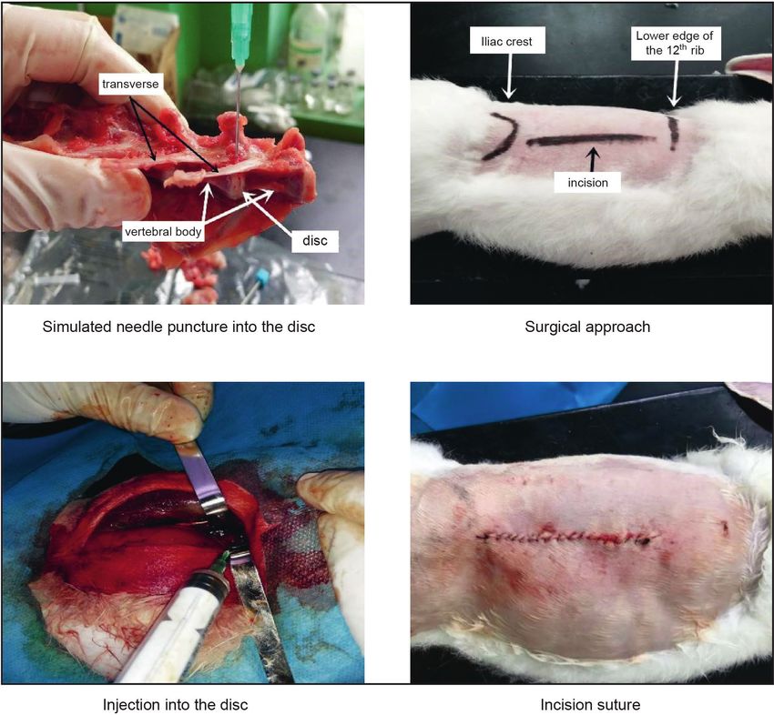
long-term regulation of matrix synthesis with the potential to the upper and lower endplates. Then, a volume of 20 μLAdv Clin Exp Med. 2020;29(6):639–647 641
Fig. 1. In vivo gene delivery. Lentiviral vectors are directly injected into the rabbit lumbar discs (L3/4, L4/5, L5/6, and L6/7) through a surgical approach
containing PBS or 107 vp of lentivirus was injected directly of the scanner were as follows: T2-weighted sequence rep-
into the NP tissue. All 25 rabbits were randomly divided into etition time – 2,000–6,000 ms; echo time – 80–120 ms;
5 groups and injected with a total volume of 20 μL of PBS bandwidth – 25 Hz; microwave – 16; matrix – 384 × 224 px;
or lentivirus, as follows: group I – PBS; group II – 107 vp nex – 4; field of vision – 16 × 16 cm; thickness – 3 mm;
of empty virus (control); group III – 107 vp of LV-MMP3- and spacing – 0.2 mm. A degenerative disc mainly emitted
shRNA; group IV – 107 vp of Lento-Sox9; and group V a low signal, that is, the T2-weighted signal intensity de-
(cocktail) – 5 × 10⁶ vp of LV-MMP3-shRNA + 5 × 10⁶ vp creased and the disc became thinner. Based on the changes
of LV-Sox9. After injection, the opening was closed with in the T2-weighted signal, the improved Thompson clas-
sutures (Fig. 1). The rabbits were administered penicillin sification method was used as the standard. The MRI data
(800,000 units) intramuscularly before and after the opera- was blindly assessed by 2 independent senior radiologists.
tion. No incidences of infection or death were reported.
Quantitative real-time polymerase
MRI assessment chain reaction
The animals were examined with MRI at 8, 12 and At 24 weeks post-operation, the animals were sacri-
24 weeks after the operation. A Siemens Avanto 3.0T ficed and the NP tissues were harvested. Total RNA was
medical superconductive MRI scanner (Siemens AG, isolated using TRIzol reagent (Amresco, Solon, USA)
Munich, Germany) was used to scan the sagittal lumbar and was reverse-transcribed to cDNA using qScript
spine of the animals with a cervical coil. The parameters cDNA SuperMix (Quanta Biosciences, Woodbridge,642 Z. Zhao et al. Combined gene therapy for disc degeneration USA). Quantitative real-time polymerase chain reaction (pH 6.8) containing 1 mM of EDTA, 2 mM of dithiothreitol (qRT-PCR) was conducted with SYBR green master mix. and 300 µg of papain at 60°C for 4 h until the tissue was The primers for rabbit genes were listed as follows: CO- soluble. Iodoacetic acid was added to a final concentration L2A1, forward 5’-GGATAGA CCCCAACCAAGGC-3’, of 10 mM and was mixed well with 1 mL 50 mM Tris/HCl reverse 5’-GCTGCTCCACCAGTTCTTCT-3’; MMP3, (pH 8.0). Both the samples and the sGAG standards were forward 5’-AGCCAATGGAAATGAAAACTCTTC-3’, mixed with DMB dye. Absorbance was read in a microplate reverse 5’-CCAGTGGATAGGCTGAGCAAA-3’; Sox9, reader at an optical density (OD) of 520 nm. 5’-GGTGCTCAAGGGCTACGACT-3’, reverse 5’-GGGTG- GTCTTTCTTGTGCT G-3’; Actin, forward 5’-CTG- Statistical analyses GAACGGTGAAGGTGACA-3’, reverse 5’-CGGCCA- CATTGC AGAACT-3’. The PCR products were analyzed The data was reported as means ± standard deviation with an ABI PRISM 7900HT Sequence Detection System (SD) (or means ± standard error of the mean (SEM); see (Applied Biosystems, Foster City, USA). The cycling condi- figure legends) of at least 3 independent trials, and was tions were 50°C for 2 min and 95°C for 10 min, followed analyzed with a two-tailed Student’s t-test for 2 groups, by 40 repetitions of 95°C for 15 s and 60°C for 1 min. One or one-way analysis of variance (ANOVA) for multiple dissociation stage (95°C for 15 s, 60°C for 15 s and 95°C for groups using GraphPad Prism software (GraphPad Soft- 15 s) was added to produce the melting curve at the end ware Inc, San Diego, USA). The variance is similar between of the cycling condition. Relative mRNA concentrations the groups in the same trial. A p-value
Adv Clin Exp Med. 2020;29(6):639–647 643
A pre-OP B 8 weeks post-OP C 12 weeks post-OP
PB con MM So MM PB con MM So MM
S tro P3 x9 P3 S tro P3 x9 P3
l -sh -sh l -sh -sh
RN RN RN RN
A A+ A A+
So So
x9 x9
D 24 weeks post-OP E
0.8
0.6
at 24 weeks post-OP
MRI signal intensity
0.4
0.2
0.0
PB con MM So MM PB con MM So MM
S x9 S tro P3 x9 P3
tro P3 P3 l -sh -sh
l -sh -sh RN RN
RN RN A A+
A A+
So So
x9 x9
Fig. 2. MRI analysis. Lumbar discs are scanned using T2-weighted MRI at different time points: (A) before the operation (pre-OP), (B) 8 weeks after operation,
(C) 12 weeks after the operation, and (D) 24 weeks after the operation. (E) MRI signal intensity at 24 weeks after the operation. Red boxes surround L3/4,
L4/5, L5/6, and L6/7 intervertebral discs. Note that lentiviral delivery of MMP3-shRNA or Sox9 greatly delays needle-puncture-induced disc degeneration
when compared to PBS or empty virus treatment, whereas combined gene delivery induces a synergistic effect in the attenuation of IDD
only appeared intact, but also displayed only a moderate in MMP3 level. Interestingly, the injection of both lenti-
decrease in MRI signal intensity (Fig. 2D,E; Table 1). viruses decreased MMP3 mRNA to a level comparable
Based on the comparison of MRI signal intensity among to that of LV-MMP3-shRNA alone (Fig. 3A). On the other
different groups, lentivirus-mediated MMP3 knockdown hand, LV-Sox9 viral vectors led to a 3.8-fold increase
or Sox9 overexpression efficiently delayed the progres- in Sox9 mRNA in comparison with the controls. Simi-
sion of needle-puncture-induced disc degeneration. larly, the cocktail of lentiviruses induced a 3.5-fold increase
Importantly, injection of combined LV-MMP3-shRNA and of Sox9 mRNA (Fig. 3B). These results not only suggest
LV-Sox9 vectors significantly exerted a greater capacity that lentiviral vectors can mediate long-term in vivo gene
than MMP3-shRNA (p = 0.026) or Sox9 (p = 0.017) single expression, but they also imply that combined transduc-
transgene to delay the development of IDD, indicating tion of 2 transgenes can stimulate higher gene expression
a synergistic effect of cocktail gene delivery. than single-gene delivery.
Lentiviral MMP3-shRNA or Sox9 gene Lentiviral MMP3-shRNA or Sox9 gene
transfer mediates long-term transgene transfer increases collagen II expression
expression Given that the loss of collagen II and proteoglycan are
The animals were sacrificed at 24 weeks after the oper the predominant pathological changes in IDD, we first
ation. The NP tissues were harvested from their lumbar examined whether in vivo gene delivery of lentivirus
discs for RNA and protein assays. We first evaluated vectors could restore the expression of collagen II in NP
transgene expression using qRT-PCR analysis and found tissues during a needle-puncture-induced degenerative
that, compared to the control group, the in vivo transfer process. Using qRT-PCR analysis, we found that inject-
of LV-MMP3-shRNA vectors resulted in a 65% decrease ing LV-MMP3-shRNA and LV-Sox9 induced a 1.82- and644 Z. Zhao et al. Combined gene therapy for disc degeneration
A B
1.5 5
control control
MMP3-shRNA 4 Sox9
relative expression
relative expression
MMP3-shRNA + Sox9 MMP3-shRNA + Sox9
1.0
3
2
0.5
1
0.0 0
MMP3 mRNA Sox9 mRNA
Fig. 3. Lentiviral gene delivery mediates long-term transgene expression. A. qRT-PCR analysis for MMP3 mRNA in NP tissue. B. qRT-PCR analysis for Sox9
mRNA in NP tissue. Data is presented as means ±SEM. Note that cocktail gene delivery induces MMP3 or Sox9 mRNA to a comparable extent as the delivery
of MMP3-shRNA or Sox9 lentivirus alone
** p < 0.01.
Table 1. MRI signal intensity (modified Thompson classification)
Group Pre-OP 8 weeks 12 weeks 24 weeks*
PBS 1.000 ±0.051 0.677 ±.097 0.367 ±0.081 0.235 ±0.037
Control 1.000 ±0.103 0.652 ±0.083 0.383 ±0.043 0.227 ±0.046
MMP3-shRNA 1.000 ±0.072 0.836 ±0.059 0.625 ±0.077 0.486 ±0.059
Sox9 1.000 ±0.067 0.845 ±0.064 0.655 ±0.062 0.498 ±0.044
MMP3-shRNA+Sox9 1.000 ±0.086 0.903 ±0.069 0.796 ±0.085 0.706 ±0.038
* Analysis between different groups as follows.
Groups Fold change p-value
MMP3-shRNA+Sox9 vs Sox9 1.42Adv Clin Exp Med. 2020;29(6):639–647 645
A COL2a1 mRNA B
3 (i) (ii) 4
S ox9
relative densitometry
relative densitometry
A A+
RN RN 3
2 o l -sh -sh
tr P 3 9 P3
P BS con M M So
x
M M
2
1 COL2A1
1
GAPDH
0 0
PB con MM So MM PB con M So MM
S tro P3 x9 P3 S tro MP3 x9 P3
l -sh -sh l -sh -sh
RN RN RN RN
A A+ A A+
So So
x9 x9
Fig. 4. MMP3 knockdown or Sox9 overexpression induces collagen II expression. A. qPCR analysis for COL2A1 mRNA. B. Western blot analysis for COL2A1
(i) and densitometry of protein bands normalized with GAPDH using ImageJ (ii). The p-values on top of the bars denote the comparison between each
lentiviral transgene group and an empty lentivirus group. For (A), data is presented as means ±SEM. For (B), data is presented as mean ±SD
* p < 0.05; ** p < 0.01.
A B
(i) (ii)
4 100
S ox9
relative densitometry
proteoglycan [µg/mL]
NA N A+ 80
R hR 3
l -sh 3-s
S ntro MP3 x9 MP 60
PB co M S o M 2
aggrecan 40
1
20
GAPDH
0 0
PB con M So MM PB con M So MM
S tro MP3 x9 P3 S tro MP3 x9 P3
l -sh -sh l -sh -sh
RN RN RN RN
A A+ A A+
So So
x9 x9
Fig. 5. MMP3 knockdown or Sox9 overexpression stimulates proteoglycan production. A. Western blot analysis for aggrecan (i) and densitometry of protein
bands normalized with GAPDH using ImageJ (ii). B. Proteoglycan assay in NP tissue. The p-values on top of the bars denote the comparison between each
lentiviral transgene group and an empty lentivirus group. For (A), data is presented as means ±SD. For (B), data is presented as means ±SEM
* p < 0.05; ** p < 0.01; *** p < 0.001.
In contrast, the injection of both lentiviruses resulted the matrix in NP mediates lumbar discs’ resistance to com-
in the highest expression of aggrecan, which was a 3.08- pressive stresses,10 the reconstitution of matrix compo-
fold increase in comparison with the controls (Fig. 5A). nents using gene therapy is of great importance in the delay
Besides the upregulation of aggrecan by lentivirus, or attenuation of IDD. The family of MMPs is thought
we found that the knockdown of MMP3 or overexpression to constitute the major catabolic enzymes in the interver-
of Sox9 resulted in a 2.1- and 2.65-fold higher synthesis tebral disc.8 Increased MMP expression and/or activity
of sGAG, respectively, compared to the controls. Strikingly, have been implicated in the pathogenesis of IDD. Although
injecting both LV-MMP3-shRNA and LV-Sox9 vectors led gene therapy using viral or non-viral vectors to overexpress
to the strongest induction of sGAG: a 4.2-fold increase growth factors or cytokines has received considerable in-
(Fig. 5B). These results suggest that lentivirus-mediated terest, a therapy that directly antagonizes MMP to prevent
MMP3 knockdown or Sox9 overexpression increases pro- disc degeneration has not been studied, and this warrants
teoglycan production, and more importantly, indicates that investigation.
the injection of a MMP3-shRNA/Sox9 lentivirus cock- In our study, we established a needle-puncture-in-
tail triggers a synergistic effect to enhance proteoglycan duced IDD model in rabbits, as reported previously.15–17
synthesis. The MRI scans revealed that the lumbar discs injected
with PBS or empty lentivirus underwent degenerative pro-
cess as early as after 8 weeks, and the disc degeneration
Discussion became extremely pronounced 24 weeks after injection.
However, injecting lentivirus carrying MMP3-shRNA
As the major cause of lower back pain, IDD is charac- greatly delayed the progression of IDD on MRI, suggest-
terized by excessive degradation of the ECM, particularly ing that the inhibition of MMP3 is effective in preventing
the type II collagen and aggrecan in NP tissues.18 Since disc degeneration. Biochemical assays further suggested646 Z. Zhao et al. Combined gene therapy for disc degeneration
that lentivirus-mediated MMP3 knockdown increased thereby leading to maximized matrix production. Notably,
the expression of collagen II at both the mRNA and protein we observed that delivery of the 2 lentiviruses reduced
levels. This finding is interesting because MMP3 mainly MMP3 – or increased Sox9 – mRNA to a level compa-
degrades collagen II and other types of collagen (e.g., III, rable to that of LV-MMP3-shRNA or LV-Sox9 gene delivery
IV, IX, and X), but it cannot degrade collagen I,19 which alone. We assume that there may be an interaction between
is the predominant type of collagen fibers in the later the 2 transgenes to further impact their expression, result-
stages of disc degeneration. As a result, the knockdown ing in augmented Sox9 or greatly reduced MMP3 expres-
of MMP3 using gene therapy not only avoids degradation sion. Our assumption is partially supported by a recent
of collagen II in the NP, but it does so without affecting study, in which Sox9 was able to inhibit the enzymatic
the degradation of collagen I by other MMPs. Additionally, activities of MMP2 and MMP13 through its binding with
lentiviral delivery of MMP3-shRNA increased the pro- the MMP gene promoter.22
duction of proteoglycan, as evidenced by the upregulated In general, our results demonstrate that lentivirus-medi-
expression of aggrecan and the synthesis of sGAG. These ated in vivo gene therapy targeting MMP3 or overexpress-
findings suggest that gene therapy targeting MMP3 rep- ing Sox9 is efficient in delaying lumbar disc degeneration
resents an efficient approach against IDD. by restoring 2 major NP matrix components, collagen II
As a master regulator for chondrogenic differentia- and proteoglycan. Our results also suggest that gene ther-
tion, Sox9 directly drives collagen II and aggrecan gene apy delivering MMP3-shRNA and Sox9 lentiviruses offers
expression.8 A loss of Sox9 is closely associated with disc a promising approach for maximizing matrix reconstitu-
degeneration.10 In our study, direct in vivo gene delivery tion with a minimal dose of lentivirus mixture.
of the Sox9 lentivirus attenuated the progression of lumbar
disc degeneration, as indicated by MRI scans. Overexpres- ORCID iDs
sion of Sox9 also increased the production of collagen II Zheng Zhao https://orcid.org/0000-0002-6523-3425
Siyuan Li https://orcid.org/0000-0003-4220-4493
and proteoglycan, suggesting that lentiviral Sox9 gene Hui Huang https://orcid.org/0000-0002-7600-2331
delivery can be used to alleviate IDD through its recon- Jing Fang https://orcid.org/0000-0002-3372-8650
stitution of major matrix components. In support of our Huawei Wei https://orcid.org/0000-0001-7483-0902
Yongming Xi https://orcid.org/0000-0002-0861-6147
findings, Paul et al. previously reported that adenoviral
vectors carrying the Sox9 gene are able to increase the pro-
References
duction of type II collagen in degenerated human disc
1. Hodgkinson T, Shen B, Diwan A, Hoyland JA, Richardson SM. Thera-
cells.20 Unfortunately, adenoviral vectors can only medi- peutic potential of growth differentiation factors in the treatment
ate transient gene expression, and their in vivo applica- of degenerative disc diseases. JOR Spine. 2019;2(1):e1045.
tion is limited due to the virus-induced immune response. 2. Lyu FJ, Cheung KM, Zheng Z, Wang H, Sakai D, Leung VY. IVD pro-
genitor cells: A new horizon for understanding disc homeostasis and
As such, lentiviral vectors carrying Sox9, which mediate repair. Nat Rev Rheumatol. 2019;15(2):102–112.
long-term transgene expression without severe immuno- 3. Gruber HE, Hoelscher GL, Ingram JA, Hanley EN Jr. Matrix metallo-
genic activity, offer greater advantages over adenoviruses. proteinase-26, a novel MMP, is constitutively expressed in the human
intervertebral disc in vivo and in vitro. Exp Mol Pathol. 2012;92(1):
Notably, our study demonstrated that a cocktail deliv- 59–63.
ery of MMP3-shRNA and Sox9 lentiviruses dramatically 4. Eser B, Eser O, Yuksel Y, et al. Effects of MMP-1 and MMP-3 gene poly-
blocked lumbar disc degeneration in rabbits. Even 24 weeks morphisms on gene expression and protein level in lumbar disc her-
niation. Genet Mol Res. 2016;15(3). doi:10.4238/gmr.15038669
after combined injection, a time point when extremely
5. Lv FJ, Peng Y, Lim FL, et al. Matrix metalloproteinase 12 is an indica-
severe disc degeneration was observed in the animals in- tor of intervertebral disc degeneration co-expressed with fibrotic
jected with PBS or an empty virus, MRI scans still identi- markers. Osteoarthr Cartil. 2016;24(10):1826–1836.
fied intact disc tissues, with the highest signal intensity 6. Zigouris A, Alexiou GA, Batistatou A, Voulgaris S, Kyritsis AP. The role
of matrix metalloproteinase 9 in intervertebral disc degeneration.
among all lentivirus-treated groups. Interestingly, Moon J Clin Neurosci. 2011;18(10):1424–1425.
et al. reported that combination therapy of TGFβ1, IGF1 7. Bachmeier BE, Nerlich A, Mittermaier N, et al. Matrix metalloprotein-
and BMP2 adenoviral vectors is better than single-gene ase expression levels suggest distinct enzyme roles during lumbar
disc herniation and degeneration. Eur Spine J. 2009;18(11):1573–1586.
therapy because multiple gene transduction enables en- 8. Lefebvre V, Angelozzi M, Haseeb A. SOX9 in cartilage development
hanced biological responses, such as proteoglycan synthe- and disease. Curr Opin Cell Biol. 2019;61:39–47.
sis.21 Likewise, in our study, the combined gene transfer 9. Sive JI, Baird P, Jeziorsk M, Watkins A, Hoyland JA, Freemont AJ.
Expression of chondrocyte markers by cells of normal and degen-
of lentiviral MMP3-shRNA and Sox9 vectors displayed erate intervertebral discs. Mol Pathol. 2002;55(2):91–97.
greater capacity than the injection of LV-MMP3-shRNA 10. Gruber HE, Norton HJ, Ingram JA, Hanley EN Jr. The SOX9 transcrip-
or LV-Sox9 alone in the induction of collagen II expres- tion factor in the human disc: Decreased immunolocalization with
age and disc degeneration. Spine (Phila Pa 1976). 2005;30(6):625–630.
sion and proteoglycan production, suggesting that com-
11. Sobajima S, Kim JS, Gilbertson LG, Kang JD. Gene therapy for degen-
bined gene delivery triggers a synergistic effect in ECM erative disc disease. Gene Ther. 2004;11(4):390–401.
synthesis. The mechanism underlying this synergy seems 12. Nishida K, Kang JD, Gilbertson LG, et al. Modulation of the biologic
likely to be mediated through the simultaneous inhibition activity of the rabbit intervertebral disc by gene therapy: An in vivo
study of adenovirus-mediated transfer of the human transform-
of matrix degradation (via MMP3 knockdown) and en- ing growth factor beta 1 encoding gene. Spine (Phila Pa 1976). 1999;
hancement of matrix induction (via Sox9 overexpression), 24(23):2419–2425.Adv Clin Exp Med. 2020;29(6):639–647 647
13. Nishida K, Suzuki T, Kakutani K, et al. Gene therapy approach for 18. Vo NV, Hartman RA, Yurube T, Jacobs LJ, Sowa GA, Kang JD. Expres-
disc degeneration and associated spinal disorders. Eur Spine J. 2008; sion and regulation of metalloproteinases and their inhibitors in inter-
17(Suppl 4):459–466. vertebral disc aging and degeneration. Spine J. 2013;13(3):331–341.
14. Ren S, Liu Y, Ma J, et al. Treatment of rabbit intervertebral disc degen- 19. Newell N, Carpanen D, Evans JH, Pearcy MJ, Masouros SD. Mechani-
eration with co-transfection by adeno-associated virus-mediated cal function of the nucleus pulposus of the intervertebral disc under
SOX9 and osteogenic protein-1 double genes in vivo. Int J Mol Med. high rates of loading. Spine (Phila Pa 1976). 2019;44(15):1035–1041.
2013;32(5):1063–1068. 20. Paul R, Haydon RC, Cheng H, et al. Potential use of Sox9 gene thera-
15. Masuda K, Aota Y, Muehleman C, et al. A novel rabbit model of mild, py for intervertebral degenerative disc disease. Spine (Phila Pa 1976).
reproducible disc degeneration by an anulus needle puncture: 2003;28(8):755–763.
Correlation between the degree of disc injury and radiological and 21. Moon SH, Nishida K, Gilbertson LG, et al. Biologic response of human
histological appearances of disc degeneration. Spine (Phila Pa 1976). intervertebral disc cells to gene therapy cocktail. Spine (Phila Pa 1976).
2005;30(1):5–14. 2008;33(17):1850–1855.
16. Kwon YJ. A minimally invasive rabbit model of progressive and repro- 22. Luo H, Wang C, Liu M, et al. Inhibition of SOX9 promotes inflam-
ducible disc degeneration confirmed by radiology, gene expression, matory and immune responses of dental pulp. J Endod. 2018;44(5):
and histology. J Korean Neurosurg Soc. 2013;53(6):323–330. 792–799.
17. Lei T, Zhang Y, Zhou Q, et al. A novel approach for the annulus nee-
dle puncture model of intervertebral disc degeneration in rabbits.
Am J Transl Res. 2017;9(3):900–909.You can also read