Mechanism of Baicalein in Brain Injury After Intracerebral Hemorrhage by Inhibiting the ROS/NLRP3 Inammasome Pathway
←
→
Page content transcription
If your browser does not render page correctly, please read the page content below
Mechanism of Baicalein in Brain Injury After
Intracerebral Hemorrhage by Inhibiting the
ROS/NLRP3 Inflammasome Pathway
Xuan Chen
The First People's Hospital of Shangqiu
Yue Zhou
Yidu Central Hospital
Shanshan Wang
Yidu Central Hospital
Wei Wang ( wanwei0311@163.com )
The Fourth Affiliated Hospital of China Medical University https://orcid.org/0000-0002-4719-7280
Research Article
Keywords: Baicalein, Brain injury, Intracerebral hemorrhage, NLRP3 inflammasome, Reactive oxygen
species, Neuron
Posted Date: June 18th, 2021
DOI: https://doi.org/10.21203/rs.3.rs-609350/v1
License: This work is licensed under a Creative Commons Attribution 4.0 International License.
Read Full License
Page 1/20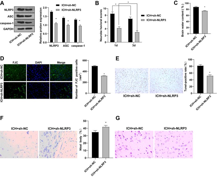
Abstract
Intracerebral hemorrhage (ICH) is a devastating subtype of stroke with high disability/mortality. Baicalein
has strong anti-inflammatory activity. This study aims to explore the mechanism of baicalein on brain
injury after ICH. The model of brain injury after ICH was established by collagenase induction, followed
by the evaluation of neurological severity, brain water content, the degenerated neurons, neuronal
apoptosis and reactive oxygen species (ROS). The ICH model was treated with baicalein and silencing
NLRP3 to detect brain injury. The expression of NLRP3 inflammasome was detected after treatment with
ROS scavenger. The expression of oxidative stress markers and inflammatory factors were detected, and
the levels of components in NLRP3 inflammasome were detected. Baicalein reduced the damage of
nervous system, lesion surface, brain water content and apoptosis. Baicalein inhibited malondialdehyde
and increased IL-10 by inhibiting ROS in brain tissue after ICH. Baicalein inhibited the high expression of
NLRP3 inflammasome in ICH. ROS scavenger inhibited the NLRP3 inflammatory response by inhibiting
ROS levels. Silencing NLRP3 alleviated the brain injury after ICH by inhibiting excessive oxidative stress
and inflammatory factors. Overall, baicalein alleviated the brain injury after ICH by inhibiting ROS and
NLRP3 inflammasome.

Introduction
Intracerebral hemorrhage (ICH) is a cerebrovascular disease with extremely high disability and mortality
rates, while existing treatments have many limitations [1, 2]. A series of inflammatory responses including
neuroinflammation, apoptosis and oxidative stress after ICH promote secondary brain injury after ICH,
and this secondary injury is the key factor of ICH-induced brain injury [3]. Therefore, research on effective
methods to reduce and eliminate secondary brain injury caused by inflammation has become the focus
and challenge of current brain injury treatment after ICH.

Baicalein is the main active component isolated from the root of scutellaria baicalensis, which has strong
anti-inflammatory activities through multi-target mechanism [4]. Baicalein has been shown to promote
neuronal and behavioral recovery after ICH by inhibiting apoptosis, oxidative stress and
neuroinflammation, and can be developed as a new drug for the clinical treatment of ICH and brain injury
related to ICH [3]. Recently, it has been documented that acute liver injury can be alleviated by baicalein
by inhibiting the nucleotide-binding domain-like receptor protein 3 (NLRP3) inflammasome [5]. Baicalein
reversed neuroinflammation in rats by inhibiting the NLRP3/caspase-1/Gasdermin D pathway [6]. Both
the activation of NLRP3 inflammasome and reactive oxygen species (ROS) could be down-regulated by
baicalein [7]. These results indicated that NLRP3 inflammasome could be inhibited by baicalein.

NLRP3 inflammasome is a multi-molecular complex that is crucial in innate immunity [8]. Its activation
can further promote the occurrence of inflammation, enhance the host's ability to remove pathogens, and
promote the repair of damaged tissues, but if the activation of inflammasome is disordered, it will cause
the development of various inflammatory diseases and metabolic disorders [9]. NLRP3 inflammasome-
mediated apoptosis is crucial in cerebral ischemia/reperfusion (I/R) injury [10]. Inhibition of NLRP3

                                                  Page 2/20
inflammasome activation ameliorates acute inflammatory injury induced by necrotizing enterocolitis in
rats [11]. NLRP3 inflammasomes is crucial in the inflammatory process that occurs in ICH-induced injury
[12]. NLRP3 inflammasomes can be activated after ICH, leading to inflammatory cascade reaction and
aggravating brain injury [13]. Blocking NLRP3 may be a therapeutic target for ICH recovery [14]. ROS
plays an important role in the activation of NLRP3 inflammasomes [15]. Previous study has reported that
anti-inflammatory properties of melatonin may inhibit the activation of ROS-NLRP3 inflammasome and
protect hippocampal neuron cells against apoptosis after ICH [8]. Many scholars have studied the effect
of NLRP3 inflammasomes on brain injury after ICH. However, the mechanism of baicalein in brain injury
after ICH by regulating the ROS/NLRP3 inflammasome signaling has not been reported. Therefore, this
study set out to identify the mechanism of baicalein inhibiting the ROS-NLRP3 inflammasome in brain
injury after ICH.

Materials And Methods
Ethics statements
Animal experiments followed the standards established by the animal experiment committee of the
Fourth Affiliated Hospital of China Medical University and approved by the ethics committee of the Fourth
Affiliated Hospital of China Medical University. All animal experiments were conducted ifollowed the
“Guidelines for the Care and Use of Laboratory Animals” [16].
Intracerebral hemorrhage (ICH) model establishment
ICH model was established in 48 female Sprague-Dawley (SD) rats (8 weeks old) purchased from Hunan
SJA laboratory animal Co., Ltd (Hunan, China).

The ICH model was processed according to the literature [17], and the specific procedure was as follows:
stereotactic intranasal injection of type VII collagenase (Sigma-Aldrich, St. Louis, Missouri, USA). After
anesthesia, a burr hole was drilled at the injection site (3.0 mm left of the midline, 0.2 mm posterior to the
bregma, and 6 mm below the skull) and type VII collagenase (dissolved in 0.23 µL brine) was slowly
injected at a rate of 0.5 µL/min into the central striatum. Then the needle was kept at the injection site for
another 10 minutes to prevent reflux. The skull was sealed using bone wax after craniotomy. Rats were
assigned into sham group, ICH group, ICH + dimethyl sulfoxide (DMSO) group (treated with DMSO after
the rat ICH modeling), ICH + Baicalein group (treated with 50mg/kg baicalein after the rat ICH modeling),
ICH + H2O group (treated with H2O after the rat ICH modeling), ICH + N-acetylcysteine (NAC) group (treated
with 5 mM NAC after the rat ICH modeling) [18], ICH + sh-negative control lentivirus (sh-NC) group (treated
with sh-NC after the rat ICH modeling), and ICH + sh-NLRP3 group (treated with sh-NLRP3 after the rat ICH
modeling),with 6 rats per group. The treatments of sh-NC, oe-NC, sh-NLRP3, and oe-NLRP3 were as
follows [16]: rats were given stereotactic microinjection of lentivirus particles at a dose of 2.0 µL (10–10
TU/mL) into the CAI region of the right hippocampus, 72 hours before ICH treatment. The treatments of
H2O and baicalein were as follows: the same volume (1 mL) of H2O or baicalein was injected

                                                  Page 3/20
intraperitoneally at an interval of 12 hours for 3 days. After 1 and 3 days of baicalein treatment, the rats
were subjected to neurology score evaluation. After 3 days of baicalein treatment, the rats were
euthanized with excessive sodium pentobarbital. Half of the tissues were used for brain edema
assessment, and the other half were used for histological staining, quantitative reverse transcription
polymerase chain reaction (qRT-PCR) and western blot (WB).

Modified neurological severity score (mNSS)
To assess neurological abnormalities in animals, a mNSS [19] was performed by two independent
investigators who were unaware of the experimental treatment. The mNSS test is composed of motor,
sensory, reflex, and balance tests. Neurological function scores ranged from 0–18 according to
supplementary table 1 (normal = 0; maximum defect score = 18).

Assessment of cerebral edema
After euthanizing animals with an overdose of pentobarbital sodium (160 mg/kg body weight) [20], the
brains were removed and divided into the contralateral and ipsilateral hemispheres and the cerebellum.
Each tissue was weighed immediately to obtain a wet weight and then dried at 160°C for 24 hours to
obtain a dry weight. The formula for calculating water content was as follows: [(wet weight - dry
weight)/(wet weight)] × 100%.

Fluoro Jade - c (FJC) staining
The number of degenerated neurons was assessed by FJC staining. ICH sections were detected using
FJC’s standby dilution staining kit (Biosensis Pty Ltd, Thebarton, South Australia). After rinsing with
phosphate buffer saline (PBS), the sections were incubated in FJC working solution for 20 minutes
according to the instructions, and then observed under a fluorescence microscope. The number of FJC-
positive neurons was calculated as follows: three brain regions of the microscope field around hematoma
were randomly selected from each rat and then the number of FJC-positive neurons was calculated using
the ImageJ software (NIH, Bethesda, MD, USA).

TUNEL staining
Apoptosis was detected by TUNEL staining. In brief, sections were paraffined, dewaxed, hydrated and
cleared in brain tissues in different treatment groups. The TUNEL staining reaction solution (Roche,
Shanghai, China) was added and the apoptotic cells were calculated under the fluorescence microscope
(Eclipse Ti-U, Nikon Co, Japan) and photographed.

Nissl staining
The brain tissues were paraffined and rehydrated, and stained with Nissl staining solution (Beyotime,
C0117) at 50–60℃ for 40 minutes. After the tissues were washed with distilled water, recrystallized with
gradient ethanol, and then cleared in 100% dimethylbenzene for 5 minutes. Then the tissues were sealed
with neutral gum or other sealant. The tissues were observed under the light microscope.

Hematoxylin-Eosin (HE) staining
                                                  Page 4/20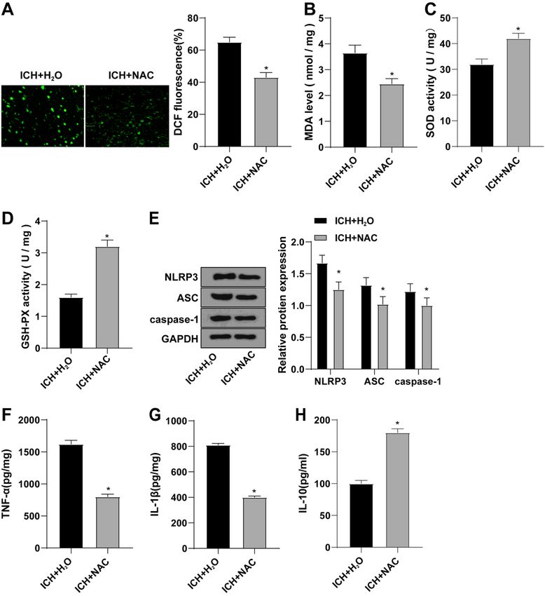
After embedding the tissue, the wax block was fixed on a conventional continuous section with a
thickness of 4 um. The wax block was spread and pasted in water at 46℃, and then baked in a toaster at
72℃ for 2 hours. The sections were cooled for 10 minutes, then dewaxed with xylene I for 10 minutes,
dewaxed with xylene II for 10 minutes, fixed with anhydrous ethanol I and II for 5 minutes, with 90%
ethanol for 2 minutes, with 80% ethanol for 2 minutes, and fixed with 70% ethanol for 2 minutes, and
flushed with water for 5 minutes, and stained with hematoxylin for 5–10 minutes. Next, the sections were
flushed with water for 5 minutes, differentiated with hydrochloric acid alcohol for 2–3 s, blued with
lithium carbonate for 10 minutes, stained with eosin for 2 minutes, dehydrated with 80% ethanol for 2
minutes, with 90% ethanol for 2 minutes, with anhydrous ethanol for 2 minutes, and cleared with xylene
for 2 minutes. The sections were finally wiped, sealed with neutral resin and observed under the
microscope.
Immunohistochemistry
The specimen was fixed with 10% formaldehyde, and paraffined embedded were sliced at 4 µm. The
tissue sections were baked at 60T for 1 hour, dewaxed with conventional xylene, then dehydrated with
gradient alcohol, incubated at 37℃ in 3% H2O2 (Sigma) for 30 minutes, washed with PBS, and boiled in
0.01 M citrate buffer at 95℃ for 20 minutes, then cooled to room temperature, and washed with PBS.
The tissue sections were be sealed with normal sheep serum working solution for 37℃ for 10 minutes.
Sections were incubated with NOD-like receptor protein 3 (NLRP3) (ab214185; 1:200, Abcam, Cambridge,
MA, USA), caspase-1 (ab62698, 1:500, Abcam), and IL-1β (ab216995, 1:200, Abcam) antibodies at 4t for
12 hours. After washing with PBS, the corresponding biotin-labeled goat anti-rabbit secondary antibody
was added, and the reaction was carried out for 10 minutes. After washing thoroughly, horseradish
peroxidase labeled streptomycidin working solution (S-A/HRP) was added to react at room temperature
for 10 minutes. The sections were visualized using Diaminobenzidine (DAB) and stored in a dark room
for 8 minutes. The sections were be rinsed with tap water, stained with hematoxylin, dehydrated, cleared,
sealed, and observed under light microscope. Nikon Imaging Software was used to count the positive
cells. Three non-overlapping fields of equal area (200 x) were selected from each section to count the
number of positive cells.

Immunofluorescence
The slides were fixed with 4% paraformaldehyde for 15 minutes, soaked in PBS 3 times, and dried with
absorbent paper. Normal goat serum was added to the slides and sealed at room temperature for 30
minutes. The blocking liquid on the slides was absorbed with absorbent paper and the slides without
washing. The primary antibodies NLRP3, Caspase-1 and apoptosis-associated speck-like (ASC) were
added to each slide for overnight incubation at 4℃. The slides were dipped and washed three times with
phosphate buffered saline with 0.05% tween 20 (PBST), 3 minutes each, and incubated with Alexa Fluor
488 labeled goat anti-rabbit IgG (ab150077, Abcam, UK) and Alexa Fluor 647 labeled goat anti-rabbit IgG
(ab150083, Abcam) at 37℃ for 1 hour in the dark. After rinsing the slides with PBS 3 times in the dark,
stained with 5ug/mL DAPI for 5 minutes and then washed with PBS for 5 minutes ×3 times. Slides were

                                                 Page 5/20
sealed and stored at 4℃ in the dark. The results were observed with the software Nis-Elements Viewer
using a confocal laser microscope (Zeiss LSM 510, Zeiss, Oberko, Germany).
Determination of reactive oxygen species (ROS)
The slides were fixed with 4% paraformaldehyde for 15 minutes, and then soaked with PBS for 3 times.
The PBS was dried with absorbent paper, and normal goat serum was added to the slides and then the
slides were sealed at room temperature for 30 minutes. The blocking solution was absorbed by absorbent
paper without washing. Each slide was dripped with DCF-DA diluent and incubated at 37℃ for 30
minutes, and observed under a light microscope (Olympus, Tokyo, Japan). Fluorescence intensity was
evaluated using Image Pro Advanced 6.0 software (NIH, Bethesda, MD, USA).
Enzyme linked immunosorbent assay (ELISA)
The expression of inflammatory cytokine interleukin-1 (IL-1) (1210122, IL-1β ELISA kit 96T, Dakewe,
Shenzhen, China), tumor necrosis factor-α (TNF-α) (1217202, TNF-α ELISA kit 96T, Dakewe), and IL-10
(1311002, IL-10 ELISA kit 96T, Dakewe) in serum of rats was detected. The levels of malondialdehyde
(MDA), superoxide dismutase (SOD) and glutathione peroxidase (GSH-Px) in serum of rats were also
measured according to the instructions of ELISA kits (Bio-Swamp, Wuhan, China). Specific steps can refer
to the operation manual. Briefly, 100 µL antibody dilution buffer were incubated with the biotinized
antibody working solution (1:100, 100 µL/well) for 2 hours, the value of optical density (OD) was
measured at 450 nm, and the results were obtained by comparing with the standard and blank control.
The experiment was repeated 3 times.
qRT-PCR
Total RNA was extracted using TRIzol (Invitrogen, Car, USA). RNA was reverse transcribed into cDNA
using PrimeScript RT kit (RR037A, Takara, Japan). The reaction system was 10 µL. Then, the reaction
solution was taken for fluorescence quantitative PCR according to the instructions of the SYBR®Premix
ExTaqTMⅡ kit (RR820A, TaKaRa) using a real-time quantitative fluorescence PCR system (ABI 7500, ABI,
Foster City, CA, USA). Using GAPDH as an internal reference, the relative expression of each target gene
was calculated by the 2−ΔΔCt method [21]. The relevant primers were designed by Shanghai Sangon Bio
(Shanghai, China) (Table 1).
Page 6/20Table 1
Primer sequence
Gene Primer sequences
NLRP3 F: 5’-AGCCTCAACAAACGCTACAC-3’
R: 5’-CATCATCGGGGTCAAACAG-3’
IL-1β F: 5’-GCAGCTATGGCAACTGTT-3’
R: 5’-GAGCCTGTAGTGCAGTTGTC-3’
ASC F: 5’-GGAGTCGTATGGCTTGGAGC-3’
R: 5’-CGTCCACTTCTGTGACCCTG-3
GAPDH F: 5’-AATCCCATCACCATCTTC-3’
R: 5’-AGGCTGTTGTCATACTTC-3’
Western blot (WB)
Tissues were collected by trypsin digestion and lysed with the enhanced radio immunoprecipitation
assay lysate (Boster, Wuhan, China) containing protease inhibitors, and then the protein concentration
was determined with the bicinchoninic acid (BCA) protein quantitative kit (Boster, Wuhan, China). Proteins
were isolated with 10% SDS-PAGE, and the isolated proteins were transferred to polyvinylidene fluoride
(PVDF) membranes. The membranes were sealed with 5% bovine serum albumin (BSA) for 2 hours to
block non-specific binding. Diluted primary antibody NLRP3 (ab214185, 1:1000, Abcam), Caspase-1
(ab62698, 1:1000, Abcam), ASC(ab180799, 1:1000, Abcam) and GAPDH (ab9485, 1:2500, Abcam) were
added, respectively, and incubated overnight at 4℃. After washing the membranes, HRP-labeled sheep
anti-rabbit secondary antibody (ab6721, 1:2000, Abcam) was added into the membranes and incubated
for 1 hour. Next, the membranes were added with enhanced chemi-luminescence (ECL) working solution
(EMD Millipore, MA, USA) at room temperature for 1 minute. Then the excess ECL reagent was removed,
the membranes were sealed with the plastic wrapped, and X-Ray film was put in the dark box for 5–10
minutes exposure for blotting development and fixation. Image J analysis software (NIH) was used to
quantify the grayscale of each band in Western blot images, and GAPDH was used as an internal
reference. Each experiment was repeated 3 times.
Statistical analysis
SPSS version 19.0 (IBM Corp. Armonk, NY, USA) was used for statistical analysis. All data were in
compliance with normal distribution and homogeneity of variance. Data were presented as mean ±
standard deviation. Unpaired t test was used to compare the data between two groups, and one-way
analysis of variance (ANOVA) was used to compare the data among multiple groups. Tukey's was used
for post hoc test. A value of P < 0.05 indicated the difference was statistically significant.
Results
Page 7/20Baicalein reduced brain injury after ICH
Baicalein has therapeutic effects on brain injury [3]. To explore the mechanism of baicalein on brain
injury after ICH, we established a brain injury model after ICH by collagenase induction. The mNSS was
used for neurological assessment. The neurological damage of ICH group was worse than that in the
sham group (Fig. 1A). Brain water content was measured by dry/wet method, and the results showed that
brain water content in ICH group was significantly increased (Fig. 1B). The number of degenerated
neurons was detected by FJC, which showed that the degenerated neurons in the ICH group were clearly
increased (Fig. 1C). Meanwhile, TUNEL staining showed that the apoptosis in ICH group was increased
(Fig. 1D). Nissl staining showed that the Nissl bodies in the ICH group were evidently reduced (Fig. 1E).
HE staining was used to detect the pathological conditions of brain tissue, which showed that the
pathological severity of the ICH group was significantly increased (Fig. 1F). These results indicated that
the animal model of ICH was successfully established, and there was brain injury after ICH. Then we
treated the ICH rat model with baicalein and scored each rat with mNSS. Compared with the ICH + DMSO
group, the ICH + baicalein group had neurological injury, and the lesion volume and brain water content
were reduced, cell apoptosis was reduced, the number of Nissl bodies was increased, and the severity of
pathology was significantly reduced (Fig. 1A ~ F). The above results indicated that baicalein treatment
can improve brain injury after ICH.
Baicalin inhibited brain injury after ICH caused by ROS
oxidative stress
In order to explore the mechanism of baicalein on the improvement of ICH brain injury, DCF fluorescence
staining was used to detect the ROS expression levels in brain tissues. Compared with the sham group,
ROS level in the ICH + baicalein group was increased, and compared with the ICH + DMSO group, ROS
level was significantly decreased in ICH + baicalein group (Fig. 2A). ELISA was used to detect oxidative
stress markers in serum of rats. Compared with the sham group, MDA content in ICH group was
obviously enhanced, and SOD and GSH-Px activities were significantly decreased. Compared with the
ICH + DMSO group, the expression of MDA in ICH + baicalein group was significantly decreased, and the
activities of SOD and GSH-Px were significantly increased (Fig. 2B-D). In short, the brain injury caused by
oxidative stress and inflammation factors can be reduced by baicalein.
NLRP3 inflammasome was inhibited by baicalein
The NLRP3 inflammasomes can be inhibited by baicalein, and ROS levels can also be reduced by
baicalein, and the inflammation mediated by NLRP3 inflammasomes can be inhibited by ROS scavenger
NAC [22, 7, 4, 6, 18]. To investigate whether baicalein inhibited brain injury after ICH by affecting NLRP3
inflammasome, we detected the levels of NLRP3, ASC and caspase-1 in ICH brain tissue by RT-qPCR and
WB. Compared with the sham group, the levels of NLRP3, ASC, caspase-1 in the ICH group were
increased, but significantly reduced in the ICH + baicalein group compared with the ICH + DMSO group
Page 8/20(Fig. 3A, B). Meanwhile, the levels of NLRP3, caspase-1 and IL-1β were detected by IHC, which showed
that compared with the sham group, these protein levels in the ICH group were significantly increased, but
significantly decreased in the ICH + baicalein group were compared with the ICH + DMSO group (Fig. 3C).
ELISA showed that compared with the sham group, IL-1β and TNF-α were elevated and IL-10 was
diminished in the ICH group, and compared with the ICH + DMSO group, IL-1β and TNF-α were decreased
and IL-10 was elevated in the ICH + baicalein group (Fig. 3D-F). The above results indicated that baicalein
can inhibit the high expression of NLRP3 inflammasome in ICH.
ROS scavenger NAC inhibited NLRP3 inflammasome-
mediated inflammation
To investigate the effect of ROS on NLRP3 inflammasomes, we tested relevant indicators after the
treatment of ICH with ROS scavenging agent (NAC). DCF fluorescence staining was used to detect ROS
expression levels. Compared with the ICH + H2O group, ROS level in the ICH + NAC group was significantly
decreased (Fig. 4A). ELISA was used to detect oxidative stress markers. Compared with ICH + H2O group,
MDA expression in ICH + NAC group was significantly decreased, while SOD and GSH-Px activities were
significantly increased (Fig. 4B-D). WB showed that NLRP3, ASC and caspase-1 proteins were
significantly downregulated in the ICH + NAC group compared with the ICH + H2O group (Fig. 4E).
Meanwhile, ELISA demonstrated that compared with ICH + H2O group, IL-1β and TNF-α were decreased in
ICH + NAC group, and IL-10 was significantly increased (Fig. 4F-H). Briefly, ROS scavenger NAC inhibited
the level of ROS, thereby inhibiting the inflammatory response mediated by NLRP3 inflammasome.
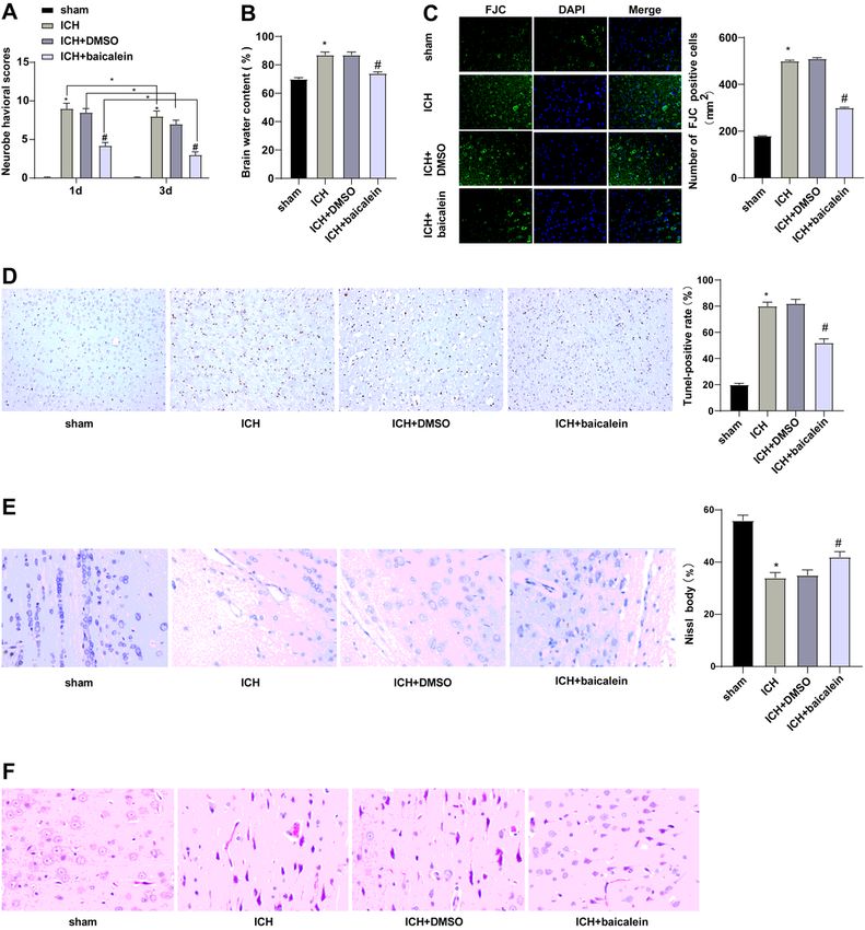
The brain injury after ICH was reduced by silencing NLRP3
To explore the mechanism of NLRP3 on brain injury after ICH, we established a brain injury model after
ICH, and treated the ICH model with silenced NLRP3. WB showed that compared with ICH + sh-NC group,
the protein levels of NLRP3, ASC and caspase-1 in ICH + sh-NLRP3 group were clearly diminished
(Fig. 5A). Compared with ICH + sh-NC group, neurological damage, brain water content, number of
degenerated neurons, apoptosis and pathological severity were significantly decreased in ICH + sh-NLRP3
group (Fig. 5B-G). Overall, silencing NLRP3 reduced brain injury after ICH.
Discussion
Baicalein has been identified to promote neuron and behavior recovery after ICH by inhibiting cell
apoptosis, oxidative stress and neuroinflammation, and can be developed as a new drug for clinical
treatment of ICH and ICH-related brain injury [3]. NLRP3 Inflammasomes are responsible for sensing a
variety of pathogenic and non-pathogenic injury signals and play an important role in neuroinflammation
and neurological diseases [23]. In this paper, a brain injury model after ICH was successfully established
and our results found that baicalein reduced the nervous system injury, lesion surface, brain water
Page 9/20content and apoptosis by inhibiting the ROS-NLRP3 inflammasome, thus alleviating the brain injury after
ICH.
Baicalein can improve sports injury, reduce brain injury, and inhibit the production of pro-inflammatory
cytokines and interleukin [24]. To explore the mechanism of baicalein on brain injury after ICH, we
established a model of brain injury after ICH by collagenase induction, and then ICH model was treated
by baicalein. Neural function and brain tissue water content are widely used to evaluate the degree of
brain injury after ICH [25]. The results showed that after baicalein treatment, nervous system damage,
lesion volume and brain water content were decreased, cell apoptosis was decreased, the number of Nissl
bodies was increased, and pathological severity significantly was decreased. Consistently, previous
reports supported that baicalein can provide neuroprotection in a variety of brain injury models [26, 3].
These results indicated that baicalein can ameliorate brain injury after ICH.
Oxidative stress is caused by the accumulation of ROS after ICH and leads to secondary damage to the
brain tissue [27]. Next, the expression of ROS in brain tissues, and oxidative stress markers in serum of
rats were determined. After baicalein treatment, MDA expression was decreased, and SOD and GSH-Px
activities were significantly increased. Previous report has also shown that baicalein can significantly
inhibit the production of ROS in lipopolysaccharide (LPS)-activated BV-2 cells [28]. Briefly, the brain injury
after ICH caused by excessive oxidative stress and inflammatory factors was inhibited by baicalein.
NLRP3 inflammasome is a key factor in ICH-induced inflammation [12]. ROS is critical for the activation
of NLRP3 inflammasomes [15]. To further investigate whether baicalein inhibits brain injury after ICH by
modulating NLRP3 inflammasome, we detected the expression of NLRP3, ASC and caspase-1 in brain
tissue of ICH rats, the levels of NLRP3, caspase-1 and IL-1β, and the serum expression of inflammatory
cytokines. The results showed that NLRP3 inflammasome was highly expressed in ICH. Baicalein
inhibited the inflammatory cytokines and NLRP3 inflammasome. Baicalein has been reported to reduce
acute liver injury by inhibiting NLRP3 inflammasomes [5]. Overall, baicalein may ameliorate brain injury
after ICH by inhibiting the expression of NLRP3 inflammasome.
To identify the effect of ROS on NLRP3 inflammasome, the ICH rat model was treated with ROS
scavenger NAC. The results showed that the ROS scavenger NAC inhibited ROS levels and thus inhibited
the inflammatory response mediated by NLRP3 inflammasomes. According the reports, melatonin may
play a direct or indirect role in anti-inflammatory and anti-apoptosis by reducing ROS levels in ICH [8].
Blocking the ROS/NLRP3 signaling helps to inhibit LPS-induced inflammation and microglia activation
[29]. Our conclusion is that the ROS scavenger NAC inhibited ROS levels and thus inhibited NLRP3
inflammasome mediated inflammation. Finally, in order to explore the mechanism of NLRP3 on brain
injury after ICH, we treated the ICH model with silencing NLRP3, and the results showed that after
silencing NLRP3, the nervous system injury, brain water content, degenerated neurons, apoptosis, and
pathological severity were significantly reduced. It has been shown that verbascoside inhibits the
inflammatory response after ICH by inhibiting NLRP3 [30]. The results showed that silencing NLRP3 can
reduce the brain injury after ICH.
Page 10/20In conclusion, baicalein can improve the brain injury after ICH by inhibiting the ROS-NLRP3
inflammasome. However, due to the limited time and funds, this paper is not thorough enough. The exact
process of action mechanism is still poorly understood. In future studies, we should conduct more
experiments to further study the effect of baicalein on brain injury after ICH through the ROS-NLRP3
signaling pathway, so as to increase the credibility of results and apply the result to clinical practice.
Authors’ Contributions
XC is the guarantor of integrity of the entire study; XC and YZ contributed to the study concepts, study
design, and definition of intellectual content, SSW contributed to the literature research, XC contributed to
the manuscript preparation and XC contributed to the manuscript editing and review; SSW and WW
contributed to the clinical studies; XC, YZ, SSW and WW contributed to the experimental studies and data
acquisition; XC and YZ contributed to the data analysis and statistical analysis. All authors read and
approved the final manuscript.
Declarations
Authors’ Contributions
XC is the guarantor of integrity of the entire study; XC and YZ contributed to the study concepts, study
design, and definition of intellectual content, SSW contributed to the literature research, XC contributed to
the manuscript preparation and XC contributed to the manuscript editing and review; SSW and WW
contributed to the clinical studies; XC, YZ, SSW and WW contributed to the experimental studies and data
acquisition; XC and YZ contributed to the data analysis and statistical analysis. All authors read and
approved the final manuscript.
Funding
Not applicable.
Acknowledgments
Not applicable.
Data Availability
All the data generated or analyzed during this study are included in this published article.
Compliance with Ethical Standards
Ethical Approval
Animal experiments followed the standards established by the animal experiment committee of the
Fourth Affiliated Hospital of China Medical University and approved by the ethics committee of the Fourth
Page 11/20Affiliated Hospital of China Medical University. All animal experiments were conducted ifollowed the
“Guidelines for the Care and Use of Laboratory Animals” [16].
Conflict of interest
The authors declare that they have no conflict of interest.
Consent for Publication
Not applicable.
References
1. Lin, X., H. Ye, F. Siaw-Debrah, S. Pan, Z. He, H. Ni, Z. Xu, K. Jin, Q. Zhuge, and L. Huang. 2018. AC-
YVAD-CMK Inhibits Pyroptosis and Improves Functional Outcome after Intracerebral Hemorrhage.
Biomed Res Int 2018:3706047.
2. Lu, Q., R. Liu, P. Sherchan, and R. Ren, et al. 2021. TREM (Triggering Receptor Expressed on Myeloid
Cells)-1 Inhibition Attenuates Neuroinflammation via PKC (Protein Kinase C) delta/CARD9 (Caspase
Recruitment Domain Family Member 9) Signaling Pathway After Intracerebral Hemorrhage in Mice.
Stroke 52 (6): 2162–2173.
3. Wei, N., Y. Wei, B. Li, and L. Pang. 2017. Baicalein Promotes Neuronal and Behavioral Recovery After
Intracerebral Hemorrhage Via Suppressing Apoptosis, Oxidative Stress and Neuroinflammation.
Neurochem Res 42 (5): 1345–1353.
4. Luo, X., Z. Yu, C. Deng, J. Zhang, G. Ren, A. Sun, S. Mani, Z. Wang, and W. Dou. 2017. Baicalein
ameliorates TNBS-induced colitis by suppressing TLR4/MyD88 signaling cascade and NLRP3
inflammasome activation in mice. Sci Rep 7 (1): 16374.
5. Xiao, T., Y. Cui, H. Ji, L. Yan, D. Pei, and S. Qu. 2021. Baicalein attenuates acute liver injury by blocking
NLRP3 inflammasome. Biochem Biophys Res Commun 534: 212–218.
6. Rui, W., S. Li, H. Xiao, M. Xiao, and J. Shi. 2020. Baicalein Attenuates Neuroinflammation by
Inhibiting NLRP3/caspase-1/GSDMD Pathway in MPTP Induced Mice Model of Parkinson's Disease.
Int J Neuropsychopharmacol.
7. Li, D., G. Shi, J. Wang, D. Zhang, Y. Pan, H. Dou, and Y. Hou. 2019. Baicalein ameliorates pristane-
induced lupus nephritis via activating Nrf2/HO-1 in myeloid-derived suppressor cells. Arthritis Res
Ther 21 (1): 105.
8. Tang, J., R. Chen, L. Wang, L. Yu, D. Zuo, G. Cui, and X. Gong. 2020. Melatonin Attenuates Thrombin-
induced Inflammation in BV2 Cells and Then Protects HT22 Cells from Apoptosis. Inflammation 43
(5): 1959–1970.
9. Chen, M. Y., X. J. Ye, X. H. He, and D. Y. Ouyang. 2021. The Signaling Pathways Regulating NLRP3
Inflammasome Activation. Inflammation.
Page 12/2010. Liu, X., M. Zhang, H. Liu, and R. Zhu, et al. 2021. Bone marrow mesenchymal stem cell-derived
exosomes attenuate cerebral ischemia-reperfusion injury-induced neuroinflammation and pyroptosis
by modulating microglia M1/M2 phenotypes. Exp Neurol 341: 113700.
11. Zhu, F., L. Wang, Z. Gong, Y. Wang, Y. Gao, W. Cai, and J. Wu. 2021. Blockage of NLRP3
inflammasome activation ameliorates acute inflammatory injury and long-term cognitive impairment
induced by necrotizing enterocolitis in mice. J Neuroinflammation 18 (1): 66.
12. Wang, L., S. Zheng, L. Zhang, H. Xiao, H. Gan, H. Chen, X. Zhai, P. Liang, J. Zhao, and Y. Li. 2020.
Histone Deacetylation 10 Alleviates Inflammation After Intracerebral Hemorrhage via the
PTPN22/NLRP3 Pathway in Rats. Neuroscience 432: 247–259.
13. Xiao, L., H. Zheng, J. Li, Q. Wang, and H. Sun. 2020. Neuroinflammation Mediated by NLRP3
Inflammasome After Intracerebral Hemorrhage and Potential Therapeutic Targets. Mol Neurobiol 57
(12): 5130–5149.
14. Kim, H., J. E. Lee, H. J. Yoo, J. H. Sung, and S. H. Yang. 2020. Effect of Pioglitazone on
Perihematomal Edema in Intracerebral Hemorrhage Mouse Model by Regulating NLRP3 Expression
and Energy Metabolism. J Korean Neurosurg Soc 63 (6): 689–697.
15. Zhao, Y., S. Jiang, J. Zhang, X. L. Guan, B. G. Sun, and L. Sun. 2021. A virulent Bacillus cereus strain
from deep-sea cold seep induces pyroptosis in a manner that involves NLRP3 inflammasome, JNK
pathway, and lysosomal rupture. Virulence 12 (1): 1362–1376.
16. Zhao, M., J. Gao, C. Cui, Y. Zhang, X. Jiang, and J. Cui. 2021. Inhibition of PTEN Ameliorates
Secondary Hippocampal Injury and Cognitive Deficits after Intracerebral Hemorrhage: Involvement of
AKT/FoxO3a/ATG-Mediated Autophagy. Oxid Med Cell Longev 2021:5472605.
17. Sun, Z., K. Wu, L. Gu, L. Huang, Q. Zhuge, S. Yang, and Z. Wang. 2020. IGF-1R stimulation alters
microglial polarization via TLR4/NF-kappaB pathway after cerebral hemorrhage in mice. Brain Res
Bull 164: 221–234.
18. Shen, R., P. Yin, H. Yao, L. Chen, X. Chang, H. Li, and X. Hou. 2021. Punicalin Ameliorates Cell
Pyroptosis Induced by LPS/ATP Through Suppression of ROS/NLRP3 Pathway. J Inflamm Res 14:
711–718.
19. Chen, J., Y. Li, L. Wang, Z. Zhang, D. Lu, M. Lu, and M. Chopp. 2001. Therapeutic benefit of
intravenous administration of bone marrow stromal cells after cerebral ischemia in rats. Stroke 32
(4): 1005–1011.
20. Omolaoye, T. S., and B. T. Skosana, and S. S. du Plessis. 2018. Diabetes mellitus- induction: Effect of
different streptozotocin doses on male reproductive parameters. Acta Histochem 120 (2):103–109.
21. Livak, K. J., and T. D. Schmittgen. 2001. Analysis of relative gene expression data using real-time
quantitative PCR and the 2(-Delta Delta C(T)) Method. Methods 25 (4): 402–408.
22. Ji, Y., J. Han, N. Lee, J. H. Yoon, K. Youn, H. J. Ha, E. Yoon, D. H. Kim, and M. Jun. 2020.
Neuroprotective Effects of Baicalein, Wogonin, and Oroxylin A on Amyloid Beta-Induced Toxicity via
NF-kappaB/MAPK Pathway Modulation. Molecules 25: (21).
Page 13/2023. Mishra, S. R., K. K. Mahapatra, B. P. Behera, and S. Patra, et al. 2021. Mitochondrial dysfunction as a
driver of NLRP3 inflammasome activation and its modulation through mitophagy for potential
therapeutics. Int J Biochem Cell Biol 136: 106013.
24. Zhang, X., Y. Yang, L. Du, W. Zhang, and G. Du. 2017. Baicalein exerts anti-neuroinflammatory effects
to protect against rotenone-induced brain injury in rats. Int Immunopharmacol 50: 38–47.
25. Zhou, W., G. Huang, J. Ye, J. Jiang, and Q. Xu. 2020. Protective Effect of miR-340-5p against Brain
Injury after Intracerebral Hemorrhage by Targeting PDCD4. Cerebrovasc Dis 49 (6): 593–600.
26. Wang, C. X., G. B. Xie, C. H. Zhou, and X. S. Zhang, et al. 2015. Baincalein alleviates early brain injury
after experimental subarachnoid hemorrhage in rats: possible involvement of TLR4/NF-kappaB-
mediated inflammatory pathway. Brain Res 1594: 245–255.
27. Yao, Z., Q. Bai, and G. Wang. 2021. Mechanisms of Oxidative Stress and Therapeutic Targets
following Intracerebral Hemorrhage. Oxid Med Cell Longev 2021:8815441.
28. Yan, J. J., G. H. Du, X. M. Qin, and L. Gao. 2020. Baicalein attenuates the neuroinflammation in LPS-
activated BV-2 microglial cells through suppression of pro-inflammatory cytokines, COX2/NF-kappaB
expressions and regulation of metabolic abnormality. Int Immunopharmacol 79: 106092.
29. Bian, H. T., G. H. Wang, J. J. Huang, L. Liang, L. Xiao, and H. L. Wang. 2020. Scutellarin protects
against lipopolysaccharide-induced behavioral deficits by inhibiting neuroinflammation and
microglia activation in rats. Int Immunopharmacol 88: 106943.
30. Zhou, H., C. Zhang, and C. Huang. 2021. Verbascoside Attenuates Acute Inflammatory Injury Caused
by an Intracerebral Hemorrhage Through the Suppression of NLRP3. Neurochem Res 46 (4): 770–
777.
Figures
Page 14/20Figure 1
Baicalein reduced brain injury after ICH. A: The mNSS was used to score the neurological severity; B:
Brain water content measured as a percentage of wet/dry weight; C: The number of degenerated neurons
was detected by FJC staining; D: TUNEL staining was used to detect brain tissue apoptosis; E: Nissl
staining was used to detect the condition of Nissl bodies; F: HE staining was used to detect the
pathological condition of brain tissue. Data were presented as mean ± standard deviation, and unpaired t
Page 15/20test was used for comparison between two groups (N=6). * compared with the sham group, P < 0.05, #
compared with the ICH + DMSO group, P < 0.05.
Figure 2
Baicalein inhibited brain damage after ICH induced by ROS. A: DCF fluorescence staining was used to
detect ROS expression levels in brain tissues of rats in each group; B-D: The expression levels of MDA,
SOD and GSH-Px in serum were detected by ELISA; Data were presented as mean ± standard deviation,
and unpaired t test was used for comparison between two groups (N=6). * compared with the sham
group, P < 0.05; # compared with the ICH + DMSO group, P < 0.05.
Page 16/20Figure 3
Baicalein inhibited the expression of NLRP3 inflammasome. A: qRT-PCR was used to detect the mRNA
expression levels of NLRP3, ASC, and caspase-1 in the brain tissues of rats in each group; B: WB was
used to detect the protein levels of NLRP3, ASC, and caspase-1 in brain tissue of ICH rats; C: The
expression levels of NLRP3, Caspase-1 and IL-1β in brain tissues of rats in each group were detected by
immunohistochemistry; D-F: The expression levels of inflammatory cytokines IL-1β, TNF-α and IL-10 in
serum of rats were detected by ELISA; Data were presented as mean ± standard deviation, and unpaired t
test was used for comparison between two groups (N=6). * compared with the sham group, P < 0.05; #
compared with the ICH + DMSO group, P < 0.05.
Page 17/20Figure 4
ROS scavenging agent NAC inhibited NLRP3 inflammasome mediated inflammation. ICH rats were
injected with ROS scavengers (NAC) and H2O was injected as control. A: DCF fluorescence staining was
used to detect ROS expression levels in brain tissues; B-D: The expression levels of oxidative stress
markers MDA, SOD and GSH-Px in brain tissues were detected by ELISA; E-H: The expression levels of
inflammatory cytokines IL-1β, TNF-α and IL-10 in brain tissues were detected by ELISA; Data were
Page 18/20presented as mean ± standard deviation, and unpaired t test was used for comparison between two
groups (N=6). * compared with the sham group, P < 0.05; # compared with the ICH + H2O group, P < 0.05.
Figure 5
Silencing NLRP3 reduced brain injury after ICH. A: The protein levels of NLRP3, ASC and Caspase-1 in
brain tissues were detected by WB; B: mNSS was used to score the neurological severity; C: Brain water
content measured as a percentage of wet/dry weight; D: The number of degenerated neurons in brain
tissue was detected by FJC staining; E: TUNEL staining was used to detect brain tissue apoptosis; F: Nissl
staining was used to detect the condition of Nissl bodies; G: HE staining was used to detect the
pathological condition of brain tissue. Data were presented as mean ±standard deviation, and unpaired t
test was used for comparison between two groups (N=6). * compared with the sham group, P < 0.05; #
compared with the ICH+sh-NC group, P < 0.05.
Supplementary Files
Page 19/20This is a list of supplementary files associated with this preprint. Click to download.
Supplementarytable1.docx
Page 20/20You can also read