A Survey on Brain Tumor Diagnosis and Edema Detection Based on Machine Learning
←
→
Page content transcription
If your browser does not render page correctly, please read the page content below

Journal of Physics: Conference Series

PAPER • OPEN ACCESS

A Survey on Brain Tumor Diagnosis and Edema Detection Based on
Machine Learning
To cite this article: K Neamah et al 2021 J. Phys.: Conf. Ser. 1892 012040

View the article online for updates and enhancements.

                               This content was downloaded from IP address 46.4.80.155 on 07/09/2021 at 10:21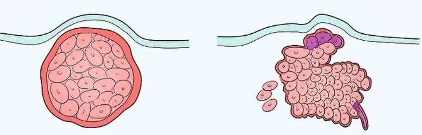
ILATOSPM 2020                                                                                                   IOP Publishing
Journal of Physics: Conference Series                         1892 (2021) 012040          doi:10.1088/1742-6596/1892/1/012040

A Survey on Brain Tumor Diagnosis and Edema Detection
Based on Machine Learning

                    K Neamah 1 , F Mohamed 2 , M M Adnan3,4 , S A Thajeel5
                    1
                     Faculty of Engineering, School of Computing, University Technology of Malaysia, Johor
                    Bahru, Malaysia
                    2
                     UTM-IRDA MaGICX, Institute of Human Centered Engineering, Universiti Teknologi
                    Malaysia, Johar Bahru, Malaysia
                    3
                     School of Computing, Faculty of Engineering, University Technology of Malaysia, Johor
                    Bahru, Malaysia
                    4
                        Islamic University, Najaf, Iraq
                    5
                     Computer Science Department, College of Education, Mustansiriyah University,
                    Baghdad, Iraq

                    E-mail: karrarneamah@yahoo.com

                    Abstract. Early brain tumor diagnosis has a significant role in reducing the risk of disease, as well
                    as led to get better treatment results. Usually, magnetic resonance imaging (MRI) images are
                    evaluated manually through visual inspection, which is difficult,time-consuming and often
                    erroneous;this process is performed by radiologists or clinical experts, and its accuracy depends on
                    their experience. Recently, computer-aided diagnosis (CAD) becomes very essential to overcome
                    these limitations. This paper provides a comprehensive assessment of the existing techniques and
                    methodologies for automated detection of brain tumor coupled with oedema detection methods
                    utilisation, with an emphasis on machine learning models. Moreover, this paper provides an analysis
                    of the integrated procedure that pertains to the retrieval of brain pictures by identifying particular
                    data sets in the procedure to recognise the stipulated attributes.

1. Introductions
This portion of the research provides a comprehensive assessment of the existing techniques and
methodologies used in medical image segmentation. This paper also incorporates a comparative review of
automated brain tumour coupled with oedema detection methods utilisation. Moreover, additionally
provides an analysis of the integrated procedure that pertains to the retrieval of brain pictures by identifying
particular data sets in the procedure to recognise the stipulated attributes. asserts that the use of a computer-
aided analysis in medical imaging affects the decision-making ability of experts in providing accurate

              Content from this work may be used under the terms of the Creative Commons Attribution 3.0 licence. Any further distribution
              of this work must maintain attribution to the author(s) and the title of the work, journal citation and DOI.
Published under licence by IOP Publishing Ltd                          1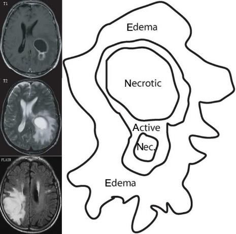
ILATOSPM 2020                                                                                 IOP Publishing
Journal of Physics: Conference Series            1892 (2021) 012040     doi:10.1088/1742-6596/1892/1/012040

images that are relevant to existing distance and volume [1-2]. Also asserts that detecting brain ailments
remains challenging among neurologists and radiologists because of the existence of related brain
abnormalities [1]. The variations in the presence of these abnormalities affect the extent of challenges
experienced by the experts with respect to the tasks because of the existence of differential analysis coupled
with the intricacy attached to physical segmentation [3]. The procedures delimit the precision levels causing
the identification of the requirement for the provision of more time considered essential in meeting the
specific needs of the procedure. The process requires the integration of extensive procedures necessary in
the identification of accurate and consistent techniques necessary in providing the solutions related to
existent questions. This portion of the research seeks to integrate a descriptive analysis, which provides a
description that recognises the existent requirements pertaining to automatic detection. The methodologies
will affect the evaluation procedures integrated relating to the existing human brain abnormalities which
include injuries, oedema and tumour. The procedure is also helpful in the identification of other
abnormalities that can be obtained from computerised pictures of the human brain. In this work, the attention
was given to four categories of machine learning these categories are Region-based methods, Neural
Networks Based Methods, Fuzzy Based Methods and Threshold-based methods. The remaining part of this
work is organized thus: Section 2 discussed 2. Medical image segmentation while section 3 described
various machine learning-based techniques. Section 4 presented the conclusion of the review.

1.1. Brain Tumour
In certain indeterminate situations, the cells of the brain may grow in an uncontrolled way, which delimits
the ability of the regulating function of the regular cells causing further uncontrolled brain cell growth [4 -
5]. The malformed mass that is developed due to the uncontrolled growth is diagnosed as the brain tumour
and it grows inside the skull region, which affects the normal brain functioning by causing an increased
heaviness on the brain. The increased burden on the brain initiates a shift among brain tissues, which pushes
the tissues towards the skull region causing more nerve damage in the healthy brain portion [4], [6]. These
brain tumours may be classified as being either malignant, which means they are cancerous or benign, which
means they are non-cancerous as shown in Figure 1, which shows the position of the tumour by the
stipulation of the origin site [4], [7]–[9].

                                  A                                        B
                 Figure 1. Types of tumour: (A) benign tumour and (B) malignant tumour

1.2. Brain Edema
Cerebral edema (or oedema) signifies the level of fluid build-up in the regions external and internal to the
brain. The detection of any variations related to cerebral oedema leads to the detection of the variations
related to the brain as it becomes smooth and soft, which causes the overfilling of the cranial cave. The
process also causes narrowing of the sulci (grooves), flattening of the gyri (ridges) and it results in
ventricular cavity compression. This abnormality may be detected through the presence of different
symptoms such as nausea, vomiting and, in serious cases, coma and seizures. In the case of herniation, the
person may undergo respiratory symptoms and possible arrest of the respiratory tract which is attributed to
high compression of the respiratory control centres in the pons as well as medulla oblongata.

                                                       2
ILATOSPM 2020 IOP Publishing
Journal of Physics: Conference Series 1892 (2021) 012040 doi:10.1088/1742-6596/1892/1/012040
2. Medical image segmentation
Maintaining that the segmentation of brain tumours includes a process that aims at distinguishing different
tumour tissues. Most studies on brain tumours suggest that detection may be influenced if abnormal tissues
are present. Nevertheless, providing precise and duplicable segmentation is complex since the prevailing
abnormalities include a complex process. The combined knowledge from several previous studies has been
used to attempt the enhancement of medical imaging techniques required for the betterment of brain tumour
segmentation. Fully automatic techniques have been suggested because of the increasing requirement for
more efficient procedures [4], [10]. A major portion of the segmentation methods considered clinically
standard has relied on the straightforward segmentation concerning the present extent of degree supervision.
The present work details the most significant aspects related to the use of segmentation techniques that are
used in combination post image acquisition. Taking into account the advantages of MRI in diagnostic
imaging, the survey is meant to formulate the process for MRI-based segmentation of brain tumours. The
process lays emphasis on the use of wholly automated methods as we showed in Figure 2, Brain tumour in
the T1 against T2 and Flair modalities.
Figure 2. Brain tumour in the T1
against T2 and Flair modalities
By specifying that the segmentation models for brain tumours are divided into three primary categories,
it depends on the level of human intervention required [11]. The categories are manual, fully automatic, and
semi-automatic segmentation. The subsequent parts contain details about each category, which have been
obtained using analysis of the specific category.
2.1. Manual segmentation
In the case of manual segmentation of brain tumours, the tumour boundaries should be defined by drawing
or through the use of painting the regions with anatomical structures using specific labels. Manual
segmentation uses the data in the image, along with the expertise and knowledge specific to anatomy.
Manual demarcation of tumours requires software with a sophisticated graphical user interface to
facilitate drawing the areas of interest in combination with the images on display. Detecting and marking
the tumour as the region of interest is the primary aspect of detection and is a computationally intensive
task. MRI scanners capture 2D slices; therefore, there is a need for a human to identify the most viable
image considering the clarity and the relevance of the recorded areas [4]. Pointed that manual segmentation
of the brain is performed when there is a single image. Contrast injection may be used during manual
segmentation to highlight regions of interest, which help in demarcating the regions. In case the individual
performing segmentation is not competent with brain anatomy, [10] highlights that the process may lead to
mediocre segmentation results. Marking tumour regions using slices may restrict the view for manual
demarcation because of blurred images. This may cause a stripping effect during image segmentation [12].
3ILATOSPM 2020 IOP Publishing
Journal of Physics: Conference Series 1892 (2021) 012040 doi:10.1088/1742-6596/1892/1/012040
2.2. Semiautomatic segmentation
Human observation is necessary during semi-automatic segmentation, especially during initialisation.
Human intervention is required to validate the accuracy of the results generated. The professional may also
manually correct any variance if detected. A large chunk of the current research suggests semi-automatic
segmentation for brain tumours with the intent of limiting expert intervention or inputs required during the
process. suggest that several essential aspects must be considered during the interactive segmentation of
brain tumours, which are the computational, the interactive, and the user interface aspects [4]. The
computational part uses algorithms to create a demarcation of the tumour using the parameter data.
The interactive aspect comprises human mediation on the data produced by the computer. The process
leads to the creation of a translation technique crafted using the incorporation of the computational inputs
as visual feedback provided to the operator, while the inputs provided by the operator are converted to
parameters that may be used by the software. The input and output devices, along with the user interface,
determine the interaction between a human and the computer, the user keys in the analysis of the visual
information displayed on the screen. User input acts as feedback for the algorithms, which then modifies
the computational process to incorporate the feedback. With respect to tumour classification, the process
comprises four primary aspects, which are initialisation, intervention or feedback, and evaluation.
2.3. Automatic Segmentation
Such methods consist of processes where the computer completely controls the tumour segmentation
process. There is an attempt to mimic human intelligence, which consists of producing deformable models
using the knowledge of algorithms, modelling techniques, and computing power. Deformable models and
fuzzy logic are classified as soft computing techniques. In the domain of segmentation of brain tumours, the
use of pattern recognition and machine learning is critical to identifying problems for which solutions may
be found by increasing human intervention in the process. Nevertheless, the creation of a program with a
high degree of automation is challenging since humans make decisions using advanced domain knowledge
and sophisticated visual processing, both of which are crucial to automate the process; therefore, formulating
highly automated methods is complex. This applies to pattern recognition too.
2.4. Segmentation methods
Detection is a crucial process adopted with relation to oncology and clinical medicine. The pre-mature
analysis and localisation of diseases, together with precise disease stage detection, may affect the
identification of variations related to patient management which is considered instrumental in providing
helpful health outcomes. Providing precise detection of the regional physiology is still dependent on precise
segmentation or delineation of the tumour structure relating to the identified area of interest combined with
the provided pictures developed. asserts that the primary roles of the process of segmentation include (1)
quantification of permit, (2) dataset reduction by placing emphasis on quantitative assessment on the
extracted areas of interest, along with the (3) development of structural connections related to physiological
information sampled within the areas [4], [10], [13]. The procedure has improved the processing of several
techniques for brain tumour-segmentation. Still, there is an absence of a standard segmentation method
necessary for providing acceptable results related to all the imaging applications. In most cases, the
techniques that are adopted use specific-imaging modalities including MRI (magnetic resonance imaging).
The existing segmentation methods have been classified into 4 main categories:
• Region-based
• Threshold-based
• Pixel classification
• Model-based
4ILATOSPM 2020 IOP Publishing
Journal of Physics: Conference Series 1892 (2021) 012040 doi:10.1088/1742-6596/1892/1/012040
2.5. Region-based methods
Region-based segmentation methods analyse the pixels within a picture through the combination of form
disjoint areas by merging the adjacent pixels with homogeneity attributes that develop based on a predefined
likeness. These techniques can be outlined in a general manner as follows: Let X represent an image which
is segmented into N areas, where each one is represented by R i where i = 1, 2…,,N
Where L(.) represents a logical predicate. The area growing as well as the watershed segmentation
techniques develop part of region-based techniques, with most of them used in the segmentation of brain
tumour. The next section describes the existent techniques together with an assessment of the existing
applications related to the available literature about brain tumour segmentation.
The region-growing technique is regarded as the simplest one, which is used in the extraction of a linked
region related to similar pixels from a picture [14]. The region-growing method is initiated with no less than
one seed that belongs to the developed formation of interest. The adjacent seeds are assessed and the ones
that meet the developing condition are adopted and included within the procedure. The similarity condition
develops from the existent range of values of pixel intensity or existent attributes related to the image. The
procedure incorporates automatic and manual selection processes with respect to the seeds. The process is
repeated to make sure that the region stayed filled to its capacity. The growth of the area is helpful in the
process of segmentation relating to the existing similar attributes combined with the creation of a connected
region. impacted the adoption of a method of region growing related to the segmentation of the MRI pictures
of brain tumours [15]. The method included the repetition of statistical categorisation that was necessary in
segmenting the image to different tissue classes that are developed on the basis of the existing value of
signal intensity. The process affected the objects of interest identification related to the categorised images
with local segmentation processes (region growing and mathematic morphology).
Others have pointed out that region growing provides a feasible and productive approach because of the
requirement of limited computationally intensive tasks when compared to non-region-based techniques for
the segmentation of brain tumours, concerning the homogeneous regions and tissues [16], [17] .
The primary drawback of the process concerns the partial volume effect [18], which restricts the accuracy
of brain segmentation using MRI images. The partial volume effect delineates the lucidity of intensity
difference between the types of tissues, especially at their border. The voxel may be in an area having more
than one type of tissue. incorporated the modified region growing method (MRGM), which facilitated the
elimination of effects of partial volume that is crucial to determine gradient data concerning boundary
detection with higher accuracy and to plug the holes after the segmentation technique has been developed
[19].
By performing an analysis and using a comparison of the segmentation performed using the region
grown method, and the MRGM concerning the segmentation of brain tumours is obtained by using 3D T1
MR images. The study indicated that MRGM offers more accurate information about the volumetric
measurement of the tumours [16]. This accuracy can be attributed to lesser errors produced by MRGM when
compared with manual segmentation.
This process caused the formulation of several approaches concerning the use of region growing as a
refining step, suggested that employing the fuzzy information fusion framework in the domain of automatic
segmentation of tumour cells from the brain using a chain of several MR images (T1, T2, and PD) [20].
5ILATOSPM 2020 IOP Publishing
Journal of Physics: Conference Series 1892 (2021) 012040 doi:10.1088/1742-6596/1892/1/012040
Edge information was retained during the process by the use of anisotropic diffusion filtering. The
process facilitates the use of a new technique that calculates the mean of the variance inside the curve while
the inverse of the mean gradient is calculated along the curve as the parameters of the study. The model
seeks to integrate aspects regarding variance and gradient. The study established that the optimal result is
obtained when the sum if minimised; the result is directly related to the required threshold. During the
process step comprising region growth, the threshold value increases gradually and facilitates the
recognition of the coarse boundary. Lastly, the optimised model facilitates the detection of accurate
segmentation using a set of boundaries [19] .
2.6. Neural Networks Based Methods
These methods employ computational models based on artificial neural networks that use ‘neurons’ as
processing elements, which are interconnected and use weights. The weights (coefficients) determine the
multipliers of the present connections and provide for the necessary training required to determine the
coefficients.
This process has exerted significant influence on the development of numerous varieties of neural
networks that are employed in the segmentation of medical images and also in other applications. Some
such techniques are back-propagation based learning, multilayer perceptron (MLP), SOM neural network,
and Hopfield Neural Networks (HNN), which are used for segmentation [21]. The significant characteristic
of neural networks is their ability to learn the segmentation process using some training process, which
makes their application lucrative in image segmentation compared to other image processing areas [22].
This study is among the initial studies using the application of MLP for the segmentation of brain
tumours. It comprised initial training of the neural networks provided through a known diagnostic image.
The training process yielded data that was used to formulate an MLP model. In the case of adaptive systems,
data from subsequent images was used as an input to create the training dataset that followed. The new data
set was then used to segment the image further. Segmentation of the image dataset used iterations. A semi-
automatic method was proposed where continuous user input was required. Tumour segmentation accuracy
is typically measured with Jaccard’s similarity measure, as applied among areas described as tumours by
medical experts. The proposed automated techniques attained a similarity index ranging from 0.6 to 0.8.
MLP networks may be trained in the segmentation of brain tumours based on textural features, with use of
numerous methods [23], such as contrast, sum variance, entropy, sum of entropy, difference of entropy,
inverse difference moment, angular second moment, and information measures of correlation. Through
MLP, supervised learning is performed by assigning labels to all anatomical designations within the image.
Fuzzy Based Methods Fuzzy logic comprises a set of mathematical rules that derive from degrees of
memberships, which replace the crisp membership criteria of conventional binary logic. Applied to brain
tumour segmentation, such fuzzy systems afford the development of algorithms and methods that perform
tasks related to intelligent human behaviours. Applied a fuzzy logic scheme to segment and detect tumours
through the extraction of features from brain MR images, which relies on human proficiency in developing
fuzzy rules [24]. This model presents unsupervised learning that is fully automated. Data is extracted
through use of intensity histogram analysis, with a new approach applied towards acquiring any membership
function appropriate to the medical MRI data. Detection and segmentation findings acquired using this
technique are of decent quality, with highest and lowest scores corresponding to 93% and 71%.
Nevertheless, experiments are conducted on just two forms of brain tumours, namely meningioma and
glioblastoma multiforme.
Introduced the use of fuzzy logic processing, with C-means clustering applied to MRI for brain tumour
segmentation tasks. Fuzzy clusters were exploited to initiate the region-based algorithm that iteratively shifts
towards the final brain tumour boundary [25] [26]. This proposed technique was experimentally trialled to
determine its efficiency at processing 15 brain MR images, wherein the manual segmentation gold-standard
6ILATOSPM 2020 IOP Publishing
Journal of Physics: Conference Series 1892 (2021) 012040 doi:10.1088/1742-6596/1892/1/012040
procedure was available for evaluation. Satisfactory results were attained, with an average value for Jaccard
coefficient of 83.19% and average sensitivity of 96.37%. An FCM clustering algorithm was utilised to
segment Glioblastoma-Multiform (GBM) brain tumour forms [19]. Intensity overlapping of tissue, noise,
and initialisation values led to inaccuracies in the segmentation of brain tumours. Nevertheless, the FCM
algorithm was shown to be straightforward, fast, and unsupervised in application [27].
Introduced an FCM approach that used the curve-let transform to eliminate noise. Detailed descriptions
of the process applied in FCM were provided without any results on the segmentation, qualitative
performance, and efficiency of the model in terms of tumour detection.
2.6.1. Threshold-based methods
Thresholding refers to an efficient region segmentation technique that develops a categorisation
model for the image objects by developing a comparison between fixed intensities with 1 or more thresholds
for intensity, which may be local or global. When the object can be detached from the image background
by integrating one threshold, it is called global thresholding. Nonetheless, when the image includes 2 or
more areas related to the various objects, the process of segmentation may be started by using local
thresholding. This process may cause segmentation of the image by applying many individual thresholds or
through a multi-thresholding method.
Global thresholding
Intensity is regarded as the simplest attribute that may be common among pixels in a particular region. Thus,
thresholding recognises a natural way, which may be used in separating light and dark regions. Thresholding
involves construction of binary images from ones with grey-level by varying all the pixels below certain
threshold to 0 along with all the pixels which are above the threshold to 1.
If g (x, y) is a threshold version of f (x, y) at some global threshold T
Furthermore, the existence of shadows of objects in the image presents a shortcoming related to the area
they fall in an object along with the included regions related to a dark object in a light backdrop. Also, the
thresholding procedure may experience many changes attributed to the intensity level relating to the
inhomogeneity across the area [28].
Local Thresholding
Contrary to global thresholding, there are different thresholding techniques that may be referred to as local
thresholding. The developed methods may be feasible in the event that the procedure remains unable to
efficiently determine a thresholding value using a histogram for the whole image or in case one threshold
may be not able to provide the expected segmentation results. Identification of local threshold can be by
estimating a threshold value related to different areas from the intensity histogram [29].
2.6.2. Combined Techniques
Hybrid system is a blend of various techniques of machine learning algorithms. This was developed to
acquire a better combined solution in place of a single approach to the same set of problems [30]. Introduced
a supervised hybrid fuzzy Bayesian clustering-based brain tumour detection system, which maps fuzzy
inputs to crisp outputs during the training phase of the Bayesian system [25]. The hybrid architecture of the
Bayesian scheme applied the Fuzzy and Learning Back Propagation Algorithm (FLBPA). Such results were
achieved with these supervised hybrid methods are promising. Nevertheless, it is difficult to determine
7ILATOSPM 2020 IOP Publishing
Journal of Physics: Conference Series 1892 (2021) 012040 doi:10.1088/1742-6596/1892/1/012040
generalised efficiency with the approach because of the unevenness of image pixel intensities among diverse
MRI scanner features, MRI modalities, and the effects of noise. Conducted a comparative review of KNN,
SVM, PNN, ISNN and Gabor Transform applications in terms of brain tumour segmentation processing. It
was proposed that with light abnormalities, the highest segmentation performance may be attained by
utilising SVM, whereas k-NN remains decent for darker abnormalities [30]. The study recommended that
the we conclude that SVM should be used for efficient segmentation of cerebrospinal fluid and skull tissue,
ISNN should be employed for gray matter whereas, PNN and ISNN should be considered for better
segmentation of white matter.
A Neural Network (NN) approach with DWT features is introduced for acquiring brain MRI
segmentation. They used discrete wavelet transform that decomposes the images and textural features were
extracted from gray-level cooccurrence matrix (GLCM) followed by morphological operation. Probabilistic
neural network (PNN) classifier is used for the classification of tumours from brain MRI images [31]. From
the observation results, it can be clearly expressed that the detection of brain tumour is fast and accurate
when compared to the manual detection carried out by clinical experts. The performance factors evaluated
also shows that it gives better outcome by improving PSNR and MSE parameters.
Combined K-means clustering together with DWT methods for medical MR image segmentation. The
DWT of MR images was used to construct the neurons according to the frequency of wavelet at each grey
level. Independency in the number of neurons for image size led to precise segmentation of regular axial
brain MR images [32].
3. Conclusion
Proper diagnosis of brain tumours in patients requires appropriate segmentation techniques in processing
brain MR images so as to deliver adequate treatment. Currently, many images from various slice orientations
are deployed for mining the information needed for planning, diagnosis, and treatment. The scale of the
information entails computation processing to expedite diagnosis and treatment processes. Since pace of the
computation process is not an issue anymore, the key emphasis is focused on enhancing the process to mine
information from the images attained through slice orientation as well as the procedure of segmentation to
obtain a precise image of the brain tumour. This paper includes a discussion on certain significant and recent
investigations in brain tumour detection and segmentation. The development of automated procedures for
brain tumour detection and segmentation from medical MR images remains a very active research area, with
many researchers involved in the field. To the best of our current knowledge, no clinically-accepted
automated technique has been reported that detects and segments tumours from brain MR images.
References
[1] Bahadure N B, Ray A K and Thethi H P 2017 Image Analysis for MRI Based Brain Tumor Detection
and Feature Extraction Using Biologically Inspired BWT and SVM Int. J. Biomed. Imaging, vol.
2017.
[2] El-Dahshan E S A, Mohsen H M, Revett K and Salem A B M 2014 Computer-aided diagnosis of
human brain tumor through MRI: A survey and a new algorithm Expert systems with
Applications, 41(11), 5526-5545.
[3] Shah N, Ziauddin S & Shahid A R 2017 Brain tumor segmentation and classification using cascaded random
decision forests. In 2017 14th International Conference on Electrical Engineering/Electronics, Computer,
Telecommunications and Information Technology (ECTI-CON) (pp. 718-721). IEEE.
[4] Eickhoff S B, Yeo B T & Genon S 2018 Imaging-based parcellations of the human brain Nature Reviews
Neuroscience, 19(11), 672-686.
[5] Saeed S & Abdullah, A B 2019 Investigation of a brain cancer with interfacing of 3-dimensional image
processing. In 2019 International Conference on Information Science and Communication Technology
(ICISCT) (pp. 1-6). IEEE.
8ILATOSPM 2020 IOP Publishing
Journal of Physics: Conference Series 1892 (2021) 012040 doi:10.1088/1742-6596/1892/1/012040
[6] Kowalska, A. A., Berus, S., Szleszkowski, Ł., Kamińska, A., Kmiecik, A., Ratajczak-Wielgomas K & Zadka
Ł 2020 Brain tumour homogenates analysed by surface-enhanced Raman spectroscopy: Discrimination
among healthy and cancer cells. Spectrochimica Acta Part A: Molecular and Biomolecular
Spectroscopy, 231, 117769.
[7] Vijayakumar B, Chaturvedi A & Kumar K M 2013 Brain Tumor in Three Dimenesional Magnetic Resonance
Images and Concavity Analysis. International Journal of Computer Application, 3.
[8] Manogaran G, Vijayakumar V, Varatharajan R, Kumar P M, Sundarasekar R & Hsu, C H 2018 Machine
learning based big data processing framework for cancer diagnosis using hidden Markov model and GM
clustering. Wireless personal communications, 102(3), 2099-2116.
[9] Noback C R, Ruggiero D A, Demarest R J & Strominger N L Eds 2005 The human nervous system: structure
and function (No. 744). Springer Science & Business Media.
[10] McNeill K A 2016 Epidemiology of brain tumors. Neurologic clinics, 34(4), 981-998.
[11] Han W S & Han I S 2019 Multimodal Brain Image Segmentation and Analysis with Neuromorphic Attention-
Based Learning. In International MICCAI Brainlesion Workshop (pp. 14-26). Springer, Cham.
[12] Wang G, Li W, Ourselin S & Vercauteren T 2018 Automatic brain tumor segmentation using convolutional
neural networks with test-time augmentation. In International MICCAI Brainlesion Workshop (pp. 61-72).
Springer.
[13] Balasooriya N M & Nawarathna R D 2017 A sophisticated convolutional neural network model for brain tumor
classification. In 2017 IEEE International Conference on Industrial and Information Systems (ICIIS) (pp.
1-5). IEEE.
[14] Adams R & Bischof L 1994 Seeded region growing. IEEE Transactions on pattern analysis and machine
intelligence, 16(6), 641-647.
[15] Makropoulos A , Aljabar P, Wright R, Hüning B, Merchant N, Arichi T& Rueckert D 2016 Regional growth
and atlasing of the developing human brain. Neuroimage, 125, 456-478.
[16] Mohammed M A, Abd Ghani M K, Hamed R I, Abdullah M K & Ibrahim D A 2017 Automatic segmentation
and automatic seed point selection of nasopharyngeal carcinoma from microscopy images using region
growing based approach. Journal of Computational Science, 20, 61-69.
[17] Raja N S M, Fernandes S L, Dey N, Satapathy S C & Rajinikanth V 2018 Contrast enhanced medical MRI
evaluation using Tsallis entropy and region growing segmentation. Journal of Ambient Intelligence and
Humanized Computing, 1-12.
[18] Deng W, Xiao W, Deng H & Liu J 2010 MRI brain tumor segmentation with region growing method based on
the gradients and variances along and inside of the boundary curve. In 2010 3rd International Conference
on Biomedical Engineering and Informatics (Vol. 1, pp. 393-396). IEEE.
[19] Sheela C J J& Suganthi G 2020 Morphological edge detection and brain tumor segmentation in Magnetic
Resonance (MR) images based on region growing and performance evaluation of modified Fuzzy C-Means
(FCM) algorithm. Multimedia Tools and Applications, 1-14.
[20] Chaibou M S, Conze P H, Kalti K, Solaiman B & Mahjoub M A 2017 Adaptive strategy for superpixel-based
region-growing image segmentation. Journal of Electronic Imaging, 26(6), 061605.
[21] Amarapur B 2020 Computer-aided diagnosis applied to MRI images of brain tumor using cognition based
modified level set and optimized ANN classifier. Multimedia Tools and Applications, 79(5), 3571-3599.
[22] Deepa S N & Devi B A 2011 Neural networks and SMO based classification for brain tumor. In 2011 World
Congress on Information and Communication Technologies (pp. 1032-1037). IEEE.
[23] Arunkumar, N., Mohammed, M. A., Abd Ghani, M. K., Ibrahim, D. A., Abdulhay, E., Ramirez-Gonzalez G &
de Albuquerque V H C 2019 K-means clustering and neural network for object detecting and identifying
abnormality of brain tumor. Soft Computing, 23(19), 9083-9096.
[24] [24]Tchoketch Kebir S, Mekaoui S & Bouhedda M 2019 A fully automatic methodology for MRI brain tumour
detection and segmentation. The Imaging Science Journal, 67(1), 42-62.
[25] Raju A R, Suresh P & Rao R R 2018 Bayesian HCS-based multi-SVNN: A classification approach for brain
tumor segmentation and classification using Bayesian fuzzy clustering. Biocybernetics and Biomedical
Engineering, 38(3), 646-660.
9ILATOSPM 2020 IOP Publishing
Journal of Physics: Conference Series 1892 (2021) 012040 doi:10.1088/1742-6596/1892/1/012040
[26] Chauhan R, Negi S & Jain S 2017 Detection and Estimation of 2-D Brain Tumor Size Using Fuzzy C-Means
Clustering. In International Conference on Next Generation Computing Technologies (pp. 88-96).
[27] Kalaivani I, Oliver A S, Pugalenthi R, Jeipratha P N , Jeena A A S & Saranya G 2019 Brain Tumor
Segmentation Using Machine Learning Classifier. In 2019 Fifth International Conference on Science
Technology Engineering and Mathematics (ICONSTEM) (Vol. 1, pp. 85-90). IEEE.
[28] Yousuf M, Khan K B, Azam M A & Aqeel M 2019 Brain Tumor Localization and Segmentation Based on
Pixel-Based Thresholding with Morphological Operation. In International Conference on Intelligent
Technologies and Applications (pp. 562-572). Springer,
[29] Agrawal D, Kulshreshtha V & Sharma P 2018 Brain tumor detection using K-means clustering and threshold
segmentation. Int. J. Adv. Res. Sci. Eng. Technol, 5(3), 5333-5340.
[30] Fiaz M, Ali K, Rehman A, Gul M J & Jung S K 2019 Brain MRI Segmentation using Rule-Based Hybrid
Approach. arXiv preprint arXiv:1902.04207.
[31] Shree N V & Kumar T N R 2018 Identification and classification of brain tumor MRI images with feature
extraction using DWT and probabilistic neural network. Brain informatics, 5(1), 23-30.
[32] Chaudhary A & Bhattacharjee V 2020 An efficient method for brain tumor detection and categorization using
MRI images by K-means clustering & DWT. International Journal of Information Technology, 12(1), 141-
148.
10You can also read