Original Article De novo cartilage growth after implantation of a 3-D-printed tracheal graft in a porcine model
←
→
Page content transcription
If your browser does not render page correctly, please read the page content below
Am J Transl Res 2020;12(7):3728-3740
www.ajtr.org /ISSN:1943-8141/AJTR0108765

Original Article
De novo cartilage growth after implantation
of a 3-D-printed tracheal graft in a porcine model
Sen-Ei Shai1,2,3, Yi-Ling Lai1, Yi-Wen Hung4,5, Chi-Wei Hsieh6, Brian J Huang7,8, Kuo-Chih Su9, Chun-Hsiang
Wang9, Shih-Chieh Hung2,7,8
1
  Department of Thoracic Surgery, Taichung Veterans General Hospital, Taiwan; 2Institute of Clinical Medicine,
National Yang-Ming University, Taipei, Taiwan; 3National Chi Nan University, Nantou, Taiwan; 4Animal Radiation
Therapy Research Center, Central Taiwan University of Science and Technology, Taichung, Taiwan; 5Terry Fox
Cancer Research Laboratory, Translational Medicine Research Center, China Medical University Hospital,
Taichung, Taiwan; 6Mathematical Gifted Class, Taichung Municipal First Senior High School, Taichung, Taiwan;
7
  Integrative Stem Cell Center, China Medical University Hospital, Taichung, Taiwan; 8Institute of New Drug
Development, China Medical University, Taichung, Taiwan; 9Department of Medical Research, Three Dimensional
Printing Research and Development Group, Taichung Veterans General Hospital, Taiwan
Received February 5, 2020; Accepted June 3, 2020; Epub July 15, 2020; Published July 30, 2020

Abstract: Background: Experiments were conducted on the assumption that vivid chondrogenesis would be boosted
in vivo following previously preliminary chondrogenesis in a mesenchymal stem cell (MSC)-rich entire umbilical cord
(UC) in vitro. Methods: Virtual 3-D tracheal grafts were generated by using a profile obtained by scanning the native
trachea of the listed porcine. Although the ultimate goal was the acquisition of a living specimen beyond a 3-week
survival period, the empirical results did not meet our criteria until the 10th experiment, ending with the sacrifice
of the animal. The categories retrospectively evolved from post-transplant modification due to porcine death using
4 different methods of implantation in chronological order. For each group, we collected details on graft construc-
tion, clinical outcomes, and results from both gross and histology examinations. Results: Three animals died due
to tracheal complications: one died from graft crush, and two died secondary to erosion of the larger graft into the
great vessels. It appeared that the remaining 7 died of tracheal stenosis from granulation tissue. Ectopic de novo
growth of neocartilage was found in three porcine subjects. In the nearby tissues, we detected neocartilage near the
anastomosis containing interim vesicles of the vascular canals (VCs), perichondrial papillae (PPs) and preresorptive
layers (PRLs), which were investigated during the infancy of cartilage development and were first unveiled in the tra-
cheal cartilage. Conclusions: 3-D-printed anatomically precise grafts could not provide successful transplantation
with stent-sparing anastomosis; nonetheless, de novo cartilage regeneration in situ appears to be promising for
tracheal graft adaptability. Further graft refinement and strategies for managing granulated tissues are still needed
to improve graft outcomes.

Keywords: De novo cartilage growth, 3-D-printed tracheal grafts, chondrogenesis

Introduction                                                   stem cell seeding, and decellularizing donor
                                                               trachea [1-8]. The trachea is considered a type
Currently, no definitive treatments are avail-                 of “complex tissue”; therefore, tracheal trans-
able in the event of extensive tracheal resec-                 plantation should not be equated with trans-
tion for either malignant or benign long-seg-                  plantation of other organs (i.e., the heart, lung,
ment stenosis. Informative experiments bas-                    liver, and kidney) with donor predominance
ed upon trial-and-error have been cumulative                   when used in combination with a rejection and
with sporadic success regarding tracheal                       anti-rejection regimen dilemma [9].
transplantation using different materials and
methods, such as allografts, autografts, pros-                 De novo generation of cartilage within cry-
thetics, autologous tissue-engineered trachea,                 opreserved stented aortic allografts has been
nanocomposites of tracheal biomaterial with                    recently reported [10]. In 2017, Gabner et al.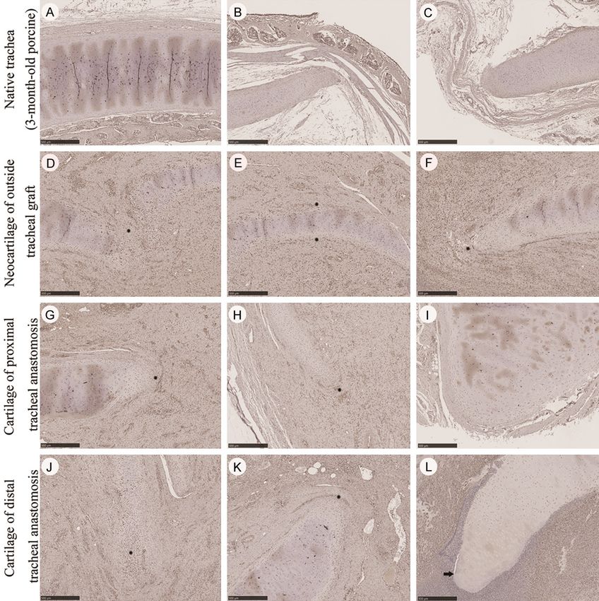
Neocartilage adjacent to implanted tracheal graft

discovered vascular canals (VCs) in the nose           practical transplantation through the use of
and rib cartilage of newborn piglets to clarify        existing software parameters each time. An
this phenomenon. They claimed that perichon-           S2 graft amended with evenly spaced holes
drial papillae (PPs), preresorptive layers (PRLs)      allowed for outside tissue ingrowth for epitheli-
and VCs are the major roles for cartilage ma-          um covering.
turation through cartilage corrosion and the
removal of degradation products. The VCs in            Tracheal transplantation designs
piglet cartilage provide metabolism for the
modulation of the outnumbered chondrocytes             Tracheal grafts of 2 cm in length were produc-
in the center of the cartilage and thus remove         ed for transplantation through various modifi-
matrix degradation products [11].                      cations of the native porcine trachea as
                                                       described below (Figure 1). Group IA: rese-
Advanced three-dimensional (3-D) printing              ction of a 2 cm anterior (A) C-shaped tra-
technology, a recipient predominant capabi-            cheal cartilage only, with the graft sutured
lity, has offered a promising solution towards         end-to-end along the resection margin to form
customizing engineered objects with a shape            a connection with the proximal and distal
compatible with a functional trachea for clini-        parts of the trachea graft wrapped with the
cal application [12, 13]. The mainstream hy-           entire UC for primitive chondrogenesis in vitro.
pothesis is that this process promotes pro-            Group IC: resection of a 2 cm circumferential
genitor or stem cell homing, leading to the de         (C) trachea, with the graft sutured end-to-end
novo generation of cartilage. In this scenario,        in anastomosed connections with the pro-
the organisms are considered to be natural             ximal and distal parts of the trachea. Group
bioreactors and facilitate in vivo airway tissue       L: resection of the trachea in the same man-
engineering [14, 15].                                  ner as group IC, followed by telescopic anasto-
                                                       mosis using a larger graft placed between the
Materials and methods
                                                       proximal and distal parts of the trachea graft,
Graft design and components                            which was then covered with gelfoam to pre-
                                                       vent friction from nearby tissue. Group S1:
We harvested fresh native listed porcine tra-          resection of the trachea in the same manner
cheae from pigs (~100 kg, 6.5 months old)              as Group IC, with a smaller graft telescopi-
and stored them in normal saline at 4°C for            cally anastomosed with the proximal and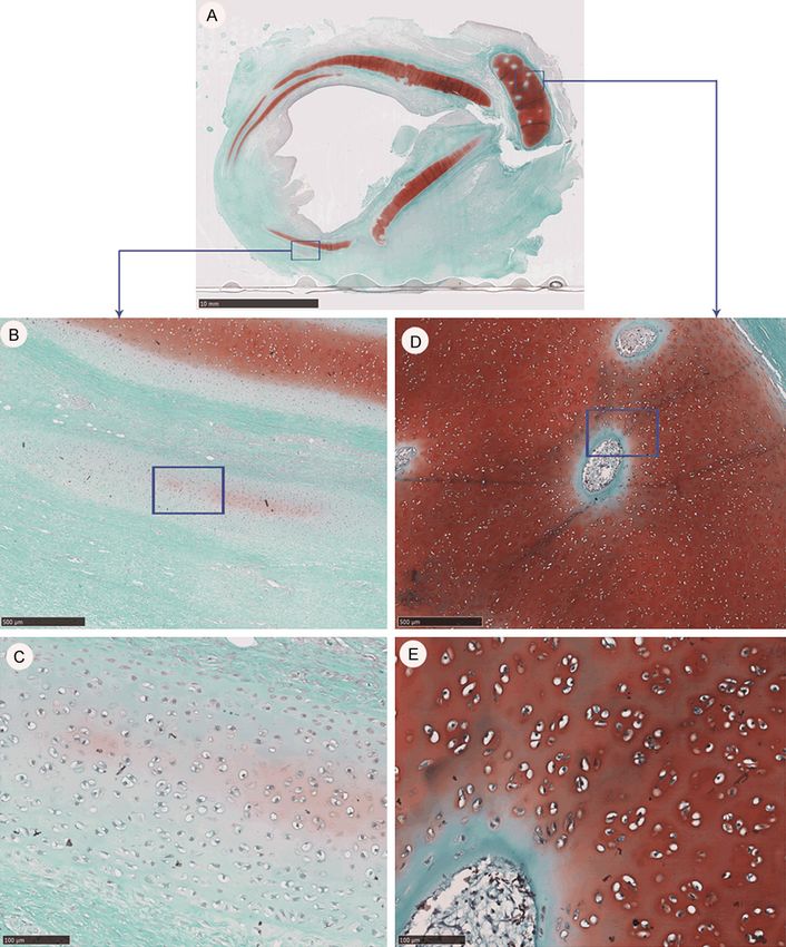
Neocartilage adjacent to implanted tracheal graft

Figure 1. Scheme of the various transplantations of
grafts. (IA) Removing the 2 cm C-shape anterior (A) tra-
cheal cartilage only, with the graft end to end anasto-
mosed to the proximal and distal trachea. (IC) Removing
the 2 cm circumferential (C) trachea, with the graft end
to end anastomosed to the proximal and distal trachea.
(L) Removing the 2 cm circumferential trachea, with the
proximal/distal trachea telescopically anastomosed
with the graft. (S1) Removing the 2 cm circumferential
trachea, with the graft telescopically anastomosed to
the proximal and distal trachea. (S2) Removing the 2
cm circumferential trachea and the graft with evenly
spaced holes telescopically anastomosed to the proxi-
mal and distal trachea.

subsequently rinsed with sterile normal saline             Virbac, Carros, France), followed by intubation
prior to transplantation.                                  with a 5.5 mm endotracheal tube, and then
                                                           they were induced under 4 % isoflurane. The
Surgical transplantation and postoperative                 animal was placed in a supine position, and
care                                                       the area of skin on the neck was prepared
                                                           with povidone iodine and 75% alcohol prior to
Surgical procedures were similar in all animal             the animal being draped with a disposable
groups (Supplementary Figure 3A-D). Each pig               sterile towel. After a cervical midline incision
was anesthetized with Zoletil 50 (8 mg/kg, IM,             and separation of the strap muscles were car-

3730                                                              Am J Transl Res 2020;12(7):3728-3740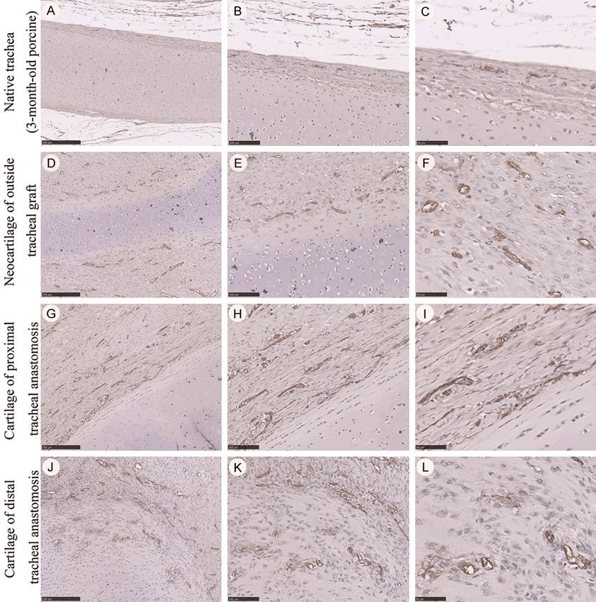
Neocartilage adjacent to implanted tracheal graft

ried out, adequate exposure of the upper tra-      els, with intensity values exceeding the thresh-
chea was achieved. A 2 cm long tracheal seg-       old as calculated by Otsu’s method [16]. The
ment was surgically removed and recons-            summed pixel area within an H&E-stained
tructed with the 3-D printed tracheal graft        cartilage section was calculated by its distinct
(Supplementary Figure 2). Cross-table ventila-     lacuna morphology. We compared the results
tion was applied during the creation of the        of the listed mature porcine trachea and the
proximal anastomosis. Once the proximal (the       experimental (3-month-old porcine) developing
same anastomosed procedure as the distal           trachea (native & neocartilage).
part) and lower posterior half of the distal
anastomosis were completed with 4-0 prolene        Statistics
for a continuous running suture, the en-
                                                   Data (mean ± SD) were analyzed statistically
dotracheal tube was readvanced through the
                                                   with the Kruskal Wallis/ANOVA test. A Dunn-
tracheal graft into the distal trachea. The
                                                   Bonferroni test/Bonferroni test was used to
distal anastomosis for the anterior half of the
                                                   compare the numbers of chondrocytes in the
trachea was then completed with interrupted
                                                   cartilage across different tissues. Values of
sutures above the endotracheal tube. Last, a
                                                   P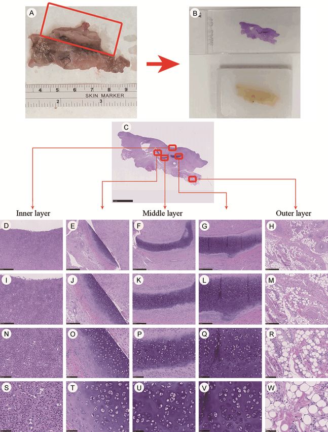
Neocartilage adjacent to implanted tracheal graft

Table 1. Summary of data on the transplantation of various grafts and the pathological outcomes
                   Weight        Post-implant         Survival Postoperation complications                                                   Pathology
Groups (N)                                                                                                                                                                                 Cause of death (Grade of stenosis %)
                    (kg)            status             (day)   (*day)                                         Proximal trachea (P)                Graft          Distal trachea (D)
IA-1                 25                                   10        Dyspnea (2~10)                                       N/A                       N/A                     N/A             Crushed graft
                                                                    Poor appetite (9~10)                                                                                                   (N/A)

IA-2                 25                                   6         Hind limb weakness (4~6)                                                       N/A                                     Occlusion of proximal and distal anastomosis
                                                                    Wound infection (Autopsy)                                                                                              (P: 10 %; D: 50 %)

IA-3                 28                                   10        N/A                                                                                                                    Occlusion of proximal and distal anastomosis
                                                                                                                                                                                           (P: 80 %; D: 80 %)

IA-4                 30                                   19        Dyspnea (9~19)                                                                                                         Occlusion of proximal and distal anastomosis
                                                                    #
                                                                      Collapse (9)                                                                                                         (P: 80 %; D: 80 %)
                                                                    Vomiting (11~15)

IC-1                 25                                   7         Dyspnea (6~7)                                        N/A                                               N/A             Occlusion of proximal and distal anastomosis
                                                                    Wound infection (Autopsy)                                                                                              (N/A)

IC-2                 25                                   14        Dyspnea (7~14)                                       N/A                       N/A                     N/A             Occlusion of proximal and distal anastomosis
                                                                    Vomiting (8)                                                                                                           (N/A)
                                                                    Poor appetite (8~14)

L1                   39                                   5         N/A                                                                                                                    Bleeding (suspected erosion of the jugular
                                                                                                                                                                                           vein)
                                                                                                                                                                                           (P: 90 %; D: 80 %)

L2                   32                                   6         Dyspnea (4~6)                                        N/A                       N/A                     N/A             Bleeding (suspected erosion of the jugular
                                                                    Poor appetite (5~6)                                                                                                    vein)
                                                                                                                                                                                           (N/A)

S1                    37                                  10        N/A                                                  N/A                                                               Occlusion of proximal and distal anastomosis
                                                                                                                                                                                           (P: 90 %; D: 70 %)

S2                   37.5                                 15        Dyspnea (9)                                                                                                            Occlusion of proximal and distal anastomosis
                                                                                                                                                                                           (P: N/A; D: 90 %)

Group IA: Resection of anterior (A) C-shaped tracheal cartilage (2 cm) with end-to-end proximal/distal anastomosis. Group IC: Circumferential resection (C) of the trachea (2 cm) with end-to-end proximal/distal anastomosis. Group L: Resection
of the whole section of the trachea (2 cm) and scaffold telescope anastomosis into the proximal/distal trachea. Group S1: Resection of the whole section of the trachea (2 cm) and proximal/distal tracheal telescope anastomosis into the
scaffold. Group S2: Resection of the whole section of the trachea (2 cm) and proximal/distal trachea telescope anastomosis into the scaffold with evenly distributed holes. P: proximal tracheal stenosis. D: distal tracheal stenosis. *Day after
surgery. N/A: none available. #CPR rescue on day 9.

3732                                                                                                                                                                             Am J Transl Res 2020;12(7):3728-3740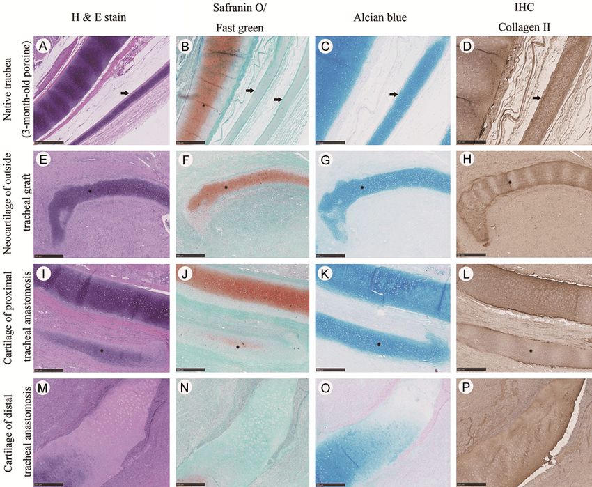
Neocartilage adjacent to implanted tracheal graft

Gross and histological analyses of the grafted       alcian blue and collagen II, with the exception
samples                                              of safranin O. The PCNA assay detected clearly
                                                     proliferated cells in the neocartilage outside
Neocartilage growth was detected in tissues          the grafts, along with anastomosis near the
outside the tracheal grafts in three of the ani-     proximal and distal tracheal cartilage, particu-
mals: IA-3, IA-4 and S2. In the S2 animal, an        larly at the tip of the cartilage (Figure 6). In
intact layer of tissue formed outside the graft      essence, different stages of chondrogenesis
after transplantation (Supplementary Figure          were observed in the developing cartilage. At
2C, 2D). Regenerative tissues displayed blood        the same time, more blood vessels (in terms of
vessels and adipose tissues in the outer layer,      CD31 expression) were formed within the peri-
along with cartilage containing perichondrium        chondrium of neocartilage outside the tracheal
in the middle layer (Figures 2, 3). Lucid angio-     graft and in the nearby proximal/distal tracheal
genesis and condensed immune cells were              cartilage compared with those in the native tra-
found in the proximal and distal granulation         cheal cartilage (Supplementary Figure 8).
areas around the tracheal anastomosis
(Figures 2, 3). In the neocartilage, PPs and         Discussion
VCs, which are regulators of cartilage develop-
ment, were found at the proximal and distal          Although our ultimate goal was the acquisition
granulation areas of the tracheal anastomosis        of a living specimen beyond a 3-week survival
(Figure 3 and Supplementary Figure 4). The           period (a span identical to in vitro chondrogen-
matrix degradation products from chondro-            esis for comparison), the empirical results did
cytes (alcian blue and collagen II expression)       not meet our criteria until the 10th experiment
were detected in the blood vessels of VCs/           ended with an animal being sacrificed. We
PPs (Supplementary Figures 5, 6, 7). Matrix          finalized the research, and analysis was then
corrosion was indicated by the presence of           performed by retrospective grouping in chrono-
PRLs and intravascular matrix degradation            logical order. We discarded the first two the
products [17]. Various stages of developing          MSC-rich UC wrapped tracheal graft for boost-
chondrocytes were found to be distributed in         ing cartilage growth as our original design in
two types of native tracheal cartilage (6.5-         terms of priority regarding graft implant suc-
and 3-month-old), in neocartilage outside the        cess, with the remaining 8 being performed
tracheal graft, and near the anastomosis of          using a pure 3-D-printed graft for implant-
the proximal and distal tracheal cartilage tis-      ation. Unprecedented dramatic growth in
sues (Figure 4A-J). The numbers of chondro-          cartilage over the tissue outside the grafts,
cytes in the 3-month-old native tracheal carti-      despite eventual failure in terms of clinical tra-
lage were higher than those in the 6.5-month-        cheal transplantation for application. Assess-
old cartilage (171.4±21.1 vs. 65.0±10.2, re-         ments of primitive cartilage growth as follows.
spectively, P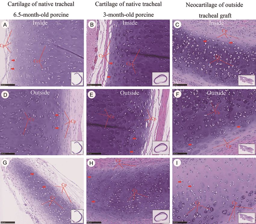
Neocartilage adjacent to implanted tracheal graft

Figure 2. Neoformation of outside tissues at the tracheal graft (three discrete band-like cartilages with a maximum
dimension of 3.78×0.79 mm) (A, B), and results of histological staining (C-W). (A) Specimen from the outside tissue
of the tracheal graft. (B) H&E stained and paraffin-embedded outside tissue of the tracheal graft. (C) Full view of
H&E stain in the outside tissue of the tracheal graft. (D, I, N, S) Immune cells condensing in the inner layer of the
outside tissue of the tracheal graft. (E-G, J-L, O-Q, S-V) Neocartilage forming in the middle layer of the outside tissue
of the tracheal graft. (H, M, R, W) Adipose tissue forming in the outer layer of the outside tissue of the tracheal graft.
Magnification: 50×/100×/200×/400×, from top to bottom.

3734                                                                      Am J Transl Res 2020;12(7):3728-3740
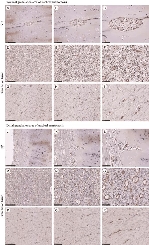
Neocartilage adjacent to implanted tracheal graft

Figure 3. Histological staining in the proximal (A-D) and distal (E-G) granulation area of tracheal anastomosis. (A, E)
Intense angiogenesis in the proximal and distal granulation area of the tracheal graft anastomosis. (B, F) Marked
chondrogenesis in the proximal and distal granulation areas of tracheal graft anastomosis. (C, G) The formation of
perichondrial papillae (PP) from the perichondrium in the proximal and distal granulation areas of tracheal anasto-
mosis. PP, showing perpendicular growth out of the perichondrium, protruding inward to the cartilage matrix. Black
arrow: preresorptive layers (PRLs). (D) The independent vascular canals (VCs) in the proximal granulation area of
tracheal anastomosis. VCs, migrated from the perichondrium and lingering around the overpopulous infant chon-
drocytes. Magnification ×100, H&E stain.

(Figure 2). Chondrogenesis, angiogenesis, and                   fusely distributed in the canal’s stroma next to
adipogenesis indicated with MSC migration to                    the PRL, within capillaries and in the associat-
the graft area for the regeneration [18]. In                    ed downstream veins of the VCs, along with the
vivo, MSCs sourcing from blood and adipose                      veins outside the perichondrium at some dis-
tissue induce chondrogenesis through stimula-                   tance from the cartilage surface. These phe-
tion with endogenous TGF-β mostly from chon-                    nomena reflect the underlying processes of
drocytes [19].                                                  vascular transport and removal [17].

Chondrogenesis in the proximal/distal gra-                      The density and distribution of chondrocytes
nulation area of the tracheal anastomosis                       were different in the porcine native tracheae
revealed that PPs, PRLs and VCs all participat-                 (6.5-month-old and 3-month-old), neocatilage
ed in cartilage development through the pro-                    outside the tracheal graft, and the proximal/
cesses of corrosion and matrix degradation by                   distal tracheal graft end neocartilage. Accord-
way of chondrolysis in order to reach homeo-                    ing to histological analysis, more oval-shaped
stasis (animal S2) (Figure 3). Besides, we                      and larger cells were found in the 6.5-month-
detected matrix degradation products (alcian                    old native porcine tracheal cartilage than in
blue and collagen II expression) within the                     its 3-month-old counterpart. H&E, safranin O,
blood vessels of VCs/PPs (Figure 3 and                          alcian blue and collagen II staining appeared
Supplementary Figures 4, 5, 6). Additional                      similar in the porcine native tracheal cartilage
degradation debris was found in both the ves-                   (3-month-old) (Figure 5A-D). Safranin O
sels and on the surface of the granulation tis-                 staining was particularly weak in both the pos-
sue of the proximal and distal trachea, particu-                terior tracheal cartilage and tracheal islands
larly the proximal trachea (Supplementary                       (Figure 5B). We recognized the neocartilage
Figure 6). Degraded matrix material can be dif-                 through the low levels of H&E, safranin O, alcian

3735                                                                     Am J Transl Res 2020;12(7):3728-3740
Neocartilage adjacent to implanted tracheal graft

Figure 4. Morphology and number of chondrocytes compared between the two ages of animals (6.5- and 3-month-
old porcine native trachea, proximal/distal tracheal cartilage, and tracheal graft neocartilage). A-E. Full view of H&E
staining in the two ages (6.5- and 3-month-old) porcine native trachea, proximal/distal tracheal cartilage, and tra-
cheal graft neocartilage. F-J. Morphology and density of chondrocytes in histological staining. MagnificationÍ 400×.
K, M. Randomly captured images and the number of chondrocytes quantified according to the Otsu method. L, N.
Randomly captured images and the number of chondrocytes quantified by the artificial method. K, L. Total numbers
of chondrocytes (N = 10 in each group) were calculated. M, N. Total numbers of chondrocytes (N = 8 in each group)
were calculated after deleting outliers. a, b, c, d: showing significant differences across groups (P
Neocartilage adjacent to implanted tracheal graft

Figure 5. Various stainings in porcine native trachea, the outside tissues of the graft, and the proximal and distal
trachea. A-D. Strong staining to H&E, alcian blue and collagen II, and weak staining to safranin O in cartilage tem-
plates of native trachea. Black arrow: cartilage templates. E-L. Strong staining to H&E, alcian blue and collagen II,
and weak staining to safranin O in neocartilages of the outside tissues of the graft and proximal trachea. Black star:
neocartilage. M-P. Weak or no staining in clusters of chondrocytes with clear lacuna. MagnificationÍ 50×.

stable chondrocytes by fading the marker. At                   Interestingly, the safranin O, alcian blue and
the same time, at the edge of the neocartilage                 collagen II stains not only distinguished neocar-
in the proximal/distal trachea (Figure 6I and                  tilage but also differentiated VCs that did not
6L), and in the outside tissues of the tracheal                show staining for safranin O and only showed
grafts (Figure 6D-H, 6J and 6K), we detected                   weak staining for alcian blue and collagen II
PCNA-positive cells with a proliferation poten-                (Supplementary Figures 4, 5). More degrada-
tial greater in number than what was found                     tion products (alcian blue expression) were
only in the native tracheal cartilage itself.                  found in both the blood vessels and on the
Furthermore, a more well-defined and well-dis-                 surface of the granulation tissue of the proxi-
tinguished perichondrium appeared in the car-                  mal and distal trachea, particularly in the over-
tilage of the native trachea than it did in either             growing granulation tissues of the proximal
the new cartilage near the proximal trachea,                   trachea. These staining patterns appeared to
outside the tracheal grafts or around the distal               positively correlate with the number of VCs
trachea. Additionally, we also found chondro-                  (Supplementary Figure 6). VCs/PPs were found
blasts extending from some areas of the neo-                   not only in the cartilage of the near proximal
cartilage (Supplementary Figure 9).                            and distal trachea but also in the 3-month-old

3737                                                                    Am J Transl Res 2020;12(7):3728-3740
Neocartilage adjacent to implanted tracheal graft

Figure 6. Immunohistochemistry stainings for proliferating cell nuclear antigen (PCNA) in porcine native trachea, the
outside tissues of graft, proximal and distal tracheae. A-C, I. Scarcity of PCNA-positive cells in the native trachea and
proximal trachea. D-H, J, K. More PCNA-positive cells in the neocartilage of the outside tissues of graft, proximal and
distal tracheae. Black star: PCNA-positive cell in the neocartilage. L. An absence of PCNA-positive cells in the distal
trachea. Black arrow: absence of PCNA-positive cells. MagnificationÍ 50×.

native tracheal cartilage. However, we found no                  not provide successful transplantation in a por-
VCs/PPs in the 6.5-month-old native tracheal                     cine model. Cartilage regeneration in situ has
cartilage. These VCs/PPs might only be found                     enlightened us towards implementing advanced
in areas of dense chondrocytes. The cell densi-                  3-D printing technology with customized tis-
ties and clusters of neocartilage were more                      sues and size-matched counterparts. This
abundant at the outside tracheal grafts (Figure                  appears to be a promising and viable option for
4), with a greater expression of the proliferated                tracheal graft adaptability.
marker PCNA than in the native tracheal carti-
lage (Figure 6).                                                 Acknowledgements

In conclusion, 3-D-printed tracheal grafts could                 The authors sincerely appreciate the assis-
readily provide de novo cartilage growth with                    tance of the Center for Translational Medicine,
stent-sparing anastomosis, although they could                   Department of Medical Research 3D Printing

3738                                                                      Am J Transl Res 2020;12(7):3728-3740
Neocartilage adjacent to implanted tracheal graft

Research and Development Group, Biostatistics                   ment with a bioabsorbable scaffold in sheep.
Task Force and Center of Quantitative Imaging                   Ann Thorac Surg 2010; 90: 1793-7.
in Medicine, Department of Medical Research,             [9]    Claesson-Welsh L and Hansson GK. Tracheo-
Taichung Veterans General Hospital, Taichung,                   bronchial transplantation: the royal swedish
Taiwan.                                                         academy of sciences’ concerns. Lancet 2016;
                                                                387: 942.
Disclosure of conflict of interest                       [10]   Zang M, Zhang Q, Davis G, Huang G, Jaffari M,
                                                                Ríos CN, Gupta V, Yu P and Mathur AB.
None.                                                           Perichondrium directed cartilage formation in
                                                                silk fibroin and chitosan blend scaffolds for tra-
Address correspondence to: Dr. Shih-Chieh Hung,                 cheal transplantation. Acta Biomater 2011; 7:
Institute of New Drug Development, China Medical                3422-31.
University, Taichung, Taiwan; Integrative Stem Cell      [11]   Gabner S, Häusler G and Böck P. Vascular ca-
Center, China Medical University Hospital, Taichung             nals in permanent hyaline cartilage: develop-
Joint PI, IBMS, Academia Sinica 7F, No. 6, Xueshi               ment, corrosion of nonmineralized cartilage
                                                                matrix, and removal of matrix degradation
Rd., North Dist., Taichung City 404, Taiwan. Tel:
                                                                products. Anat Rec (Hoboken) 2017; 300:
+886-4-2205-2121 Ext. # 7728, +886-952-53-
                                                                1067-1082.
2728; E-mail: hung3340@gmail.com
                                                         [12]   Bhora FY, Lewis EE, Rehmani SS, Ayub A,
                                                                Raad W, Al-Ayoubi AM and Lebovics RS.
References
                                                                Circumferential three-dimensional-printed tra-
                                                                cheal grafts: research model feasibility and
[1]   Lano CF Jr, Duncavage JA, Reinisch L, Ossoff
      RH, Courey MS and Netterville JL. Laryngo-                early results. Ann Thorac Surg 2017; 104: 958-
      tracheal reconstruction in the adult: a ten year          963.
      experience. Ann Otol Rhinol Laryngol 1998;         [13]   Rehmani SS, Al-Ayoubi AM, Ayub A, Barsky M,
      107: 92-7.                                                Lewis E, Flores R, Lebovics R and Bhora FY.
[2]   Tan A, Cheng S, Cui P, Gao P, Luo J, Fang C and           Three-dimensional-printed bioengineered tra-
      Zhao Z. Experimental study on an airway pros-             cheal grafts: preclinical results and potential
      thesis made of a new metastable β-type tita-              for human use. Ann Thorac Surg 2017; 104:
      nium alloy. J Thorac Cardiovasc Surg 2011;                998-1004.
      141: 888-894.                                      [14]   Martinod E, Chouahnia K, Radu DM, Joudiou P,
[3]   Kojima K, Bonassar LJ, Roy AK, Vacanti CA and             Uzunhan Y, Bensidhoum M, Santos Portela
      Cortiella J. Autologous tissue-engineered tra-            AM, Guiraudet P, Peretti M, Destable MD, Solis
      chea with sheep nasal chondrocytes. J Thorac              A, Benachi S, Fialaire-Legendre A, Rouard H,
      Cardiovasc Surg 2002; 123: 1177-1184.                     Collon T, Piquet J, Leroy S, Vénissac N, Santini
[4]   Tsukada H, Gangadharan S, Garland R, Herth                J, Tresallet C, Dutau H, Sebbane G, Cohen Y,
      F, DeCamp M and Ernst A. Tracheal replace-                Beloucif S, d’Audiffret AC, Petite H, Valeyre D,
      ment with a bioabsorbable scaffold in sheep.              Carpentier A and Vicaut E. Feasibility of bioen-
      Ann Thorac Surg 2010; 90: 1793-1797.                      gineered tracheal and bronchial reconstruc-
[5]   Shin YS, Choi JW, Park JK, Kim YS, Yang SS,               tion using stented aortic matrices. JAMA 2018;
      Min BH and Kim CH. Tissue-engineered tra-                 319: 2212-2222.
      cheal reconstruction using mesenchymal stem        [15]   Martinod E, Paquet J, Dutau H, Radu DM,
      cells seeded on a porcine cartilage powder                Bensidhoum M, Abad S, Uzunhan Y, Vicaut E
      scaffold. Ann Biomed Eng 2015; 43: 1003-13.               and Petite H. In vivo tissue engineering of hu-
[6]   Kim DY, Pyun J, Choi JW, Kim JH, Lee JS, Shin             man airways. Ann Thorac Surg 2017; 103:
      HA, Kim HJ, Lee HN, Min BH, Cha HE and Kim                1631-1640.
      CH. Tissue-engineered allograft tracheal carti-    [16]   Jungebluth P, Go T, Asnaghi A, Bellini S,
      lage using fibrin/hyaluronan composite gel                Martorell J, Calore C, Urbani L, Ostertag H,
      and its in vivo implantation. Laryngoscope                Mantero S, Conconi MT and Macchiarini P.
      2010; 120: 30-38.                                         Structural and morphologic evaluation of a
[7]   Kamil SH, Eavey RD, Vacanti MP, Vacanti CA                novel detergent-enzymatic tissue-engineered
      and Hartnick CJ. Tissue-engineered cartilage              tracheal tubular matrix. J Thorac Cardiovasc
      as a graft source for laryngotracheal recon-              Surg 2009; 138: 586-93.
      struction: a pig model. Arch Otolaryngol Head      [17]   Ponte AL, Marais E, Gallay N, Langonné A,
      Neck Surg 2004; 130: 1048-51.                             Delorme B, Hérault O, Charbord P and
[8]   Tsukada H, Gangadharan S, Garland R, Herth                Domenech J. The in vitro migration capacity of
      F, DeCamp M and Ernst A. Tracheal replace-                human bone marrow mesenchymal stem cells:

3739                                                              Am J Transl Res 2020;12(7):3728-3740
Neocartilage adjacent to implanted tracheal graft
comparison of chemokine and growth factor [19] Chen MJ, Whiteley JP, Please CP, Ehlicke F,
chemotactic activities. Stem Cells 2007; 25: Waters SL and Byrne HM. Identifying chondro-
1737-1745. genesis strategies for tissue engineering of
[18] Baumann K. Stem cells: moving out of the articular cartilage. J Tissue Eng 2019; 10:
niche. Nat Rev Mol Cell Biol 2014; 15: 79. 2041731419842431.
3740 Am J Transl Res 2020;12(7):3728-3740Neocartilage adjacent to implanted tracheal graft Supplementary Figure 1. Native tracheal tissue of listed porcine. A. A long segment of fresh native listed porcine trachea acquired and immersed in 0.9% normal saline >24 hours at 4°C (an approximately 100 kg, 6.5-month-old pig). B. 3-D printed using a crystal ingredient with an imitation of the fresh native listed porcine trachea as comput- er-scanned. Tracheal size: 120×25×3 mm. Supplementary Figure 2. Tracheal graft implantation in animal S2 and its autopsy picture afterwards. A. Resected porcine trachea fitted to the tracheal graft prior to implantation in Group S. B. Finalizing implantation of the graft with evenly-spaced holes under a telescopic view. Red arrow: tracheal graft. C. External view of the porcine tracheal tissue. D. Integrated tissue covering the graft with easy detachment. Unfolding tissue outside the tracheal graft. 1
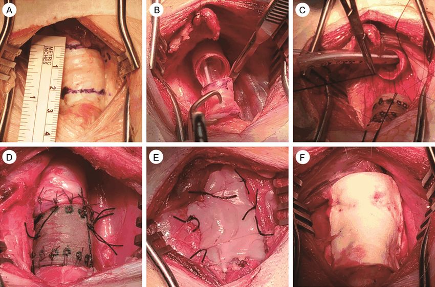
Neocartilage adjacent to implanted tracheal graft

Supplementary Figure 3. Surgical procedure for tracheal graft transplantation. A. Marking a 2 cm-long region on
the trachea. B. Cutting the marked distal trachea. C. Anastomosis of the proximal trachea was performed using 4-0
sutures. D. The anastomosis to the proximal and distal tracheae after graft transplantation. E. Covering the porcine
umbilical cord (UC) after graft transplantation to induce mesenchymal stem cells (MSCs) for UC migration. F. Cover-
ing with hemostatic gelfoam to minimize friction between the bulging tracheal graft and internal carotid artery in
the L group.

2
Neocartilage adjacent to implanted tracheal graft

Supplementary Figure 4. Safranin O/fast green staining in the proximal granulation area of the tracheal anastomo-
sis. (A) Holoscopic view of safranin O/fast green staining in the proximal granulation area of the tracheal anastomo-
sis. (B) Weak safranin O expression in the neocartilage of the proximal granulation area of the tracheal anastomosis
×50×. (C) Safranin O/fast green staining at high magnification (200×) - same area as the blue box in (B). Note the
abundance of chondrocytes with weak staining for glycosaminoglycans (GAGs) and the absence of VCs in this area
of neocartilage. (D) Strong safranin O expression in the mature cartilage of the proximal granulation area of the
tracheal anastomosis ×50×. (E) Safranin O/fast green staining at high magnification (200×) - same area as the blue
box in (D). Note the abundance of chondrocytes with large amounts of GAGs and the presence of multiple VCs in this
area of primitive cartilage. Magnification: 100×/200×/400×, from left to right.

3
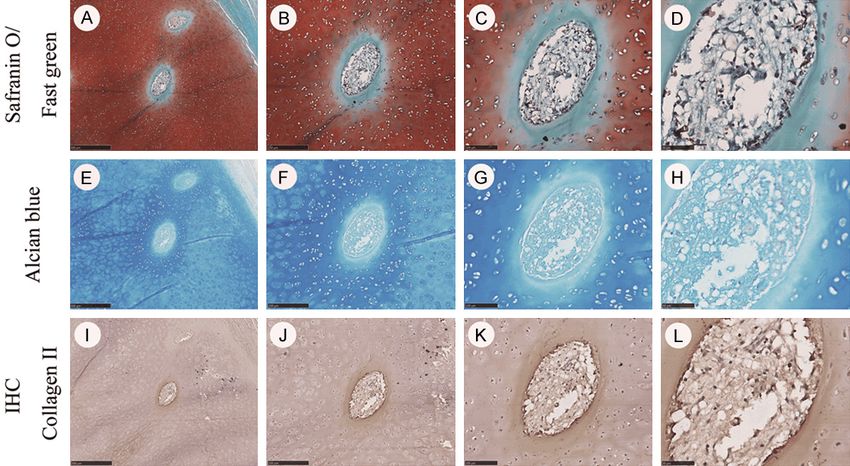
Neocartilage adjacent to implanted tracheal graft

Supplementary Figure 5. Various levels of staining in VCs from proximal tracheal cartilage. A. Two separate migrato-
ry VCs that act like icebreakers for the modified mature chondrocytes. B. A ruby-chestnut-colored background filled
with GAG matrix distributed with unduly chondrocytes, contrasted with the corona-like elliptical VC-fading radiant
blue-tinged ring due to vanishing GAGs. C. A magnified view of a solitary VC ×200×. D. Views of the inner VC showing
capillaries, extravasation of erythrocytes, and fibroblasts ×400×. A-D. Strong safranin O staining in the cartilage and
an abundance of chondrocytes. Note VCs without safranin O staining in the process of lysing the cartilage matrix
(green area around VCs). E-H. Matrix (GAG) degradation products detected weakly within the VCs with alcian blue
staining. Matrix degradation products can be transported via the blood vessels in VCs. I-L. Fibers of VCs detected
weakly with collagen II staining. Magnification: 50×/100×/200×/400×, from left to right.

4
Neocartilage adjacent to implanted tracheal graft

Supplementary Figure 6. Matrix degradation products found within the mucosa and submucosa vessels beneath the cartilage of the native porcine trachea (6.5-
and 3-month-old), in the area of the proximal/distal trachea, and in the area outside of the tracheal graft-like sewage work. A. Scarcity of matrix degradation prod-
ucts within vessels near the surface of the 6.5-month-old porcine tracheal epithelium. B. Scarcity of matrix degradation products within vessels around the cartilage
of the 6.5-month-old porcine trachea. C. Scarcity of matrix degradation products within vessels near the surface of the 3-month-old porcine tracheal epithelium. D.
Scarcity of matrix degradation products within vessels around the cartilage of the 3-month-old porcine trachea. E. Abundance of matrix degradation products within
vessels near the surface of the granulation tissue area of the proximal trachea. F. Abundance of matrix degradation products within vessels around the cartilage in
the area of the proximal trachea. G. Scarcity of matrix degradation products within vessels near the surface of the outside tissue of the tracheal graft. H. Scarcity
of matrix degradation products within vessels around the neocartilage area outside the tracheal tissue graft. I. Abundance of matrix degradation products within
vessels near the surface of the granulation tissue area of the distal trachea. J. Abundance of matrix degradation products within vessels around the cartilage area
of the distal trachea. Magnification ×50×.

5
Neocartilage adjacent to implanted tracheal graft

6
Neocartilage adjacent to implanted tracheal graft

Supplementary Figure 7. CD31 staining by IHC in the proximal (A-I) and distal (J-R) granulation areas of the tracheal
anastomosis. (A-C) CD31 expressions of blood vessels in the proximal distal granulation area of tracheal graft
anastomosis. (D-I) CD31 expressions of blood vessels in the granulation tissue of proximal area of tracheal graft
anastomosis. (J-L) CD31 expressions of blood vessels in the PP of distal distal granulation area of tracheal graft
anastomosis. (M-R) CD31 expressions on blood vessels in the granulation tissue of distal area of tracheal graft
anastomosis. CD31 expression: brown. Magnification: 100×/200×/400×, from left to right.

Supplementary Figure 8. CD31 staining by IHC in porcine native trachea, the outside tissue of the graft, proximal
and distal tracheae. A-C. Scarcity of blood vessels in the perichondrium of native porcine trachea. D-L. Abundance of
blood vessels in the perichondrium neocartilage of the outside tissues of the graft, cartilage of proximal and distal
tracheae. CD31 expression: brown. Magnification: 100×/200×/400×, from left to right.

7
Neocartilage adjacent to implanted tracheal graft

Supplementary Figure 9. Evolution in tapered spear-like neocartilage with elongation in the trachea. De novo carti-
lage growth after tracheal transplantation in a porcine preparation using a mock-up three-dimensional-printed graft,
with access to its tissue regeneration and early structural integrity.

Supplementary Figure 10. Distribution of chondroblasts (Cb), chondrocytes (Cc) and chondroprogenitor cells (Cp) in
the 6.5- and 3-month-old native tracheal cartilage or neocartilage tissues outside the tracheal graft. A, D, G. Limited
numbers of Cbs, Ccs and Cps in 6.5-month-old native tracheal cartilage. B, E, H. Abundance of Cbs, Ccs and Cps in
3-month-old native tracheal cartilage. C, F, I. Abundance of Cbs, Ccs, Cps and clusters of chondrocytes in the neo-
cartilage of the outside tracheal graft. Red arrows: chondroblasts. Magnification ×200×, H&E staining.

8
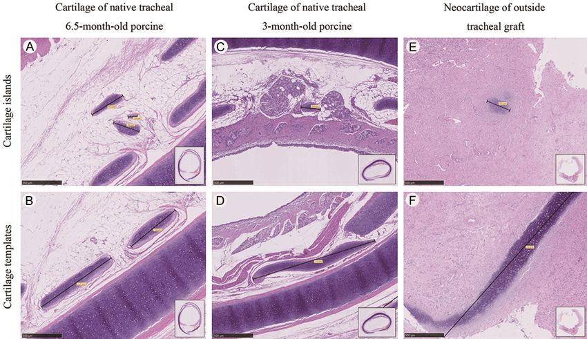
Neocartilage adjacent to implanted tracheal graft

Supplementary Figure 11. Cartilage islands and templates in native (6.5- and 3-month-old) trachea and neocarti-
lage of the outside tracheal graft. A, B. Cartilage islands and templates with well-distinguished perichondrium in the
6.5-month-old porcine native trachea. C, D. Cartilage islands and templates with well-distinguished perichondrium
in 3-month-old porcine native trachea. E, F. Cartilage islands and templates without well-distinguished perichon-
drium in the neocartilage of the outside tracheal graft. Magnification ×50×.

9
You can also read