Fibrinogen like protein 2 promotes the accumulation of myeloid derived suppressor cells in the hepatocellular carcinoma tumor microenvironment
←
→
Page content transcription
If your browser does not render page correctly, please read the page content below
ONCOLOGY LETTERS 21: 47, 2021

            Fibrinogen‑like protein 2 promotes the accumulation of
             myeloid‑derived suppressor cells in the hepatocellular
                     carcinoma tumor microenvironment
                               BO‑QIAN LIU1,2, ZHI‑YE BAO1, JIA‑YI ZHU1 and HAO LIU1

              1
              Department of Transplant and Hepatobilliary Surgery, The First Hospital of China Medical University;
   2
       Department of Anorectal Surgery, The People's Hospital of Liaoning Province, Shenyang, Liaoning 110000, P.R. China

                                         Received July 7, 2020; Accepted October 19, 2020

                                                      DOI: 10.3892/ol.2020.12308

Abstract. The tumor microenvironment in hepatocellular                 cells, therefore promoting MDSC accumulation. Furthermore,
carcinoma can be classified into cellular and non‑cellular             in the orthotopic hepatocellular carcinoma mouse model, we
components. Myeloid‑derived suppressor cells (MDSCs) are               observed that overexpression of FGL2 could promote tumor
cellular components of this microenvironment that serve an             growth and significantly increase the number of MDSCs in the
important role in the progression of hepatocellular carcinoma.         tumors and spleen. Taken together, these findings suggested
Fibrinogen‑like protein 2 (FGL2) has been demonstrated to              that FGL2 may promote hepatocellular carcinoma tumor
promote tumor progression by regulating cellular compo‑                growth by promoting the accumulation of MDSCs in the
nents of the tumor microenvironment in various types of                tumor microenvironment.
malignant tumor. The present study aimed to determine the
expression of FGL2 in hepatocellular carcinoma and its effect          Introduction
on the tumor microenvironment in order to determine novel
targets for liver cancer treatment. Immunohistochemistry and           Primary liver cancer is a common malignant tumor in
reverse transcription quantitative PCR were performed to               China (1). Liver cancer can be divided into three types as
determine the expression level of FGL2 and the correlation             follows: Hepatocellular carcinoma, bile duct cell carcinoma
with surface markers of human MDSCs in hepatocellular                  and a mixture of both types. Hepatocellular carcinoma repre‑
carcinoma. Furthermore, a mouse hepatocellular carcinoma               sents 70‑90% of all primary liver cancers worldwide. Primary
cell line overexpressing FGL2 was established by stable                liver cancer is the second leading cause of tumor‑associated
transfection of a lentivirus expressing FGL2. In addition,             mortality worldwide and in China. The main cause is
fresh bone marrow cells extracted from mouse femurs were               chronic infection of hepatitis B virus (HBV) and hepatitis C
in vitro cultured using conditioned medium derived from the            virus (HCV) (2).
cell line overexpressing FGL2. An orthotopic hepatocellular                The tumor microenvironment is the major site for tumor cell
carcinoma mouse model was also established. The results                proliferation and differentiation. Previous studies have demon‑
demonstrated that FGL2 expression level in hepatocellular              strated that the tumor microenvironment significantly affects
carcinoma tissues was closely associated with tumor size.              tumorigenesis and development, metastasis, and response to
FGL2 level was positively correlated with the expression level         immunotherapy and chemotherapy (3,4). The hepatocellular
of the MDSC surface markers CD11b and CD33 in hepatocel‑               carcinoma tumor microenvironment is divided into cellular
lular carcinoma. The in vitro results demonstrated that FGL2           and non‑cellular components (5). The cellular components
could maintain the undifferentiated state of bone marrow               of this microenvironment include liver sinusoidal endothelial
                                                                       cells and hepatic stellate cells (6,7). In addition, infiltrating
                                                                       immune cells are present in hepatocellular carcinoma, and
                                                                       mainly include neutrophils, lymphocytes, tumor‑associated
                                                                       macrophages, dendritic cells and myeloid‑derived suppressor
Correspondence to: Professor Hao Liu, Department of Transplant
                                                                       cells (8‑10).
and Hepatobilliary Surgery, The First Hospital of China Medical
University, 155 Nanjing North Street, Shenyang, Liaoning 110000,           Myeloid‑derived suppressor cells (MDSCs) form a group of
P.R. China                                                             heterogeneous cells composed of bone marrow progenitor cells
E‑mail: liuhaodoctor@hotmail.com                                       and immature bone marrow cells. These cells are precursors
                                                                       of dendritic cells, granulocytes and macrophages. In healthy
Key words: hepatocellular carcinoma, fibrinogen‑like protein 2,        individuals, immature bone marrow cells produced by the bone
myeloid‑derived suppressor     cells,   orthotopic    hepatocellular   marrow can rapidly differentiate into mature dendritic cells,
carcinoma model                                                        macrophages and granulocytes (11). However, under certain
                                                                       pathological conditions, such as cancer, infection, inflamma‑
                                                                       tion, sepsis, trauma, transplantation or autoimmune diseases,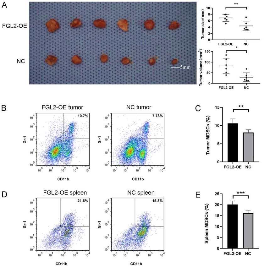
2                                    LIU et al: FGL2 PROMOTES THE ACCUMULATION OF MDSC

the differentiation of immature bone marrow cells into mature       neutral resin counterstained and imaged using a microscope
bone marrow cells is blocked (12,13). This phenomenon even‑         (magnification, x200; Nikon Eclipse Ti‑S; Nikon Corporation).
tually leads to the expansion of MDSCs.                             The expression level of FGL2 was evaluated using the Engers
    In humans, MDSCs express CD11b and CD33 on their                scoring method (28). In addition, tumor tissue cases were
surface, whereas in mouse, MDSCs express CD11b and gluco‑           divided into high and low/medium groups according to the
corticoid receptor 1 Gr‑1, which includes two types, Ly6G and       median expression level using the Engers scoring method.
Ly6C (14,15). MDSCs express high levels of the immunosup‑
pressive molecule arginase 1 (Arg‑1) and inducible nitric oxide     Reverse transcription quantitative (RT‑q) PCR. TRIZOL
synthase (iNOS) that exert immunosuppressive functions              (Invitrogen; Thermo Fisher Scientific, Inc.) were used to
in humans and mice (16,17). MDSCs are one of the main               extract total RNA from human hepatocellular carcinoma
components of the tumor microenvironment. They migrate to           tissues. cDNA was generated using 5x Prime Script RT Master
peripheral lymphoid organs, such as the spleen, to infiltrate       Mix (Takara Bio, Inc.) on PCR System 9700 (GeneAmp)
into the tumor, contributing therefore to the formation of the      under the following conditions: 37˚C for 15 min, 85˚C for
tumor microenvironment (18,19).                                     5 sec and 4˚C. The sequences of the primers were as follows:
    Fibrinogen‑like protein 2 (FGL2) comprises two forms, a         FGL2, forward, 5'-ACAAAGGTGTCCGTAATGGG‑3' and
membrane‑associated form and a secreted form (20,21). The           reverse, 5'‑GTTTGAAGGAGGACTTGTAGCC‑3'; CD11b,
cleavage of the amino‑terminal hydrophobic segment results          forward, 5'‑CTGTTTACCTGTTTCACGGAAC‑3' and
in the secreted form of FGL2 that has immunoregulatory              reverse, 5'-GATTGCCTTGACTCTCAGTACT‑3'; and CD33,
functions. The carboxyl terminus of FGL2 contains a fibrin‑         forward, 5'‑CGATCTTCTCCTGGTTGTCAG‑3' and reverse,
ogen‑related domain (FRED) that is similar to the β and γ           5'‑GATGGTTCTCTCCGCGTAGTCAC‑3'. Human GAPDH
chains of fibrinogen (22). This domain has been demonstrated        Endogenous Reference Genes Primers (cat. no. B662104;
to have immunoregulatory effects (23). The immunoregula‑            Sangon Biotech Co., Ltd.) and Mouse GAPDH Endogenous
tory function of FGL2 serves crucial roles in certain diseases,     Reference Genes Primers (cat. no. B662304; Sangon Biotech
including viral hepatitis, transplant rejection and cancers (24).   Co, Ltd.) were used as the internal controls. The mRNA
Previous studies have reported that FGL2 is involved in the         expression was determined using TB Green Premix Ex TaqII
promotion of tumor progression by regulating the cellular           (Takara Bio, Inc.) on Applied Biosystems® 7500 (Applied
components of the tumor microenvironment in some types of           Biosystems; Thermo Fisher Scientific, Inc.) according to the
cancer, including renal clear cell carcinoma, gliomas and lung      following thermocycling conditions: 95˚C for 30 sec, followed
cancer (25‑27). However, the expression of FGL2 in hepatocel‑       by 40 cycles of 95˚C for 3 sec and 60˚C for 30 sec. The relative
lular carcinomas and its effect on the tumor microenvironment       expression levels were normalized to endogenous control and
remain unknown.                                                     were expressed as 2‑ΔCt or 2‑ΔΔCt (29).

Materials and methods                                               Cell line experiments. The C57BL/6 mouse‑derived Hepa1‑6
                                                                    hepatocellular carcinoma cell line was purchased from
Clinical samples and clinicopathological data. A total of 40        The Cell Bank of Type Culture Collection of the Chinese
pairs of hepatocellular carcinoma cancer and peritumor fresh        Academy of Sciences. Cells were cultured in DMEM medium
and FFPE tissue samples were collected between October 2017         (cat. no. SH30243.01; HyClone; Cytiva) supplemented with
and December 2017 from patients who were pathologi‑                 10% FBS (cat. no. VS500T; Ausbian) and penicillin‑strepto‑
cally diagnosed with hepatocellular carcinoma at The First          mycin (100 U/ml penicillin and 100 µg/ml streptomycin; Gibco;
Hospital of China Medical University. The clinicopathological       Thermo Fisher Scientific, Inc.; cat. no. 15070063) and placed at
characteristics of patients are presented in Table I. The age       37˚C in a humidified incubator containing 5% CO2. Cells were
of the patients ranged from 39 to 72 years, and the mean            transfected with LV5‑FGL2 [overexpressing (OE) FGLV2] and
age was 54 years. All clinical samples were collected after         LV5‑Negative Control (NC) lentivirus (Shanghai GenePharma
obtaining written informed consent from patients. The study         Co., Ltd.) using standard protocols. The transfection efficiency
was approved by the Ethics Committee of the China Medical           was verified in 293T cells. The MOI of Hepa1‑6 was 100, and
University.                                                         1x10⁸ lentivirus were used to transfect 1x10⁶ Hepa1‑6 cells for
                                                                    24 h. After 48 h of transfection, light and green fluorescence
Immunohistochemistry. Serial pathological tissue sections           microscopy (magnification x4; Nikon Eclipse Ti‑S) images were
(5 µm‑thick) were dewaxed using xylene and rinsed with              taken to verify the proportion of transfected cells. After 1 week
gradient ethanol (100, 95, 90, 80 and 70%, 5 min each). Antigen     of transfection, the mRNA and protein expression of FGL2 was
retrieval was performed using citrate buffer at high pressure.      tested by RT‑qPCR, western blotting and ELISA. Transfected
Sections were incubated with rabbit anti‑FGL2 polyclonal            cells were subsequently used for further experiments. To
antibody (1:100; cat. no. ab198029; Abcam) overnight at 4˚C,        generate conditioned media (CM) for MDSC culture, ~1x10⁶
washed with PBS and incubated with secondary antibody (goat         Hepa1‑6 cells were cultured in serum‑free DMEM medium for
anti‑rabbit; ready to use; MaxVision; cat. no. KIT‑5004) for        48 h. CM was then collected, centrifuged at 300 x g at 37˚C and
30 min at room temperature. Streptavidin‑peroxidase solution        for 5 min to remove any debris and was freshly used.
(ready to use; MaxVision; cat. no. KIT‑5004) was then added
and incubated for 30 min at room temperature. Sections were         Western blotting and ELISA assay. Western blotting was
color developed using DAB, stained with hematoxylin for 5 min       performed to detect the FGL2 expression in Hepa1‑6 cells
at room temperature and dried. Transparent sections were            transfected with LV5‑FGL2 and LV5‑NC.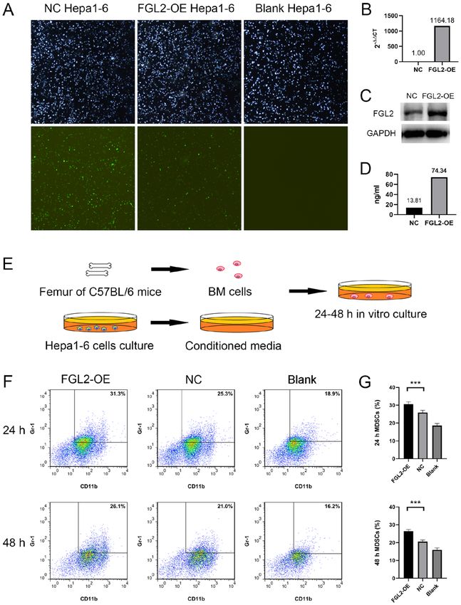
ONCOLOGY LETTERS 21: 47, 2021                                                   3

Table I. Association between FGL2 expression level and the clinicopathological characteristics of patients with hepatocellular
carcinoma.

Clinicopathological		                                     Low/medium expression
characteristics     Number of patients                          of FGL2                       High expression of FGL2        P‑value

Total cases                           40                              23                                  17
Age, years
 ≤53                                  22                              11                                  11                  0.348
 >53                                  18                              12                                   6
Sex
 Male                                 32                              20                                  12                  0.250
 Female                                8                               3                                   5
Tumor size, cm
 ≤5                                   23                              17                                   6                  0.024a
 >5                                   17                               6                                  11
Number of tumors
 Single                               28                              16                                  12                  1.000
 Multiple                             12                               7                                   5
HBsAg expression
 +                                    30                              16                                  14                  0.471
 ‑                                    10                               7                                   3
Cirrhosis
 +                                    32                              19                                  13                  0.702
 ‑                                     8                               4                                   4
AFP level, ng/dl
 ≤200                                 19                              13                                   6                  0.216
 >200                                 21                              10                                  11
TNM stage
 I                                    23                              14                                   9                  0.749
 II‑IV                                17                               9                                   8

P
4 LIU et al: FGL2 PROMOTES THE ACCUMULATION OF MDSC
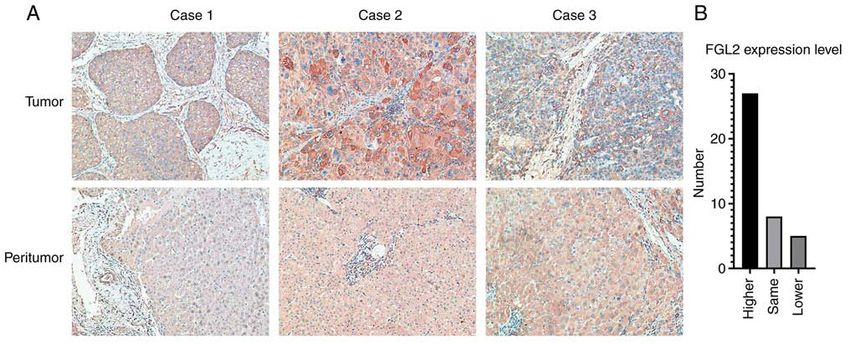
Figure 1. FGL2 expression in tumor and peritumor tissues from patients with hepatocellular carcinoma. (A) FGL2 expression in tumor tissues was higher
compared with their corresponding matched peritumor tissues (three representative tumor/peritumor sample pairs; magnification, x200). (B) Number of
patients with higher, same or lower expression level of FGL2. FGL2, fibrinogen‑like protein 2.
Flow cytometry. Two flow cytometry tubes containing were measured with a caliper using the following formula:
single‑cell suspensions (from bone marrow cells, tumor or Length x width x height x 0.52 (mm3).
spleen) in cell staining buffer (CSB; cat. no. 00‑4222‑57;
Invitrogen; Thermo Fisher Scientific, Inc.) were prepared Preparation of single‑cell suspensions. A total of 12
per sample, one serving as the negative control. Each tube tumor‑bearing C57BL/6 mice (6 per group) were euthanized
contained ~1x10⁶ cells, and 400 µl CSB solution was added by cervical dislocation. For tumor single‑cell suspension, liver
to the tube 1 that was stored on ice. Mouse Fc Block (2 µl; tumors in situ were excised and washed with PBS. Tumors
BD Biosciences) was added to the tube 2 of each sample. were cut into small pieces and washed with PBS. Tumor
Then, APC rat anti‑mouse CD11b (1 µl; BD Pharmingen; tissues were then digested with 0.1% collagenase solution in
BD Biosciences; cat. no. 557657) and PE rat anti‑mouse a 37˚C water bath for 30 min. Cell suspensions were filtered
Ly‑6G and Ly‑6C (1 µl; BD Pharmingen; BD Biosciences; using a 200‑mesh nylon strainer, washed twice with PBS and
cat. no. 561084) were added to the tube 2 of each sample. resuspended into CSB solution.
After incubation on ice for 20 min in the dark, cells were For spleen single‑cell suspension, spleens were excised
washed twice with CSB solution, diluted in 500 µl CSB and placed in a 24‑well plate containing 2 ml PBS. Tissues
and analyzed by flow cytometry (BD FACSAria III; BD were crushed using a 1 ml syringe core and filtered with a
Biosciences) and data were analyzed by Flowjo (v10.0.7; BD 200‑mesh nylon strainer. Erythrocytes were lysed using eryth‑
Biosciences). rocyte‑lysing solution, and the cell suspension was washed
twice with PBS solution and resuspended with CSB solution.
Establishment of the hepatocellular carcinoma orthotopic
mouse model. FGL2‑OE Hepa1‑6 cells and NC Hepa1‑6 Statistical analysis. SPSS 22.0 (IBM Corp.) was used for
cells were harvested, washed twice with PBS, resuspended in statistical analysis. χ2 test was used for clinical‑pathological
PBS and counted. Cells were diluted in PBS to the density data analysis, linear Pearson correlation coefficient was
of 5x10⁷/ml. A total of 12 C57BL/6 mice (age, 5‑6 weeks) used for correlation analysis, and unpaired t‑test was used to
purchased from the Experimental Animal Center of China compare means between groups. PONCOLOGY LETTERS 21: 47, 2021 5 Figure 2. Correlation analysis between the expression level of FGL2 and surface markers of myeloid‑derived suppressor cells in hepatocellular carcinoma tissues. (A and B) Linear Pearson's correlation analysis between FGL2 expression levels and (A) CD11b and (B) CD33 in 40 hepatocellular carcinoma tumor samples. (C) Linear Pearson's correlation analysis between CD11b and CD33. FGL2, fibrinogen‑like protein 2. pairs of tumor tissues expressed the same level of FGL2 rela‑ FGL2 mRNA expression level in FGL2‑OE Hepa1‑6 cells tive to peritumor tissues, whereas five pairs of tumor tissues was significantly upregulated (Fig. 3B). FGL2 protein had lower FGL2 expression level relative to peritumor tissues expression was increased in FGL2‑OE Hepa1‑6 cells and in (Fig. 1B). Furthermore, the results from association analysis the culture supernatants compared with NC Hepa1‑6 cells between FGL2 expression level and the clinicopathological (Fig. 3C and D). Bone marrow cells harvested from the femurs characteristics of patients demonstrated that FGL2 expression of C57BL/6 mice were cultured in vitro using CM from either level was only associated with hepatocellular carcinoma tumor FGL2‑OE Hepa1‑6 or NC Hepa1‑6 cells for 24‑48 h (Fig. 3E). size (P
6                                         LIU et al: FGL2 PROMOTES THE ACCUMULATION OF MDSC

Figure 3. Conditioned media from FGL2‑OE Hepa1‑6 cells increased the proportion of MDSCs in mouse BM cells. (A) Hepa1‑6 cells stably transfected with
LV5‑FGL2 or LV5‑NC lentivirus (48 h post‑transfection). Light and green fluorescence microscopy images (magnification, x4). (B) One week post LV5‑FGL2
lentivirus transfection, FGL2 mRNA expression level in FGL2‑OE Hepa1‑6 cells was upregulated 1168 fold compared with NC Hepa1‑6 cells according to
reverse transcription quantitative PCR experiment. (C) FGL2 protein expression was increased in FGL2‑OE Hepa1‑6 cells compared with NC Hepa1‑6 cells
according to western blotting experiment. (D) FGL2 protein level was significantly higher in FGL2‑OE Hepa1‑6 cell culture supernatants compared with NC
Hepa1‑6 cell supernatants according to ELISA assay. (E) BM cells extracted from C57BL/6 mice femurs were cultured for 24‑48 h using conditioned media
from FGL2‑OE Hepa1‑6 cell, NC Hepa1‑6 cell or conventional medium. (F and G) Proportion of CD11b+/Gr‑1+ cells in BM cells cultured in FGL2‑OE Hepa1‑6
conditioned media for 24 and 48 h was higher compared with BM cells cultured in NC Hepa1‑6 conditioned media (n=8). ***P
ONCOLOGY LETTERS 21: 47, 2021                                                             7

Figure 4. Overexpression of FGL2 promotes tumor growth through the accumulation of MDSCs in mice tumors and spleen. (A) Tumor size and volume
in the FGL2‑OE Hepa1‑6 orthotopic hepatocellular carcinoma model group was significantly higher compared with tumors in the NC Hepa1‑6 group.
(B and C) Proportion of CD11b+/Gr‑1+ cells in the tumors of mice in the FGL2‑OE Hepa1‑6 orthotopic hepatocellular carcinoma model group was higher
compared with mice in the NC Hepa1‑6 group (n=6). (D and E) Proportion of CD11b+/Gr‑1+ cells in the spleen of mice in the FGL2‑OE Hepa1‑6 ortho‑
topic hepatocellular carcinoma model group was higher compared with spleen in the NC Hepa1‑6 group (n=6). *P
8                                    LIU et al: FGL2 PROMOTES THE ACCUMULATION OF MDSC

organs, such as the spleen (33). Previous studies reported that     with anti‑MDSCs may significantly increase the efficacy of
the suppression of T cells in peripheral lymphoid organs by         this therapy (57). Because of the unique immunosuppressive
MDSCs requires direct cell contact (19,34). Furthermore,            microenvironment of the liver and hepatocellular carcinoma
the number of MDSCs in the peripheral blood and tumors of           insensitivity to traditional chemotherapy and radiotherapy,
patients with hepatocellular carcinoma is significantly higher      precise cell therapy and associated cytokines targeted to
compared with healthy people (35) and is associated with            the immunosuppressive microenvironment may represent a
tumor stage, size and Child‑Pugh classification (36). In addi‑      successful therapeutic strategy for the treatment of hepatocel‑
tion, MDSCs presence has been associated with poor overall          lular carcinoma. Further investigation is required to better
survival and early relapse in patients with hepatocellular          understand the role of FGL2 on the tumor microenvironment
carcinoma (37‑40).                                                  of hepatocellular carcinomas.
    In the present study, a mouse hepatocellular carcinoma cell
line overexpressing FGL2 was established by lentivirus stable       Acknowledgements
transfection. Furthermore, fresh bone marrow cells harvested
from the femurs of C57BL/6 mice were cultured in vitro              Not applicable.
using conditioned media from hepatocellular carcinoma cells
overexpressing FGL2. The results demonstrated that FGL2             Funding
could maintain the undifferentiated state of bone marrow cells
in vitro, thereby promoting the accumulation of MDSCs. In vivo,     The present study was funded by the National Natural Science
the overexpression of FGL2 could promote tumor growth. In           Foundation of China (grant no. 81373173).
addition, MDSC levels in the tumors and spleen of mouse over‑
expressing FGL2 were significantly increased. It was therefore      Availability of data and materials
hypothesized that FGL2 may promote the accumulation of
MDSCs to stimulate tumor growth. The original low expression        All data generated or analyzed during this study are included
of FGL2 in mouse hepatocellular carcinoma cell line makes           in this published article.
it not suitable for silencing FGL2. Therefore, FGL2 was only
overexpressed in Hepa1‑6 cells. FGL2 knockdown in mice will         Authors' contributions
be performed in the future. Previous studies have demonstrated
that FGL2 could specifically bind to FcγR receptors, including      BQL performed the main experimental work and wrote
FcγRIIB (41‑43). FcγRIIB is a transmembrane protein that is         the manuscript. ZYB performed in vitro experiments. JYZ
abundantly expressed on the surface of myeloid cells (44,45).       contributed to animal experiments. HL performed the impor‑
FGL2 in hepatocellular carcinoma may help maintaining the           tant experimental design work and contributed to the writing
undifferentiated state of bone marrow cells directly via FcγRIIB,   and revising of the manuscript. All authors read and approved
and therefore promote the accumulation of MDSCs required            the final version.
for tumor growth. FGL2 may also upregulate the expression of
several cytokines, including tumor growth factor β and inter‑       Ethics approval and consent to participate
leukin 6 (46,47), in the human hepatocellular carcinoma tumor
microenvironment in order to promote the maintenance and            All clinical samples were collected after obtaining written
recruitment of MDSCs. FGL2 may also induce the accumula‑            informed consent from patients. The study was approved
tion of MDSCs by direct and indirect mechanisms. Further            by the Ethics Committee of the China Medical University.
investigation is required to test this hypothesis.                  Animal experiments were approved by the Laboratory Animal
    The liver is exposed to large amounts of antigen from the       Management and Use Committee of the China Medical
gastrointestinal tract via the portal vein blood flow. To prevent   University (approval no. 2020016).
continuous immune stimulation and autoimmune damage
induced by antigen exposure, the liver undergoes innate and         Patient consent for publication
adaptive immune responses and develops an intrinsic tolerance
mechanism (48,49). The immune characteristics of the tumor          Not applicable.
microenvironment, the immune checkpoint molecules and the
prognosis differ between patients with hepatocellular carci‑        Competing interests
noma (50,51). Immunotherapy for hepatocellular carcinoma
has been rapidly developed due to the crucial role that immu‑       The authors declare that they have no competing interests.
nosuppressive cells play in anti‑tumor immune response (52).
Recently, immunotherapy has become the first‑line treatment         References
method to treat solid tumors. This includes anti‑cytotoxic
T lymphocyte antigen 4 (CTLA‑4) and anti‑programmed                  1. An L, Zeng HM, Zheng RS, Zhang SW, Sun KX, Zou XN,
                                                                        Chen R, Wang SM, Gu XY, Wei WW and He J: Liver cancer
cell death 1 (PD‑1/PD‑L1) monoclonal antibodies (53,54).                epidemiology in China, 2015. Zhonghua Zhong Liu Za Zhi 41:
Targeted therapy for tumor‑associated macrophages has                   721‑727, 2019 (In Chinese).
rapidly progressed in the recent years (55), and targeted            2. Torre LA, Bray F, Siegel RL, Ferlay J, Lortet‑Tieulent J and
                                                                        Jemal A: Global cancer statistics, 2012. CA Cancer J Clin 65:
therapy for MDSCs shows great potential (56). Since the                 87‑108, 2015.
accumulation of MDSCs in tumors could increase resistance            3. Eggert T and Greten TF: Tumor regulation of the tissue environ‑
to anti‑CTLA‑4/anti‑PD‑1 therapy, combined targeted therapy             ment in the liver. Pharmacol Ther 173: 47‑57, 2017.
ONCOLOGY LETTERS 21: 47, 2021 9
4. Wu T and Dai Y: Tumor microenvironment and therapeutic 25. Tang M, Cao X, Li P, Zhang K, Li Y, Zheng QY, Li GQ, Chen J,
response. Cancer Lett 387: 61‑68, 2017. Xu GL and Zhang KQ: Increased expression of fibrinogen‑like
5. Sevic I, Spinelli FM, Cantero MJ, Reszegi A, Kovalszky I, protein 2 is associated with poor prognosis in patients with clear
García MG and Alaniz L: The role of the tumor micro‑ cell renal cell carcinoma. Sci Rep 7: 12676, 2017.
envi ron ment in t he development a nd prog ression of 26. Yan J, Kong LY, Hu J, Gabrusiewicz K, Dibra D, Xia X,
hepatocellular carcinoma. In: Hepatocellular Carcinoma Heimberger AB and Li S: FGL2 as a multimodality regulator of
[Internet]. Tirnitz‑Parker J (ed). Codon Publications, Brisbane tumor‑mediated immune suppression and therapeutic target in
(AU), Chapter 2, 2019. Available from: https://www.ncbi.nlm. gliomas. J Natl Cancer Inst 107: djv137, 2015.
nih.gov/books/NBK549192/. 27. Zhu Y, Zhang L, Zha H, Yang F, Hu C, Chen L, Guo B and
6. Yin Z, Dong C, Jiang K, Xu Z, Li R, Guo K, Shao S and Wang L: Zhu B: Stroma‑derived fibrinogen‑like protein 2 activates
Heterogeneity of cancer‑associated fibroblasts and roles in the cancer‑associated fibroblasts to promote tumor growth in lung
progression, prognosis, and therapy of hepatocellular carcinoma. cancer. Int J Biol Sci 13: 804‑814, 2017.
J Hematol Oncol 12: 101, 2019. 28. Engers R, Mueller M, Walter A, Collard JG, Willers R and
7. Thomas H: LSEC stretch promotes fibrosis during hepatic Gabbert HE: Prognostic relevance of Tiam1 protein expression
vascular congestion. Nat Rev Gastroenterol Hepatol 16: 262‑263, in prostate carcinomas. Br J Cancer 95: 1081‑1086, 2006.
2019. 29. Schmittgen TD and Livak KJ: Analyzing real‑time PCR data by
8. Tesi RJ: MDSC; the most important cell you have never heard of. the comparative C(T) method. Nat Protoc 3: 1101‑1108, 2008.
Trends Pharmacol Sci 40: 4‑7, 2019. 30. Vetsika EK, Koukos A and Kotsakis A: Myeloid‑derived
9. Tahmasebi Birgani M and Carloni V: Tumor microenvironment, suppressor cells: Major figures that shape the immunosuppres‑
a paradigm in hepatocellular carcinoma progression and therapy. sive and angiogenic network in cancer. Cells 8: 1647, 2019.
Int J Mol Sci 18: 405, 2017. 31. Law AMK, Valdes‑Mora F and Gallego‑Ortega D: Myeloid-
10. Novikova MV, Khromova NV and Kopnin PB: Components of derived suppressor cells as a therapeutic target for cancer.
the hepatocellular carcinoma microenvironment and their role Cells 9: 561, 2020.
in tumor progression. Biochemistry (Mosc) 82: 861‑873, 2017. 32. Tian X, Shen H, Li Z, Wang T and Wang S: Tumor‑derived
11. Condamine T, Mastio J and Gabrilovich DI: Transcriptional exosomes, myeloid‑derived suppressor cells, and tumor microen‑
regulation of myeloid‑derived suppressor cells. J Leukoc Biol 98: vironment. J Hematol Oncol 12: 84, 2019.
913‑922, 2015. 33. Andreu P, Johansson M, Affara NI, Pucci F, Tan T, Junankar S,
12. Porembka MR, Mitchem JB, Belt BA, Hsieh CS, Lee HM, Korets L, Lam J, Tawfik D, DeNardo DG, et al: FcRgamma
Herndon J, Gillanders WE, Linehan DC and Goedegebuure P: activation regulates inflammation‑associated squamous carcino‑
Pancreatic adenocarcinoma induces bone marrow mobilization genesis. Cancer Cell 17: 121‑134, 2010.
of myeloid‑derived suppressor cells which promote primary 34. Gabrilovich DI, Ostrand‑Rosenberg S and Bronte V: Coordinated
tumor growth. Cancer Immunol Immunother 61: 1373‑1385, regulation of myeloid cells by tumours. Nat Rev Immunol 12:
2012. 253‑268, 2012.
13. Sinha P, Okoro C, Foell D, Freeze HH, Ostrand‑Rosenberg S and 35. Hoechst B, Ormandy LA, Ballmaier M, Lehner F, Krüger C,
Srikrishna G: Proinflammatory S100 proteins regulate the accu‑ Manns MP, Greten TF and Korangy F: A new popula‑
mulation of myeloid‑derived suppressor cells. J Immunol 181: tion of myeloid‑derived suppressor cells in hepatocellular
4666‑4675, 2008. carcinoma patients induces CD4 + CD25 + Foxp3 + T cells.
14. Talmadge JE and Gabrilovich DI: History of myeloid‑derived Gastroenterology 135: 234‑243, 2008.
suppressor cells. Nat Rev Cancer 13: 739‑752, 2013. 36. Wang D, An G, Xie S, Yao Y and Feng G: The clinical and prog‑
15. Ugel S, De Sanctis F, Mandruzzato S and Bronte V: Tumor‑induced nostic significance of CD14(+) HLA‑DR(‑/low) myeloid‑derived
myeloid deviation: When myeloid‑derived suppressor cells meet suppressor cells in hepatocellular carcinoma patients receiving
tumor‑associated macrophages. J Clin Invest 125: 3365‑3376, radiotherapy. Tumour Biol 37: 10427‑10433, 2016.
2015. 37. Zhang S, Ma X, Zhu C, Liu L, Wang G and Yuan X: The role of
16. Bergenfelz C and Leandersson K: The generation and identity myeloid‑derived suppressor cells in patients with solid tumors: A
of human myeloid‑derived suppressor cells. Front Oncol 10: 109, meta‑analysis. PLoS One 11: e0164514, 2016.
38. Gao XH, Tian L, Wu J, Ma XL, Zhang CY, Zhou Y, Sun YF,
2020. Hu B, Qiu SJ, Zhou J, et al: Circulating CD14 + HLA‑DR‑/low
17. Hsieh CC, Hung CH, Lu L and Qian S: Hepatic immune toler‑ myeloid‑derived suppressor cells predicted early recurrence of hepa‑
ance induced by hepatic stellate cells. World J Gastroenterol 21: tocellular carcinoma after surgery. Hepatol Res 47: 1061‑1071, 2017.
11887‑11892, 2015. 39. Mizukoshi E, Yamashita T, Arai K, Terashima T, Kitahara M,
18. Xu Y, Fang F, Jiao H, Zheng X, Huang L, Yi X and Zhao W: Nakagawa H, Iida N, Fushimi K and Kaneko S: Myeloid‑derived
Activated hepatic stellate cells regulate MDSC migration suppressor cells correlate with patient outcomes in hepatic arte‑
through the SDF‑1/CXCR4 axis in an orthotopic mouse model rial infusion chemotherapy for hepatocellular carcinoma. Cancer
of hepatocellular carcinoma. Cancer Immunol Immunother 68: Immunol Immunother 65: 715‑725, 2016.
1959‑1969, 2019. 40. Zhang X, Fu X, Li T and Yan H: The prognostic value of myeloid
19. Kumar V, Patel S, Tcyganov E and Gabrilovich DI: The nature derived suppressor cell level in hepatocellular carcinoma: A system‑
of myeloid‑derived suppressor cells in the tumor microenviron‑ atic review and meta‑analysis. PLoS One 14: e0225327, 2019.
ment. Trends Immunol 37: 208‑220, 2016. 41. Liu H, Shalev I, Manuel J, He W, Leung E, Crookshank J, Liu MF,
20. Yuwaraj S, Ding J, Liu M, Marsden PA and Levy GA: Genomic Diao J, Cattral M, Clark DA, et al: The FGL2‑FcgammaRIIB
characterization, localization, and functional expression of pathway: A novel mechanism leading to immunosuppression.
FGL2, the human gene encoding fibroleukin: A novel human Eur J Immunol 38: 3114‑3126, 2008.
procoagulant. Genomics 71: 330‑338, 2001. 42. Morris AB, Farley CR, Pinelli DF, Adams LE, Cragg MS,
21. Marazzi S, Blum S, Hartmann R, Gundersen D, Schreyer M, Boss JM, Scharer CD, Fribourg M, Cravedi P, Heeger PS
Argraves S, von Fliedner V, Pytela R and Rüegg C: and Ford ML: Signaling through the inhibitory Fc receptor
Characterization of human fibroleukin, a fibrinogen‑like protein FcgammaRIIB induces CD8+T cell apoptosis to limit T cell
secreted by T lymphocytes. J Immunol 161: 138‑147, 1998. immunity. Immunity 52: 136‑150.e6, 2020.
22. Yang G and Hooper WC: Physiological functions and clinical 43. Selzner N, Liu H, Boehnert MU, Adeyi OA, Shalev I, Bartczak AM,
implications of fibrinogen‑like 2: A review. World J Clin Infect Xue‑Zhong M, Manuel J, Rotstein OD, McGilvray ID, et al:
Dis 3: 37‑46, 2013. FGL2/fibroleukin mediates hepatic reperfusion injury by induc‑
23. Chan CW, Kay LS, Khadaroo RG, Chan MW, Lakatoo S, tion of sinusoidal endothelial cell and hepatocyte apoptosis in
Young KJ, Zhang L, Gorczynski RM, Cattral M, Rotstein O and mice. J Hepatol 56: 153‑159, 2012.
Levy GA: Soluble fibrinogen‑like protein 2/fibroleukin exhibits 44. Takai T, Li M, Sylvestre D, Clynes R and Ravetch JV: FcR
immunosuppressive properties: Suppressing T cell proliferation gamma chain deletion results in pleiotrophic effector cell defects.
and inhibiting maturation of bone marrow‑derived dendritic Cell 76: 519‑529, 1994.
cells. J Immunol 170: 4036‑4044, 2003. 45. Ravetch JV and Bolland S: IgG Fc receptors. Annu Rev
24. Liu H, Yang PS, Zhu T, Manuel J, Zhang J, He W, Shalev I, Immunol 19: 275‑290, 2001.
Zhang L, Cybulsky MI, Grant DR, et al: Characterization 46. Pan G, Zhao Z, Tang C, Ding L, Li Z, Zheng D, Zong L and
of fibrinogen‑like protein 2 (FGL2): Monomeric FGL2 has Wu Z: Soluble fibrinogen‑like protein 2 ameliorates acute rejec‑
enhanced immunosuppressive activity in comparison to oligo‑ tion of liver transplantation in rat via inducing Kupffer cells M2
meric FGL2. Int J Biochem Cell Biol 45: 408‑418, 2013. polarization. Cancer Med 7: 3168‑3177, 2018.10 LIU et al: FGL2 PROMOTES THE ACCUMULATION OF MDSC
47. Jin SJ, Liu Y, Deng SH, Liao LH, Lin TL, Ning Q and Luo XP: 53. Liao H, Chen W, Dai Y, Richardson JJ, Guo J, Yuan K, Zeng Y
Neuroprotective effects of activated protein C on intrauterine and Xie K: Expression of programmed cell death‑ligands in
inflammation‑induced neonatal white matter injury are associated hepatocellular carcinoma: Correlation with immune microenvi‑
with the downregulation of fibrinogen‑like protein 2/fibroleukin ronment and survival outcomes. Front Oncol 9: 883, 2019.
prothrombinase and the inhibition of pro‑inflammatory cytokine 54. Hilmi M, Vienot A, Rousseau B and Neuzillet C: Immune
expression. Int J Mol Med 35: 1199‑1212, 2015. therapy for liver cancers. Cancers (Basel) 12: 77, 2019.
48. Buonaguro L, Mauriello A, Cavalluzzo B, Petrizzo A and 55. van der Heide D, Weiskirchen R and Bansal R: Therapeutic
Tagliamonte M: Immunotherapy in hepatocellular carcinoma. targeting of hepatic macrophages for the treatment of liver
Ann Hepatol 18: 291‑297, 2019. diseases. Front Immunol 10: 2852, 2019.
49. Brown ZJ and Greten TF: Hepatocellular carcinoma: Translational 56. Lu LC, Chang CJ and Hsu CH: Targeting myeloid‑derived
precision medicine approaches (Internet). In: Hoshida Y (ed). suppressor cells in the treatment of hepatocellular carcinoma:
Immune Therapies. Cham (CH), Humana Press, Chapter 12, 2019. Current state and future perspectives. J Hepatocell Carcinoma 6:
50. Ma L, Hernandez MO, Zhao Y, Mehta M, Tran B, Kelly M, 71‑84, 2019.
Rae Z, Hernandez JM, Davis JL, Martin SP, et al: Tumor cell 57. Chesney JA, Mitchell RA and Yaddanapudi K: Myeloid‑derived
biodiversity drives microenvironmental reprogramming in liver suppressor cells‑a new therapeutic target to overcome resistance
cancer. Cancer Cell 36: 418‑430.e6, 2019. to cancer immunotherapy. J Leukoc Biol 102: 727‑740, 2017.
51. Li W, Wang H, Ma Z, Zhang J, Ou‑Yang W, Qi Y and Liu J:
Multi‑omics analysis of microenvironment characteristics and This work is licensed under a Creative Commons
immune escape mechanisms of hepatocellular carcinoma. Front Attribution-NonCommercial-NoDerivatives 4.0
Oncol 9: 1019, 2019. International (CC BY-NC-ND 4.0) License.
52. Lu C, Rong D, Zhang B, Zheng W, Wang X, Chen Z and
Tang W: Current perspectives on the immunosuppressive tumor
microenvironment in hepatocellular carcinoma: Challenges and
opportunities. Mol Cancer 18: 130, 2019.You can also read