Similar Image Search for Histopathology: SMILY - arXiv
←
→
Page content transcription
If your browser does not render page correctly, please read the page content below
Similar Image Search for Histopathology: SMILY
Narayan Hegde1*, Jason D. Hipp1*, Yun Liu1, Michael Emmert-Buck2 ,Emily Reif1, Daniel
Smilkov1, Michael Terry1, Carrie J. Cai1, Mahul B. Amin3, Craig H. Mermel1, Phil Q. Nelson1, Lily
H. Peng1, Greg S. Corrado1, Martin C. Stumpe1
Abstract
The increasing availability of large institutional and public histopathology image datasets is
enabling the searching of these datasets for diagnosis, research, and education. Though these
datasets typically have associated metadata such as diagnosis or clinical notes, even carefully
curated datasets rarely contain annotations of the location of regions of interest on each image.
Because pathology images are extremely large (up to 100,000 pixels in each dimension), further
laborious visual search of each image may be needed to find the feature of interest. In this
paper, we introduce a deep learning based reverse image search tool for histopathology
images: Similar Medical Images Like Yours (SMILY). We assessed SMILY’s ability to retrieve
search results in two ways: using pathologist-provided annotations, and via prospective studies
where pathologists evaluated the quality of SMILY search results. As a negative control in the
second evaluation, pathologists were blinded to whether search results were retrieved by
SMILY or randomly. In both types of assessments, SMILY was able to retrieve search results
with similar histologic features, organ site, and prostate cancer Gleason grade compared with
the original query. SMILY may be a useful general-purpose tool in the pathologist’s arsenal, to
improve the efficiency of searching large archives of histopathology images, without the need to
develop and implement specific tools for each application.
1
Google AI Healthcare, Mountain View, CA 94043, USA
2
Avoneaux Medical Institute, Baltimore, MD 21215, USA
3
Department of Pathology and Laboratory Medicine, University of Tennessee Health Science
Center, Memphis, TN 38163, USA
*
Equal Contributions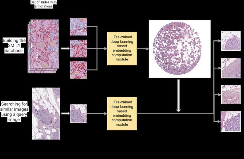
Introduction
The growing adoption of digital pathology1 provides opportunities to archive and search large
databases of pathology images for diagnosis, research, and education. Histopathology is the
examination of biological tissue specimens for diagnostic purposes and is traditionally
performed using microscopes. After digitization, images “tagged” (annotated) with clinical data
such as diagnoses and patient demographics can be searched based on the text-based tags.
For example, searching for “breast” and “carcinoma” in the clinical notes could yield a list of
images that were diagnosed or suspected to contain breast cancer.
A relatively unique aspect of histopathology images is that they are typically much larger than
those found in other imaging specialties: a typical pathology slide might be 100,000×100,000
pixels when digitized at high magnification. Since clinical annotations such as text reports apply
to the entire image or sets of images rather than specific locations within the image, matching a
search “query” with the location in the image that the search is relevant to can be challenging.
For instance, a tumor in a pathology image may be only 100 pixels across, comprising
one-millionth of the image area. A clinician, researcher, or trainee who has found this image or
set of images via searching based on text would still need to visually search the image to locate
the lesion before any subsequent analysis. This problem is further compounded because like
many disciplines, real-world pathology cases contain multiple (e.g. 5-100) images, and the
available text labels might not be specific enough in terms of a particular disease subtype of
interest.
In non-medical domains, a potential solution is reverse image search, also termed
content-based image retrieval (CBIR)2, to find visually “similar” images. In the diagnostic
workflow for example, a clinician may want to search a database for similar lesions to determine
if a feature of interest is malignant or a benign histologic mimic, for example in basal cell
carcinoma3. Relevant tools in non-medical domains include “search by image” for general
images4, visual search5 for retail products, and other tools for faces6 and art7. In medical
imaging, related works include CBIR for radiology8–10 and pathology11–18. Prior machine learning
based CBIR systems have employed application-specific models, which require collecting
labeled data for each application, creating a significant burden to their implementation.
Furthermore, “similarity” in these works were defined along specific axes, whereas the intended
meaning could vary based on the use case. For example, two images could be similar in that
they originate from the same organ, same cancer, similar staining, or similar histologic features.
In this paper, we developed a histopathology similar image search tool (Similar Medical Images
Like Yours, SMILY) without using labeled histopathology images. We then evaluated
2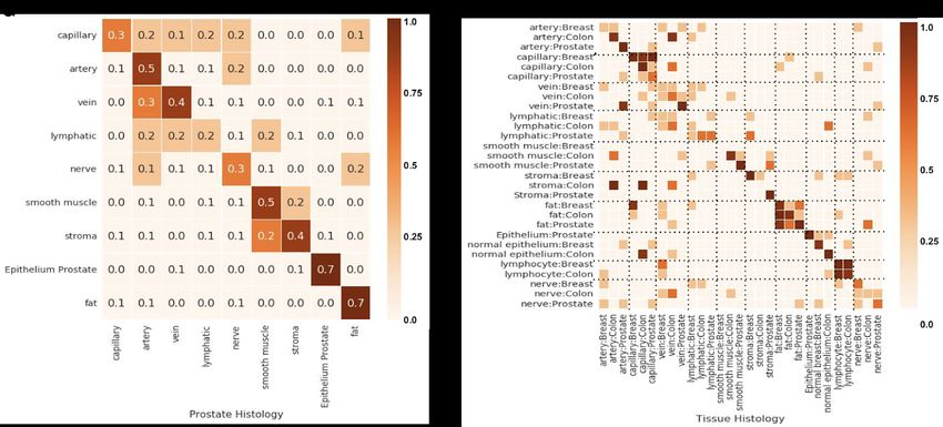
histopathology image search quality in several organs: breast, prostate, and colon, representing three of the four most common non-cutaneous cancer sites. Our evaluation had two components. First, we quantitatively evaluated how often a query image would be matched to an appropriate result from a dataset that was pre-annotated by pathologists. Second, in a blinded prospective study, we had pathologists evaluate how a query image compared to search results from a dataset that were selected either using SMILY or a random image patch. In both evaluations, we assessed SMILY’s ability to retrieve similar tissue types, histologic features and even disease states such as prostate cancer Gleason grading. Results Fig. 1 provides an overview of the development and usage of our proposed tool, SMILY. The first step is to create the SMILY database for searching. The core algorithm of this step is a convolutional neural network that condenses image information into a numerical feature vector, termed an embedding. When computed across image patches cropped from slides, this created a database of patches and a numerical summary of each patch’s information content. When a region of interest is selected for searching (termed the query image), the embedding for the query image is computed and compared with those in the database to retrieve the most similar patches (Methods). An example of the user interface for SMILY is presented in Fig. 2. In the following evaluations, the database was constructed using images at medium (10X) magnification from the publicly available TCGA (The Cancer Genome Atlas)19. In total, the evaluations used 127,000 image patches from 45 slides and the query set consisted of 22,500 patches from another 15 slides. Labels for the first type of evaluation were prepared by pathologists annotating various histologic features in the images, such as arteries, nerves, smooth muscle, and fat. Despite non-exhaustive annotations, this process produced a total annotated area exceeding 150,000 8×8 μm regions (each roughly equivalent to a lymphocyte). After sampling to ensure a balanced dataset with respect to classes of interest in each analysis, this produced thousands of image patches per class (Methods, Table 1). Next, for each query patch, we evaluated the performance of SMILY in retrieving patches of the same histologic feature in the database. We used the top-5 score, which evaluates the ability of SMILY to correctly present at least one correct result in the top 5 search results. This metric was chosen to mimic the standard search process, where a user evaluates a small number of search results to find matches of interest. The subsequent evaluations used image patches extracted at 10X “medium power” magnification, which is commonly used for reviewing images. The results on other magnifications and using other performance metrics are presented in Supp Figs. 1 and 2, respectively. Fig. 3A illustrates the results of this large-scale quantitative analysis. When we used query images from prostate specimens, SMILY had a 63.9% top-5 score at retrieving images of the same histologic feature. This was significantly higher than a traditional image feature extractor (scale-invariant feature transform, SIFT) used in related work17 (45.2%, p
(28.3%). When SMILY retrieved results that did not exactly match the histologic feature, it
commonly returned a similar feature, such as another fluid-transporting vessel: capillaries,
arteries, veins, and lymphatics (Fig. 4A). Next, we expanded to queries from multiple organs:
breast, colon, and prostate. For most histologic features, errors tended to occur between the
same histologic features, but across organs (Fig. 4B). For example, the histologic feature match
score was at 65.3%, but the combined histologic feature and organ match was lower at 40.0%.
Finally, we evaluated the ability of SMILY to retrieve images of the same prostate cancer
Gleason pattern (Fig. 3B). SMILY was significantly more accurate than the SIFT baselines at
retrieving images with the correct Gleason patterns (76.0% vs. 65.2%, p
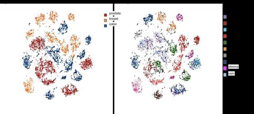
6B). For example, the bottom left prostate cluster in Fig 6A is composed of a mixture of
histologic features such as arteries, lymphatic vessels and capillaries in Fig 6B.
Finally, to investigate the computational efficiency of SMILY for datasets of realistic sizes, we
created a database for all prostate, breast, lung, and colon specimens from TCGA, at 4
magnifications: 40X, 20X, 10X, and 5X. This generated about 109 image patches. Using 400
computers with 10 compute threads each, and some optimizations to use a hash table instead
of the kd-tree depending on the local embedding density21, queries had a median query time of
1.3 seconds. This can in principle be further accelerated using text-based search to filter
images, and real-time updating of search results to present preliminary results before the search
completes. By contrast, a naive implementation on a single machine with 107 image patches
(100 times fewer than above) required a significantly slower 25 seconds per query.
Discussion
This study presents SMILY, a tool to search for similar histopathology images using an image
as the query. To our knowledge, we have performed the most comprehensive evaluation of a
reverse image search tool for histopathology. SMILY retrieves image search results with similar
histologic features, organ site, and cancer grades, based on both large-scale quantitative
analysis using annotated tissue regions and prospective studies with pathologists blinded to the
source of the search results. In the rest of this discussion, we will discuss some nuanced issues
regarding similar image search: what ‘similarity’ means; what a tool like SMILY can be used for;
comparison with “traditional” application-specific approaches; how SMILY was developed and
what that means for future applications not covered in our evaluations; comparison with prior
work; and finally technical implementation considerations.
First, the meaning of “accuracy” in the setting of a similar image search tool deserves some
thought. From first principles, the ideal search tool displays what you are searching for.
However, this goal is ambiguous because the intent of the search depends on the use case:
searching for other images with the same stain, similar stain intensity, same histologic feature,
or similar lesion in the most general sense. As such, in the absence of information about search
intent, the ideal tool should surface a breadth of search results instead of focusing on any single
axis of similarity. To address the lack of algorithmic awareness for the search intent, advances
in human-computer interaction may enable interactive refinement of search results based on
certain desired axes of similarity22.
The potential use cases for a tool like SMILY can be categorized into diagnosis, research, and
education. In diagnosis, SMILY could be a helpful tool to search for similar lesions within the
same slide or in other patients. For example, when a lesion or histologic feature (e.g. mitotic
figure, prominent nucleoli, necrosis, etc) is found, the pathologist might want to know the
frequency of that lesion’s or histologic feature’s occurrence in the specimen. For example,
counting the frequency of mitosis is clinically relevant in breast cancer23. Searching for rare
features in other slides may be helpful in rare diagnoses, to better understand the prognosis of
5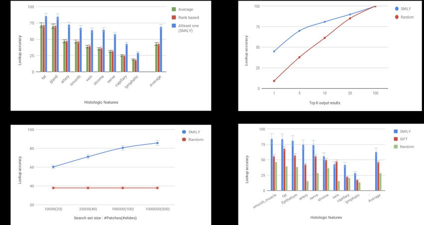
other patients with similar features, including potentially rare pathologies from historic cases with
known intervention and treatment response. For research, a clinician might have a hypothesis:
occurrence of a certain histopathological feature in the slide is correlated with clinical endpoints.
However, an adequately powered study may require a large number of patients, rendering the
manual search for these features highly labor intensive. SMILY could enable significant
speedups in this search via computer-assisted search. Finally, trainees are frequently
confronted with unknown lesions. Manual searching of pathology textbooks, atlases, and other
resources for similar lesions can be time-consuming; SMILY could reduce this process to an
image-based query and manual assessment for the most relevant result. Importantly, these
searches could also leverage large publicly available databases such as the TCGA, as we have
done here.
Indeed, with respect to specific applications such as mitotic counting24, approaches that have
been developed specifically for that application may result in higher accuracy for that purpose.
However, developing and implementing specific but separate approaches for every possible
task of interest is impractical. Some challenges are: expensive data collection and labeling,
difficulty of workflow integration and potential legal or commercial issues, and lack of machine
learning, software or hardware expertise for development or implementation. As such, the
availability of a general-purpose tool like SMILY that can be used in multiple applications, can
be helpful despite having lower accuracy than an application-specific tool.
An interesting aspect of SMILY is that the core neural network algorithm was not trained using
histopathology images. Instead, the network was trained using a dataset of images including
people, animals, and man-made and natural objects (see Methods). Thus our approach does
not require the use of large, pixel-annotated datasets such as those used for breast cancer
mitotic figure detection25, breast cancer metastasis26, or “image-level” labels such as those
extracted from pathology reports27. Because the network is not optimized for the image
characteristics of any given laboratory or slide scanner, the network is likewise not overfit to
these aspects, and can be expected to generalize to other datasets. In principle, the
development of a similar histopathology “similarity” dataset could further improve the
embeddings learned by the model, and is the subject of future work. The results of using several
other “pre-trained” neural networks are presented in Supp Fig. 4.
CBIR has been studied extensively in medical imaging9,10,28, and in histopathology for both
slides14,18 and image patches11–13,15,29. However, the models underlying these CBIR systems
require pathologist-annotated labels for development, which is both costly and non-scalable.
These annotations also in turn restricts the concept of similarity to be along a few predetermined
axes, such as cancer grade and histologic features. In prior work, lookup accuracies ranged
from 60-80% for breast, prostate, necrotic and leukocyte organ sites and histologic features13,15.
By contrast, SMILY achieved comparable performance without the use of application-specific
labeled data for development (though we collected annotations for evaluation purposes), and
thus can be applied to applications without labeled training data. Alternative histopathology
CBIR approaches in the absence of labeled training data include SIFT, kernel and Fourier
6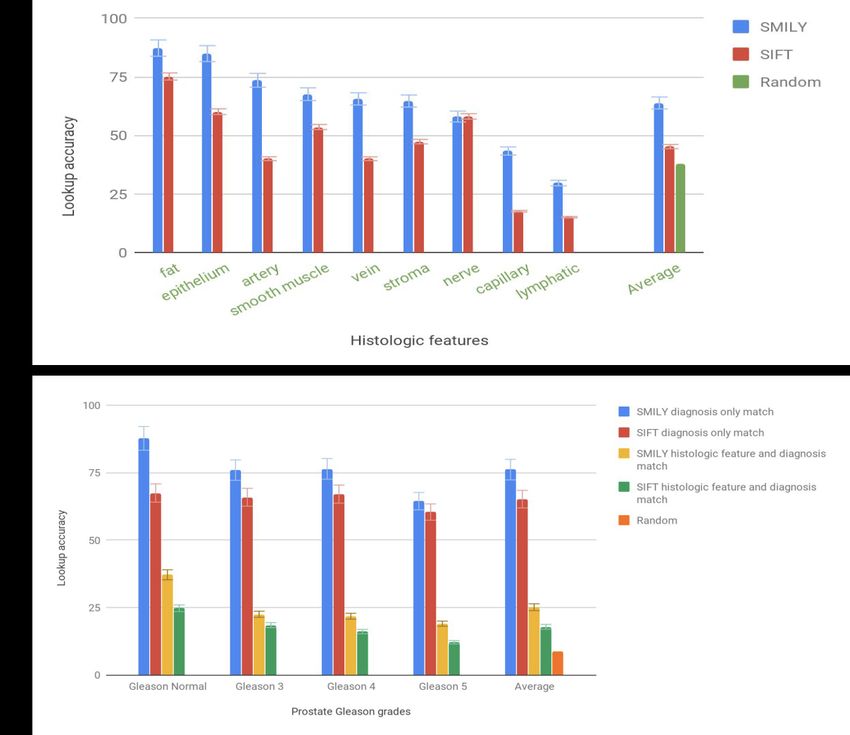
functions17,30. We have performed a quantitative comparison with SIFT to evaluate the added
value of using a neural-network-based system like SMILY for automatic feature extraction,
showing a significant improvement in lookup accuracy across multiple image retrieval
experiments.
The large size of each histopathology image and the scale of typical histopathology databases
(103-106 images) raise important technical considerations for real world use. First, the
embedding of each patch needs to be calculated as a one-time computation cost. This incurs a
delay to compute the embeddings for the 103-106 patches in each newly digitized slide before
the slide can be searched across. Second, these embeddings need to be stored to avoid
repeated embedding recomputation. Though this overhead was only 0.4% per gigabyte-sized
image in our studies, this storage requirement increases with the number of magnifications of
interest, and density of sampling each slide. Finally, the search phase requires comparing the
query image embedding with millions or billions of other embeddings. For example, a naive
implementation of this process on the entirety of the publicly-available The Cancer Genome
Atlas (TCGA, contains over 33,000 slides) dataset31 will incur an impractical, half-minute latency
on a modern desktop computer. To support real-world usage, we have optimized this process to
require only seconds on a web interface (Methods).
This study contains limitations, such as those discussed in-depth above regarding accuracy of a
similar image search tool and limitation of a general-purpose search tool versus application
specific tools. In addition, the number of slides could be increased to better capture the breadth
of tissue processing conditions and resulting images. We have evaluated ‘similarity’ in terms of
histologic features, organ site, and prostate cancer grade, but there are other axes of similarities
that SMILY will need to be further validated on. Lastly, future work will also need to tackle the
‘refinement’ of search results along specific similarity axes of interest, to enable more targeted
image-based search for histopathology.
Methods
The SMILY tool
Neural network architecture
SMILY is based on a convolutional neural network architecture called a deep ranking network32.
Briefly, the deep ranking network is based on an embedding-computing module that
compresses input image patches (of dimensions width x height x channels) into a fixed-length
vector. This module contains layers of convolutional, pooling, and concatenation operations.
During training, the network was fed labeled sets of 3 images: a reference image I of a certain
class, a second image I+ of the same class, and a third image I- of a different class. The network
then uses the modules to compute the embeddings of each of the 3 images. The network is
then trained to assign a lower distance between the embeddings of (I, I+) than the embedding
7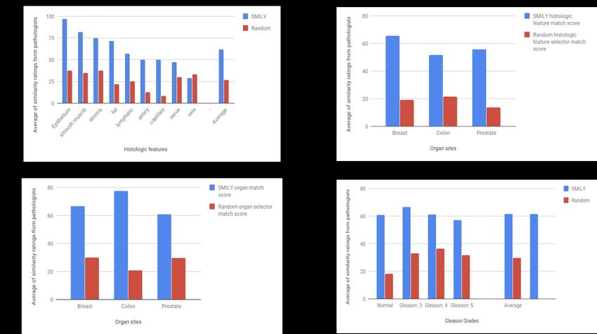
distances of (I, I- ). Our network was trained on about 500,000,000 “natural images” (e.g. dogs,
cats, trees, man-made objects etc) from 18,000 distinct classes. In this way, the network learned
to distinguish similar images from dissimilar ones by computing and comparing the embeddings
of input images.32,33
Building the SMILY embedding database
For the experiments described in this paper, we used slides from The Cancer Genome Atlas
(TCGA)19. TCGA was used because it is publicly available and widely used for histopathology
studies. TCGA tissue samples were collected with approval of local Institutional Review Boards
(IRBs), with the informed consent of patients. Ethics review and IRB exemption for the use of
de-identified images in this study was obtained from Quorum Review IRB (Seattle, WA).
Additional details about each experiment’s dataset are provided in the respective study
sections. SMILY uses the embedding-computing module from the deep ranking network (Fig. 1)
to compress input image patches (300×300 pixels 3-channel RGB (red-green-blue) images) into
embeddings vectors of size 128. Because histopathology images are orientation-independent,
we additionally generate the four 90-degree rotations of each input image, and the mirrored and
rotated versions for a total of 8 orientations, and correspondingly eight 128-sized embeddings
per image patch, a 260-fold dimensionality reduction. Even in the absence of any additional
compression, storing these embeddings required a reasonable additional 0.4% storage
overhead compared to storing the original images alone.
To create image patches at a given image magnification (e.g. high magnifications like 40X and
20X, or medium magnification like 10X), we extracted thousands of non-overlapping patches
per category (Table 1). For a real use case, overlaps can be used to ensure each histologic
feature is contained entirely in a patch of the appropriate magnification, instead of being
potentially bisected into two patches.
Querying the database
To retrieve matches from the database, SMILY first computes the embedding for a selected
query image patch, and then compares that embedding with the embeddings stored in the
database. For this work, our comparison function was the L2 distance across pairs of 128-sized
embedding vectors. To handle the 8 orientations (see above), we filtered the search results
such that the most similar orientation was returned, and only one orientation for each distinct
image patch was presented in each set of search results. In addition, to enhance diversity of the
search results, we filtered the results to ensure that no results were within 1,000 pixels of each
other.
Our experiments (described below) required large numbers of embedding comparisons, ranging
from 40,000 to 90,000. To enable efficient lookups, we used k-dimensional (k-d) trees34 with a
leaf size of 40 and depth of 6; this is customizable to fit computation resources and speed
requirements. To further optimize lookup speed, we parallelized the comparisons across
8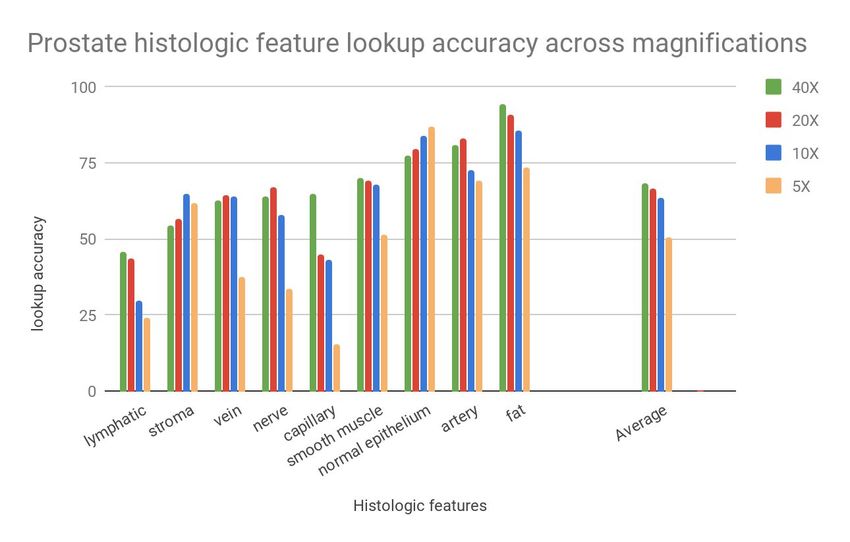
multiple machines (Results). These steps provided a lookup time sublinear in the number of
comparisons.
SMILY’s user interface
SMILY was implemented as a web-based whole-slide viewer (Fig. 2). To conduct a search, a
user selects a rectangular image patch between 200 and 400 pixels in height and width. For a
query patch that is not 300×300 pixels, SMILY resizes to 224×224 pixels using bilinear
interpolation before computing the embedding. The embedding is then used to search the
database based on the current magnification in the selected region, and the results are
displayed as a customizable number of image patches. Optionally, any existing pixel-level
annotations or slide-level metadata such as the original diagnosis can be displayed as well.
Evaluations
To evaluate the utility of SMILY, we conducted several experiments by building a SMILY
database using the TCGA dataset, and then conducting studies to examine the quality of SMILY
image search results.
Large-scale quantitative studies
Our large-scale quantitative experiments were based on regions annotated by pathologists with
various labels (Table 1). In each case, pathologists annotated slides with various histologic
features (up to 11 categories) or Gleason patterns (4 categories: non-tumor and 3 Gleason
patterns). Annotations were performed by three pathologists using the Hamamatsu NDPview2
whole-slide image viewer 35
, using the free-hand outlining and labeling tool. These annotations
were used only for evaluating SMILY, and not for developing the SMILY embedding neural
network. Because of the large size of each slide and the complexity in the appearances of each
feature of interest, annotations were not required to be exhaustive.
Patches of size 300×300 pixels for each histologic feature or Gleason pattern category were
then extracted based on the annotations and stored in the SMILY database along with their
embeddings computed at the appropriate magnification. To avoid class imbalance for these
experiments, we subsampled hundreds to thousands of patches without replacement for each
category (Table 1).
Prospective studies with pathologists
In addition to the large-scale studies, we conducted similar studies by asking pathologists to
rate the quality of search results. Because our annotations (for the large-scale quantitative
studies) were non-exhaustive, these studies with pathologists allowed regions containing
multiple labels (but annotated only as one label) to be assessed correctly. The same database
from the large-scale quantitative studies were used, but the query data were subsampled to
hundreds instead of thousands, to retain a tractable number of search results for manual
evaluation by pathologists. To mimic the use case of a user assessing multiple search results
9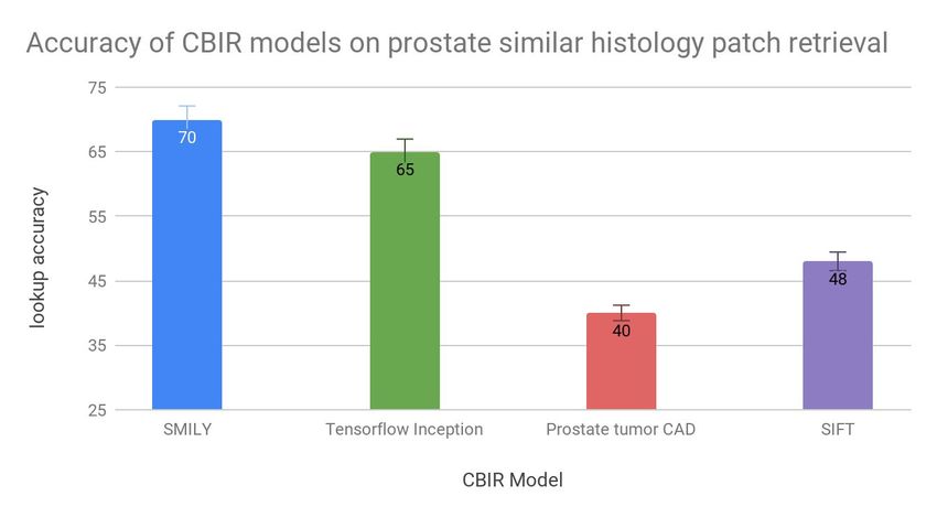
for a single query, each query generated 4 search results, and the pathologist rated each of the
4 results (the scoring system for each experiment is described below). The final average score
for each study is the average score across all search results for all queries.
Similarity along histologic feature, organ, and Gleason pattern were assessed analogously to
the large-scale studies using binary scores (100 for “match” and 0 for “not-match”). For an
overall “match quality score” combining multiple axes, we devised a 100-point score in 25-point
increments (Table 3). A few samples of the user interface for the histologic feature and organ
similarity assessment is presented in Supp Fig. 3.
In total, 3 anatomic pathologists from diverse backgrounds participated in this study: 1 U.S.
board-certified, 1 non-U.S. board-certified, and 1 U.S. residency-trained. As a negative control
to ensure that our pathologists were not artificially rating SMILY search results highly, 25% of
the queries returned search results from random selection (i.e. all 4 images were from SMILY or
all 4 images were randomly selected). The pathologists were blinded to the source of the search
results: SMILY versus random.
Statistical analysis
To assess the statistical significance of our results, we used a two-tailed Chi-squared test.
Because of the large size of each study, most differences were statistically significant.
Acknowledgements
For technical advice and discussion, we thank the following, who are all employees of Alphabet
Inc: Alvin Rajkomar, MD, Daniel Tse, MD. For software infrastructure, logistical support, and
slide digitization services, we thank members of the Google AI Healthcare Pathology team.
Lastly, we are deeply grateful to the pathologists Isabelle Flament, Trissia Brown, and Stanislaw
Krajewski who provided annotations for this study, or participated in the “prospective study with
pathologists”.
Author Contributions
L.H.P. and M.C.S. conceived the idea; N.H., J.D.H., Y.L., and M.T. designed the experiments;
N.H., E.R., D.S., and C.J.C. wrote code for various parts of the work; L.H.P., M.C.S., P.Q.N.,
and G.S.C. acquired the tissue samples for use in the study and provided strategic support;
M.B.A., M.E.B., and J.D.H. created the guidelines for the studies with pathologists and designed
the evaluation scoring systems; C.H.M. and M.C.S. supervised the project; N.H., Y.L., C.H.M.,
and J.D.H. wrote the manuscript with the assistance and feedback of all other co-authors.
10Competing Interests
N.H., J.D.H., Y.L., E.R., D.S., M.T., C.J.C., C.H.M., P.Q.N., L.H.P., G.S.C., and M.C.S. are
employees of Google LLC and own Alphabet stock. M.E.B. and M.B.A. were compensated for
their expertise and time as pathologists. The authors declare no competing non-financial
interests.
Data Availability
Our study used images accessible from the Genomic Data Commons portal36, which is based
upon data generated by the TCGA Research Network31.
11Figures
Fig. 1 | Overview of Similar Medical Images Like Yours (SMILY). First, a database of image
patches and a numerical characterization of each patch’s image contents (termed the
embedding) is created. SMILY uses a convolutional neural network to compute this embedding
(schematic used for illustration purposes only, see Methods for architecture descriptions). Next,
when a query image is selected, SMILY computes the embedding of that query image and
compares the embedding with those in the database in a computationally efficient manner.
Finally, SMILY returns the k most similar patches, where k is customizable.
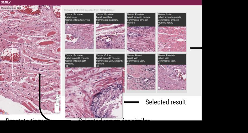
12Fig. 2 | Sample view of the SMILY user interface. Sample query from a prostate specimen
and search results. One of the search results has been magnified for better visualization.
Additional examples of queries and search results are presented in Supp Fig. 3, including an
additional interface for scoring the quality of each search result for the prospective studies with
pathologists.
13Fig. 3 | SMILY search accuracy from large-scale quantitative evaluation using
pathologist-provided annotations. (a) Results for histologic feature match in prostate
specimens, in comparison with a traditional image feature extractor (scale-invariant feature
transform, SIFT) and random search. (b) Results for prostate cancer Gleason grade and
histologic feature match, in comparison with the same baselines.
14Fig. 4 | Confusion matrix from SMILY search. An element in row i, column j indicates the
fraction of search results for query i that result in a “hit” based on the top-5 score for the
category j. (a) Confusion matrix for the results from Fig. 3a: histologic feature match in prostate
specimens. (b) Confusion matrix for histologic feature match across prostate, breast, and colon
specimens. To improve visual contrast and highlight trends better, only discrete colors and
rounded-off values are used.
15Fig. 5 | Evaluation of SMILY from studies with pathologists. The pathologists evaluated
search results, blinded to whether the results were retrieved by SMILY versus a negative
control, random selection. (a) Histologic feature match in prostate specimens. (b) Histologic
feature match in prostate, breast, and colon specimens. (c) Organ site match in prostate,
breast, and colon specimens. (d) Overall match score (Methods, Table 3) in prostate specimens
for similarity in histology and prostate cancer Gleason grade.
16Fig. 6 | Visualizations of the embeddings of image patches in the SMILY database. Each
dot represents an image patch. (a) Colored by organ site, indicating that patches from the same
organ were distributed among different clusters. (b) Colored by histologic feature, indicating a
more distinct separation between histologic features.
17Tables
Table 1 | Summary of data used in large-scale quantitative study. To avoid biases in the
evaluation, we randomly subsampled the original annotated regions, resulting in 5,000 patches
per histologic feature per organ.
Database Query set
Dataset Organ Categories
site(s) assessed Number of Number of Number of Number of
slides patches slides patches
Organ-spe Prostate 9 histologic 20 45,000 5 9,000
cific features (5,000 per (1,000 per
feature) feature)
Multi-organ Prostate, 10 60 87,000 15 14,500
breast, histologic (3,000 per (500 per
colon features feature/org feature/org
an*) an*)
Gleason Prostate Non-tumor 20 40,000 5 8,000
grading and (10,000 in (2,000 in
Gleason each each
Patterns category) category)
3,4,5 (NT,
GP3, GP4,
GP5)
*In our study, no lymphocytes were found upon non-exhaustive review of the prostate
specimens, so the number of patches exclude this.
18Table 2 | Summary of data used in the studies with pathologists. The database used for
this study is identical to Table 1, while the query set was subsampled to retain a tractable
number for manual evaluations.
No. of patches for query Scoring
Dataset Organ site Categories system (see
assessed SMILY Random Methods)
(negative
control)
Organ-specific Prostate 9 histologic 270 by 2 90 by 2 0 or 100 for
features pathologists pathologists histologic
feature match
Multi-organ Prostate, 10 histologic 410 by 2 145 by 2 2 scores: 0 or
breast, colon features pathologists pathologists 100 for
histologic
feature
match, and 0
or 100 or
“unclear” for
organ match
Gleason Prostate Non-tumor 250 by 2 120 by 2 0 to 100 for
grading and Gleason pathologists pathologists tumor grade
Patterns and histologic
3,4,5 (NT, feature match
GP3, GP4,
GP5)
19Table 3 | Overall match quality score for multi-aspect similarity evaluation.
Score Criteria
If the presence/absence of tumor in both patches don't match and they look
0 visually different
If the presence/absence of tumor in both patches don't match and but
25 histological features match
50 If the presence of tumor in both patches match but not the tumor grade
If the diagnostic grades match or both patches are normal (ex: Gleason grade
75 for prostate)
If the diagnostic grades match or both patches are normal (ex: Gleason
100 Grades for Prostate) in addition to at least one histological feature match
20References
1. Mukhopadhyay, S. et al. Whole Slide Imaging Versus Microscopy for Primary Diagnosis in
Surgical Pathology: A Multicenter Blinded Randomized Noninferiority Study of 1992 Cases
(Pivotal Study). Am. J. Surg. Pathol. 42, 39–52 (2018).
2. Lew, M. S., Sebe, N., Djeraba, C. & Jain, R. Content-based Multimedia Information
Retrieval: State of the Art and Challenges. ACM Trans. Multimedia Comput. Commun.
Appl. 2, 1–19 (2006).
3. Stanoszek, L. M., Wang, G. Y. & Harms, P. W. Histologic Mimics of Basal Cell Carcinoma.
Arch. Pathol. Lab. Med. 141, 1490–1502 (2017).
4. Google Images. Available at: https://images.google.com/. (Accessed: 17th January 2019)
5. Amazon Flow. A9 Available at: https://a9.com/what-we-do/visual-search.html. (Accessed:
17th January 2019)
6. Borovikov, E. & Vajda, S. FaceMatch: real-world face image retrieval. U.S. National Library
of Medicine
7. Ivanova, K. Content-Based Image Retrieval in Digital Libraries of Art Images Utilizing
Colour Semantics. in Research and Advanced Technology for Digital Libraries -
International Conference on Theory and Practice of Digital Libraries, TPDL 2011, Berlin,
Germany, September 26-28, 2011. Proceedings 515–518 (2011).
8. Sklan, J. E. S., Plassard, A. J., Fabbri, D. & Landman, B. A. Toward Content Based Image
Retrieval with Deep Convolutional Neural Networks. Proceedings of SPIE--the International
Society for Optical Engineering 9417, (2015).
9. Ahmad, J., Sajjad, M., Mehmood, I. & Baik, S. W. SiNC: Saliency-injected neural codes for
representation and efficient retrieval of medical radiographs. PLoS One 12, e0181707
21(2017).
10. Müller, H., Michoux, N., Bandon, D. & Geissbuhler, A. A review of content-based image
retrieval systems in medical applications-clinical benefits and future directions. Int. J. Med.
Inform. 73, 1–23 (2004).
11. Wang, J. Z. Pathfinder: multiresolution region-based searching of pathology images using
IRM. Proc. AMIA Symp. 883–887 (2000).
12. Komura, D. et al. Luigi: Large-scale histopathological image retrieval system using deep
texture representations. bioRxiv 345785 (2018). doi:10.1101/345785
13. Qi, X. et al. Content-based histopathology image retrieval using CometCloud. BMC
Bioinformatics 15, 287 (2014).
14. Zheng, Y. et al. Histopathological Whole Slide Image Analysis Using Context-Based CBIR.
IEEE Trans. Med. Imaging 37, 1641–1652 (2018).
15. Sridhar, A., Doyle, S. & Madabhushi, A. Content-based image retrieval of digitized
histopathology in boosted spectrally embedded spaces. J. Pathol. Inform. 6, 41 (2015).
16. Mosquera-Lopez, C., Agaian, S., Velez-Hoyos, A. & Thompson, I. Computer-Aided Prostate
Cancer Diagnosis From Digitized Histopathology: A Review on Texture-Based Systems.
IEEE Rev. Biomed. Eng. 8, 98–113 (2015).
17. Mehta, N., Alomari, R. S. & Chaudhary, V. Content based sub-image retrieval system for
high resolution pathology images using salient interest points. Conf. Proc. IEEE Eng. Med.
Biol. Soc. 2009, 3719–3722 (2009).
18. Kwak, J. T., Hewitt, S. M., Kajdacsy-Balla, A. A., Sinha, S. & Bhargava, R. Automated
prostate tissue referencing for cancer detection and diagnosis. BMC Bioinformatics 17, 227
(2016).
19. The Cancer Genome Atlas Home Page. The Cancer Genome Atlas - National Cancer
22Institute (2011). Available at: https://cancergenome.nih.gov/. (Accessed: 13th December
2018)
20. Maaten, L. van der & Hinton, G. Visualizing Data using t-SNE. J. Mach. Learn. Res. 9,
2579–2605 (2008).
21. Jegou, H., Douze, M. & Schmid, C. Hamming Embedding and Weak Geometric
Consistency for Large Scale Image Search. in Proceedings of the 10th European
Conference on Computer Vision: Part I 304–317 (Springer-Verlag, 2008).
22. Cai, C. J. et al. Human-Centered Tools for Coping with Imperfect Algorithms during Medical
Decision-Making. in Proceedings of the 2019 CHI Conference on Human Factors in
Computing Systems (ACM, 2019).
23. Elston, C. W. & Ellis, I. O. Pathological prognostic factors in breast cancer. I. The value of
histological grade in breast cancer: experience from a large study with long-term follow-up.
Histopathology 19, 403–410 (1991).
24. Veta, M. et al. Assessment of algorithms for mitosis detection in breast cancer
histopathology images. arXiv [cs.CV] (2014).
25. Veta, M. et al. Predicting breast tumor proliferation from whole-slide images: the TUPAC16
challenge. arXiv [cs.CV] (2018).
26. Litjens, G. et al. 1399 H&E-stained sentinel lymph node sections of breast cancer patients:
the CAMELYON dataset. Gigascience 7, (2018).
27. Campanella, G., Silva, V. W. K. & Fuchs, T. J. Terabyte-scale Deep Multiple Instance
Learning for Classification and Localization in Pathology. arXiv [cs.CV] (2018).
28. Akgül, C. B. et al. Content-based image retrieval in radiology: current status and future
directions. J. Digit. Imaging 24, 208–222 (2011).
29. Sparks, R. & Madabhushi, A. Out-of-Sample Extrapolation utilizing Semi-Supervised
23Manifold Learning (OSE-SSL): Content Based Image Retrieval for Histopathology Images.
Sci. Rep. 6, 27306 (2016).
30. Caicedo, J. C., González, F. A. & Romero, E. Content-based histopathology image retrieval
using a kernel-based semantic annotation framework. J. Biomed. Inform. 44, 519–528
(2011).
31. The Cancer Genome Atlas Home Page. The Cancer Genome Atlas - National Cancer
Institute (2011). Available at: https://cancergenome.nih.gov/. (Accessed: 13th December
2018)
32. Wang, J. et al. Learning Fine-grained Image Similarity with Deep Ranking. arXiv [cs.CV]
(2014).
33. Image Similarity Data. Available at: https://sites.google.com/site/imagesimilaritydata.
(Accessed: 17th January 2019)
34. Friedman, J. H., Bentley, J. L. & Finkel, R. A. An Algorithm for Finding Best Matches in
Logarithmic Expected Time. ACM Trans. Math. Softw. 3, 209–226 (1977).
35. NDP.view2 Viewing software U12388-01. Available at:
https://www.hamamatsu.com/jp/en/product/type/U12388-01/index.html. (Accessed: 13th
December 2018)
36. GDC. Available at: https://portal.gdc.cancer.gov/. (Accessed: 1st February 2019)
24Supplementary Material
Supp. Fig. 1 | Top-5 score for histology match using SMILY at different magnifications.
The 5X magnification results are lower because there are substantially fewer image patches per
slides at that magnification, of which even fewer pass the 1,000-pixel filter used to enhance
search result diversity.
25Supp. Fig. 2 | Additional performance metrics for SMILY. (a) Comparison different metrics:
averaging the histology match (1/0), weighted based on rank in the search results, and
at-least-one (top-5 score). (b) Comparison of different values of “k” in the top-k score (results for
histologic feature match in prostate specimens). (c) Effect of search database size (results for
gleason cancer grade match in prostate specimens). (d) Top-5 score when searching the
original annotations in prostate specimens (without resample to ensure uniform distribution of
histologic features).
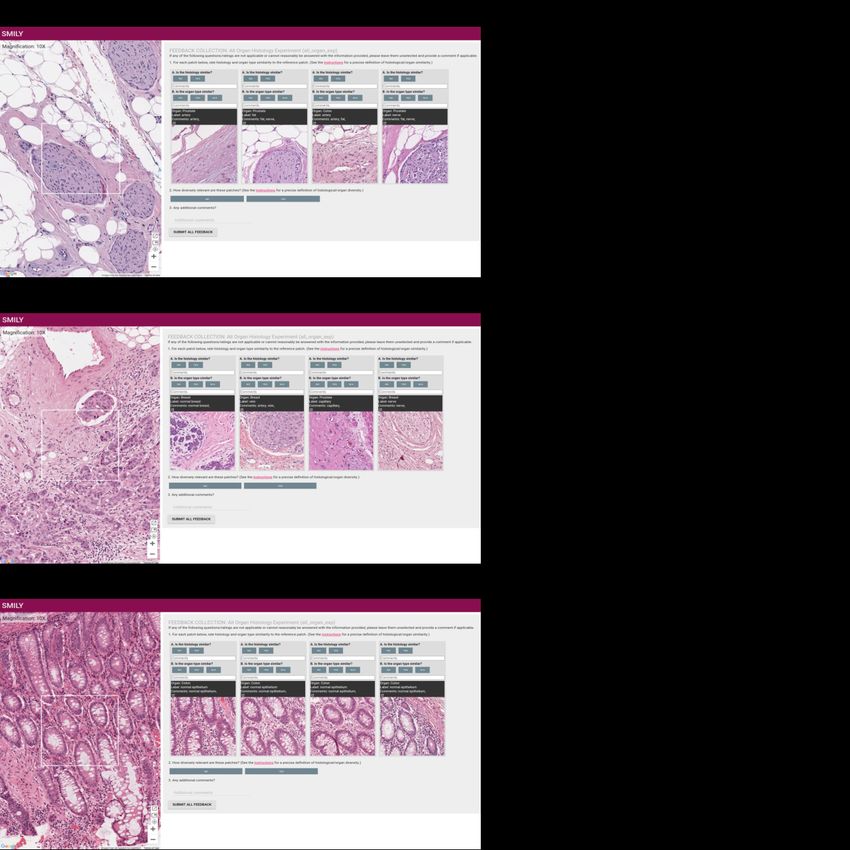
26Supp Fig. 3 | Samples of SMILY similar image retrieval (a) Query consists of a nerve from a
prostate specimen, alongside stromal connective tissue, adipocytes, and blood vessels. The
search results contained (1) connective tissue adjacent to a blood vessel, (2) a nerve adjacent
to fat, (3) a blood vessel adjacent to fat, and (3) a nerve adjacent to fat and connective tissue.
(b) Query consists of nerve, stroma, tumor, and a lymphatic vessel containing metastatic tumor
from a breast specimen. The search results contained (1) a large tumor focus and stroma, (2) a
nerve adjacent to stroma and muscle connective tissue, (3) a lymphatic vessel with stroma, and
(4) a nerve adjacent to stroma and a blood vessel. (c) Query consists of colonic glands cut in
cross and longitudinal sections. The search results all contained colonic glands with similar
architecture.
27Supp. Fig. 4 | Comparison of using different pre-trained embedding generators: SMILY’s
deep ranking network, Inception (V3)1,2, a network trained for Gleason grading3, and
scale-invariant feature transform (SIFT)4.
28Supplemental References
1. Szegedy, C., Vanhoucke, V., Ioffe, S., Shlens, J. & Wojna, Z. Rethinking the Inception
Architecture for Computer Vision. arXiv [cs.CV] (2015).
2. TensorFlow Hub. Available at:
https://tfhub.dev/google/imagenet/inception_v3/classification/1. (Accessed: 23rd January
2019)
3. Nagpal, K. et al. Development and Validation of a Deep Learning Algorithm for Improving
Gleason Scoring of Prostate Cancer. arXiv [cs.CV] (2018).
4. Mehta, N., Alomari, R. S. & Chaudhary, V. Content based sub-image retrieval system for
high resolution pathology images using salient interest points. Conf. Proc. IEEE Eng. Med.
Biol. Soc. 2009, 3719–3722 (2009).
29You can also read