The breast surgeons' approach to mastectomy and prepectoral breast reconstruction - Gland Surgery
←
→
Page content transcription
If your browser does not render page correctly, please read the page content below
Review Article

The breast surgeons’ approach to mastectomy and prepectoral
breast reconstruction
Toni Storm-Dickerson1, Noemi M. Sigalove2
1
Department of Surgical Oncology, Compass Oncology, PeaceHealth Medical Center, Washington State University School of Medicine, Vancouver,
WA, USA; 2Department of Breast Surgery, Comprehensive Breast Center of Arizona, Arizona Center for Cancer Care, Scottsdale, AZ, USA
Contributions: (I) Conception and design: All authors; (II) Administrative support: None; (III) Provision of study materials or patients: None; (IV)
Collection and assembly of data: All authors; (V) Data analysis and interpretation: All authors; (VI) Manuscript writing: All authors; (VII) Final
approval of manuscript: All authors.
Correspondence to: Toni Storm-Dickerson, MD, FACS. Department of Surgical Oncology, Compass Oncology, PeaceHealth Medical Center, Washington
State University School of Medicine, 210 SE 136th Ave., Vancouver, WA 98664, USA. Email: Toni.Storm-Dickerson@Compassoncology.com.

                Abstract: The purpose of this review article is to discuss and highlight the data, techniques and our
                experience performing mastectomies in the setting of prepectoral breast reconstruction. Using a systematic
                review of the approach to mastectomy in the oncologic setting encompassing patient selection, safety,
                anatomy and methods including a literature review of mastectomy trends, safety data and outcomes, anatomy
                and our experience, we are able to illustrate the safety and utility of this technique. The literature strongly
                supports the oncologic safety of these methods. This review also supports the use of these techniques
                as a surgical approach to any mastectomy, with or without reconstruction, and addresses many of the
                factors involved in improving and maximizing outcomes. While, there are multiple and equally efficacious
                approaches to mastectomy, several surgical techniques can be used to improve outcomes and ensure optimal
                flap viability.

                Keywords: Nipple sparing mastectomy; prepectoral reconstruction; oncologic safety; perfusion; incision
                placement

                Submitted Oct 01, 2018. Accepted for publication Nov 19, 2018.
                doi: 10.21037/gs.2018.11.06
                View this article at: http://dx.doi.org/10.21037/gs.2018.11.06

Good planning for any oncologic surgery, starts with an                      cancer survival after both breast conserving surgery (BCS)
understanding of the disease for which surgery is being                      and mastectomy is based on stage at diagnosis (tumor size
performed along with a clear definition of the goal of the                   and characteristics, lymph node status, and presence of
procedure. Regardless of the specific surgical procedure,                    systemic disease) and not the type surgery performed, is
for the treatment of breast cancer, these principles mark the                frequently overlooked. Regardless of the surgical approach,
foundation of our approach to every patient, every time.                     the oncologic principles of complete tumor removal,
   There were an estimated 252,710 new cases of breast                       appropriate lymph node assessment and patient selection
cancer in 2017 (1), with 40,610 deaths, translating to 1                     remain paramount in achieving optimal oncologic results.
death for every 37 women diagnosed (2). In the United                           While BCS is the treatment of choice for most women
States, as of January 2018, there were more than 3.4 million                 with early stage breast cancer (59% of early stage vs. 13% of
women either with a history of breast cancer, or being                       advanced stage), mastectomies have retained an important
actively treated for the disease (3). Not including benign                   role for multi-centric, locally advanced disease (59% of
breast biopsies and cosmetic breast surgery, there are over                  advanced stage vs. 36% of late stage), tumors not visualized
half a million-breast cancer related surgeries performed                     on imaging and for deleterious gene carriers (5). Without
per year in the United States (4). The fact that that breast                 addressing the axilla, the three main types of mastectomy

© Gland Surgery. All rights reserved.                              gs.amegroups.com                                     Gland Surg 2019;8(1):27-35
28 Storm-Dickerson and Sigalove. The breast surgeon’s approach
performed today are total mastectomy (TM), skin sparing breast cancers (14-16). In 2013, Agarwal et al. used
mastectomy (SSM) and nipple sparing mastectomy (NSM). Surveillance, Epidemiology, and End Results (SEERs) data
TM with or without reconstruction entails removal of to compare the DFS and OS following NSM and modified
the breast with maximal removal of the overlying breast radical mastectomy (MRM) and found no significant
skin. SSM preserves the skin envelope less the nipple difference between the procedures (17). Although their
areolar complex (NAC) and includes reconstruction. NSM data supported the oncological safety of NSM, they
preserves the entire skin envelope and requires some form acknowledged possible selection bias (lower tumor grade,
of reconstruction, typically immediate. negative nodes, no lympho-vascular invasion, etc.), and
In the United States, we have seen an increasing suggested careful patient selection, as reflected in the
preference toward mastectomies since 2005, especially among NCCN guidelines (8).
patients with early stage breast cancer (6). Between 2002 Newer data supports preservation of uninvolved nipples
and 2012, rates of contralateral prophylactic mastectomies regardless of tumor size. Several studies have shown, that
(CPM) more than doubled (3.9% to 12.7%) (7). This trend while locoregional recurrence is clearly elevated in locally
occurred despite evidence that in most cases, CPM does advanced disease, preservation of the uninvolved nipple
not increase overall survival (OS), and under National does not increase the risk of local recurrence and when
Comprehensive Cancer Network (NCCN) guidelines, CPM local regional recurrence does occur, it is not at the nipple
is not routinely recommended (8). In spite of improved (18-20). Smith et al. published their data September 2017,
survival, due in large part to better systemic therapies, showing that in 2,182 patients with stage 0–III breast cancer
improved early diagnosis (9,10) and improved BCS cosmetic who underwent NSM at Massachusetts General Hospital
outcomes with oncoplastic techniques, we continue to between 2007–2016, at a mean follow-up of 51 months,
see a trend toward more aggressive breast surgery (11). there was a 2.7% distant recurrence with two deaths and no
The preference for mastectomies is likely to be fueled by recurrences at the nipple (19).
the availability of improved reconstructive techniques in Li et al., used SEERS data to look at cancer specific
combination with increased overall patient awareness of survival (CSS) and OS in 2,440 patients undergoing NSM
surgical options. Appropriate oncologic counseling should between 1998–2013 (20). Median age was 50, and they
be provided for all patients in order to alleviate the fear and included Tis, T1–3 (79.6% T2–3) and N0–3. Twenty-six
anxiety normally associated with their diagnosis. It is our percent of the NSM cases were T2–3, 20% were N1–3
belief that the choice in surgery ultimately lies in the hands and 13.8% were estrogen receptor (ER) negative. For
of a well-informed patient who is aware of the survival patients diagnosed between 1998–2010 (N=763), median
benefit and makes their choice based on understanding not follow-up was 69 months, 5- and 10-year CSS were
fear or a false sense of safety. 96.9% and 94.9% respectively, while OS was 94.1% and
88.0% respectively. Ethnicity, T-stage and N-stage were
independently associated with CSS, and age and T-stage
Oncologic safety of nipple and skin sparing
were factors independently associated with OS. They
mastectomies
showed a 10-year OS of 72% for N2–3 patients after NSM.
Data on the oncologic safety of NSM and SSM has This was significantly better than the reported outcomes
been accumulating for the last 50 years. Early data from following traditional mastectomy in patients with a similar
Barbara Freeman in 1962 showed a 0.4% incidence of tumor burden per National Cancer Data-base (8-year OS
breast cancer at 10-year follow-up in 1,500 subcutaneous was 66.6% and 53.5% respectively) following complete
mastectomies performed for benign symptomatic fibro- mastectomy in N2–3 patients with and without RT,
cystic breast disease (12). In 1984, Hinton et al., published respectively (20).
their work showing no difference in disease-free survival The above data repeatedly demonstrates that the primary
(DFS) and OS in patients with stage I and II breast cancer predictor of both locoregional and distant recurrence in
undergoing subcutaneous mastectomies (13). There have breast cancer is the extent of disease at diagnosis, not the
been multiple large population studies evaluating the safety surgical approach utilized. Therefore, preservation of
of prophylactic mastectomies in patients considered to be the nipple in NSM is a safe option for patients without
high risk secondary to family history showing significant pathologic evidence of nipple involvement, extensive skin
decreased incidence of both recurrent and contralateral involvement, or the presence of inflammatory cancer.
© Gland Surgery. All rights reserved. gs.amegroups.com Gland Surg 2019;8(1):27-35Gland Surgery, Vol 8, No 1 February 2019 29
Table 1 Contraindications to prepectoral reconstruction
Reconstructive Oncologic
Thin flaps Large tumors (>5 cm)
Poorly vascularized/ischemic flaps Late cancer stage
History of prior radiation therapy (unless latissimus is used) Deep tumors
2
BMI >40 kg/m * immunocompromised HbA1c >7.5% Chest wall involvement
Active smokers Grossly positive axillary involvement
Lack fat donor sites High risk of recurrence (based on multidisciplinary approach)
*, with other associated comorbidities (e.g., diabetes mellitus, hypertension). BMI, body mass index; HbA1c, hemoglobin A1c.
Prepectoral reconstruction-preoperative of discussion are the timing of expander or implant
selection criteria placement and possible benefit from using additional flap-
based coverage. In our experience, to date, complications
Prepectoral reconstruction is not suitable for all patients.
associated with adjuvant radiation are not significantly
Selection criteria for prepectoral reconstruction
different than those observed with partial tissue or dual
combine variables that are unique to each individual
plane reconstruction (23). Although complications of skin
with conservatively defined general and oncologic safety
necrosis can occur, more commonly, complications tend to
parameters. In our experience, general disqualifiers for
be skin tightening and stiffness with associated reduction
prepectoral reconstruction include patients with ischemic
in breast size. Since the modern prepectoral approach is
or poorly vascularized mastectomy skin flaps, a history of
relatively new, oncologic guidelines continue to evolve as
preoperative breast irradiation (without use of a flap at
long-term data becomes available. We feel that it is prudent
reconstruction), poorly controlled diabetes (hemoglobin
to err on the side of caution until data from randomized,
A1c >7.5%), active smoking, immunocompromise, morbid
prospective clinical trials and results of oncologic outcomes
obesity [body mass index (BMI) >40] and lack fat donor sites
become available.
(Table 1) (21).
In addition, we have identified a number of oncologic
contraindications to the prepectoral reconstructive approach Prepectoral reconstruction and patient benefit
(Table 1) (21). Patients with late-stage breast cancer,
Prepectoral reconstruction is not a new concept, however,
posterior tumors involving the pectoralis major muscle,
prior versions of this operative technique were limited by
and those at high risk of locoregional recurrence, such as
aggressive mastectomy dissection, paucity of reconstructive
skin nodules or inflammatory disease, should be excluded
materials and lack of innovative technology. We have made
from prepectoral reconstruction. It is important to note
technical advances in the treatment of breast cancer due to
that the oncologic safety of this procedure has not yet been
the recognition that breast cancer is a disease defined by
documented over time. As with subpectoral reconstruction,
biology rather than anatomy alone. Bigger surgery does not
caution should be exercised for large tumors, and for
necessarily translate to better outcome. A new appreciation
clinically positive lymph nodes (22). We encourage the use
for existing anatomical divisions between the breast and
of neoadjuvant chemotherapy to downstage the tumor when
skin flap hypodermis and a desire for improved mastectomy
possible and thus, allow for prepectoral reconstruction in
flap viability, have led to a quest for improving surgical
cases of a favorable clinical and radiographic response.
technique. Better understanding of skin flap perfusion and
Although a history of radiation therapy negatively
the importance of maintaining appropriate vascular integrity
impacts prepectoral reconstruction due to compromised
for the success of reconstruction have led to surgical
tissue perfusion and flap viability, the converse has not
changes that minimize skin flap necrosis. The availability of
been the case. Radiation therapy can and should be used
tissue perfusion assessment devices also allows for objective
for all prepectoral reconstruction patients with oncologic
determination of perfusion and skin flap viability. With
indication for post-mastectomy radiation. Issues worthy
their advent, immediate evaluation and decision making
© Gland Surgery. All rights reserved. gs.amegroups.com Gland Surg 2019;8(1):27-3530 Storm-Dickerson and Sigalove. The breast surgeon’s approach
regarding a patient’s candidacy for type of reconstruction preserve the NAC and expectation of adjuvant radiation.
are now possible. Important to these quantum shifts has The multi-disciplinary discussion should extend to the
also been the use of acellular dermal matrices and materials medical and radiation oncologists who will administer
that stabilize the reconstructed breast and serve as a layer adjuvant or neo-adjuvant treatment. The timing of all
of vascularized regenerative tissue between the implant and therapies should be discussed in advance, as recovery
mastectomy flap. from surgery may interfere with additional treatments
A critical question is, why change to prepectoral planned. Surgical pathology results are best reviewed with
reconstruction if partial muscle coverage or retropectoral a team approach, especially when pathology influences
reconstruction are sufficient and have worked well in further therapy such as radiation. The timing of second
the past? The answer is derived from patient benefit and stage reconstruction, if needed, will also be affected by
satisfaction resulting from an undisturbed pectoralis muscle additional treatments such as chemo or radiation therapy.
(24,25). Less trauma to the muscle leads to less pain with When members of the patient care team are in agreement
decreased need for narcotics and faster recovery. Implant regarding the treatment plan, patients benefit from focused
placement over the muscle eliminates animation deformity, discussion and the recognition of personalized care.
improves long term comfort, and lends to a more natural Patient expectations and involvement in “Enhanced
appearing breast (21,24,25). The newly reconstructed breast Recovery After Surgery” (ERAS) is also key to optimizing
lies in the natural anatomical position of the surgically outcomes and patient satisfaction (30,31). Patient education
removed breast. Further, post-operative complications have regarding survival benefit, surgical options, unilateral versus
been found to be similar for both prepectoral and partial bilateral mastectomy and oncoplastic surgery are integral to
muscle coverage techniques (26-28). assuring knowledge and preference driven decision making.
Breast surgeons are also foundational in setting proper
expectations and covering basic reconstructive techniques at
Preoperative planning
the time of cancer consultation. A well-informed patient has
Pre-operative surgical planning relies on the input of a realistic expectations and is able to ask pertinent questions at
multidisciplinary team. In order to maximize outcomes and the time of the reconstructive consultation. Surgeons should
the patient experience, each specialty must be aware of what be familiar with each other’s postoperative patient instructions
the other plans, as our different treatment modalities have in order to avoid conflicting information. Postoperative
potential additive impact on our patients. A team approach outpatient visits should be coordinated in a manner that
leads to better oncologic and aesthetic results as well as minimizes patient discomfort and inconvenience.
improved patient satisfaction. The multidisciplinary team Discussion of post-operative expectations is also
is held by certifying organizations such as the Commission important and in line with ERAS recommendations (30,31).
on Cancer (CoC) and National Accreditation Program for We encourage our patients to start pre-operative enteric
Breast Centers (NAPBC) as the sine qua non for the care coated probiotics, 1,000 mg vitamin C, and 81 mg buffered
of the oncologic patient (29). It is imperative that surgeons aspirin daily. The aspirin is held 5 days prior to surgery and
work in concert with each other in order to achieve the resumed on post-operative day 1. We discuss expectation
most optimal oncologic outcome along with the best of drains and drain care, showering, and negative pressure
possible aesthetic appearance of the reconstructed breast. dressings (32). We encourage use of minimal narcotics
While surgeons of the past commonly worked in sequence, combined with nonsteroidal medications for the first few
today’s surgical oncologist and reconstructive surgeon days post-operatively, discussing the expectation of “some
benefit from working in collaboration. discomfort” but minimal “pain”. We also encourage non-
Communication between the oncologic surgeon and particulate liquids up to arrival at hospital rather than
plastic surgeon should start before the mastectomy and the traditional nothing to eat or drink after midnight
continue well beyond the completion of reconstruction. before surgery. We educate the patient on postoperative
The breast surgeon should convey information to the constipation exacerbated by narcotics and encourage a
plastic surgeon regarding oncologic parameters that define bowel regimen to start 3 days preoperatively. With these
reconstructive options. These include the presence of measures, we have found that many of our patients take few
anterior or peripheral tumors potentially compromising to no postoperative narcotics.
the flap or requiring more extensive dissection, ability to Supportive services such as physical and occupational
© Gland Surgery. All rights reserved. gs.amegroups.com Gland Surg 2019;8(1):27-35Gland Surgery, Vol 8, No 1 February 2019 31
therapy, as well lymphedema assessment and treatment necrosis. Severe allergic reactions have been observed with
are included in the post-operative recovery plan. These isosulfan (33-35). Diluting blue dye and using a limited
disciplines are well informed of post-operative limitations in amount with a slightly subdermal injection will minimize
movement so that exercise regimens do not interfere with necrosis. From our experience, both dyes can be diluted
optimal reconstructive recovery. 1:3 with 0.9 normal saline and work as effectively as full
concentration with 0.5 cc single site injected volume.
Placement of the injection away from the planned incision
Maximizing the mastectomy and placement of
lines will further minimize the risk of tissue ischemia. For
incisions
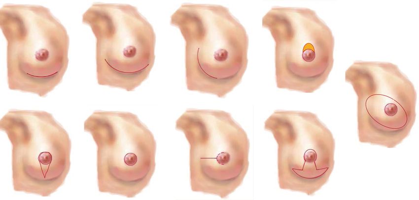
NSM, place the injection slightly lateral to the NAC and
By the time of surgery, decisions regarding the procedure with SSM, place the injection into skin that will be excised.
have been made in concert with the patient and derived Incisions for NSM include inframammary, extended
from vigorous patient education, consideration for patient inframammary, lateral inframammary, horizontal pericentral
preference and recommendations of the oncologic team. On (extending from the lateral aspect of the areola), and
the operative day, the breast and plastic surgeon ideally will superior areolar crescent also known as the modified
work in concert. We have found that time spent together nipple sparing (Figure 1). Our personal preference is the
in the operating room tends to be mutually educational and inframammary approach, which has proven to be both
enjoyable. effective and safe (36,37). In our opinion, the inframammary
In order to achieve optimal skin flaps while working incision leads to the most pleasing aesthetic outcome
through limited incisions, the breast surgeon should with little or no visible scar with the breast in the upright
be aware of the exact location of the tumor and breast position. With smaller, ptotic breasts the superior areolar
boarders. Out lining the breast and anticipated mastectomy crescent incision allows for removal of superior pole skin
field as well as the tumor location with a simple skin marker resulting in a small lift, while minimizing nipple loss. The
can be utilized to prevent inadvertent over or under- best results/nipple viability are seen when the peri-areolar
dissection of flaps and unrelated benign tissue such as the incision is limited to the 9-3 o’clock position. Important
lateral chest wall. These techniques are especially useful issues to consider when choosing incision type are surgeon
for nonpalpable malignancies, or cancers that are located comfort level with the procedure and the ability to properly
near resection margins. Depending on surgeon comfort remove the breast gland while preserving flap viability.
and preference, superficial, posterior or peripheral lesions Careful patient selection and use of larger incisions should
can be identified by intra-operative ultrasound, a marking be encouraged until the operating surgeons masters the
pin at the overlying skin, or localization of previously procedure and feels comfortable with the technique.
placed markers. The ability to confirm complete removal Incisions for SSM include: horizontal central breast,
of a peripheral, posterior or axillary tail tumor focus can be vertical, vertical reduction pattern, and peri-areolar
critical. If during the procedure, the level of concern for a (Figure 1). With the peri-areolar approach, the incision
focal area of involvement extending to a resection margin is made along the areolar border, such that the NAC is
is high, with potential need for additional resection, send removed en bloc with the underlying breast gland, and
an immediate intraoperative pathology assessment and/ the breast is extracted through this circular incision. If
or place a small nonabsorbable suture through the full necessary, this incision can be extended superiorly in
thickness of the flap at the site of concern. With the suture an elliptical manner, allowing for greater visibility and
placement technique, if the final pathology is positive for access, or removal of a larger breast. The peri-areolar
residual disease, you have marked the area for re-excision. If approach allows for maximum skin preservation, leaving
the pathology reveals clear margins, the suture can be easily the reconstructive surgeon the ability to use the entire
removed in the office. skin envelope. If needed, reduction of the skin envelope
Plastic surgeons should plan incision length and location can be achieved by de-epithelialization of the lower pole
allowing for comfortable dissection by the breast surgeon. for overlapping of flaps. For skin-sparing mastectomy,
Unnecessarily small incisions increase surgical time, tension the incisional approach can vary to include the reduction
on flaps, as well as the potential for errors. If blue dye is pattern mastectomy, the low horizontal or the vertical
to be used, both isosulfan blue and methylene blue are incision. The reduction pattern mastectomy allows for a
acceptable, with the understanding that each can cause skin more natural shape of the breast with improved anterior
© Gland Surgery. All rights reserved. gs.amegroups.com Gland Surg 2019;8(1):27-3532 Storm-Dickerson and Sigalove. The breast surgeon’s approach
IMF E-IMF L-IMF M-NSM
Trans
V-SSM P-NAC H-Cent RDP
Figure 1 Nipple- and skin-sparing mastectomy skin incisions. IMF, inframammary fold incision; E-IMF, extended IMF; L-IMF, lateral IMF;
P-NAC, peri-nipple areolar complex, H-Cent, horizontal-pericentral; RDP, reduction pattern; Trans, transverse.
projection and less horizontal expanse. The low horizontal be placed to avoid disruption of flow and the surgeon can
mastectomy allows for the entire incision line to be hidden take care to not over dissect thus minimizing the risk of flap
along the inframammary fold with no flap confluence, necrosis.
decreasing risk for skin necrosis. The vertical incision
closely encompassing the NAC and is very useful when the
Understanding the mastectomy flap and tricks
breast is smaller in size, less ptotic or the patient is not a
to maximize success
candidate for NSM
If available, perfusion assessment devices allow viewing of Handling of the mastectomy flap can make the difference
post mastectomy perfusion in real-time. These devices have between viable and nonviable, success and failure.
been beneficial in increasing our understanding of the impact Retraction damage can be minimized by a gentler touch
of our mastectomy technique and incision placement on flap and the use of non-metal, non-conducting materials.
perfusion. They have also contributed to our appreciation Tumescence, sharp dissection and low thermal conduction
of important perforators, blood flow and areas of increased devices minimize over-dissection. Leaving extra tissue
ischemia susceptibility. Perfusion assessment devices are not at the start of the incisions, as well as the area under the
only used in real-time decision-making regarding proceeding nipple will minimize over-thinning during the procedure.
with prepectoral reconstruction, but also a teaching tool to Tissue at the edge of incisions tends to be under the most
help the breast surgeon recognize and avoid future dissection tension and suffers from retraction and friction from the
errors and eventually be able to identify patients at high risk surgeon’s hand. Areas that were purposely left thick at the
for failure. For patients with a history of prior breast surgery beginning of the procedure are trimmed once the breast
such as reductions and lumpectomies, these devices can also has been removed. Use of proper equipment such as lighted
be used prior to mastectomy to help with optimal incision retractors, headlights and appropriately sized instruments
placement based on perfusion of the pre-mastectomy flap. ensure a successful operation. The importance of reliable
For instance, a patient with a history of lumpectomy with assistance and retraction cannot be overly emphasized.
radiation may have disruption of the medial perforators and Overall flap thickness varies by patient weight and
have developed a blood flow based on lateral collaterals. By size. A very large breasted and obese woman will have a
understanding the location of these collaterals, incisions can thicker natural flap than a thin, small breasted woman.
© Gland Surgery. All rights reserved. gs.amegroups.com Gland Surg 2019;8(1):27-35Gland Surgery, Vol 8, No 1 February 2019 33
Adipocytes express hyperplasia and hypertrophy and in Footnote
obesity both components increase. This leads to an overall
Conflicts of Interest: The authors have no conflicts of interest
enlargement of the thickness of the hypodermis, while the
to declare.
dermal layer remains relatively unchanged (38,39). Frey
et al., performed magnetic resonance imagings (MRIs) on
420 NSMs, 379 preoperative and 60 postoperative. The References
average total preoperative skin/subcutaneous tissue “flap”
1. ACS. Cancer Treatment & Survivorship Facts & Figures
thickness was 11.4 mm and the average total postoperative
2016-2017. Atlanta: American Cancer Society; 2016.
flap thickness was 8.7 mm. They found that a flap thickness
2. ACS. Surgery for Breast Cancer. 2016. Available online:
of less than 8 mm was an independent predictor of ischemic
https://www.cancer.org/cancer/breast-cancer/treatment/
complications. By MRI, the overall postoperative flap
surgery-for-breast-cancer.html
thickness was 68.2% of preoperative measurements, and
3. De Angelis R, Tavilla A, Verdecchia A, et al. Breast cancer
ranged from 52.0% to 74.0% (P34 Storm-Dickerson and Sigalove. The breast surgeon’s approach
Plast Reconstr Surg Transplant Bull 1962;30:676-82. year experience with tissue expander/implant breast
13. Hinton CP, et al. Subcutaneous Mastectomy for Primary reconstruction: Part I. A prospective analysis of early
Operable Breast Cancer, Br J Surg 1984;71:469-72. complications. Plast Reconstr Surg 2006;118:825-31.
14. Hartmann LC, Sellers TA, Schaid DJ, et al. Efficacy of 27. Hunsicker LM, Ashikari AY, Berry C, et al. Short-term
bilateral prophylactic mastectomy in BRCA1 and BRCA2 complications associated with acellular dermal matrix-
gene mutation carriers. J Natl Cancer Inst 2001;93:1633-7. assisted direct-to-implant breast reconstruction. Ann Plast
15. Hartmann LC, Schaid DJ, Woods JE, et al. Efficacy Surg 2017;78:35-40.
of Bilateral Prophylactic Mastectomy in Women with 28. Kim JY, Davila AA, Persing S, et al. A meta-analysis of
a Family History of Breast Cancer. N Engl J Med human acellular dermis and submuscular tissue expander
1999;340:77-84. breast reconstruction. Plast Reconstr Surg 2012;129:28-41.
16. Rebbeck TR, Friebel T, Lynch HT, et al. Bilateral 29. Fennell ML, Das IP, Clauser S, et al. The Organization
Prophylactic Mastectomy Reduces Breast Cancer Risk of Multidisciplinary Care Teams: Modeling Internal and
in BRCA1 and BRCA2 Mutation Carriers: The PROSE External Influences on Cancer Care Quality. J Natl Cancer
Study Group. J Clin Oncol 2004;22:1055-62. Inst Monogr 2010;2010:72-80.
17. Agarwal S, Neumayer L, Agarwal JP. Therapeutic Nipple- 30. Meyer LA, Lasala JL, IniestaMD, et al. Effect of an
Sparing Mastectomy: Trends based on a national cancer Enhanced Recovery After Surgery Program on Opioid
database. Am J Surg 2014;208:93-8. Use and Patient-Reported Outcomes. Obstet Gynecol
18. Burdge EC, Yuen J, Hardee M, et al. Nipple-Sparing 2018;132:281-90.
Mastectomy is Feasible for Advanced disease. Ann Surg 31. Astanehe A, Temple-Oberle C, Nielsen M, et al.
Oncol 2013;20:3294-302. An Enhanced Recovery after Surgery Pathway for
19. Smith BL, Tang R, Rai U, et al. Oncologic Safety of Microvascular Breast Reconstruction Is Safe and Effective
Nipple-Sparing Mastectomy in Women with Breast Plastic and Reconstructive Surgery. Plast Reconstr Surg
Cancer. J Am Coll Surg 2017;225:361-5. Glob Open 2018;6:e1634.
20. Li M, Chen K, Liu F, et al. Nipple-Sparing Mastectomy 32. Gabriel A, Sigalove S, Sigalove N, et al. Can Closed
in Breast Cancer Patients and Long-Term Survival Incision Negative Pressure Therapy Impact Post Operative
Outcomes: An analysis of the SEER database. PLoS One Outcomes in Breast Reconstruction? Plast Reconstr Surg
2017;12:e0183448. Glob Open 2017;5:46-7.
21. Sigalove S, Maxwell PG, Sigalove NM, et al. Prepectoral 33. Mathes SJ, Nahai F. Reconstructive Surgery: Principles,
Implant-Based Breast Reconstruction: Rationale, Anatomy, and Technique. New York: Churchill
Indications and Preliminary Results. Plast Reconstr Surg Livingstone and Quality Medical Publishing Inc.; 1997.
2017;139:287-94. 34. Schulz S, Zeiderman M, Gunn JS, et al. Safe Plastic
22. Maxwell GP, Storm-Dickerson T, Whitworth P, et al. Surgery of the Breast II: Saving Nipple Sensation. Eplasty
Advances in nipple-sparing mastectomy: Oncological safety 2017;17:e33.
and incision selection. Aesthet Surg J 2011;31:310-9. 35. Bircan HY, Ozcelik U, Koc B, et al. Cutaneous Necrosis
23. Sigalove S, Maxwell, GP, Sigalove N, et al. Prepectoral as a Result of Isosulphane Blue Injection in Mammarian
Implant-Based Breast Reconstruction and Postmastectomy Sentinel Lymph Node Mapping: Report of two Cases.
Radiotherapy: Short-Term Outcomes. Plast Reconstr Surg Clin Med Insights Case Rep 2014;7:79-81.
Glob Open 2017;5:e1631. 36. Farahat AM, Hashim T, Soliman HO, et al. Skin sparing
24. Gabriel A, Sigalove S, Sigalove NM, et al. Abstract P4: mastectomy: technique and suggested methods of
Can Surgical Technique Impact Length of Stay and Post- reconstruction. J Egypt Natl Canc Inst 2014;26:153-9.
Operative Outcomes in Breast Reconstruction? Plast 37. Rawlani V, Fiuk J, Johnson SA, et al. The effect of incision
Reconstr Surg Glob Open 2017;5:104-5. choice on outcomes of nipple-sparing mastectomy
25. Gabriel A, Sigalove S, Sigalove NM, et al. Does reconstruction. Can J Plast Surg 2011;19:129-33.
Surgical Technique Impact Post-Operative Outcomes of 38. Burns DA, Breathnach SM, Cox N, et al. Rook’s Textbook
Breast Reconstruction? Available online: https://www. of Dermatology. 7th edition. Malden, Mass: Blackwell
breastsurgeons.org/docs2017/posters/ASBrS_2017_ Science, 2004.
Poster_255754.pdf 39. Jo J, Oksana O, Pack S, et al. Hypertrophy and/or
26. Cordeiro PG, McCarthy CM. A single surgeon’s 12- Hyperplasia: Dynamics of Adipose Tissue Growth. PLoS
© Gland Surgery. All rights reserved. gs.amegroups.com Gland Surg 2019;8(1):27-35Gland Surgery, Vol 8, No 1 February 2019 35
Comput Biol 2009;5:e1000324. Resonance Imaging. Plast Reconstr Surg Glob Open
40. Frey JD, Salibian AA, Choi M, et al. Mastectomy 2017;5:e1439.
Flap Thickness and Complications in Nipple-Sparing 41. Sun K, Kusminski CM, Scherer PE. Adipose tissue
Mastectomy: Objective Evaluation using Magnetic remodeling and obesity. J Clin Invest 2011;121:2094-101.
Cite this article as: Storm-Dickerson T, Sigalove NM. The
breast surgeons’ approach to mastectomy and prepectoral breast
reconstruction. Gland Surg 2019;8(1):27-35. doi: 10.21037/
gs.2018.11.06
© Gland Surgery. All rights reserved. gs.amegroups.com Gland Surg 2019;8(1):27-35You can also read