Complete thymectomy for myasthenia gravis - Journal of ...
←
→
Page content transcription
If your browser does not render page correctly, please read the page content below
Review Article
                                                                                                                                        Page 1 of 8

Complete thymectomy for myasthenia gravis
Jens-C. Rückert1#, Hongbin Zhang1#, Feng Li1, Deniz Uluk1, Mahmoud Ismail1, Andreas Meisel2
1
    Department of Surgery, Competence Center of Thoracic Surgery, 2Department of Neurology Berlin, Charité University Hospital Berlin, Berlin,
Germany
Contributions: (I) Conception and design: H Zhang, F Li, JC Rückert, D Uluk; (II) Administrative support: JC Rückert; (III) Provision of study
materials or patients: JC Rückert, D Uluk; (IV) Collection and assembly of data: None; (V) Data analysis and interpretation: None; (VI) Manuscript
writing: All authors; (VII) Final approval of manuscript: All authors.
#
These authors contributed equally to this work.
Correspondence to: Jens-C. Rückert, MD, PhD. Department of Surgery, Competence Center of Thoracic Surgery, Charité University Hospital Berlin,
Charitéplatz 1, 10115 Berlin, Germany. Email: jens-c.rueckert@charite.de.

                  Abstract: The therapeutic value of transsternal extended thymectomy for patients with myasthenia gravis
                  (MG) has been determined by the results of the Myasthenia Gravis Thymectomy Trial (MGTX). The most
                  crucial point of thymectomy lies in the completeness of the resection with the thymus, mediastinal fat,
                  lower cervical fat and thymoma (if any), the more complete the resection the better neurological outcome.
                  In clinical practice, the minimally invasive approaches for thymectomy have become a “usual” approach for
                  thymectomy in many centers worldwide. Robotic thymectomy, as the latest advance in minimally invasive
                  thymectomy, has gained much popularity and has been regarded as a promising approach for MG patients
                  with encouraging neurological outcomes. Although several approaches for robotic thymectomy have been
                  described, the authors recommend a unilateral approach from the left side with 3 trocars only for most of
                  the cases, this is for several anatomical reasons, e.g., access to the common anatomical locations with ectopic
                  thymic tissues; dissection of the upper poles and thymic veins; atypical anatomy of the left thymic main
                  lobe, visualization of the contralateral phrenic nerve favor this technique. Herein, the authors describe the
                  left-sided robotic extended thymectomy for MG patients in detail including patient selection and workup,
                  preoperative preparation, equipment preference cart, procedure and tips, tricks and pitfalls.

                  Keywords: Left-sided; robotic extended thymectomy; myasthenia gravis (MG); thymoma

                  Received: 03 January 2020. Accepted: 06 March 2020; Published: 20 January 2021.
                  doi: 10.21037/jovs.2020.03.08
                  View this article at: http://dx.doi.org/10.21037/jovs.2020.03.08

Introduction                                                                  early results of six patients with MG after the removal of the
                                                                              thymus gland and recommended total thymectomy to alter
Myasthenia gravis (MG) is a relatively rare autoimmune
                                                                              the course of MG (4). Following these inspiring preliminary
disorder with different severities of muscle weakness.
                                                                              investigations, many surgeons performed numbers of
Although it can be life-threatening in some cases, MG is                      observational retrospective studies to evaluate the role of
generally treatable and even curable with multidisciplinary                   thymectomy in the treatment of MG (5,6). In 2016, the first
treatments (1). In 1912, Schumacher and Roth first reported                   randomized controlled trial evaluating thymectomy in MG
an improvement of myasthenic weakness after thymus                            patients seropositive for anti-acetylcholine receptor (anti-
removal for the treatment of thyrotoxicosis in an 18-year-                    AChR) autoantibody demonstrated that significant benefits
old female patient (2). Later in 1939, Blalock et al. noticed                 in the improvement of neurological outcome and steroid-
a durable remission of generalized MG in a 24-year-old                        sparing effect favors extended transsternal thymectomy
woman after thymectomy for the treatment of thymic cystic                     within a three-year follow-up (7). Recently, the 5-year
tumor (3). Two years later, Blalock described encouraging                     results from MGTX still favor thymectomy for these

© Journal of Visualized Surgery. All rights reserved.                                J Vis Surg 2021;7:8 | http://dx.doi.org/10.21037/jovs.2020.03.08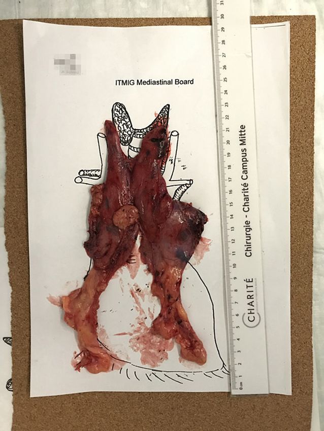
Page 2 of 8                                                                                   Journal of Visualized Surgery, 2021

patients (8). It is now common knowledge that thymectomy         consensus and guidelines recommend that thymectomy
is an indispensable and effective option for patients with       should be performed after MG is well controlled to avoid
MG.                                                              postoperative respiratory failure (22,28).
   The use of the da Vinci surgical system in thymectomy            Furthermore, a computed tomography (CT) scan
for the treatment of MG was first reported by Ashton et al.      of the chest should be performed in each MG patient
in 2003 (9). Since then many institutes have adopted robotic     before thymectomy, and if there is a mass with soft-tissue
technology for thymectomy and reported their experience          attenuation, contrast-enhanced CT will be performed to
and clinical outcomes of patients undergoing robotic             assess the enhancement characteristics and the adjacent
thymectomy for MG (10-13). Up to now, many approaches            structures of the lesion (29). If the lesion is adherent
for robotic thymectomy have been developed and described,        to adjacent tissues like major vessels, phrenic nerves or
including unilateral, bilateral and subxiphoid (11,14-16).       the sternum, the robotic extended thymectomy will not
With the 3-dimensional (3-D) visualization, high dexterity       be suitable. In differentiating the hyperplasia from the
and enhanced precision provided by the da Vinci surgical         thymoma, magnetic resonance imaging (MRI) can be better
system, robotic thymectomy has been proved not only to be        than CT, as thymoma may sometimes show diffuse thymic
safe and feasible (17-21), but also to provide MG patients       enlargement and may be diagnosed as hyperplasia in CT
with encouraging neurological outcomes even comparing            scan (30). Positron emission tomography-CT (PET-CT)
with the extended transsternal thymectomy (19-21).               can help to differentiate thymic cancer from other thymic
   Although controversies exist as to the best approach          diseases, but overlapping uptake value exists between
for robotic thymectomy in the treatment of MG patients,          normal and abnormal findings (31). Both MRI and PET-CT
we have been performing robotic thymectomy via a left-           can be performed in need, but not routinely. In addition,
sided unilateral approach for MG patients since 2003 and         pulmonary function tests and routine blood examinations
consider it a perfect combination of minimal invasiveness        for surgery as well as the electrocardiogram should also be
and maximal exposure for resection. Furthermore, it could        used in the assessment of MG patients before the surgery.
be shown that left-sided robotic extended thymectomy
is significantly beneficial for MG patients according to
                                                                 Preoperative preparation
cumulative complete remission comparing with non-robotic
thoracoscopic thymectomy (21). Herein, we describe the           The position of the three trocars is usually marked on the
left-sided robotic extended thymectomy for MG patients in        skin for each case on the ward. Our usual approach for
detail, including patient selection and workup, preoperative     robotic extended thymectomy is performed through the
preparation, equipment preference cart, procedure and tips,      left side under general anesthesia with a left-sided double-
tricks and pitfalls.                                             lumen endotracheal tube. Before the operation, the patient
                                                                 is placed supine and moved to the left edge of the operating
                                                                 table, the left arm is placed lower than the table plane,
Patient selection and workup
                                                                 providing enough space for the motion of the instruments
The main indications of robotic extended thymectomy              (Figure 1A). Thereafter, the operative field is prepared,
for MG are patients with resectable thymomas or patients         sterilized and draped, exposing the adequate area to make
without thymomas aiming to reduce or avoid the use of            sure a conversion to open or an introduction of a right-
immunosuppressive medications (22). The diagnosis of MG          sided trocar could be easily performed if necessary (Figure
is usually confirmed by relevant symptoms and a positive         1B). Then, the 8mm trocar, to which the 30-degree robotic
test for antibodies against acetylcholine receptors (AChR),      camera is mounted, is inserted at the 4th intercostal space
muscle-specific kinase (MuSK), and lipoprotein receptor-         on the midaxillary line, followed by insufflating carbon
related protein 4 (LRP4) (23). If the antibodies are negative,   dioxide (CO2) to pressure about 8 mmHg. However, the
neurophysiological tests and a Tensilon test would be            insufflation of CO2 should be used only for the placement of
performed to confirm the diagnosis (24). In anti-MuSK            the resting trocars or even be avoided if possible so that an
and anti-LRP4 MG patients, previous studies have shown           exact incision of the mediastinal pleura can be performed.
that thymectomy has little effects on the improvement of         Subsequently, the 8mm cranial and caudal special working
MG symptoms if any (22,25). Although early removal of            trocars are inserted at the 3 rd intercostal space in the
the thymus seems to be beneficial for MG patients (26,27),       anterior axillary line and the 5th intercostal space between

© Journal of Visualized Surgery. All rights reserved.                 J Vis Surg 2021;7:8 | http://dx.doi.org/10.21037/jovs.2020.03.08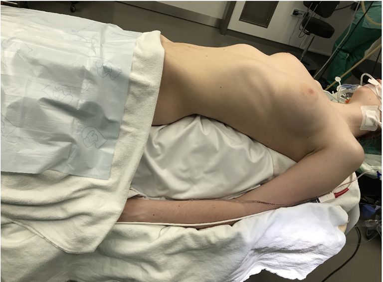
Journal of Visualized Surgery, 2021                                                                                              Page 3 of 8

                       A                                           C

                       B

Figure 1 Modified surgical position, operation field preparation, and docking of the robotic arms. (A) A supine position with the body
moved to the left edge of the operating table, the left arm is placed as low as possible; (B) operation field is prepared for conversion to
median sternotomy or thoracotomy; (C) three robotic arms are docked.

the anterior axillary line and the middle clavicular line               connecting with an ultrasonic dissector and a precise
respectively. The patient cart with four arms is docked                 bipolar forcep, respectively, they can wrist and rotate much
on the right side of the patient (Figure 1C). The surgeon               more dexterously than human hands. The surgeon sits at
console is positioned at some distance from the patient                 the console with his fingers grasping the master controls to
while the vision cart is placed at the bottom of the operating          operate with extreme precision.
table.

                                                                        Procedure
Equipment preference cart
                                                                        We present here a complete process of robotic assisted
Robotic extended thymectomy was performed initially with                thymectomy (Video 1).
the basic model of the da Vinci system, later on refined                   The positioning of the trocars always follows a rule
accordingly with the development of the further models                  of measuring adapted to the very different anatomy of
of the S-, and Si-system, and is actually performed with                the patients and to the sternum. The initial use of CO 2-
the da Vinci system Xi (Intuitive Surgical, Sunnyvale,                  insufflation may be useful or even necessary at this point.
CA), the fourth generation of the da Vinci system, which                The next step is the inspection of the thymic gland and the
consists of a surgeon console, a patient cart with four                 straight view at the left phrenic nerve with the 30° optic
robotic arms and a 3-D vision system. Comparing to the                  looking down. For MG patients with thymoma, meticulous
former Si and X generations, the Xi system has smaller,                 inspection of the tumor is necessary before any dissection
thinner arms, longer instrument shafts, allowing a greater              to re-check if the thymoma is adherent to any major vessels,
range of motion and greater operational reach. Besides,                 nerves or adjacent organs. Furthermore, thoracoscopic
the computerized automatic positioning system can help                  exploration should be made to exclude or prove stage IV
to shorten the docking process. Three robotic arms are                  in thymoma cases. During the operation, the “no-touch”
connected with the surgical instruments, the middle                     surgical technique should be obeyed to make sure the
one holding the camera, the cranial and the caudal ones                 tumor is not directly manipulated, minimizing the risk of

© Journal of Visualized Surgery. All rights reserved.                         J Vis Surg 2021;7:8 | http://dx.doi.org/10.21037/jovs.2020.03.08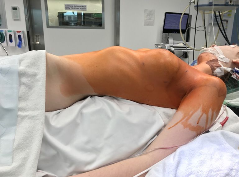
Page 4 of 8 Journal of Visualized Surgery, 2021
tumor seeding. Furthermore, according to the International Subsequently, the dissection of the cervical pleural fold
Thymic Malignancy Interest Group criteria (32), the is continued to the median retrosternal line. The right lung
surgical margins should be assessed by the pathologist with is visualized coved only by the mediastinal pleural which
microscopic inspection after an en bloc resection of the is kept closed. After taking down the right upper pole,
thymus, thymoma and the mediastinal and lower cervical fat. the right mammary vein is found. At its entrance into the
The dissection usually starts at approximately the middle venous confluence, the right phrenic nerve will be presented
of the pericardium along the left phrenic nerve where the at the lateral side of the superior vena cava. Following
thymus gland is regularly visible. In elder and/or obese the nerve along the vena cava down allows for complete
cases where this area is frequently covered by fat tissue, the dissection of the tissue in the aortocaval groove. On the
CO2 insufflation can be applied to get a better view. There right side, continue the dissection towards the subxiphoid
is always a gap between the upper part of the main thymic pleural fold until the right lung is visible. The right phrenic
lobe – even if covered by large amounts of fat and the lower nerve can be frequently found at the root of the right lung.
cardiophrenic fatty tissue. Then mobilize the tissue block from the pericardium.
The dissection is extended caudally along the left phrenic Attention should be paid to keep the right pleural cavity
nerve until the phrenic bundle is isolated and the tissue in closed here, otherwise, the effect of CO2 insufflation on
the aortopulmonary window is completely mobilized. Care enlarging the operative field will be lost.
should be taken of the left phrenic nerve while isolating, in The whole median retrosternal tissue is mobilized, then
some cases, the thymus gland can extend across the nerve the right thymic lobe is visualized with surrounding fat
or completely include the nerve. Dissection is continued tissue in the aortocaval groove. Then, the tissue in the left
upwards along the nerve until the cervical pleura is reached cardiophrenic angle is dissected and continue upwards.
at the entrance to the innominate vein. The last dissection proceeds from the left cardiophrenic
Then, the pleural incision is extended up to the jugular angle to the subxiphoid area and extends to the right
fold of the mediastinal pleura. Further dissection of the cardiophrenic area. The right pleura is opened at last and
neck area at the left phrenic nerve allows for visualizing the the cardiophrenic tissue is dissected completely.
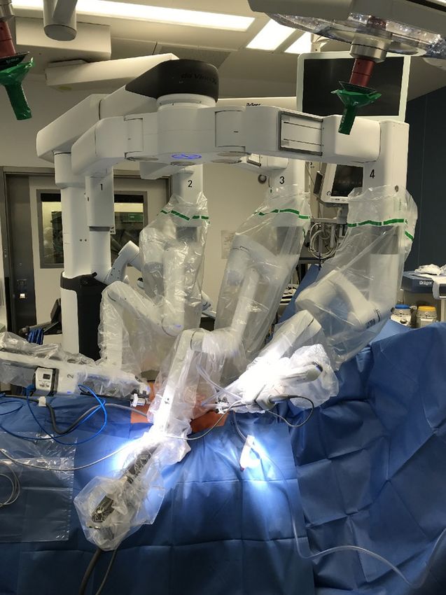
innominate vein, regularly medial to the left phrenic nerve. In conclusion, the en bloc resected thymus specimen,
Thereafter, the pressure of CO2 can be increased to get a including the mediastinal fat and thymoma (if any), is
better view for the dissection of the neck area. However, removed out in an endo bag through the camera incision.
attention should be paid to the hemodynamic monitoring. The whole specimen is weighed immediately after the
After that, mobilization of the upper poles of the thymus removal, then is placed on a mediastinal board as in situ and
is continued above the left innominate vein, but sometimes photographed before sending it to the pathologists (Figure 2).
the anatomical variations of the upper poles do exist, e.g.,
the upper poles run behind or around the innominate vein.
Role of team members
The upper poles are mobilized by a combination of blunt
dissection shifting the tissue with open branches of the The surgical team usually consisted of a board-certified
grasping instruments (long bipolar or Maryland dissector). console surgeon, a board-certified table surgeon, an
Intermediate grasping, mechanical and harmonic dissection anesthesiologist, a scrub nurse, and an operating room
add to the isolating of the thyrothymic ligament where both nurse. The console surgeon controlled the robot to perform
of the poles are divided. an extended thymectomy. The table surgeon, on the left
The upper poles are managed by grasping and bringing side of the patient, should excel at inserting the trocars and
them down to get a complete resection. They are divided connecting, adjusting and changing the surgical instruments,
bluntly with spread branches of the grasping instrument also have a thorough knowledge of managing emergencies
by ultrasonic dissection from the inferior portion of the like major bleeding that may require a conversion to
thyroid gland. transsternal thymectomy. A scrub nurse should be familiar
In most cases, two to four thymic veins are collecting with the robotic materials and qualified for the adjustment
all the venous blood into the innominate vein. Thymic and change of the robotic instruments. An operating room
veins are served with a harmonic scalpel without using clip nurse should know how to connect and calibrate the robotic
ligatures. It is worth mentioning that anatomical variations components. An experienced anesthesiologist in this team
of thymic veins should be always kept in mind, albeit rare. was supposed to be familiar with the management of both
© Journal of Visualized Surgery. All rights reserved. J Vis Surg 2021;7:8 | http://dx.doi.org/10.21037/jovs.2020.03.08Journal of Visualized Surgery, 2021 Page 5 of 8
and to guide the insertion of the trocars can make sure the
insertion is under control all the time. Furthermore, we
have introduced this method into many institutes where the
surgeons are doing quite well.
Another main reason why many surgeons favor the
right-sided approach is that the visualization of the superior
vena cava is beneficial for identifying the left innominate
vein at the venous confluence. However, through the left-
sided approach, the left innominate vein can also be easily
identified after opening the cervical pleura fold at the
point medial to the phrenic nerve. Before dissecting the
upper poles from the inferior portion of the thyroid gland,
grasping and pulling them downward is always necessary
for a radical resection, and in this way, the overall anatomy
of the left innominate vein is exposed, then identifying
and dissecting the thymic veins which drain into the left
innominate vein is followed without too much difficulty.
The authors prefer left-sided approach for robotic
thymectomy, not only because the left lobe of the thymus
is usually larger, but also because it is easy to handle
Figure 2 Specimen is placed on an ITMIG mediastinal board and
the upper poles, manage the thymic veins, approach to
photographed.
the contralateral phrenic nerve and access the common
anatomical locations with ectopic thymic tissue from the
left side. The thymic tissue can extend to or even across
robotic-assisted thoracic surgery and the general anesthesia
the left phrenic nerve in many cases, which can hardly
in MG patients.
be addressed from the right side (33). Sometimes, the
Each member of the robotic thymectomy team is crucial
upper poles can hide behind or run circularly around the
for the success of the robotic extended thymectomy. They
innominate vein, which is easier to manage through the
should learn more than just professional knowledge but also
left-sided approach. Besides, the left innominate vein
be educated to be familiar with the da Vinci system, in case
mainly runs on the left side, which provides benefits
any technical problems occur during the procedure.
for managing the thymic veins as well as protecting the
innominate vein. Recently, our team did a close review
Tips, tricks, and pitfalls on the presence of ectopic thymic tissue in patients
undergoing thymectomy for MG (34), the results showed
The proper approach for robotic thymectomy has been some preferred anatomical locations of ectopic thymic
an ongoing debate for decades among thoracic surgeons tissue: anterior mediastinal fat, pericardiophrenic angles,
worldwide. Some surgeons supposed it was dangerous aortopulmonary window, cervical region (pretracheal
doing the left-sided approach for thymectomy, concerning fat) and lateral to phrenic nerves (mostly at the left side).
about the indeed reports of cardiac injuries with trocar Importantly, our previous study has shown that an extended
insertion. But this can be perfectly avoided by visual control thymectomy favors the left-sided approach (35).
guided the trocar insertion with scissors and adequate During the last few years, subxiphoid robotic
CO2 insufflation at the beginning. In fact, more than 900 thymectomy has attracted increasing attention worldwide
cases have been performed in the Charité without any (16,36). This procedure offers surgeons a direct visualization
pericardium or heart injury during the placement of trocars. of the mediastinum similar to median sternotomy in a
The insufflated CO2 can help to enlarge the space at the minimally invasive way. Although the operative view of
anterior mediastinum, providing adequate space for trocar the neck region is good, when it comes to the anatomical
placement. On the other hand, using stitch scissors with variation of the upper poles running behind or even
round tips to open the intercostal space by blunt dissection surround the innominate vein and ectopic thymic tissue
© Journal of Visualized Surgery. All rights reserved. J Vis Surg 2021;7:8 | http://dx.doi.org/10.21037/jovs.2020.03.08Page 6 of 8 Journal of Visualized Surgery, 2021
behind the left innominate vein, the left approach could be thymectomy, wherein the operative space is confined and
more direct. Besides, long-term follow-up data are needed the working angles are difficult for human hands. The left-
to explicit the neurological outcomes of MG. sided robotic extended thymectomy is safe and feasible with
International consensus and guidelines recommend all encouraging clinical outcomes for both MG and thymoma.
MG patients with resectable thymoma undergo thymectomy However, further researches with long-term follow up is
after MG is well controlled, and thymectomy is performed still required before the robotic approach becomes the gold
for the treatment of thymoma with the resection of thymus standard for thymectomy.
and the mediastinal and lower cervical fat (22,28,37).
Although many studies have shown robotic thymectomy is
Acknowledgments
safe and feasible for MG patients with early-stage thymoma,
the proper size of thymoma for robotic thymectomy and Funding: None.
the long-term oncologic outcome after robotic thymectomy
are still under debate (38-40). Available data has shown
Footnote
encouraging early and mid-term oncologic outcomes in MG
patients with thymoma after robotic thymectomy (21,41). Provenance and Peer Review: This article was commissioned
As for the proper size of thymoma for robotic thymectomy, by the Guest Editor (Jean-Marc Baste) for the series
although no international consensus or guideline is “Robotic Assisted Thoracic Surgery: Advanced
dictating the size of thymoma as a criterion for robotic procedures in lung and mediastinum: From post-
thymectomy, most surgeons consider thymomas smaller induction TTT (immunotherapy) to sleeve resection,
than 5 cm are proper and acceptable for robotic thymectomy Complex segmentectomies and Extended Thymectomy
(38,42,43). However, recent studies have shown that robotic for Myasthenia Gravis” published in Journal of Visualized
thymectomy is also safe and feasible for thymomas larger Surgery. The article has undergone external peer review.
than 5cm and the perioperative outcomes of patients are
better when compared with open surgery (44,45). The Conflicts of Interest: All authors have completed the ICMJE
authors also had some experience in handling large-sized uniform disclosure form (available at http://dx.doi.
thymomas in a robotic-assisted way, usually, a fourth trocar org/10.21037/jovs.2020.03.08). The series “Robotic Assisted
would be introduced at the subxiphoid to assist exposure Thoracic Surgery: Advanced procedures in lung and
and to take out the specimen. Thus, the authors recommend mediastinum: From post-induction TTT (immunotherapy)
that robotic extended thymectomy is indicated in all patients to sleeve resection, Complex segmentectomies and
with resectable thymoma, typically Masaoka Koga I and II. Extended Thymectomy for Myasthenia Gravis” was
High cost has long been considered as a pitfall for commissioned by the editorial office without any funding or
robotic surgery, also docking and undocking time needed sponsorship. MI serves as an unpaid editorial board member
for the relatively newly established team, especially when of Journal of Visualized Surgery from Jun 2019 to May 2021.
an emergency occurs. Since the console surgeon is not JCR reports and declares as a proctor for Intuitive Surgical.
able to handle the emergency immediately due to the non- The authors have no other conflicts of interest to declare.
sterilized situation, there should always be an experienced
surgeon sitting by the patient during the robotic surgery. Ethical Statement: The authors are accountable for all
Besides, conversion to open should be performed if major aspects of the work in ensuring that questions related
bleeding occurs or the oncologic principles (including to the accuracy or integrity of any part of the work are
incomplete resection, not en bloc resection or perforation appropriately investigated and resolved. Written informed
of the capsule) are violated. Systemic training is needed for consent was obtained from the patient for publication of
the whole team in the OR to avoid the unexpected accident, this manuscript and any accompanying images.
and a contingency plan should always be prepared before
the robotic-assisted surgery. Open Access Statement: This is an Open Access article
distributed in accordance with the Creative Commons
Attribution-NonCommercial-NoDerivs 4.0 International
Conclusions
License (CC BY-NC-ND 4.0), which permits the non-
The robotic approach is perfect for application to commercial replication and distribution of the article with
© Journal of Visualized Surgery. All rights reserved. J Vis Surg 2021;7:8 | http://dx.doi.org/10.21037/jovs.2020.03.08Journal of Visualized Surgery, 2021 Page 7 of 8
the strict proviso that no changes or edits are made and the 14. Goldstein SD, Yang SC. Assessment of Robotic
original work is properly cited (including links to both the Thymectomy Using the Myasthenia Gravis Foundation
formal publication through the relevant DOI and the license). of America Guidelines. Ann Thorac Surg 2010;89:1080-5;
See: https://creativecommons.org/licenses/by-nc-nd/4.0/. discussion 1085-6.
15. Kawaguchi K, Fukui T, Nakamura S, et al. A bilateral
approach to extended thymectomy using the da Vinci
References
Surgical System for patients with myasthenia gravis. Surg
1. Gilhus NE. Myasthenia Gravis. N Engl J Med Today 2018;48:195-9.
2016;375:2570-81. 16. Suda T, Tochii D, Tochii S, et al. Trans-subxiphoid
2. Schumacher E, Roth J. Thymektomie bei einem fall von robotic thymectomy. Interact Cardiovasc Thorac Surg
morbus basedowii mit myasthenie (in German). Med Chir 2015;20:669-71.
1912;25:746. 17. Orsini B, Santelmo N, Pages PB, et al. Comparative
3. Blalock A, Mason MF, Morgan HJ, et al. Myasthenia study for surgical management of thymectomy for
gravis and tumours of the thymic region - Report of non-thymomatous myasthenia gravis from the French
a case in which the tumor was removed. Ann Surg national database EPITHOR. Eur J Cardiothorac Surg
1939;110:544-61. 2016;50:418-22.
4. Blalock A, Harvey AM, Ford FR, et al. The treatment 18. Suda T, Kaneda S, Hachimaru A, et al. Thymectomy via
of myasthenia gravis by removal of the thymus gland - a subxiphoid approach: single-port and robot-assisted. J
Preliminary report. J Amer Med Assoc 1941;117:1529-33. Thorac Dis 2016;8:S265-71.
5. Keynes G. The results of thymectomy in myasthenia 19. Renaud S, Santelmo N, Renaud M, et al. Robotic-assisted
gravis. Br Med J 1949;2:611-6. thymectomy with Da Vinci II versus sternotomy in the
6. Perlo VP, Poskanzer DC, Schwab RS, et al. Myasthenia surgical treatment of non-thymomatous myasthenia gravis:
gravis: evaluation of treatment in 1,355 patients. Early results. Rev Neurol 2013;169:30-6.
Neurology 1966;16:431-9. 20. Cakar F, Werner P, Augustin F, et al. A comparison
7. Wolfe GI, Kaminski HJ, Aban IB, et al. Randomized Trial of outcomes after robotic open extended thymectomy
of Thymectomy in Myasthenia Gravis. N Engl J Med for myasthenia gravis. Eur J Cardiothorac Surg
2016;375:511-22. 2007;31:501-4.
8. Wolfe GI, Kaminski HJ, Aban IB, et al. Long-term effect 21. Rückert JC, Swierzy M, Ismail M. Comparison of robotic
of thymectomy plus prednisone versus prednisone alone in and nonrobotic thoracoscopic thymectomy: a cohort study.
patients with non-thymomatous myasthenia gravis: 2-year J Thorac Cardiovasc Surg 2011;141:673-7.
extension of the MGTX randomised trial. Lancet Neurol 22. Sanders DB, Wolfe GI, Benatar M, et al. International
2019;18:259-68. consensus guidance for management of myasthenia gravis:
9. Ashton RC Jr, McGinnis KM, Connery CP, et al. Totally Executive summary. Neurology 2016;87:419-25.
endoscopic robotic thymectomy for myasthenia gravis. 23. Zisimopoulou P, Brenner T, Trakas N, et al. Serological
Ann Thorac Surg 2003;75:569-71. diagnostics in myasthenia gravis based on novel assays
10. Rea F, Marulli G, Bortolotti L. Robotic video-assisted and recently identified antigens. Autoimmun Rev
thoracoscopic thymectomy. Multimed Man Cardiothorac 2013;12:924-30.
Surg 2005;2005:mmcts 2004 000422. 24. Chiou-Tan FY, Gilchrist JM. Repetitive nerve stimulation
11. Rückert JC, Ismail M, Swierzy M, et al. Thoracoscopic and single-fiber electromyography in the evaluation of
thymectomy with the da Vinci robotic system for patients with suspected myasthenia gravis or Lambert-
myasthenia gravis. Ann N Y Acad Sci 2008;1132:329-35. Eaton myasthenic syndrome: Review of recent literature.
12. Bodner J, Wykypiel H, Wetscher G, et al. First experiences Muscle Nerve 2015;52:455-62.
with the da Vinci operating robot in thoracic surgery. Eur 25. Pompeo E, Tacconi F, Massa R, et al. Long-term
J Cardiothorac Surg 2004;25:844-51. outcome of thoracoscopic extended thymectomy for
13. Fleck T, Fleck M, Muller M, et al. Extended videoscopic nonthymomatous myasthenia gravis. Eur J Cardiothorac
robotic thymectomy with the da Vinci telemanipulator for Surg 2009;36:164-9.
the treatment of myasthenia gravis: the Vienna experience. 26. Mineo TC, Pompeo E, Lerut TE, et al. Thoracoscopic
Interact Cardiovasc Thorac Surg 2009;9:784-7. thymectomy in autoimmune myasthenia: Results of left-
© Journal of Visualized Surgery. All rights reserved. J Vis Surg 2021;7:8 | http://dx.doi.org/10.21037/jovs.2020.03.08Page 8 of 8 Journal of Visualized Surgery, 2021
sided approach. Ann Thorac Surg 2000;69:1537-41. a systematic review and a proposal by the guideline
27. Nakamura H, Taniguchi Y, Suzuki Y, et al. Delayed committee of the Japanese Association for Chest Surgery
remission after thymectomy for myasthenia gravis 2014. Gen Thorac Cardiovasc Surg 2015;63:201-15.
of the purely ocular type. J Thorac Cardiovasc Surg 38. Marulli G, Rea F, Melfi F, et al. Robot-aided
1996;112:371-5. thoracoscopic thymectomy for early-stage thymoma: a
28. Sussman J, Farrugia ME, Maddison P, et al. Myasthenia multicenter European study. J Thorac Cardiovasc Surg
gravis: Association of British Neurologists' management 2012;144:1125-30.
guidelines. Pract Neurol 2015;15:199-206. 39. Qian L, Chen X, Huang J, et al. A comparison of
29. Priola AM, Priola SM. Imaging of thymus in myasthenia three approaches for the treatment of early-stage
gravis: from thymic hyperplasia to thymic tumor. Clin thymomas: robot-assisted thoracic surgery, video-assisted
Radiol 2014;69:e230-45. thoracic surgery, and median sternotomy. J Thorac Dis
30. Inaoka T, Takahashi K, Mineta M, et al. Thymic 2017;9:1997-2005.
hyperplasia and thymus gland tumors: differentiation with 40. Seong YW, Kang CH, Choi JW, et al. Early clinical
chemical shift MR imaging. Radiology 2007;243:869-76. outcomes of robot-assisted surgery for anterior mediastinal
31. Sadato N, Tsuchida T, Nakaumra S, et al. Non-invasive mass: its superiority over a conventional sternotomy
estimation of the net influx constant using the standardized approach evaluated by propensity score matching. Eur J
uptake value for quantification of FDG uptake of tumours. Cardiothorac Surg 2014;45:e68-73; discussion e73.
Eur J Nucl Med 1998;25:559-64. 41. Keijzers M, Dingemans AM, Blaauwgeers H, et al. 8 years'
32. Toker A, Sonett J, Zielinski M, et al. Standard terms, experience with robotic thymectomy for thymomas. Surg
definitions, and policies for minimally invasive resection of Endosc 2014;28:1202-8.
thymoma. J Thorac Oncol 2011;6:S1739-42. 42. Toker A, Erus S, Ozkan B, et al. Does a relationship exist
33. Mulder DG, White K, Herrmann C, Jr. Thymectomy. between the number of thoracoscopic thymectomies
Surgical procedure for myasthenia gravis. AORN J performed and the learning curve for thoracoscopic
1986;43:640-6. resection of thymoma in patients with myasthenia gravis?
34. Li F, Tao Y, Bauer G, et al. Unraveling the role of ectopic Interact Cardiovasc Thorac Surg 2011;12:152-5.
thymic tissue in patients undergoing thymectomy for 43. Schwartz GS, Yang SC. Robotic thymectomy for thymic
myasthenia gravis. J Thorac Dis 2019;11:4039-48. neoplasms. Thorac Surg Clin 2014;24:197-201, vii.
35. Rückert JC, Czyzewski D, Pest S, et al. Radicality of 44. Kneuertz PJ, Kamel MK, Stiles BM, et al. Robotic
thoracoscopic thymectomy--an anatomical study. Eur J Thymectomy Is Feasible for Large Thymomas: A
Cardiothorac Surg 2000;18:735-6. Propensity-Matched Comparison. Ann Thorac Surg
36. Zhang H, Chen L, Zheng Y, et al. Robot-assisted 2017;104:1673-8.
thymectomy via subxiphoid approach: technical details and 45. Wilshire CL, Vallieres E, Shultz D, et al. Robotic
early outcomes. J Thorac Dis 2018;10:1677-82. Resection of 3 cm and Larger Thymomas Is Associated
37. Kadota Y, Horio H, Mori T, et al. Perioperative With Low Perioperative Morbidity and Mortality.
management in myasthenia gravis: republication of Innovations 2016;11:321-6.
doi: 10.21037/jovs.2020.03.08
Cite this article as: Rückert JC, Zhang H, Li F, Uluk D, Ismail
M, Meisel A. Complete thymectomy for myasthenia gravis. J
Vis Surg 2021;7:8.
© Journal of Visualized Surgery. All rights reserved. J Vis Surg 2021;7:8 | http://dx.doi.org/10.21037/jovs.2020.03.08You can also read