Nerve Compression Syndromes in the Posterior Cranial Fossa
←
→
Page content transcription
If your browser does not render page correctly, please read the page content below
MEDICINE

Review Article

Nerve Compression Syndromes in the
Posterior Cranial Fossa
Diagnosis and Treatment

Jörg Baldauf, Christian Rosenstengel, Henry W. S. Schroeder

                                                                                          N
                                                                                                 eurovascular compression syndromes are clinically
Summary                                                                                          characterized by functional disturbances of
                                                                                                 individual cranial nerves. The most common com-
Background: Nerve compression syndromes in the posterior cranial fossa can
                                                                                          pression syndrome affects the trigeminal nerve and leads
severely impair patients’ quality of life. There is often uncertainty about the best
                                                                                          to trigeminal neuralgia, followed by hemifacial spasm,
treatment. In this article, we provide an overview of these conditions and the
                                                                                          which is caused by vascular compression of the facial
corresponding treatment strategies.
                                                                                          nerve. Less well-known nerve compression syndromes
Methods: This review is based on pertinent publications retrieved by a selective          affect the glossopharyngeal nerve, the nervus intermedius
search in PubMed and on a scientific analysis of the authors’ patient collective.         and the vestibulocochlear nerve. Very rarely, the
                                                                                          oculomotor nerve or the abducens nerve is involved.
Results: These syndromes are caused by compression of a cranial nerve by an                  A single pathophysiological mechanism underlies
artery or vein at the zone of the nerve’s entry to or exit from the brainstem. The        all of these compression syndromes. In the area of the
best-known neurovascular compression syndrome is trigeminal neuralgia, followed
                                                                                          root entry zone or root exit zone (REZ) of the relevant
by hemifacial spasm. Less well known are glossopharyngeal neuralgia, nervus
                                                                                          cranial nerve at the brainstem, the nerve comes into
intermedius neuralgia, and vestibular paroxysmia. The initial treatment of trigeminal
                                                                                          contact with a blood vessel—usually an artery, less
neuralgia is medical: the first line of treatment is with sodium-blocking anticon-
                                                                                          commonly a vein. This is a natural weak point of the
vulsants, such as carbamazepine. For patients with hemifacial spasm, botulinum
                                                                                          nerve, the site of transition between central and
toxin injection is the recommended initial treatment and often leads to a satisfactory
                                                                                          peripheral myelin. The nerve is especially sensitive to
regression of the spasms. If these treatments fail, a microvascular decompression
                                                                                          mechanical irritation here, which provokes the
operation is indicated. The aim of the procedure is to separate the irritating vessel
from the nerve and to keep these structures apart permanently. There is hardly any
                                                                                          clinical symptoms of nerve compression.
available evidence on these treatment strategies from randomized controlled trials.          In this review, we discuss the individual neuro-
                                                                                          vascular compression syndromes of the posterior
Conclusion: Nerve compression syndromes in the posterior cranial fossa can                fossa and various corresponding modes of
generally be treated nonsurgically at first. Over the course of the condition, however,   treatment.
treatment failure or intolerable side effects may arise. In such cases, a microvascu-
lar decompression operation is indicated. This is a causally directed form of treat-      Methods
ment that generally yields very good results.                                             A literature search was carried out in PubMed with the
                                                                                          following search terms: “neurovascular compression syn-
Cite this as:
                                                                                          drome,” “cranial neuralgia,” “trigeminal neuralgia,”
Baldauf J, Rosenstengel C, Schroeder HWS: Nerve compression syndromes in
                                                                                          “hemifacial spasm,” “glossopharyngeal neuralgia,”
the posterior cranial fossa—diagnosis and treatment. Dtsch Arztebl Int 2019; 116:
                                                                                          “vestibular nerve compression,” “vestibular paroxy-
54–60. DOI: 10.3238/arztebl.2019.0054
                                                                                          smia,” “intermedius neuralgia” and “microvascular de-
                                                                                          compression.” For this review, we mainly considered
                                                                                          large-scale studies carried out in the past 20 years. There
                                                                                          is hardly any evidence available from randomized con-
                                                                                          trolled trials on treatment strategies for these compression
                                                                                          syndromes. In particular, the evidence for the utility of in-
                                                                                          terventional and invasive treatments is based mainly on
                                                                                          longitudinal follow-up studies evaluating the long-term
                                                                                          results in patient cohorts of variable sizes.

                                                                                          Diagnostic evaluation
                                                                                          Whenever a nerve compression syndrome is suspected,
                                                                                          magnetic resonance imaging (MRI) of the brain should
Department of Neurosurgery, University Medicine Greifswald: PD Dr. med. Jörg Baldauf,     be performed, with particular attention to the posterior
Dr. med. Christian Rosenstengel, Prof. Dr. med. Henry W. S. Schroeder                     fossa. High-resolution 3D-T2-weighted sequences,

54                                                                                        Deutsches Ärzteblatt International | Dtsch Arztebl Int 2019; 116: 54–60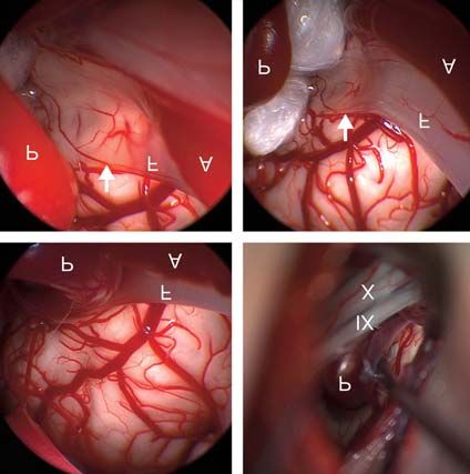
MEDICINE

  a                                                                         b
Figure 1: Magnetic resonance imaging in hemifacial spasm.
a) The TOF (time of flight) and b) CISS (constructive interference in steady-state) sequences in the axial plane clearly reveal vascular compres-
sion of the facial nerve (arrow, F) by the posterior inferior cerebellar artery (arrowhead, P)

such as the CISS (constructive interference in                             demyelinating foci in the brainstem (e3, e4). It can also
steady-state) sequence, should be obtained, as well as                     be caused by a tumor.
3D-TOF (time-of-flight) MR angiography, in order to                           The primary treatment of both types of trigeminal
distinguish reliably between arterial and venous com-                      neuralgia is with drugs, primarily sodium-channel
pression (Figure 1) (1, 2, e1). MRI also serves to rule                    blockers that are also used as antiepileptic drugs. The
out other processes that might cause the same clinical                     scientific evidence supports the use of carbamazepine
manifestations, such as a tumor or aneurysm.                               as the drug of first choice (6, 7). In symptomatic
                                                                           trigeminal neuralgia, the underlying disease should be
Trigeminal neuralgia                                                       treated if possible (e.g., resection of a tumor). If
Trigeminal neuralgia is the most common neurovascu-                        pharmacotherapy brings inadequate relief or causes
lar compression syndrome in the posterior fossa, with                      unacceptable side effects (e.g., fatigue, dizziness,
an incidence of 4–5 cases per 100 000 persons per year                     cognitive disturbances), then a neurosurgical pro-
(among persons over age 60: up to 20 per 100 000 per-                      cedure can be recommended, such as microvascular
sons per year ) (3, 4). It can thus be estimated that some                 decompression (MVD), percutaneous procedures
4200 people newly develop symptoms of trigeminal                           (thermocoagulation, glycerol rhizolysis, balloon com-
neuralgia in Germany each year. The pain is generally                      pression of the gasserian ganglion), or a radiosurgical
strictly unilateral, lasting a few seconds to minutes. It is               procedure (e5, e6). In our opinion, percutaneous
generally described as attacks of stabbing (lancinating)                   techniques should only be considered if there is no
pain and is usually located in the distribution of the                     demonstrable neurovascular conflict, because, when
second or third division of the trigeminal nerve (the                      such a conflict is present, MVD provides better and
maxillary or mandibular nerve). The attacks arise spon-                    longer-lasting success and is associated with fewer com-
taneously and can also be evoked by external stimuli,                      plications (8–10).
including touching the face or chewing. Spontaneous                           MVD (also called “the Jannetta procedure,” after
remission for months or years can occur. Over the                          the American neurosurgeon Peter Jannetta
course of time, the attacks may become more frequent                       [1932–2016]) is considered a causally directed
and the pain longer-lasting (5). A distinction is drawn                    treatment for classic trigeminal neuralgia and has the
between classic (idiopathic) trigeminal neuralgia and                      highest long-term success rate (11). In large-scale
the much less common symptomatic form of the condi-                        case series, MVD yielded relief or improvement of
tion. Classic trigeminal neuralgia is usually due to a                     pain in 68% to more than 90% of cases (12–15,
neurovascular conflict in which the trigeminal nerve is                    e7–e9). Moreover, meta-analyses have shown MVD
compressed at its REZ by a blood vessel, often the su-                     to be clearly superior to radiosurgical treatment with
perior cerebellar artery (e2). Symptomatic trigeminal                      respect to early as well as long-term pain relief (16,
neuralgia is defined as trigeminal neuralgia due to                        17). Well over 80% of patients are still free of pain
another disease; it is found, for example, among pa-                       five years after MVD, and the success rate at 10 years
tients with multiple sclerosis, who often have typical                     has been reported at over 70% (14, 15). Our experience

Deutsches Ärzteblatt International | Dtsch Arztebl Int 2019; 116: 54–60                                                                                   55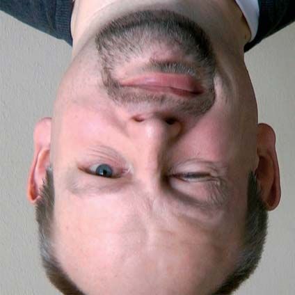
MEDICINE
TABLE 1 gical procedure. We have had favorable results from it
in patients with multiple sclerosis (among others). In
Results of microvascular decompression for trigeminal neuralgia* PSR, up to two-thirds of the sensory fibers of the
Trigeminal neuralgia Overall: 175 patients trigeminal nerve are cut (13, e12). The first division of
the trigeminal nerve must not be cut, because of the
Sex distribution Men: 72 Women: 103
danger of neuroparalytic keratitis; thus, PSR should
Mean age (range) 62.3 years (20–89 years) only be used to treat trigeminal neuralgia of the second
Mean duration of pain 7.8 years (1–33 years) and/or third divisions. The patient must be informed
(range) preoperatively that PSR will cause numbness in the
Outcome Immediately after surgery Late follow-up at >1 year corresponding area of the face. Our patients have not
(175 patients) (113 pts.); mean, 53 months found this distressing. There is a risk that PSR may be
No pain 155 (88.6%) 91 (80.5%) followed by burning dysesthesia in the numb cutaneous
50% relief of pain 4 (2%) 12 (10%)
area (anesthesia dolorosa) or by difficulty eating or
chewing (20), but our patients have not experienced
Permanent morbidity these negative sequelae to date. Anesthesia dolorosa, a
Hearing loss 3 (1.7%) rare complication of neuroablative techniques, is
Hypesthesia 5 (2.9%) difficult to treat (6).
In summary, it must be stated that the current
*Results from the authors’ own series of microvascular decompression surgery in 175 patients with trigemi- evidence base is inadequate for a satisfactory direct
nal neuralgia. Pain relief immediately after surgery and on late follow-up is shown, as is the frequency of comparison of the invasive and operative treatments
permanent morbidity (hearing loss and sensory loss)
for trigeminal neuralgia (21). An extensive Cochrane
analysis published in 2011 additionally documents
this fact (22).
Figure 2
Babinski-2 sign in
hemifacial spasm. Hemifacial spasm
During the spasms, The epidemiology of hemifacial spasm has not been ex-
the eyebrow rises tensively studied to date. In a study from Olmsted
while the eye simul- County, Minnesota, the mean annual incidence was
taneously closes found to be 0.81 and 0.74 cases per 100 000 persons per
year among women and men, respectively, with a mean
prevalence of 11 per 100 000 persons (23). It can thus
be calculated that some 9000 persons in Germany now
suffer from this condition. Hemifacial spasm is charac-
terized by involuntary, strictly unilateral tonic and/or
clonic contractions of the facial musculature, typically
including the platysma (e13). The spasms cannot be
voluntarily suppressed, and they persist during sleep.
Examination reveals the Babinski-2 sign: during the
contractions, the eyebrow rises while the eye is closed
(Figure 2). This pattern of movement cannot be
reproduced in persons without the condition (e14). The
diagnosis of hemifacial spasm can generally be made
immediately on inspection, but it can also be confirmed
electrophysiologically by the demonstration of patho-
to date has shown comparably good to very good re- logical lateral spreading of activation (24). A source of
sults (Table 1) (e10). There is no age limit to MVD additional distress to persons with this condition is that
surgery; good results are achievable even in elderly the facial spasms are often wrongly thought to be
patients (>75 years old) (e11). Complications of the psychogenic (e15). The differential diagnosis of hemi-
operation are rare, including ipsilateral hearing loss or facial spasm includes blepharospasm, facial dystonia,
deafness and sensory deficits. Multiple studies with tardive dyskinesia, motor tics, and synkinesia after
long-term follow-up have revealed permanent impair- facial nerve injury, among other conditions (25, 26,
ment of hearing in 0.1–3% of patients (18). The risk e16, e17).
of cerebellar infarction is reported as less than 1% in The current state of the evidence on the treatment
most case series (12, 15, 19). The mortality, across of hemifacial spasm is poor. For drug therapy,
multiple studies, is approximately 0.3% (12, 15, 18). anticonvulsants are usually recommended, but they
Finally, partial sensory rhizotomy (PSR) should be often fail to provide adequate benefit. Botulinum
mentioned as well. This is, in our opinion, a treatment of neurotoxin (BTX) injections have become the
last resort for intractable trigeminal neuralgia. Patients standard symptomatic treatment. The efficacy of BTX
who have already undergone multiple types of treatment is based on blockade of calcium-dependent acetyl-
without lasting relief of pain may benefit from this sur- choline release at the neuromuscular junction, leading
56 Deutsches Ärzteblatt International | Dtsch Arztebl Int 2019; 116: 54–60MEDICINE
to reversible paralysis of the affected muscles (e18). TABLE 2
BTX is injected into the affected muscles directly; the
clinically visible effect is first seen 3–6 days after in- Results of microvascular decompression for hemifacial spasm*
jection and lasts approximately three months. Tran- Hemifacial spasm Overall: 320 patients
sient side effects arise in approximately 20% of pa-
Sex distribution Men: 120 Women: 200
tients, including ptosis, facial palsy, (rarely) diplopia,
and headache (e19). Mean age (range) 54.89 years (22–81 years)
The only causally directed form of treatment is Mean duration of symptoms 7.84 years (1–35 years)
MVD. In patients with marked symptoms, we recom- Follow-up (FU) Immediately after surgery FU >12 months
mend early surgery, because some of our patients who (320 patients) (201 patients)
already displayed structural damage of the facial – No spasms 178 (55.63%) 156 (77.61%)
nerve did not experience satisfactory regression of the
– Spasms reduced by 90% 53 (16.56%) 22 (10.95%)
spasms. Large-scale studies have yielded very good
success rates, with postoperative freedom from – Spasms reduced by 50% 62 (19.38%) 20 (9.95%)
spasms in 85–90% of cases (27). These figures are – No improvement 27 (8.44%) 3 (1.49%)
also consistent with our experience (Table 2). The Morbidity (in a total of 339 operations)
main risk of MVD for hemifacial spasm is ipsilateral
Transient Permanent
hearing loss or deafness, with a reported frequency of
1.5–8% in large case series (28, 29, e20). The rate of Vertigo 11 (3.2%) 2 (0.6%)
deafness among our patients was 3%. We have not Partial hearing loss 34 (10%) 8 (2.4%)
seen a permanent, high-grade facial palsy over the
Deafness 9 (2.7%) 9 (2.7%)
long term in any of our patients, but 7% of our pa-
tients have had a delayed postoperative facial palsy Facial palsy (immediate) due 5 (1.5%) 2 (0.6%, mild)
to surgical manipulation
arising 10–14 days after surgery—a phenomenon also
reported by other authors (30, 31, e20). It is con- Facial palsy arising after a 22 (6.5%) 1 (0.3%, mild)
delay of >10 days
ceivable that reactivation of herpes zoster plays a role
in this phenomenon, but its cause has not yet been * Results from the authors’ own series of microvascular decompression surgery in 320 patients with hemifa-
clearly identified (31). Fortunately, this delayed facial cial spasm (a total of 339 operations, including 19 second procedures because of inadequate initial relief
palsy generally regresses completely. In current re- or because of recurrence). The relief of spasms immediately after surgery and on late follow-up (>12
months) is shown, as is the frequency of transient and permanent morbidity on the affected side
views of MVD for hemifacial spasm including data
from large numbers of patients, the risk of stroke is
estimated at 0.1%, and the mortality is also estimated
at 0.1% (27, 32, e21). nerve to keep these two structures apart (Figure 4). In
some cases, a Teflon sling containing the compressing
The microvascular decompression operation vessel is sewn up to the dura mater to keep the vessel
In our institution, microvascular decompression is per- away from the nerve (Figure 4 d).
formed as an endoscopically assisted microsurgical
procedure (33). This means that the main operative Glossopharyngeal neuralgia
moves are carried out under the operating microscope, (vago-glossopharyngeal neuralgia)
while the endoscope is used at certain points during the This is a rare entity, accounting for only 0.2–1.3% of all
operation as an aid to the inspection of the local cases of “facial pain syndrome” (34, e22). Its incidence
anatomy at the REZ, which often (mainly in operations has been estimated at 0.7 cases per 100 000 persons per
for hemifacial spasm) is less well seen under the micro- year (4). A neurovascular conflict at the REZ of the
scope alone (Figure 3). The goal of microvascular glossopharyngeal nerve can lead to attacks of sudden,
decompression surgery is to eliminate the neurovascu- lancinating pain in the sensory distribution of the
lar conflict by separating the nerve and the compressing auricular and pharyngeal branches of the ninth and
vessel from each other and preventing recurrent contact tenth cranial nerves (the glossopharyngeal nerve and
over the long term. the vagus nerve). The pain is typically felt in the
The procedure is carried out under general anes- posterior portion of the tongue, the tonsils, the pharynx,
thesia through a retrosigmoid craniotomy. Continuous the larynx, the middle ear, and the angle of the jaw. It
neuromonitoring with facial nerve EMG and auditory can be precipitated by triggers such as swallowing or
evoked potentials is mandatory (26). This enables the chewing. In rare cases, vagus involvement leads to
early recognition of, and response to, any changes in attacks of bradycardia, asystole, cramps, or syncopal
the function of the facial or auditory nerve. Under episodes (35). Like trigeminal neuralgia, this disorder can
microscopic vision, the cerebellopontine angle is go into remission and possibly relapse at a later time (36).
opened, and the target cranial nerve is exposed. Endo- The diagnosis of (vago-)glossopharyngeal neuralgia
scopic inspection at this point in the operation reveals is made on clinical grounds; it can be additionally con-
the vascular compression very well (Figure 4). The firmed by immediate suppression of pain during an
vessel is mobilized away from the affected nerve and attack by local anesthesia of the throat. In differential
a Teflon pledget is inserted between the vessel and the diagnosis, this disorder may be difficult to distinguish
Deutsches Ärzteblatt International | Dtsch Arztebl Int 2019; 116: 54–60 57MEDICINE
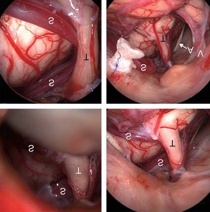
Figure 3: Endoscopically assisted microvascular decompression for
hemifacial spasm.
a) Inspection through the endoscope reveals vascular compression of
the facial nerve (F) by a loop of the posterior inferior cerebellar
artery (P), while the anterior inferior cerebellar artery (A) courses
parallel to the nerve.
b) Inspection through the operating microscope reveals the posterior
inferior cerebellar artery (P) but does not directly reveal the site of
compression, which is hidden behind the lower cranial nerve group
(cranial nerve IX, the glossopharyngeal nerve, and cranial nerve X, a b
the vagus nerve).
c) After mobilization of the artery (P), inspection through the endoscope
reveals that the facial nerve (F) is paper-thin after years of compres-
sion (14 years of symptoms) (arrow). The brainstem vessels are
visible through the flattened, translucent nerve.
d) Two Teflon pledgets have been interposed between the posterior in-
ferior cerebellar artery (P) and the facial nerve (F) to hold these two
structures apart without touching the nerve at the site of its previous
compression. The hemifacial spasm finally regressed 13 months
after surgery; the long delay may be due to the severe structural
change of the facial nerve as a result of its longstanding compres-
c d
sion. The anterior inferior cerebellar artery (A) is marked as well.
Figure 4: Endoscopically assisted microvascular decompression for
trigeminal neuralgia.
a) View of the trigeminal nerve (T) and the superior cerebellar artery
(S) through the operating microscope.
b) Inspection through the endoscope (0° optics), revealing the entire
cisternal course of the trigeminal nerve (T) and the superior
cerebellar artery (S).
c) Inspection through the endoscope with 30° optics reveals severe
vascular compression of the trigeminal nerve (T) by the elongated
loop of the superior cerebellar artery (S). a b
d) The superior cerebellar artery (S) has been transposed and sewn
upward toward the tentorium with a loop of Teflon. Ideally, the tri-
geminal nerve (T) should be entirely free after the decompression,
without any contact either with the vessel or with the Teflon pledget.
The abducens nerve (arrow A) and the vestibulocochlear nerve (V)
can be seen in the vicinity.
c d
from trigeminal neuralgia, because the affected areas MVD procedure in the 1970s and -80s (11, e24). He
are adjacent and the pain is of similar quality. Further found that some patients with vertigo profit from the
differential diagnoses include superior laryngeal neural- operation, but that it was ineffective against tinnitus. It
gia and nervus intermedius neuralgia. The main form of is still debated whether either of these symptoms is ever
conservative treatment is with anticonvulsant drugs, caused by neurovascular compression. Any potential
which can also be combined with antidepressants (e23). case requires very precise neurophysiological, neu-
Surgical treatment by microvascular decompression roradiological, and clinical evaluation (e25). Only a
should be considered the gold standard of treatment few studies have been dedicated to this topic (e26, e27).
(37); further options include radiofrequency ablation A clinically significant entity is vestibular paroxysmia,
and radiosurgery (38). The reported success rate of which is characterized by attacks of rotational or sway-
microvascular decompression is over 90% (34, 39, 40). ing vertigo lasting from a few seconds to one minute
(e26). Drugs (mainly carbamazepine) are the first line
Neurovascular compression of the eighth cranial of treatment, and MVD can be considered in intractable
nerve cases. No statement can be made about a potential
A series of microvascular decompression operations to association of chronic tinnitus with a neurovascular
relieve compression of the eighth cranial nerve was conflict. In a systematic review by Nash et al., the suc-
already reported on by Jannetta, who popularized the cess rate for the treatment of tinnitus as an isolated
58 Deutsches Ärzteblatt International | Dtsch Arztebl Int 2019; 116: 54–60MEDICINE
recommendation. A neurovascular conflict involving
Key messages the eighth cranial nerve that has been revealed by an
imaging study should be considered critically, as such
● The most common nerve compression syndrome in the findings are common in asymptomatic persons as
posterior cranial fossa is trigeminal neuralgia, followed by well. The current state of knowledge suggests that
hemifacial spasm. nervus intermedius neuralgia and carbamazepine-
● Neurovascular compression syndromes are generally due to responsive vestibular paroxysmia are potential indi-
compression of a cranial nerve by an artery or vein at the cations for MVD.
root entry or exit zone (REZ) near the brainstem. Acknowledgement
The authors thank Marc Matthes for processing the images and for the
● In patients with typical clinical manifestations, the diagnosis statistical analysis of the authors’ own data, as presented in this paper.
can be confirmed by high-resolution 3D-MRI.
Conflict of interest statement
● Most nerve compression syndromes are treated with drugs at Dr. Baldauf has received reimbursement of travel and accommodation
first, e.g., trigeminal neuralgia with carbamazepine and expenses as well as lecture honoraria from the Karl Storz company.
hemifacial spasm with botulinum toxin injections. Prof. Schroeder has served as a paid consultant for the Karl Storz
company, from which he has also recieved reimbursement of travel and
● Microvascular decompression is the only causally directed accommodation expenses as well as lecture honoraria.
treatment of these conditions and should be considered early Dr. Rosenstengel states that he has no conflict of interest.
in patients for whom pharmacotherapy is insufficiently effec-
Manuscript received on 29 March 2018; revised version accepted on
tive or causes unacceptable side effects. 4 October 2018.
Translated from the original German by Ethan Taub, M.D.
References
1. Donahue JH, Ornan DA, Mukherjee S: Imaging of vascular
symptom was 60%—a figure based on a total of only compression syndromes. Radiol Clin North Am 2017; 55: 123–38.
43 cases culled from an extensive literature search 2. El Refaee E, Langner S, Baldauf J, Matthes M, Kirsch M, Schroeder
(e27). HW: Value of 3-dimensional high-resolution magnetic resonance
imaging in detecting the offending vessel in hemifacial spasm:
comparison with intraoperative high definition endoscopic
Nervus intermedius neuralgia visualization. Neurosurgery 2013; 73: 58–67.discussion 67.
Nervus intermedius neuralgia, also called geniculate 3. Katusic S, Williams DB, Beard CM, Bergstralh EJ, Kurland LT:
Epidemiology and clinical features of idiopathic trigeminal neuralgia and
neuralgia, is a very rare condition. Only 174 cases were glossopharyngeal neuralgia: similarities and differences. Rochester,
described from 1932 to 2018 (e28–e30). It is character- Minnesota, 1945–1984. Neuroepidemiology 1991; 10: 276–81.
ized by brief attacks of stabbing deep-seated ear pain 4. Manzoni GC, Torelli P: Epidemiology of typical and atypical craniofacial
neuralgias. Neurol Sci 2005; 26(Suppl 2): s65–s67.
(e31). The pain may be accompanied by lacrimation
5. Fromm GH: Trigeminal neuralgia and related disorders. Neurol Clin
and taste sensations. The condition is usually due to 1989; 7: 305–19.
vascular compression of the nervus intermedius, but 6. DGN: Leitlinie Trigeminusneuralgie. 2012. www.dgn.org/leitlinien/
can also be caused by herpes zoster. Aside from micro- 2287-ll-58–012-trigeminusneuralgie (last accessed on 30 October
2018).
vascular decompression, transection of the nervus
7. Gronseth G, Cruccu G, Alksne J, et al.: Practice parameter: the diag-
intermedius is also recommended as a treatment for nostic evaluation and treatment of trigeminal neuralgia (an evidence-
medically intractable cases. Unfortunately, because of based review): report of the Quality Standards Subcommittee of the
the rarity of nervus intermedius neuralgia, the evidence American Academy of Neurology and the European Federation of
Neurological Societies. Neurology 2008; 71: 1183–90.
base for its treatment is very small (e28–30).
8. Spatz AL, Zakrzewska JM, Kay EJ: Decision analysis of medical and
surgical treatments for trigeminal neuralgia: how patient evaluations of
Overview benefits and risks affect the utility of treatment decisions. Pain 2007;
131: 302–10.
The most common neurovascular compression syn-
9. Degn J, Brennum J: Surgical treatment of trigeminal neuralgia. Re-
drome is trigeminal neuralgia, followed by hemifacial sults from the use of glycerol injection, microvascular decompression,
spasm. High-resolution 3D-MRI is now available and and rhizotomia. Acta Neurochirurgica 2010; 152: 2125–32.
enables reliable confirmation of the diagnosis in 10. Tatli M, Satici O, Kanpolat Y, Sindou M: Various surgical modalities for
trigeminal neuralgia: literature study of respective long-term outcomes.
patients with typical clinical manifestations. Micro- Acta Neurochirurgica 2008; 150: 243–55.
vascular decompression can then be carried out as a 11. Jannetta PJ: Arterial compression of the trigeminal nerve at the pons
causally directed treatment. Patients for whom conser- in patients with trigeminal neuralgia. J Neurosurg 1967; 26 (Suppl):
vative therapy is insufficiently effective should be in- 159–62.
12. Barker FG, Jannetta PJ, Bissonette DJ, Larkins MV, Jho HD: The
formed early about the option of surgical treatment. long-term outcome of microvascular decompression for trigeminal
(Vago-)glossopharyngeal neuralgia, a rare condition, neuralgia. N Engl J Med 1996; 334: 1077–83.
can also be treated surgically with MVD; the operation 13. Zakrzewska JM, Lopez BC, Kim SE, Coakham HB: Patient reports of
is indicated for patients with medically intractable satisfaction after microvascular decompression and partial sensory
rhizotomy for trigeminal neuralgia. Neurosurgery 2005; 56: 1304–11;
symptoms. discussion 11–2.
Vestibular paroxysmia is due to compression of the 14. Sindou M, Leston J, Howeidy T, Decullier E, Chapuis F: Micro-vascular
eighth cranial nerve. Tinnitus, too, has been relieved decompression for primary trigeminal neuralgia (typical or atypical).
Long-term effectiveness on pain; prospective study with survival
by MVD in a few reported cases, but the evidence analysis in a consecutive series of 362 patients. Acta Neurochir (Wien)
available from the literature does not permit any clear 2006; 148: 1235–45; discussion 45.
Deutsches Ärzteblatt International | Dtsch Arztebl Int 2019; 116: 54–60 59MEDICINE
15. Zhang H, Lei D, You C, Mao BY, Wu B, Fang Y: The long-term out- 30. El Damaty A, Rosenstengel C, Matthes M, et al.: A new score to
come predictors of pure microvascular decompression for primary predict the risk of hearing impairment after microvascular decom-
trigeminal neuralgia. World Neurosurg 2013; 79: 756–62. pression for hemifacial spasm. Neurosurgery 2017; 81: 834–43.
16. Mendelson ZS, Velagala JR, Kohli G, Heir GM, Mammis A, Liu JK: 31. Lovely TJ, Getch CC, Jannetta PJ: Delayed facial weakness after
Pain-free outcomes and durability of surgical intervention for microvascular decompression of cranial nerve VII. Surg Neurol 1998;
trigeminal neuralgia: a comparison of gamma knife and microvascular 50: 449–52.
decompression. World Neurosurg 2018; 112: e732–e746.
32. Mizobuchi Y, Muramatsu K, Ohtani M, et al.: The current status of
17. Gubian A, Rosahl SK: Meta-analysis on safety and efficacy of micro- microvascular decompression for the treatment of hemifacial spasm in
surgical and radiosurgical treatment of trigeminal neuralgia. World Japan: an analysis of 2907 patients using the Japanese diagnosis
Neurosurg 2017; 103: 757–67. procedure combination database. Neurologia medico-chirurgica 2017;
18. Franzini A, Ferroli P, Messina G, Broggi G: Surgical treatment of 57: 184–90.
cranial neuralgias. Handb Clin Neurol 2010; 97: 679–92. 33. Rosenstengel C, Matthes M, Baldauf J, Fleck S, Schroeder H:
19. Broggi G, Ferroli P, Franzini A, Servello D, Dones I: Microvascular Hemifacial spasm: conservative and surgical treatment options.
decompression for trigeminal neuralgia: comments on a series of 250 Dtsch Arztebl Int 2012; 109: 667–73.
cases, including 10 patients with multiple sclerosis. J Neurol
Neurosurg Psychiatry 2000; 68: 59–64. 34. Chen J, Sindou M: Vago-glossopharyngeal neuralgia: a literature
review of neurosurgical experience. Acta Neurochir (Wien) 2015; 157:
20. Jafree DJ, Williams AC, Zakrzewska JM: Impact of pain and postoper- 311–21; discussion 21.
ative complications on patient-reported outcome measures 5 years
after microvascular decompression or partial sensory rhizotomy for 35. Blumenfeld A, Nikolskaya G: Glossopharyngeal neuralgia. Curr Pain
trigeminal neuralgia. Acta Neurochir (Wien) 2018; 160: 125–34. Headache Rep 2013; 17: 343.
rd
21. Cruccu G, Gronseth G, Alksne J, et al.: AAN-EFNS guidelines on 36. The International Classification of Headache Disorders. 3 edition
trigeminal neuralgia management. Eur J Neurol 2008; 15: 1013–28. (beta version). Cephalalgia 2013; 33: 629–808.
22. Zakrzewska JM, Akram H: Neurosurgical interventions for the 37. Rey-Dios R, Cohen-Gadol AA: Current neurosurgical management of
treatment of classical trigeminal neuralgia. Cochrane Database Syst glossopharyngeal neuralgia and technical nuances for microvascular
Rev 2011: CD007312. decompression surgery. Neurosurg Focus 2013; 34: E8.
23. Auger RG, Whisnant JP: Hemifacial spasm in Rochester and Olmsted 38. Kano H, Urgosik D, Liscak R, et al.: Stereotactic radiosurgery for idio-
County, Minnesota, 1960 to 1984. Arch Neurol 1990; 47: 1233–4. pathic glossopharyngeal neuralgia: an international multicenter study.
24. El Damaty A, Rosenstengel C, Matthes M, Baldauf J, Schroeder HW: J Neurosurg 2016; 125: 147–53.
The value of lateral spread response monitoring in predicting the clinical 39. Ma Y, Li YF, Wang QC, Wang B, Huang HT: Neurosurgical treatment
outcome after microvascular decompression in hemifacial spasm: of glossopharyngeal neuralgia: analysis of 103 cases. J Neurosurg
a prospective study on 100 patients. Neurosurg Rev 2016; 39: 455–66. 2016; 124: 1088–92.
25. Deluca C, Tommasi G, Moretto G, Fiaschi A, Tinazzi M: Focal motor 40. Xia L, Li YS, Liu MX, et al.: Microvascular decompression for glossop-
seizures mimicking hemifacial spasm. Parkinsonism Relat Disord haryngeal neuralgia: a retrospective analysis of 228 cases. Acta
2008; 14: 649–51. Neurochir (Wien) 2018; 160: 117–23.
26. Yaltho TC, Jankovic J: The many faces of hemifacial spasm: differen-
tial diagnosis of unilateral facial spasms. Mov Disord 2011; 26: Corresponding author
1582–92. PD Dr. med. Jörg Baldauf
27. Sindou M, Mercier P: Microvascular decompression for hemifacial Klinik für Neurochirurgie der Universitätsmedizin Greifswald
spasm: outcome on spasm and complications. A review. Neurochirur- Sauerbruchstr.
gie 2018; 64: 106–16.
17475 Greifswald, Germany
28. Barker FG, Jannetta PJ, Bissonette DJ, Shields PT, Larkins MV, Jho baldauf@uni-greifswald.de
HD: Microvascular decompression for hemifacial spasm. J Neurosurg
1995; 82: 201–10.
►Supplementary material
29. Jo KW, Kim JW, Kong DS, Hong SH, Park K: The patterns and risk
factors of hearing loss following microvascular decompression for For eReferences please refer to:
hemifacial spasm. Acta Neurochir (Wien) 2011; 153: 1023–30. www.aerzteblatt-international.de/ref0419
60 Deutsches Ärzteblatt International | Dtsch Arztebl Int 2019; 116: 54–60MEDICINE
Supplementary material to:
Nerve Compression Syndromes in the Posterior Cranial Fossa
Diagnosis and Treatment
by Jörg Baldauf, Christian Rosenstengel, and Henry W. S. Schroeder
Dtsch Arztebl Int 2019; 116: 54–60. DOI: 10.3238/arztebl.2019.0054
eReferences e14. Babinski J: “Hémispasme facial périphérique”. Nouvelle icono-
e1. Haller S, Etienne L, Kovari E, Varoquaux AD, Urbach H, Becker M: graphie de la Salpétrière 1905; 18: 418–23.
Imaging of neurovascular compression syndromes: trigeminal e15. Tan EK, Jankovic J: Psychogenic hemifacial spasm. J Neuropsychiatry
neuralgia, hemifacial spasm, vestibular paroxysmia, and glosso- Clin Neurosci 2001; 13: 380–4.
pharyngeal neuralgia. Am J Neuroradiol 2016; 37: 1384–92. e16. Tan EK, Fook-Chong S, Lum SY, Lim E: Botulinum toxin improves
e2. Love S, Coakham HB: Trigeminal neuralgia: pathology and patho- quality of life in hemifacial spasm: validation of a questionnaire
genesis. Brain 2001; 124: 2347–60. (HFS-30). J Neurol Sci 2004; 219: 151–5.
e3. Meaney JF, Watt JW, Eldridge PR, Whitehouse GH, Wells JC, Miles e17. Jankovic J: Tourette syndrome. Phenomenology and classification of
JB: Association between trigeminal neuralgia and multiple sclerosis: tics. Neurol Clin 1997; 15: 267–75.
role of magnetic resonance imaging. J Neurol Neurosurg Psychiatry e18. Lim EC, Seet RC: Use of botulinum toxin in the neurology clinic. Nat
1995; 59: 253–9. Rev Neurol 2010; 6: 624–36.
e4. Gass A, Kitchen N, MacManus DG, Moseley IF, Hennerici MG, Miller e19. Defazio G, Abbruzzese G, Girlanda P, et al.: Botulinum toxin A treat-
DH: Trigeminal neuralgia in patients with multiple sclerosis: lesion ment for primary hemifacial spasm: a 10-year multicenter study.
localization with magnetic resonance imaging. Neurology 1997; 49: Arch Neurol 2002; 59: 418–20.
1142–4.
e20. Dannenbaum M, Lega BC, Suki D, Harper RL, Yoshor D: Microvas-
e5. Udupi BP, Chouhan RS, Dash HH, Bithal PK, Prabhakar H: cular decompression for hemifacial spasm: long-term results from
Comparative evaluation of percutaneous retrogasserian glycerol 114 operations performed without neurophysiological monitoring. J
rhizolysis and radiofrequency thermocoagulation techniques in the Neurosurg 2008; 109: 410–5.
management of trigeminal neuralgia. Neurosurgery 2012; 70:
e21. Lee MH, Jee TK, Lee JA, Park K: Postoperative complications of
407–12; discussion 12–3.
microvascular decompression for hemifacial spasm: lessons from
e6. Noorani I, Lodge A, Vajramani G, Sparrow O: Comparing percuta- experience of 2040 cases. Neurosurgical Review 2016; 39: 151–8.
neous treatments of trigeminal neuralgia: 19 years of experience in e22. Ceylan S, Karakus A, Duru S, Baykal S, Koca O: Glossopharyngeal
a single centre. Stereotact Funct Neurosurg 2016; 94: 75–85. neuralgia: a study of 6 cases. Neurosurg Rev 1997; 20: 196–200.
e7. Kondo A: Follow-up results of microvascular decompression in e23. Headache Classification Subcommittee of the International Head-
trigeminal neuralgia and hemifacial spasm. Neurosurgery 1997; 40: ache Society: The international classification of headache disorders:
46–51; discussion –2. nd
2 edition. Cephalalgia 2004; 24(Suppl 1): 9–160.
e8. Tronnier VM, Rasche D, Hamer J, Kienle AL, Kunze S: Treatment of e24. Jannetta PJ: Neurovascular compression in cranial nerve and
idiopathic trigeminal neuralgia: comparison of long-term outcome systemic disease. Ann Surg 1980; 192: 518–25.
after radiofrequency rhizotomy and microvascular decompression.
Neurosurgery 2001; 48: 1261–7; discussion 7–8. e25. Best C, Gawehn J, Kramer HH, et al.: MRI and neurophysiology in
vestibular paroxysmia: contradiction and correlation. J Neurol
e9. Sarsam Z, Garcia-Finana M, Nurmikko TJ, Varma TR, Eldridge P: Neurosurg Psychiatry 2013; 84: 1349–56.
The long-term outcome of microvascular decompression for trige-
minal neuralgia. Br J Neurosurg 2010; 24: 18–25. e26. Brandt T, Strupp M, Dieterich M: Vestibular paroxysmia: a treatable
neurovascular cross-compression syndrome. J Neurol 2016; 263
e10. Baldauf J, Rosenstengel C, Matthes M, Fleck S, Marx S, Schroeder (Suppl 1): S90–6.
HWS: Microvascular decompression and partial sensory rhizotomy
for trigeminal neuralgia with special reference to endoscopic- e27. Nash B, Carlson ML, van Gompel JJ: Microvascular decompression
assisted microsurgery. J Neurol Surg B 2016; 77: LFP-12–03. for tinnitus: systematic review. J Neurosurg 2017; 126: 1148–57.
e11. Gunther T, Gerganov VM, Stieglitz L, Ludemann W, Samii A, Samii e28. Tang IP, Freeman SR, Kontorinis G, et al.: Geniculate neuralgia: a
M: Microvascular decompression for trigeminal neuralgia in the systematic review. J Laryngol Otol 2014; 128: 394–9.
elderly: long-term treatment outcome and comparison with younger e29. Peris-Celda M, Oushy S, Perry A, et al.: Nervus intermedius and the
patients. Neurosurgery 2009; 65: 477–82; discussion 82. surgical management of geniculate neuralgia. J Neurosurg 2018:
e12. Abhinav K, Love S, Kalantzis G, Coakham HB, Patel NK: Clinico- 1–9.
pathological review of patients with and without multiple sclerosis e30. Holste KG, Hardaway FA, Raslan AM, Burchiel KJ: Pain-free and
treated by partial sensory rhizotomy for medically refractory pain-controlled survival after sectioning the nervus intermedius in
trigeminal neuralgia: a 12-year retrospective study. Clin Neurol nervus intermedius neuralgia: a single-institution review. J Neur-
Neurosurg 2012; 114: 361–5. osurg 2018: 1–8.
e13. Wang A, Jankovic J: Hemifacial spasm: clinical findings and treat- e31. O‘Neill F, Nurmikko T, Sommer C: Other facial neuralgias. Cephalalgia
ment. Muscle Nerve 1998; 21: 1740–7. 2017; 37: 658–69.
Deutsches Ärzteblatt International | Dtsch Arztebl Int 2019; 116: 54–60 | Supplementary material IYou can also read