Heat profiles of laser-irradiated nails - Uwe Paasch Pietro Nenoff Anna-Theresa Seitz Justinus A. Wagner Michael Kendler Jan C. Simon Sonja ...
←
→
Page content transcription
If your browser does not render page correctly, please read the page content below
Heat profiles of laser-irradiated nails

                 Uwe Paasch
                 Pietro Nenoff
                 Anna-Theresa Seitz
                 Justinus A. Wagner
                 Michael Kendler
                 Jan C. Simon
                 Sonja Grunewald

Downloaded From: https://www.spiedigitallibrary.org/journals/Journal-of-Biomedical-Optics on 18 Apr 2021
Terms of Use: https://www.spiedigitallibrary.org/terms-of-use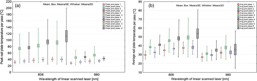
Journal of Biomedical Optics 19(1), 018001 (January 2014)

                Heat profiles of laser-irradiated nails
                Uwe Paasch,b,* Pietro Nenoff,a Anna-Theresa Seitz,b Justinus A. Wagner,b Michael Kendler,b Jan C. Simon,b
                and Sonja Grunewaldb
                a
                 Labor für medizinische Mikrobiologie, Partnerschaft Prof. Pietro Nenoff & Dr. Constanze Krüger, Mölbis 04579, Germany
                b
                 Klinik und Poliklinik für Dermatologie, Venerologie und Allergologie, Universitätsklinikum Leipzig AöR und Medizinische Fakultät der Universität
                Leipzig, 04103, Germany

                                Abstract. Onychomycosis is a worldwide problem with no tendency for self-healing, and existing systemic
                                treatments achieve disease-free nails in only 35 to 76% of cases. Recently, treatment of nail fungus with
                                a near-infrared laser has been introduced. It is assumed that fungal eradication is mediated by local heat.
                                To investigate if laser treatment has the potential to eradicate fungal hyphae and arthrospores, laser heat appli-
                                cation and propagation needs to be studied in detail. This study aimed to measure nail temperatures using
                                real-time videothermography during laser irradiation. Treatment was performed using 808- and 980-nm linear
                                scanning diode lasers developed for hair removal, enabling contact-free homogeneous irradiation of a human
                                nail plate in one pass. Average and peak temperatures increased pass by pass, while the laser beam moved
                                along the nail plates. The achieved mean peak temperatures (808 nm: 74.1 to 112.4°C, 980 nm: 45.8 to 53.5°C),
                                as well as the elevation of average temperatures (808 nm: 29.5 to 38.2°C, 980 nm: 27.1 to 32.6°C) were asso-
                                ciated with pain that was equivalent to that of hair removal procedures and was not significantly different for
                                various wavelengths. The linear scanning laser devices provide the benefits of contact-free homogeneous heat-
                                ing of the human nail while ensuring adequate temperature rises. © 2014 Society of Photo-Optical Instrumentation Engineers
                                (SPIE) [DOI: 10.1117/1.JBO.19.1.018001]

                                Keywords: laser; fungi; thermography; temperature; nails.
                                Paper 130647R received Sep. 6, 2013; revised manuscript received Nov. 21, 2013; accepted for publication Dec. 9, 2013; published
                                online Jan. 9, 2014.

                1       Introduction                                                                water decreases. Overall, a nail does have a water content of
                Dermatophytosis is found in ∼20 to 25% of the world’s popu-                         9 to 35%21,22 and arthrospores are protected by proteins.18
                lation.1 An estimated 2 to 13% of the population suffers from                       However, our in vitro study on the heating effects of common
                onychomycosis (OM),2,3,4 which is the most common nail dis-                         dermatological lasers demonstrated that hair removal lasers
                ease worldwide and is responsible for approximately half of                         operating at 808, 980, or 1064 nm are able to heat liquid patho-
                all nail abnormalities.5 This condition has a huge impact on                        gens in liquid cultures efficiently if certain parameters are
                the quality of life.6,7 To treat the dermatophytes T. rubrum                        adopted.23 This finding is of practical importance because lasers
                and T. interdigitale (formerly T. mentagrophytes) that are the                      using wavelengths of ∼800 nm are widely used for hair
                main causative agents of OM, near-infrared lasers have been                         removal.24 Moreover, lasers operating at a 1064-nm wavelength
                introduced because standard systemic terbinafine administration                     are frequently used for vascular treatments and skin rejuvenation
                achieves disease-free nails in only ∼35 to 76% of cases.8,9,10 In                   in addition to hair removal, and therefore, all of these laser
                addition, the relapse rates are up to 22.3% within 3 years after                    systems have been proven safe for use on human skin if precau-
                completion of the systemic treatment.11                                             tions are taken. Finally, the 1064-nm systems are most often
                    Previously, CO2 lasers were found to be effective but unpre-                    Food and drug administration (FDA)-approved for the “tempo-
                dictable in terms of efficacy and side effects. Therefore, longer                   rary increase of clear nails in patients with onychomycosis.”
                pulsed nonablative near-infrared lasers were thought to have                        However, the reported clearance rates vary substantially, from
                a much better side-effect profile while maintaining their effi-                     50 to ∼100%.13,25–27 In line with this, the pathogen eradication
                cacy.12,13 Due to their absorption characteristics, potential targets               effects observed in vitro were less impressive.28 To date, many
                are both water and melanin. This absorption is of interest                          systems that can operate with diverse parameter settings are
                because T. rubrum, the most common causative agent of                               available, making clinical comparisons difficult.23
                OM, expresses a pigment called xanthomegnin that provides                               This situation reflects the lack of knowledge of a highly inter-
                a typical color in agar-based culture systems and in nails.14                       esting clinical laser application. Assuming that heat is the under-
                Earlier studies located the pigments into the outer microconidia                    lying mechanism, the application and propagation of heat via
                walls of T. interdigitale.15 Approximately 0.2% of the wall com-                    lasers needs to be studied. The peak and average temperatures
                pounds reflect pigment.16 It is assumed that the fungal eradica-                    should be investigated to answer the question of whether
                tion effect is mediated by the heat absorption of water and/or                      the proposed laser treatment regimens have the potential to
                melanin,17–19 although heat-resistant (up to 80°C) strains of                       eradicate the fungi and spores within the entire nail plate.
                fungi have been detected recently20 (Table 1). With lower wave-                     Because spores are known to survive at 60 to 80°C, the laser
                lengths, the absorption of melanin increases, whereas that of                       must be able to heat the entire area to this threshold
                                                                                                    value.18,29 However, heat generates pain. Pain is inflicted by
                *
                    Address all correspondence to: Uwe Paasch, E-mail: uwe.paasch@medizin
                    .uni-leipzig.de                                                                 0091-3286/2014/$25.00 © 2014 SPIE

                Journal of Biomedical Optics                                               018001-1                                          January 2014   •   Vol. 19(1)

Downloaded From: https://www.spiedigitallibrary.org/journals/Journal-of-Biomedical-Optics on 18 Apr 2021
Terms of Use: https://www.spiedigitallibrary.org/terms-of-use
Paasch et al.: Heat profiles of laser-irradiated nails
Table 1 Published evidence of heat susceptibility of pathogens that 2.1 Pain Evaluation
cause onychomycosis in humans.
Because the method used is based on heat application to nails to
eradicate the pathogens that cause OM, pain determined the
Pathogen Finding Reference clinical endpoint of the treatments performed in earlier studies.
Pain was quantified using a visual analogue scale (1 to 10).
T. rubrum Conidia (measurements 2.0 to 17, 18, and
Patients were asked to report the highest pain score during
T. interdigitale 3.3 by 2.9 to 3.8 μm) are 29
extremely susceptible to moderate each treatment per foot.
heat and desiccation
2.2 Thermography Measurements
T. interdigitale Germination can be triggered by 30
sublethal heating, e.g., 45°C Thermography was performed by using a device for measuring
for 30 min the power of incident electromagnetic radiation due to the heat-
ing of a given structure with a temperature-dependent electrical
T. interdigitale Dormant and germinated 30
microconidia can be eradicated resistance. This method was invented by the American astrono-
to the same extend if temperatures mer Samuel Pierpont Langley in 1878.
are elevated up to 55°C The thermography system (InfraTec mobileIR E9, InfraTec,
Germany) used was a bolometric camera equipped with a
M. gypseum 15 min in vitro exposure to 55°C is 23 and 25-mm lens field of view (FOV) (22 × 16)/instantaneous FOV
lethal to macroconidia and mycelia 29
1.0 mrad and an uncooled microbolometric focal plane array
T. mentagrophytes 100% eradication at 18 detector with a spectral range of 8 to 14 μm. The measurement
60°C∕2 min 90% eradication accuracy was given as 2 K for 0 to 100°C, and 2% for 100°C at a temperature measuring range of −20 to 250°C.
at 48°C∕30 min The temperature resolution at 30°C was determined to be better
than 0.06 K (thermal sensitivity). The thermograms had an
image format of 384 × 288 pixels at an IR frame rate of 50 Hz.
the current OM laser treatments, and this physiological reaction Real-time video recording was performed in all of the treatment
determines the clinical endpoint of treatment. Therefore, tem- sessions. The Irbis3Plus software (InfraTec) was further used
perature profiles for individual laser systems are of interest to for processing the primary images. The video streams were
define safe and effective heating regimens for larger and smaller uploaded and examined for quality control. Then, the video
nails that ensure the lowest pain intensity. Finally, homogenous streams were analyzed manually by frame-by-frame analysis
heat distribution is highly desirable to achieve complete patho- to note the temperatures of interest by setting a continuously
gen clearance. adjusted region of interest for calculation of the following:
To address these issues, this study aimed to measure nail tem- (1) peak temperatures of all of the toes during laser irradiation
peratures during laser irradiation (1) to estimate the peak tem- in all of the passes, (2) average nail temperatures of all of the
peratures using two wavelengths, (2) to establish temperature toes immediately before and after laser irradiation during all of
profiles for all of the toes immediately before and after laser the passes, and (3) qualitative analysis of heat propagation
irradiation during consecutive treatment passes, (3) to analyze during the laser treatments.
the heat propagation during laser treatment, and (4) to investi-
gate histological changes in nail explants. These investigations 2.3 Temperature Measurements
will help to rank the value of the investigated wavelengths for
Foot nails of 11 patients were evaluated using an infrared ther-
their suitability in OM laser treatment, to define concepts for
mometer (Voltcraft IR-1000L, −50.0 to 1000.0°C, Germany) in
application, and to analyze the potential risk of insufficient treat-
a fixated position at 13 cm distance from digitus I of both feet to
ment due to inhomogeneous irradiation. To address these ques-
ensure measuring at the whole nail plate. Measurements were
tions, an advanced real-time videothermography system was
taken before intervention (t0 ), immediately after the last laser
used. Additionally, nail explants were subjected to histological
pass to measure the temperature maximum (Temp. max), and
investigation.
30 s postintervention (Temp. post).
2 Materials and Methods 2.4 Laser Treatment
The objective of this study was to define the ability of 808- and Laser treatment was performed using two different systems: an
980-nm linear scanning lasers, using proven safe and effective 808-nm linear scanning diode laser (Alma Lasers, formerly
parameter settings established for hair removal procedures, to Quantel-Derma and Wavelight Aesthetic, Erlangen, Germany)
deliver heat to nails on human feet in vivo to treat OM. The and a 980-nm linear scanning diode laser (Alma Lasers, for-
patients were selected after informed consent was given to merly Quantel-Derma and Wavelight Aesthetic). Both systems
also have a thermographic (EasyIR-9™, using software IRBIS are routinely used for hair removal. Both systems are therefore
3plus, InfraTec GmbH, Dresden, Germany) video record made tested to ensure that they would be safe and efficient in clinical
during the routine treatment procedure using CE certified routine treatments using a fluence of 30 J∕cm2 , with a pulse
devices. To compare temperatures additionally a contact-free duration of 12 ms and a spot size of 12 × 12 mm.31
temperature measurement (Voltcraft IR-1000L, Germany) was The laser beam itself is made of a rectangular array of diodes
performed in another group of patients treated with either forming a spot of 1 × 12 mm. Using a mirror system, this rec-
980-nm linear scanning laser or a long pulsed 1064-nm Nd:YAG tangular spot is moved linearly to cover an area of 12 × 50 mm.
laser with a cooled contact hand piece. Scattering of the light along the 10-mm side of the rectangular
Journal of Biomedical Optics 018001-2 January 2014 • Vol. 19(1)
Downloaded From: https://www.spiedigitallibrary.org/journals/Journal-of-Biomedical-Optics on 18 Apr 2021
Terms of Use: https://www.spiedigitallibrary.org/terms-of-usePaasch et al.: Heat profiles of laser-irradiated nails
spot allows a deep penetration in one dimension. At the 1-mm tests were two-tailed, and significance was indicated by
side, the scattering is also present within the second dimension p < 0.05.
since the spot is moved continuously over the nail. Each area is
therefore preheated by scattered photons, and immediately after 3 Results
this, the full beam is heating up the whole area.
In total, 187 toes of 11 patients (nine males, two females, all
The parameter settings used were the following: 808 and
Caucasian, Fitzpatrick skin types I-II, age 61.7 14.2 years)
980 nm: fluence of 30 J∕cm2 , pulse duration of 12 ms, spot
were treated for toe nail fungus confirmed by mycology
size of 12 × 12 mm, five (808 nm) or three (980 nm) passes
using a linear scanning diode laser emitting at 808 nm
for digits I to V. A fixed number of passes applied was chosen
(n ¼ 125) or 980 nm (n ¼ 62). During the treatment, real-
based on the in vitro temperature profiling of earlier studies.23
time thermographic monitoring was performed at a frame
The patients were asked to allow an extra pass from the standard
rate of 50 frames per second (fps). A total of 42.268
treatment in case they had no clear feeling of pain. The treatment
(1.083 374) video frames were subjected to analysis using
was performed by starting pass one at digitus one on a given
Irbis3Plus software.
foot. Then, the laser was moved to the next toe, allowing a cool-
Overall, the treatment procedures were well tolerated.
ing period for the recently treated one. After all five toes had
However, in selected cases, the development of a single subun-
been treated, the second pass was begun at toe one. In case
gual hematoma was noted as a side effect separate from the
of a severe pain sensation, extra time for cooling was given
ubiquity of pain.
until the patient felt comfortable to continue.
For comparison of temperature measurement results using
videothermography and conventional infrared thermometer, 3.1 Pain Evaluation
a 980-nm linear scanning diode laser (Alma Lasers, formerly
Quantel-Derma and Wavelight) and a long pulsed 1064-nm Pain, quantified using a visual analog scale, was reported as
Nd:YAG-laser (Alma Lasers, formerly Quantel-Derma and 6.2 2.2. There was no significant difference (p > 0.05) with
Wavelight) with a cooled hand piece operated in nail contact regard to the application of either the 808-nm (6.1 2.2) or
were in use. The parameter settings used were the following: the 980-nm (6.4 2.3) laser.
980 nm: fluence of 30 J∕cm2 , pulse duration of 12 ms, spot
size of 12 × 12 mm, three passes for digits I to V and 1064 nm: 3.2 Thermographic Measurements
fluence of 70 J∕cm2 , pulse duration of 40 ms, spot size 5 mm,
three passes for digits I to V. The number of passes applied was Thermographic video recording was performed in such a way
chosen based on the in vitro temperature profiling of earlier that the linear scan of the laser beam could be followed over
studies.23 The treatment with the 980-nm system was performed time and over the total area of each toe. In case of incomplete
as described above, while the 1064-nm treatment was performed visibility (time- or area-wise), the data were not subjected to
by starting pass one at digitus one on a given foot. The whole evaluation. The larger the nail plate was, the easier it was to
nail plate was covered with 30% overlap three times having perform thermographic recording.
a 5- to 10-s break in between the treatments. Then, the laser
was moved to the next toe. In case of a severe pain sensation, 3.3 Peak Temperatures
extra time for cooling was given until the patient felt comfort-
able to continue. In general, the peak temperatures measured during the move-
ment of the laser beam along the nail plates increased pass-
2.5 Histological Analysis of Laser–Nail Interaction by-pass, starting at a mean of 74.1°C and reaching a mean of
112.4°C after five consecutive passes using the 808-nm linear
Basic histological investigation was performed in the human scanning laser (Fig. 1). Between the passes, while the remaining
nail explant after six shots with the linear scanning 808-nm toes were being treated, the temperatures decreased substantially
diode laser (fluence: 30 J∕cm2 ; pulse duration: 12 ms). An addi- (Table 2). Despite this decrease, the absolute peak temperatures
tional nail with mycologically proven infection was subjected to measured ranged from 260 to 290°C starting with the very first
histology to visualize growth pattern of fungi within the nail treatment pass. The relatively high SD can most likely be
plate. The specimens were decalcified and then subjected to attributed to the fact that at 50 fps, the recording rate of the
buffered 4% formalin for 24 h for fixation. Tissue blocks were thermographic system is relatively slow compared to the
embedded in paraffin, cut into 5- to 8-μm slices, and stained pulse durations of 12 ms. As a consequence, the increase in
with hematoxylin and eosin [H&E and periodic acid schiff the mean peak temperatures did not reach the level of signifi-
(PAS)] according to standard procedures. Slides were evaluated cance. With regard to the different size of the nails, plotting
under a calibrated microscope (BX41, Olympus Germany, the peak temperature profiles toe-wise showed higher peak
Hamburg, Germany) equipped with a digital camera (DP70, nail plate temperatures post first to fifth pass of the laser inter-
Olympus Germany). Dimensions were measured using cali- vention in digitus I compared to all of the other toes (p < 0.05).
brated CellF software (Olympus Germany). In comparison, the 980-nm treatment showed the same trend
of stepwise increasing temperatures over four passes, although
2.6 Statistics the trend started at 45.8°C, reached the peak temperature after
the third pass (53.5°C), and ended at 42.6°C after the fourth pass.
The statistical analysis of the thermography data was performed The temperatures reached using the two laser systems were
using Statistica 8.0 software for Windows (StatSoft Inc., OK). significantly different at each pass. The peak temperatures
The normality of the distribution was investigated using the reached 161.5°C after the third pass. Digitus I showed a signifi-
Shapiro-Wilkes test. A Mann-Whitney U test was performed cantly higher peak temperature after the second pass compared
to investigate the differences between the groups. Both of the to all of the other toes. Comparing the two laser systems
Journal of Biomedical Optics 018001-3 January 2014 • Vol. 19(1)
Downloaded From: https://www.spiedigitallibrary.org/journals/Journal-of-Biomedical-Optics on 18 Apr 2021
Terms of Use: https://www.spiedigitallibrary.org/terms-of-usePaasch et al.: Heat profiles of laser-irradiated nails
Fig. 1 Average and peak nail plate temperature profiles showing a higher average p > 0.05 (a) and peak
(b) nail plate temperature after each pass of an 808-nm laser compared to 980-nm irradiation after each
pass (p < 0.05).
pass-by-pass revealed that the 808-nm system always resulted in first three passes. As early as pass 4, no significant increase in
significantly higher peak temperatures on the nail surface. temperature was detected for the 980-nm system (p < 0.01). The
same trend was demonstrated for the 808-nm system beginning
at pass 6, whereas the temperature elevation was significantly
3.4 Average Temperature Profiles lower at pass 6 than at pass 5 (p < 0.01). The cooling rates
The average temperature measured immediately before laser were always lower during laser pass 1 to 5, whereas the opposite
treatment within a continuously adjusted region of interest was true for the last pass when the 808-nm system was used.
increased significantly (p < 0.01) stepwise from pass to pass The highest cooling rate was visible between passes 2 and 3 in
using the 808-nm linear scanning diode laser, increasing the 980-nm group (Table 2).
from 29.5°C (prepass 1) to 38.2°C (prepass 5). The average tem-
peratures measured immediately after a laser pass were higher
and increased stepwise from pass to pass (38.4°C postpass 1 and 3.5 Heat Distribution
53.8°C postpass 6). With regard to the different sizes of the nails,
plotting the temperature profiles toe-wise showed higher aver- In general, the linear scanning laser devices with a spot size of
age nail plate temperatures after each pass of laser treatment in 12 × 12 mm were easy to handle in terms of the nail treatments
digitus I compared to all of the other toes. performed in this study and clearly had the advantage of
The laser energy emitted by the 980-nm system also resulted allowing a contact-free and very rapid procedure (Fig. 2).
in a stepwise significant (p < 0.01) elevation of the average tem- Real-time evaluation of the thermal effects in >40 video
peratures measured before laser irradiation increasing from 27.1 streams revealed that exact positioning of the laser is crucial
to 32.6°C. Immediately after each laser irradiation, the nail tem- to achieve stepwise homogeneous heating of the nail plates.
perature was slightly higher than the mean value (31.0 to 35.6° If placed correctly, uniform heating was observed as long as
C). However, the maximum average temperatures reached 57.7° the nail plate was free of rough areas. With regard to the
C. The temperature profiles plotted toe-wise showed slightly wavelength, there was some delay in lateral heat diffusion
higher average nail plate temperatures after each pass of laser within the toe correlated with the higher wavelength. Although
irradiation for digitus I compared to all of the other toes. the result was not statistically significant, the 980-nm system
The average temperature elevation per pass of laser irradia- was rated as more painful, resulting in a lower number of
tion did not differ significantly between the laser systems for the passes applied.
Table 2 Temperature reduction (mean values) between passes of laser irradiation.
808 nm 980 nm
n Mean SD n Mean SD p
Δ Avg postpass 1 prepass 2 125 −3.8 9.5 62 −0.0 9.5Paasch et al.: Heat profiles of laser-irradiated nails
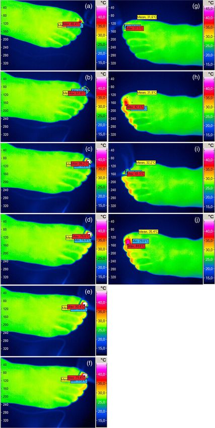
Fig. 2 Frames of interest from a videothermographic recording of six passes of an 808-nm [right foot,
(a) to (f)] and four passes [left foot, (g) to (j)] of a 980-nm linear scanning laser using a spot size of
12 × 12 mm.
Journal of Biomedical Optics 018001-5 January 2014 • Vol. 19(1)
Downloaded From: https://www.spiedigitallibrary.org/journals/Journal-of-Biomedical-Optics on 18 Apr 2021
Terms of Use: https://www.spiedigitallibrary.org/terms-of-usePaasch et al.: Heat profiles of laser-irradiated nails
Table 3 Nail temperatures measured after laser irradiation (Digitus I foot left, 1064 nm, 70 J∕cm2 , 40 ms, 5 mm spot, three passes having a 5- to
10-s break in between the treatments, ultrasound gel coupling, contact cooling, 30% overlap; Digitus I foot right, 980 nm, 30 J∕cm2 , 12 ms, 12 ×
10 mm spot, three passes having a 5- to 10-s break in between the treatments, no cooling) using an infrared thermometer (Voltcraft IR-1000L,
−50.0 to 1000.0°C) at a fixed distance of 13 cm. Measurements were taken before intervention (t 0 ), immediately after the last laser irradiation pass
(Temp. max), and 30 s post last treatment (Temp. post).
n ¼ 11 Temp. T 0 Temp. max ΔT max p T 0 versus Tmax Temp. post ΔT post p Tmax versus Tpost
1064 nm 25.0 2.9 42.5 4.9 17.5 4.7 0.05
3.6 Alternative Temperature Measurements The nail explant subjected to six passes of 808-nm laser dis-
played changes in the nail plate structure. The relatively high
Nail temperature was measured after laser irradiation (Digitus I temperatures caused disruptions and condensed hypereosino-
foot left, 1064 nm, 70 J∕cm2 , 40 ms, 5 mm spot, three passes philic areas (Fig. 4).
having a 5- to 10-s break in between the treatments, ultrasound
gel coupling, contact cooling, 30% overlap; Digitus I foot right, 4 Discussion
980 nm, 30 J∕cm2 , 12 ms, 12 × 10 mm spot, three passes hav-
ing a 5- to 10-s break-in between the treatments, no cooling) Recently, the option of near-infrared laser treatment of nail fun-
using a high-temperature infrared thermometer (Voltcraft gus has become available. Generally, the 1064-nm systems
IR-1000L, −50.0 to 1000.0°C) at a fixed distance of 13 cm are FDA-approved for the “temporary increase of clear nails in
(Table 3). Measurements were taken before intervention (t0 ), patients with onychomycosis.” The reported clearance rates vary
immediately after the last laser irradiation pass (Temp. max), substantially from 50 to ∼100%,13,25–27 although the eradication
and 30 s post last treatment (Temp. post). effects observed in vitro are less convincing.28 However, recent
While temperatures measured before laser interventions were in vitro studies suggested that systems operating at 808 to
lower at t0 compared to the videothermography, Temp. max was 980 nm may be effective if temperatures >50°C are achieved.23
∼10 deg lower after three passes of 980 nm measured with the The assumed unifying mechanism is that the heat is delivered to
infrared thermometer compared to the values of videothermog- the nail plate and nail bed due to absorption by water and/or
raphy (53.5 26.3 versus 44.3 7.0°C). In general, there was melanin. The wide range of reported clinical efficacy might
a significant increase of the nail temperatures in both laser sys- result from the lack of knowledge of how much heat is generated
tems (1064 nm 17.5 4.7 versus 980 nm 19.4 6.1) as well as and propagated throughout the nail and nail bed area. However,
cooling 30 s post treatment (p > 0.05 between the systems). it is crucial that certain temperature levels be kept constant over
The temperature rise after three passes of 980 nm measured a certain time to ensure secure pathogen eradication and to avoid
by thermography resulted in a 24.9°C elevation. growth induction.
In general, the fast, contact-free treatment at 808 and 980 nm
using the linear scanning laser devices with a 12 × 12 mm spot
3.7 Histological Analysis of Laser–Nail Interaction not only ensured the prevention of pathogen transmission, but
also allowed the study of temperature development over time
Basic histological investigation of a human nail explant clini- and over the area of the entire nail plate.
cally diagnosed with OM revealed rather long septed hyphae
with a small diameter of ∼1 μm (Fig. 3) located everywhere
from the surface down to the nail bed within the nail plate.
Fig. 4 Impact of 808-nm diode laser treatment (six passes at a flu-
ence of 30 J∕cm2 and 12 ms pulse duration) on nail morphology.
Disruptions and coagulations of the nail plate (hematoxylin and
Fig. 3 Histological specimen stained with PAS, 100× magnification. eosin, 40×) have been observed. The changes of the nail structure
Septed hyphae are found as rather long structures up to 100 μm with do reflect the enormous heat action and may explain that living con-
a diameter of ∼1 μm within the whole nail plate. ditions for pathogens stop do be ideal for further growth.
Journal of Biomedical Optics 018001-6 January 2014 • Vol. 19(1)
Downloaded From: https://www.spiedigitallibrary.org/journals/Journal-of-Biomedical-Optics on 18 Apr 2021
Terms of Use: https://www.spiedigitallibrary.org/terms-of-usePaasch et al.: Heat profiles of laser-irradiated nails
On examining the peak temperatures achieved using both skin, i.e., the soles and palms, which are the sources of nail
systems, huge differences between the two wavelengths were infections.
noted. In general, we conclude that the temperatures, at least
those at the nail surface, were high enough to kill spores
when the laser energy was safely administered to a human toe. Acknowledgments
However, it is still not known how long those temperatures need The authors wish to thank their colleagues in InfraTec GmbH,
to be maintained to achieve complete pathogen eradication. Germany, for assistance in generating thermograms. Uwe
While in vitro arthrospores as well as microconidia of T. rubrum Paasch and Jan C. Simon received unrestricted research grants
and T. interdigitale did not survive heat applications >60 to 80° from Quantel-Derma, now Alma Lasers.
C for as short as 2 to 10 min, the protection by nail keratin might
decrease eradication rates and therefore direct us to apply higher
peak temperatures or longer heat applications. Specifically, References
it seems to be important to avoid sublethal temperatures in 1. B. Havlickova, V. A. Czaika, and M. Friedrich, “Epidemiological trends
order to prevent growth induction30 and to apply temperatures in skin mycoses worldwide,” Mycoses 51(Suppl. 4), 2–15 (2008).
that do kill heat-resistant strains.20 Interestingly, the shorter 2. B. Amichai et al., “A rationale for systemic treatment in onychomycosis
wavelength resulted in consistently higher temperatures, with negative results on fungal examination,” Clin. Exp. Dermatol.
although the patients reported a slightly lower pain level and 36(7), 724–727 (2011).
could tolerate more passes. This phenomenon might be attrib- 3. N. Hamnerius, J. Berglund, and J. Faergemann, “Pedal dermatophyte
infection in psoriasis,” Br. J. Dermatol. 150(6), 1125–1128 (2004).
uted to the fact that the higher wavelength may penetrate deeper. 4. R. K. Scher et al., “Onychomycosis: diagnosis and definition of cure,”
Because this leads to a higher pain level, a lower number of J. Am. Acad. Dermatol. 56(6), 939–944 (2007).
passes can be administered. To what extent this is important 5. P. Nenoff, G. Ginter-Hanselmayer, and H. J. Tietz, “[Fungal nail infec-
to reach subungual fungi needs to be evaluated in clinical studies tions—an update: part 1 prevalence, epidemiology, predisposing condi-
or by invasive temperature measurement. Also, our approach to tions, and differential diagnosis],” Der Hautarzt 63(1), 30–38 (2012).
measure temperatures by thermography helps to determine nail 6. B. E. Elewski, “Onychomycosis. Treatment, quality of life, and eco-
nomic issues,” Am. J. Clin. Dermatol. 1(1), 19–26 (2000).
surface heat, but it fails to tell us how much heat is generated 7. A. K. Gupta et al., “Prevalence and epidemiology of onychomycosis in
within the nail. On a histological level, changes of the nail struc- patients visiting physicians’ offices: a multicenter Canadian survey of
ture with typical heat-induced coagulation zones were visible 15,000 patients,” J. Am. Acad. Dermatol. 43(2 Pt 1), 244–248 (2000).
using the 808-nm system. This implies that at least the whole 8. E. Epstein, “How often does oral treatment of toenail onychomycosis
nail plate will be heated up, although the water content of produce a disease-free nail? An analysis of published data,” Arch.
a nail plate is lower than that of skin.21,22 Microscopical effects Dermatol. 134(12), 1551–1554 (1998).
9. A. K. Gupta, J. E. Ryder, and A. M. Johnson, “Cumulative meta-analy-
made by the 1064-nm long pulsed laser are characterized by
sis of systemic antifungal agents for the treatment of onychomycosis,”
a dissection of the nail plate from the nail bed, confirming Br. J Dermatol. 150(3), 537–544 (2004).
a deeper heat propagation.23 Because to date the 1064-nm 10. G. L. Van Duyn and B. E. Elewski, “Recent updates in oral terbinafine:
systems are most commonly used to clear nails suffering from its use in onychomycosis and tinea capitis in the US,” Mycoses 54(6),
OM,13,25–27 clinical studies comparing the efficacy of various e679–e685 (2011).
wavelengths would be of interest. 11. A. Tosti et al., “Relapses of onychomycosis after successful treatment
This study adds knowledge to the field by demonstrating with systemic antifungals: a three-year follow-up,” Dermatology
197(2), 162–166 (1998).
the usefulness of real-time thermographic recording during
12. M. Hiruma et al., “Hyperthermic treatment of sporotrichosis: experi-
laser interventions. However, there are important limitations of mental use of infrared and far infrared rays,” Mycoses 35(11–12),
the specific system used. Due to the very short pulse duration 293–299 (1992).
and a rather slow recording rate, data acquisition might have 13. U. Kimura et al., “Treating onychomycoses of the toenail: clinical effi-
been biased. If possible, high-speed cameras should be utilized cacy of the sub-millisecond 1,064 nm Nd: YAG laser using a 5 mm spot
in future. The comparison to a conventional standalone infrared diameter,” J. Drugs Dermatol. 11(4), 496–504 (2012).
thermometer measurement showed most probably an underesti- 14. A. K. Gupta et al., “Detection of xanthomegnin in epidermal materials
infected with Trichophyton rubrum,” J. Invest. Dermatol. 115(5), 901–
mation of temperatures reached. The value of an in-built meas- 905 (2000).
urement system should be determined. On top of this not only 15. T. Hashimoto, C. D. Wu-Yuan, and H. J. Blumenthal, “Isolation and
planar temperature profiles are of interest. Heat propagation to characterization of the rodlet layer of Trichophyton mentagrophytes
the depth is also of importance. Model calculations might microconidial wall,” J. Bacteriol. 127(3), 1543–1549 (1976).
further help to develop advanced laser systems. 16. C. D. Wu-Yuan and T. Hashimoto, “Architecture and chemistry of
microconidial walls of Trichophyton mentagrophytes,” J. Bacteriol.
129(3), 1584–1592 (1977).
5 Conclusion 17. D. J. Bibel et al., “Development of arthrospores of Trichophyton
mentagrophytes,” Infect. Immun. 15(3), 958–971 (1977).
Recently, a new generation of large-area linear scanning hair 18. T. Hashimoto and H. J. Blumenthal, “Survival and resistance of
removal laser operating at 808 and 980 nm has been introduced Trichophyton mentagrophytes arthrospores,” Appl. Environ. Microbiol.
and extensively studied with regard to safety and efficacy.31 On 35(2), 274–277 (1978).
top of this, its suitability to treat common pathogens of OM in 19. H. Paldrok, “The effect of temperature on the growth and development
vitro has been established.23 Here, we show for the first time by of dermatophytes,” Acta Derm. Venereol. 35(1), 1–30 (1955).
real-time thermographic video recording a contact-free stepwise 20. J. P. Essien et al., “Heat resistance of dermatophyte’s conidiospores
from athletes kits stored in Nigerian University Sport’s Center,” Acta
homogeneous heating of the human nail, most likely hot enough Microbiol. Immunol. Hung. 56(1), 71–79 (2009).
and acting long enough to eradicate pathogens with high effi- 21. G. B. Jemec and J. Serup, “Ultrasound structure of the human nail
cacy. However, the latter assumption must be confirmed clini- plate,” Arch. Dermatol. 125(5), 643–646 (1989).
cally. Once the concept is proven, this approach might be 22. M. Johnson and S. Shuster, “Continuous formation of nail along the
extended to fungal infections of hair-free areas of the human bed,” Br. J. Dermatol. 128(3), 277–280 (1993).
Journal of Biomedical Optics 018001-7 January 2014 • Vol. 19(1)
Downloaded From: https://www.spiedigitallibrary.org/journals/Journal-of-Biomedical-Optics on 18 Apr 2021
Terms of Use: https://www.spiedigitallibrary.org/terms-of-usePaasch et al.: Heat profiles of laser-irradiated nails
23. U. Paasch et al., “Antifungal efficacy of lasers against dermatophytes Special fields of scientific interests are medical fungi, e.g., dermato-
and yeasts in vitro,” Int. J. Hyperthermia 29(6), 544–550 (2013). phytes, and, modern diagnostic methods.
24. M. O. Bodendorf et al., “Efficacy and safety of laser shields to prevent
radiant transmission onto pigmented nevi during laser epilation: an ex Anna-Theresa Seitz studied medicine at Carl-Gustav-Carus
vivo histology study,” Int. J. Hyperthermia 29(6), 539–543 (2013). University Dresden in Germany until 2010. She has conferred a doc-
25. L. G. Hochman, “Laser treatment of onychomycosis using a novel 0.65- torate in August 2010 at the Carl-Gustav-Carus University Dresden,
millisecond pulsed Nd:YAG 1064-nm laser,” J. Cosmet. Laser Ther. Germany. Currently, she is serving as a junior house officer at
13(1), 2–5 (2011). the Department of Dermatology, University of Leipzig, while being
involved in a number of clinical and experimental studies investigating
26. J. Kozarev and Z. Vizintin, “Novel laser therapy in treatment of
skin laser treatments.
onychomycosis,” J. Laser Health Acad. 2010(1), 1–8 (2010).
27. R. N. Zhang et al., “Long-pulse Nd:YAG 1064-nm laser treatment for Justinus A. Wagner studed medicine at Graz University until 2006.
onychomycosis,” Chin. Med. J. (Engl.) 125(18), 3288–3291 (2012). Hi is a board certified dermatologist, who received his academic
28. H. Hees, C. Raulin, and W. Baumler, “Laser treatment of onychomy- degree (thesis, Dr. med. univ.) at the University Graz, Austria, in
cosis: an in vitro pilot study,” J. Dtsch. Dermatol. Ges. 10(12), 913–918 2006. His specific interests are dermatologic surgery and aesthetic
(2012). dermatology. Besides this, he initiated, performed, and published
29. H. J. Tietz and P. Nenoff, “[Onychomycosis: a crown jewel of derma- clinical trials, to improve laser scar treatments. To date, eight peer-
tology],” Der Hautarzt 63(11), 842–847 (2012). reviewed papers are published.
30. T. Hashimoto, C. D. Wu, and H. J. Blumenthal, “Characterization of
L-leucine-induced germination of Trichophyton mentagrophytes micro- Michael Kendler received his degree as medical doctor at the
conidia,” J. Bacteriol. 112(2), 967–976 (1972). University of Vienna, Austria. Since 2010, he is serving as a senior
31. M. O. Bodendorf, S. Grunewald, and U. Paasch, Epilationslaser, Vol. physician at the Department of Dermatology of the University of
Band 3, KVM, Berlin (2013). Leipzig, Germany. Special fields of clinical and scientific interests
are dermatologic surgery and phlebology.
Uwe Paasch received his academic degree (thesis, Dr. med.) at Jan C. Simon received his academic degree (thesis, Dr. med.) at
the Leipzig University, Germany, in 1996 followed by PhD thesis
the Department of Dermatology, Freiburg University Medical Center,
(Dr. med. habil., habilitation) in 2001. Since 2008, he serves as pro- Freiburg, in 1988 followed by PhD thesis (habilitation) in 1994. Since
fessor supervising clinical and experimental andrology, dermatopa- 2003, he is the professor and chairman of the Department of
thology as well as lasers and aesthetics. He has published more than Dermatology, Venerology, and Allergology. He has published more
135 peer-reviewed papers and a series of standard laser text books. than 400 papers.
Pietro Nenoff received his degree as medical doctor (thesis) from Sonja Grunewald received her doctorate degree at the Leipzig
the Department of Dermatology of the Leipzig University, Germany,
University in 2003 followed by postdoctoral lecture qualification
followed by PhD thesis (habilitation) in 2000. Since 2002, he is (habilitation) in 2009. Since 2013, she serves as a senior physician
directing his own lab together with the microbiologist Dr. Constanze in dermatological surgery as well as in lasers and aesthetics. She
Krüger. As an associate professor, he lectures in dermatology at has published more than 70 peer-reviewed papers and a series of
the Leipzig University and at the Department of Dermatology, standard laser text books.
Mbarara University of Science and Health, Uganda, East Africa.
Journal of Biomedical Optics 018001-8 January 2014 • Vol. 19(1)
Downloaded From: https://www.spiedigitallibrary.org/journals/Journal-of-Biomedical-Optics on 18 Apr 2021
Terms of Use: https://www.spiedigitallibrary.org/terms-of-useYou can also read