Radiology in the COVID-19 Pandemic: Current role, recommendations for structuring the radiological report and our Departments experience
←
→
Page content transcription
If your browser does not render page correctly, please read the page content below
Rev Chil Radiol 2020; 26(3): 88-99.

Radiology in the COVID-19 Pandemic: Current role,
recommendations for structuring the radiological
report and our Departments experience

Felipe Castillo A.1*, Diego Bazaes N. 2, Álvaro Huete G.3

1. Radiologist, Thoracic-Abdominal Imaging Unit, Red de Salud UC-Christus. Santiago, Chile.
2. Medicine Intern, Pontificia Universidad Católica de Chile. Santiago, Chile.
3. Radiologist, Associate Professor, Radiology Department, Pontificia Universidad Católica de Chile. Santiago, Chile.

Radiología en la Pandemia COVID-19: Uso actual, recomendaciones para la estructuración del informe
radiológico y experiencia de nuestro departamento

Abstract:
The pandemic caused by the new coronavirus (SARS-CoV-2) has imposed new challenges to the way
radiology supports referring clinicians and provides timely services. This article reviews the main radiolo-
gical publications on COVID-19 to date, with an emphasis on structured reporting schemes in computed
tomography and chest radiography. The modifications to clinical practice and academic activities made in
our Radiology Department to face the pandemic are also provided.
Keywords: Computed tomography; Coronavirus; COVID-19; Radiography.

Resumen:
La pandemia causada por el nuevo coronavirus (SARS-CoV-2) ha derivado en nuevos desafíos en la ma-
nera que radiología apoya el trabajo clínico y presta servicios oportunos. El presente artículo revisa las
principales publicaciones en la literatura radiológica a la fecha, con énfasis en los sistemas de informe
estructurado en tomografía computada y radiografía de tórax. Se relata además nuestra experiencia en
las modificaciones realizadas en el Departamento de Radiología para hacer frente a la pandemia.
Palabras clave: Coronavirus; COVID-19; Radiografía; Tomografía computada.

Castillo F. et al. Radiología en la Pandemia COVID-19: Uso actual, recomendaciones para la estructuración del informe
radiológico y experiencia de nuestro departamento. Rev Chil Radiol 2020; 26(3): 88-99.
*Email address: Felipe Castillo A./ fncastil@uc.cl
Work sent 24th May 2020. Accepted for publication 03 July 2020.

Introduction                                                  test used to confirm the disease - reverse transcrip-
    The new coronavirus infection SARS-CoV-2, de-             tase polymerase chain reaction (RT-PCR): it has a
clared a pandemic by the World Health Organization            moderate diagnostic sensitivity, a delay of up to 4
on March 11, 20201, has caused a high number of               days in converting to a positive result and there is a
infections and deaths since its initial outbreak in Wu-       variable latency in delivering the result.
han, China. The disease resulting from the infection,             The objective of this article is to review the main
called COVID-19, is characterized by life-threatening         topics published in the radiological literature on CO-
respiratory failure.                                          VID-19 existing at the date of writing the manuscript
    The emergence of this new pandemic forces us              (June 14), with an emphasis on the structured report
to define the role of imaging in the management of            standards in chest radiography and chest computed
patients with suspected or confirmed COVID-19. In             tomography. In addition, the local experience of our
the context of the initial diagnosis, it is also important    Radiology Department is shared during the develo-
to consider the reported limitations of the laboratory        pment of the pandemic.

88
Rev Chil Radiol 2020; 26(3): 88-99. ARTÍCULO DE REVISIÓN
Imaging indications a matter of debate as new scientific evidence is
At the beginning of the pandemic, the main ra- published (See Imaging indications).
diological societies, including the American College
of Radiology (ACR), demonstrated their concern Tomographic findings
about the increase in imaging studies performed Frequent
on patients with suspected or confirmed COVID-19, The most frequently reported tomographic
noting in their statements 2 that both chest radio- findings in published series 6 of patients with confir-
graphy (CXR) and chest computed tomography med disease with COVID-19, correspond to dense
(CT) are not recommended tests for screening or pulmonary ground-glass opacities (GGO) (53%
confirmation of the disease. The foregoing with -100%), GGO associated with foci of condensa-
an emphasis on avoiding unnecessary exposure tion (27% -72%) and interstitial thickening with a
of health personnel during the obtaining of these polygonal pattern (crazy-paving) (19%) (Figure 1).
studies. The morphology of the opacities is usually round or
This approach, however, has been subsequently rectangular, and the zonal distribution in the lung
reinterpreted by the Fleischner Society, which, parenchyma occurs predominantly bilaterally and
through a consensus published on April 73 argues peripherally (93%) and towards the posterior and
that, given that there are significant differences in inferior areas (93%).
the amount of resources and prevalence of disease
between the different health systems around the Infrequent
world, a potential role is generated for the use of Other less frequently reported signs correspond
CXR and CT in management algorithms, particu- to the inverse halo sign (10%), air bronchogram,
larly in three possible clinical scenarios, which also linear parenchymal bands and vascular parietal
consider the severity of clinical symptoms. thickening, being even less frequent the presence
The first scenario consists of a patient presen- of pleural effusion (4-7%) and mediastinal lympha-
ting with mild respiratory symptoms, with any pre- denopathy (2%)6 (Figure 2)
test probability of infection, and in an environment
without resource limitations. The second scenario Temporal evolution
contemplates a patient with moderate to severe There is a relationship between the frequency of
respiratory symptoms, independent of the pre-test tomographic alterations and the time of acquisition
probability of COVID-19 infection and without critical of the examination in the course of the disease7.
resource limitations. The last scenario presented Before the onset of symptoms, up to 60% of CT
consists of a patient with the same characteristics scans may show no alterations, 20% show GGO
as the second, but in an environment with a high and the remaining 20% can show foci of conden-
quantity of community disease and with critical sation. There is also an estimated period of up
resource limitations. to 2 to 6 days from the onset of symptoms where
The Fleischner Society recommends in this alterations may not be observed.
context the use of CXR and/or CT in: 1) confirmed In early stages (0 - 5 days), the predominant
cases with COVID-19 suffering clinical decomposition pattern is GGO (62%), followed by foci of conden-
and 2) patients with moderate or severe respiratory sation (23%), and as the days of illness progress,
symptoms in whom the disease is suspected, in a the prevalence of GGO decreases (45%) at the
system with limited resources and a high pre-test expense of an increase in the percentage of a
probability of COVID-19 (third scenario). On the mixed pattern determined by GGO and foci of
other hand, they discourage its use in patients who condensation (38%), the latter becoming the most
present with mild respiratory symptoms, except for prevalent pattern after 12 days of illness. Unilateral
those at risk of disease progression. involvement is rare and is only seen in the onset
and very late stages of the disease7.
Computed tomography
The performance of CT in COVID-19 has been Structured report on CT
reported in different series, with a recent meta- RSNA / STR / ACR
analysis showing sensitivity and specificity values On March 25, the Radiology Society of North
of 94% and 37% respectively4. The positive and America (RSNA), in conjunction with the Society
negative predictive values for infection in a second for Thoracic Radiology (STR) and the American
study were 92% and 42% 5, assuming a pre-test College of Radiology (ACR), publishes a consensus
probability in the population of 85%. These diag- to guide the use of structured reports in reporting
nostic performance values give weight to CT as the probability of COVID-19 infection in CT8. This
a valid study method in patients with suspected consensus establishes 4 categories and has been
or confirmed disease, and its indications are still widely disseminated in the radiological community,
89Rev Chil Radiol 2020; 26(3): 88-99.
including the Chilean Society of Radiology, which greater frequency and specificity, reported in
recently, through its chapter on Thorax, proposed pneumonia due to COVID-19 (see frequent
a structured report model 9. tomographic findings), the main differential
The 4 categories of the RSNA consensus and diagnoses being viral pneumonia due to
the terminology suggested by our group (Table 1) non-COVID-19 agents (e.g.: influenza) and
correspond to: patterns of acute lung injury (e.g., organizing
A) Typical pattern: Findings that have been, with pneumonia) (Figures 3A-B).
A B C
D E F
G H I
Figure 1: Frequently reported tomographic findings in COVID-19 lung disease, in three patients with PCR-confirmed disease.
Patient 1 (A-C): Bilateral dense pulmonary ground-glass opacities (arrowheads), rounded morphology and predominantly
peripheral distribution. Patient 2 (D-F): Mixed pulmonary opacities, with a significant condensation component, peripheral
predominance and rounded morphology. Patient 3 (G-H): Dense ground-glass opacities of predominantly peripheral and
inferior distribution. Patient 4 (I): Mixed pulmonary opacity in the left lower lobe, which in its anterior aspect (arrowhead)
presents interstitial thickening, configuring a cobblestone-like pattern (crazy paving).
90Rev Chil Radiol 2020; 26(3): 88-99. ARTÍCULO DE REVISIÓN
A B C
Figure 2: Uncommon tomographic findings in COVID-19 infection. Patient 1 (A): Linear opacity of semi-round morphology
in the lower right lobe that surrounds a central area with a slight density of “ground-glass”, configuring the so-called inverse
halo sign (arrow), described in approximately 10%. Patient 2 (B-C): bilateral dense pulmonary ground-glass opacities of
peripheral distribution in the pulmonary window, (B) compatible with frequent findings in COVID-19 infection, however, in
the mediastinal window (C) a slight pleural effusion is also observed, a rare finding that has been reported in series with a
frequency of approximately 4 to 7% of CT scans of patients with COVID-19
Table 1. Suggested terminology for structured reporting on COVID-19 in computed tomography, based on
the North American Society of Radiology8 consensus. Abbreviations: GGO = Dense ground-glass opacities.
Patternb Findings Suggested opinion
Typical GGO of bilateral and peripheral distribution: “Tomographic (frequently reported)
+/- foci of condensation (highly suggestive) (classic) findings
+/- intralobular lines (crazy-paving pattern) of/in COVID-19 viral pneumonia. The
or differential diagnosis corresponds to viral
Multifocal GGO with rounded morphology: pneumonia due to different agents (e.g.,
+/- foci of condensation influenza) and organizing pneumonia“
+/- intralobular lines (crazy-paving pattern)
Indeterminate Absence of typical findings and: “Tomographic findings possible to observe
Presence of: in COVID-19 type viral pneumonia,
Multifocal, diffuse, perihilar or however, nonspecific and that can be a
unilateral peripheral GGO or rounded manifestation of another infectious or
morphology non-infectious process.”
or
Few and small GGO without a peripheral
distribution or rounded morphology.
Atypical Absence of typical and indeterminate findings and “Atypical or rarely reported tomographic
Presence of: findings in COVID-19 type viral pneumonia.
Single lobar or segmental condensation, It is suggested to consider an
without GGO. alternative diagnosis for the
Centrilobular nodules with tree-in-bud morphology. imaging findings“.
Pulmonary cavitation
Smooth interlobular septal thickening
with pleural effusion
Negative Absence of tomographic findings “Computed tomography without
suggestive of pneumonia findings suggestive of pneumonia.
Note: Consider that in early stages
of COVID-19 disease, tomographic
alterations may not be observed“.
91Rev Chil Radiol 2020; 26(3): 88-99.
Suggested opinion: Tomographic [frequently another infectious or non-infectious process.
reported] [highly suggestive] [classical] findings C) Atypical pattern: Findings reported as uncommon
of/in COVID-19 viral pneumonia. The differential in COVID-19 pneumonias, more typical of other
diagnosis corresponds to viral pneumonia due to diseases, such as bacterial pneumonia, necrotizing
different agents (e.g. influenza) and organizing pneumonia, among others. Examples: centrilo-
pneumonia. bular nodules with tree-in-bud morphology, lung
B) Indeterminate pattern: Findings reported in cavitation, pleural effusion (Figure 3E and F).
COVID-19 pneumonia, but lacking sufficient Suggested opinion: Atypical or rarely reported
specificity for a definite diagnosis of the disease. tomographic findings in COVID-19 type viral pneu-
Example: GGO with a diffuse distribution and monia. It is suggested to consider an alternative
without clear zonal predominance downwards diagnosis for the imaging findings.
or rounded morphology, since it also occurs in D) Negative for pneumonia: Studies without to-
other etiologies (e.g.: alveolar hemorrhage, P. mographic findings suggestive of infection. It is
jiirovecii pneumonia, among others) (Figure 3C relevant to mention that in the early stages of
and D). the disease, tomographic alterations may not be
Suggested opinion: Possible tomographic findings observed (see Temporal Evolution) and therefore
to observe in COVID-19 viral pneumonia, however, a CT without alterations does not rule out the
nonspecific and that may be a manifestation of presence of COVID-19 infection (Figure 3G).
B C
A
E F
D
Figure 3: Examples of North American Radiology Society consensus tomographic
patterns for structured reporting on COVID-19. Typical pattern (AB): Multiple
peripheral dense ground-glass opacities in a patient with COVID-19 infection (A)
versus multiple peripheral distribution foci of condensation in a patient with a final
diagnosis of organizing pneumonia (B), which corresponds together with other viral
G pneumonias to the main differential diagnoses of the typical pattern for COVID-19.
Indeterminate pattern (C-D): Multiple bilateral dense ground-glass opacities of
diffuse distribution, without peripheral predominance or rounded morphology, in a
patient confirmed with COVID-19 (C) and in a patient confirmed with infection by
P. jiirovecii (D). Atypical pattern (E-F): (E) Small centrilobular nodules compatible
with cellular bronchiolitis in the right upper lobe (arrowhead) in a patient confirmed
with COVID-19, a finding that is also recognized in a patient with mycobacterial
infection (F) in the middle and lower lobes (arrow), with the presence also of
bronchoceles with mucous content in the middle lobe (arrowhead). Negative
pattern (G): Patient with two days of symptoms evolution, without tomographic
findings on examination, confirmed with COVID-19 (+) infection by PCR test.
92Rev Chil Radiol 2020; 26(3): 88-99. ARTÍCULO DE REVISIÓN
Suggested opinion: CT scan without findings limited due to its recent publication, but it constitutes
suggestive of pneumonia. Note: consider that in a potential alternative to consider.
early stages of COVID-19 disease, tomographic
alterations may not be observed. Severity
Studies have sought to correlate CT findings with
It seems relevant to us to note the effect that the unfavorable clinical outcomes, as well as to establish a
pretest probability for COVID-19 has on the predictive severity score and prognostic value12. These have shown
values of the consensus. Specifically, given the high that patients with mild disease present a lower number
quantity of SARS-CoV-2 infection in our country to of affected lung segments (average of 7.5 segments in
date, and the low relative incidence of other respi- one study), alterations that are distributed mainly in the
ratory diseases, the “indeterminate”, “atypical” and periphery of the parenchyma and a higher prevalence
“negative” tomographic patterns finally present a not of GGO above that of foci of condensation. On the
negligible percentage of confirmation of infection by other hand, in more severe cases a greater number
COVID-19. This is partly demonstrated in a recent of affected segments has been observed (average
study10 that reported a positive PCR test in appro- 17.5), distribution of opacities preferably central and
ximately 50% of patients with an “indeterminate” peripheral (95%), foci of condensation, and a higher
tomographic pattern, 5% in patients with an “atypical” proportion of cases with air bronchogram, interlobular
pattern, and 20% with a “negative” pattern. septal thickening, pleural effusion and mediastinal
lymphadenopathy13.
CO-RADS Yang R et al. developed a Chest CT severity score14
On April 2711, the Netherlands Society of Ra- to differentiate mild from severe cases based on the
diology published a new reporting and information extent of lung damage from COVID-19. To do this, they
system for COVID-19 called CO-RADS (Table 2), divided the 18 lung segments into 20 regions, assigning
which based on tomographic findings establishes a each of these a score according to the compromised
probability of infection by COVID-19, from very low area: 0 points if there were no opacities present, 1 point
level of suspicion (CO-RADS 1) to very high (CO- for a compromise less than 50%, and 2 points if the
RADS 5). CO-RADS values 0 and 6 correspond compromise is greater than or equal to 50. The total
respectively to insufficient image quality or incom- sum of points indicates the disease score. Thus, they
plete images, and COVID-19 confirmed by RT-PCR. demonstrated differences in scores between mild and
For the construction of this system, 105 CT scans of severe clinical disease, the former with a mean of 13
patients analyzed by 8 independent observers were points; and the second, 23.5, establishing an optimal
used, demonstrating a good performance to predict cut-off score of 19.5 that reaches a sensitivity of 83%
the presence of COVID-19 in patients with moderate and a specificity of 94% to differentiate mild from severe
to severe symptoms. Experience in its use is still cases, with a high negative predictive value of 96%.
Table 2. CO-RADS classification for computed tomography11. Abbreviations: RT-PCR = reverse transcriptase
polymerase chain reaction.
CO-RADS Level of suspicion for lung Summary
Category damage attributable to
COVID-19
0 Not interpretable Technically insufficient exam to assign a score.
1 Very low Normal or non-infectious etiology
2 Low Typical findings for another infection but not COVID-19
3 Equivocal Findings compatible with COVID-19, but also for other diseases
4 High Suspicious COVID-19 findings
5 Very high Typical Findings for COVID-19
6 Confirmed RT-PCR (+) for SARS-CoV-2
93Rev Chil Radiol 2020; 26(3): 88-99.
Pulmonary embolism Infrequent findings corresponded to pleural effusion
The incidence of pulmonary embolic disease (3%) and pulmonary nodules (0 cases) (Figure 4).
(PED) in COVID-19 patients undergoing computed
tomography angiography (CTA) has been reported Structured report
in approximately 30%15, which in turn correlates with British Society for Thoracic Imaging (BSTI)
higher levels of D-dimer than patients without PED On March 16, the BSTI proposes a structured
(average 6110 ug/L vs 1920 ug/L). The postulated reporting classification for chest radiography in CO-
physiopathological phenomenon corresponds to the VID-19 based on the characteristics, location and
development of thrombo-inflammatory processes se- zonal predominance of the radiological alterations20
condary to infection, previously described in MERS16. (Table 3).
This rate of PTE is higher than for critically ill patients For its application, it is required to arbitrarily
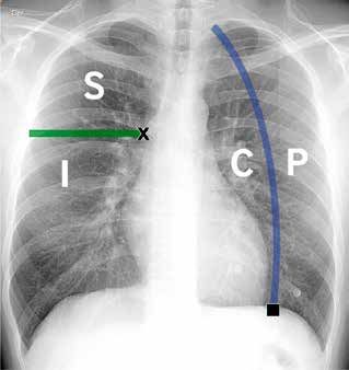
and those in emergency services without COVID-19. divide both lung volumes into central and peripheral
Other thromboembolic events such as renal vein regions and into upper and lower halves. For the
thrombosis, cerebral infarcts and limb ischemia have first division, our group uses an oblique line parallel
also been reported17. to the external pleural border, from the midpoint of
the hemidiaphragm towards the cephalic, and for the
Chest X-ray second division a horizontal line from the superior
The American College of Radiology considers margin of the pulmonary hila (Figure 5).
chest radiography as a recommended study in its To define zonal predominance in this system, we
criteria of relevance for the indication of examinations use as a rule a lung involvement greater than 50%
in patients with acute respiratory disease18. It has com- of a location in the case of a single opacity, or more
parative advantages compared to CT in the context than 50% of a location with respect to the total lung
of the COVID-19 pandemic, which include its easy involvement, in the case of more than one opacity
accessibility, lower radiation dose and the possibility present.
of carrying out a portable examination, reducing the The 4 radiological patterns of the BSTI system
probability of contagion from health personnel. correspond to:
A) Classic / Probable COVID-19: Multiple bilateral
Radiological findings pulmonary opacities, either foci of condensation
The findings of COVID-19 infection in CXR des- and/or ground-glass, with a predominance of lower
cribed to date reflect the same findings described for and peripheral areas. Also included are multiple
CT. In a study of 64 patients with confirmed disease19, bilateral opacities that present peripheral zone
initial radiographs showed both foci of condensation predominance, but equivalent between upper
(59%) and GGO (41%), which were distributed bila- and lower halves (without lower predominance)
terally (63%), basally (63%), and peripherally (51%). (Figure 6A and B).
A B
Figure 4: Frequent radiological alterations in patients with COVID-19 infection. The alterations in chest radiography are
similar to those described in computed tomography, the most frequent alterations being the presence of bilateral dense
ground-glass opacities (arrowheads) that adopt a peripheral distribution. Foci of condensation of similar distribution can
also be observed. Infrequent findings correspond to pleural effusion, pulmonary nodules and adenopathies.
94Rev Chil Radiol 2020; 26(3): 88-99. ARTÍCULO DE REVISIÓN
Table 3. BSTI classification of structured report for COVID-19 in chest radiography 20. Abbreviations:
RT-PCR = reverse transcriptase polymerase chain reaction. GGO = dense ground-glass opacities.
Pattern Findings
Classic / Probable COVID-19 Foci of condensation and/or multiple, bilateral, predominantly
peripheral and inferior GGO.
Foci of condensation and/or multiple, bilateral, predominantly
peripheral GGO, with no inferior or superior dominance.
Indeterminate for COVID-19 Alterations that do not meet the criteria of a classic pattern or a
Non-COVID-19 pattern.
Non-COVID-19 Alterations suggestive of another pathology with greater probability to
COVID-19:
Single lobar or segmental pneumonia
Pleural effusion
Interstitial edema
Pneumothorax
Others: masses, lobar atelectasis, pulmonary fibrosis.
B) Indeterminate for COVID-19: Alterations present,
but that do not meet the criteria for a classic pat-
tern or for a Non-COVID-19 pattern. Examples:
Multiple opacities of diffuse distribution, without
zonal predominance; Multiple, peripheral, and
predominantly upper foci of condensation; Mul-
tiple unilateral and predominantly peripheral foci
of condensation (Figure 6C).
C) Non-COVID-19: Alterations present, but su-
ggestive of another pathology. Examples: Single
focus of condensation, reticular interstitial pattern
(interstitial edema, lymphangitis carcinomatosa),
bronchiolitis type reticular pattern, pulmonary
fibrosis, pulmonary emphysema, lung nodules /
masses, lobar atelectasis, pneumothorax, pleural
effusion, cardiomegaly (Figures 6D and E).
D) Normal: Exam without radiological findings or not
correlated with clinical symptoms (example: lung
scars, calcified granulomas, linear atelectasis). As
in CT, a chest X-ray without pathological findings
does not rule out the possibility of COVID-19 infec-
X : Superior margin of the pulmonary hila. tion, so we suggest concluding with the following
█ : Midpoint of the hemidiaphragm statement: Exam without radiological findings of
pneumonia. Indispensable correlation with PCR
S : Superior/upper - I : Inferior/lower test (Figure 6F).
C : Central - P : Peripheral
Prognostic value
Figure 5: Division lines used by our group for the BSTI Recently, Toussie et. al investigated chest radio-
classification system. To divide the upper/superior (S) and graphy as a method to predict clinical outcomes21
lower/inferior (I) halves, a horizontal line is drawn from the in patients with COVID-19 infection. To do this, they
upper margin of the pulmonary hila, and to divide the central used data from patients between 21 and 50 years
(C) and peripheral (P) regions an oblique line, parallel to
old, dividing each lung into 3 zones (upper, middle
the external pleural border, drawn from the midpoint of the
hemidiaphragm towards the cephalic. and lower) and assigning a total score according to
95Rev Chil Radiol 2020; 26(3): 88-99.
the presence (1) or absence (0) of opacities. With a mentary tool, especially in a clinical setting with
cut-off score of 2 or more, it achieved a sensitivity little availability of other imaging resources. There
(S) and specificity (E) of 66% and 79% respectively is also the need to clean the equipment properly
to predict hospitalization, while, for those hospita- to avoid the spread of the infection.
lized patients, the commitment of 3 or more zones
achieved an S and E of 68% and 67% respectively Local experience in the Radiology Service and
to predict intubation. For other outcomes, such as Department of the UC-Christus Health Network
prolonged stay and sepsis, a statistically significant (Red de Salud UC-Christus)
number could not be identified. This could place Since the declaration of phase 4 of the pandemic
CXR as an independent prognostic indicator in in Chile (March 16)23, our Service / Department has
patients with COVID-19. been taking measures aimed at reducing the risk
of contagion by the professionals, technicians and
Ultrasonography administrators who work in its facilities (Table 4) .
The role of ultrasonography in COVID-19 pa- These measures are in line with those adopted by
tients is under development. Among the findings other university radiological centers in the United
described in series, there are abnormalities in pleu- States (University of Washington, New York Uni-
ral lines (100%), B-lines (100%) and consolidation versity, University of Wisconsin, etc.)24, with the
(64%)22. Pleural effusion is seen less frequently. objectives of maintaining a continuous operation
These findings are rather nonspecific and may be during the emergency, supporting patient care
in the context of non-COVID respiratory distress, and maintaining a diagnostic and interventional
so their usefulness would be rather as a comple- radiological support.
A B C
D E F
Figure 6: Examples of chest radiographs for each pattern of the BSTI structured reporting system in COVID-19. Classic
/ probable pattern (A-B): Patient 1 (A) Bilateral dense ground-glass opacities of peripheral and inferior distribution (black
arrowheads). Patient 2 (B): Bilateral ground-glass opacities (black arrowheads) associated with multiple foci of condensation
in the distribution described (white arrowheads) Indeterminate pattern (C): Diffuse distribution of dense ground-glass opacities,
without lower or peripheral zonal predominance. Non-COVID-19 (D-E) pattern: Patient (D) with a unifocal, retrocardiac
focus of condensation, consistent with bacterial pneumonia. Patient (E) with signs of diffuse bilateral interstitial and alveolar
edema, associated with bilateral pleural effusion, consistent with decompensated heart failure. Normal pattern (F): Exam
without radiological findings suggestive of pneumonia in a patient with COVID-19 confirmed by PCR test. It is relevant to
mention that this pattern does not rule out the presence of disease.
96Rev Chil Radiol 2020; 26(3): 88-99. ARTÍCULO DE REVISIÓN
Table 4. Measures adopted by the Radiology Service and Department of the UC-Christus Health Network (Red
de Salud UC-Christus) during the COVID-19 pandemic period.
Field / Area Measures
Social distancing Restricted number of staff / residents per report room.
Individual and non-shared use of report stations, with frequent cleaning
before and after use.
Reduction in the number of people in reporting areas, restricting to only essential
personnel, staff / residents: suspension of rotations of interns and visiting
residents; radiological and MT consultations by telephone only; PACS upload
of previous exams only via email.
Rotations “on / off” of staff and residents.
Support to implement workstations at home.
Shift deliveries, classes and seminars of the residency program transformed into
virtual format: use of Canvas and Zoom platforms.
Virtual Inter-departmental clinical meetings.
Contact management / Local management of personal protection items (PPE) stock.
high risk interactions Access to PPE online course.
Patients transferred to the radiology service with use of a face mask.
Attention of patients with PPE by staff / residents.
Cleaning protocols for ultrasound equipment after each use.
Upon resuming outpatient care: prior triage by support personnel identifying
respiratory or infectious symptoms.
Diagnosis of possible COVID-19 in images: notification as a diagnostic alert.
Retrospective alert: review in PACS of the list of examinations of a patient at
the time of being diagnosed as COVID-19 in chest x-ray or CT, with the aim
of identifying imaging studies performed in the 2 weeks prior to diagnosis
that may have meant contact with staff / residents (e.g.: ultrasound, imaging
biopsy, etc.) and thus notify those involved.
Immediate notification system of respiratory symptoms by staff / residents
for taking RT-PCR, identification of contacts and preventive isolation.
Development and review of Guidelines for the appropriate use of images for COVID-19 in emergency services.
information related to UC-Christus Health Network Integrated training sessions on COVID-19 (broadcast
COVID-19 for its on YouTube).
circulation in the medical Weekly intra-departmental teaching sessions on COVID-19 updates.
community Creation of a Research group on COVID-19 to look at the teaching and
research mission of our institution. Lines of work:
- Chest X-ray and structured report for COVID-19.
- Inter-observer variability in CXR for diagnosis of COVID-19.
- Signs of response to treatment with convalescent plasma in CT.
- Development of Artificial Intelligence algorithms in medical images associated
with COVID-19.
Conclusion the way of teaching and the lines of research
The current SARS-CoV-2 pandemic has led to the new scenario. A global example of these
to reorganizing the way in which academic ra- changes are the consensus proposed by the main
diology services and departments carry out their radiological societies to define correct indica-
work, not only contributing to the diagnosis and tions for imaging in different clinical scenarios
management of these cases, but also promoting of the pandemic and the standardization of the
a safe environment for other patients and the structure of CT and CXR reports for patients with
radiological work group, as well as restructuring suspected COVID-19.
97Rev Chil Radiol 2020; 26(3): 88-99.
Acknowledgment 10. De Jaegere TMH, Krdzalic J, Fasen BACM, Kwee
The authors wish to thank Dr. Rodrigo San RM. Radiological Society of North America Chest CT
Martín Bachmann for his collaboration in the Classification System for Reporting COVID-19 Pneu-
preparation of the document. monia: Interobserver Variability and Correlation with
RT-PCR. Radiol Cardiothorac Imaging. 2020 Jun 1;
2(3): e200213. Disponible en: https://doi.org/10.1148/
Bibliography ryct.2020200213
1. WHO. Director-General’s opening remarks at the 11. Prokop M, van Everdingen W, van Rees Vellinga
media briefing on COVID-19-11 March. (Fecha de T, Quarles van Ufford J, Stöger L, Beenen L, et al.
citación 14 de junio 2020). Disponible en: https://www. CO-RADS-A categorical CT assessment scheme
who.int/dg/speeches/detail/who-director-general- for patients with suspected COVID-19: definition and
s-opening-remarks-at-the-media-briefing-on-covid- evaluation. Radiology. 2020 Apr 27; 201473. Disponible
19---11-march-2020. en: https://doi.org/10.1148/radiol.2020201473
2. ACR Recommendations for the use of Chest Radio- 12. Tabatabaei SMH, Talari H, Moghaddas F, Rajebi H.
graphy and Computed Tomography (CT) for Sus- Computed Tomographic Features and Short-term
pected COVID-19 Infection. (Fecha de citación: 14 Prognosis of Coronavirus Disease 2019 (COVID-19)
de junio 2020). Disponible en: https://www.acr.org/ Pneumonia: A Single-Center Study from Kashan,
Advocacy-and-Economics/ACR-Position-Statements/ Iran. Radiol Cardiothorac Imaging. 2020 Apr 1; 2(2):
Recommendations-for-Chest-Radiography-and-CT- e200130. Disponible en: https://doi.org/10.1148/
for-Suspected-COVID19-Infection. ryct.2020200130
3. Rubin GD, Ryerson CJ, Haramati LB, Sverzellati N, 13. Yu M, Xu D, Lan L, Tu M, Liao R, Cai S, et al. Thin-
Kanne JP, Raoof S, et al. The Role of Chest Imaging section Chest CT Imaging of Coronavirus Disease
in Patient Management during the COVID-19 Pande- 2019 Pneumonia: Comparison Between Patients
mic: A Multinational Consensus Statement from the with Mild and Severe Disease. Radiol Cardiothorac
Fleischner Society. Radiology. 2020 Apr 7; 296(1): Imaging. 2020 Apr 1; 2(2): e200126. Disponible en:
172-180. Disponible en: https://doi.org/10.1148/ra- https://doi.org/10.1148/ryct.2020200126
diol.2020201365 14. Yang R, Li X, Liu H, Zhen Y, Zhang X, Xiong Q, et
4. Kim H, Hong H, Yoon SH. Diagnostic Performance al. Chest CT Severity Score: An Imaging Tool for
of CT and Reverse Transcriptase-Polymerase Chain Assessing Severe COVID-19. Radiol Cardiothorac
Reaction for Coronavirus Disease 2019: A Meta- Imaging. 2020 Mar 30; 2(2): e200047. Disponible en:
Analysis. Radiology. 2020 Apr 17;201343. Disponible https://doi.org/10.1148/ryct.2020200047
en: https://doi.org/10.1148/radiol.2020201343 15. Leonard-Lorant I, Delabranche X, Severac F, Helms
5. Wen Z, Chi Y, Zhang L, Liu H, Du K, Li Z, et al. Coro- J, Pauzet C, Collange O, et al. Acute Pulmonary
navirus Disease 2019: Initial Detection on Chest CT Embolism in COVID-19 Patients on CT Angiography
in a Retrospective Multicenter Study of 103 Chinese and Relationship to D-Dimer Levels. Radiology. 2020
Subjects. Radiol Cardiothorac Imaging. 2020 Apr 1; Apr 23; 201561. Disponible en: https://doi.org/10.1148/
2(2): e200092. Disponible en: https://doi.org/10.1148/ radiol.2020201561
ryct.2020200092 16. Oudkerk M, Büller HR, Kuijpers D, van Es N, Ou-
6. Caruso D, Zerunian M, Polici M, Pucciarelli F, Polidori dkerk SF, McLoud TC, et al. Diagnosis, Prevention,
T, Rucci C, et al. Chest CT Features of COVID-19 in and Treatment of Thromboembolic Complications in
Rome, Italy. Radiology. 2020 Apr 3; 201237. Disponible COVID-19: Report of the National Institute for Public
en: https://doi.org/10.1148/radiol.2020201237 Health of the Netherlands. Radiology. 2020 Apr
7. Wang Y, Dong C, Hu Y, Li C, Ren Q, Zhang X, et al. 23; 201629. Disponible en: https://doi.org/10.1148/
Temporal Changes of CT Findings in 90 Patients with radiol.2020201629
COVID-19 Pneumonia: A Longitudinal Study. Radiolo- 17. Lushina N, Kuo JS, Shaikh HA. Pulmonary, Cerebral,
gy. 2020 Mar 19; 200843. Disponible en: https://doi. and Renal Thromboembolic Disease Associated with
org/10.1148/radiol.2020200843 COVID-19 Infection. Radiology. 2020 Apr 23; 201623.
8. Simpson S, Kay FU, Abbara S, Bhalla S, Chung JH, Disponible en: https://doi.org/10.1148/radiol.2020201623
Chung M, et al. Radiological Society of North America 18. Kirsch J, Ramirez J, Mohammed T-LH, Amorosa JK,
Expert Consensus Statement on Reporting Chest Brown K, Dyer DS, et al. ACR Appropriateness Cri-
CT Findings Related to COVID-19. Endorsed by the teria Acute Respiratory Illness in Immunocompetent
Society of Thoracic Radiology, the American College Patients. J Thorac Imaging. 2011; 26(2): W42-W44.
of Radiology, and RSNA. Radiol Cardiothorac Imaging. 19. Wong HYF, Lam HYS, Fong AH-T, Leung ST, Chin
2020 Mar 25; 2(2): e200152. Disponible en: https:// TW-Y, Lo CSY, et al. Frequency and Distribution of
doi.org/10.1148/ryct.2020200152 Chest Radiographic Findings in COVID-19 Positive
9. Capítulo de Tórax toma como modelo propuesta Patients. Radiology. 2020 Mar 27; 201160. Disponible
elaborada por la RSNA, SRT y la ACR en informes en: https://doi.org/10.1148/radiol.2020201160
Covid19, para sugerir formato de informe. (Fecha de 20. COVID-19 BSTI Reporting templates. (Fecha de
citación 14 de junio 2020). Disponible en: https://www. citación 14 de junio 2020). Disponible en: https://
sochradi.cl/2020/05/noticias/capitulo-de-torax-toma- www.bsti.org.uk/covid-19-resources/covid-19-bsti-
como-modelo-propuesta-elaborada-por-la-rsna-str-y- reporting-templates.
la-acr-en-informes-covid19-para-sugerir-formato-de- 21. Toussie D, Voutsinas N, Finkelstein M, Cedillo MA,
informe. Manna S, Maron SZ, et al. Clinical and Chest Ra-
98Rev Chil Radiol 2020; 26(3): 88-99. ARTÍCULO DE REVISIÓN
diography Features Determine Patient Outcomes In anuncia cierre de fronteras. (Fecha de citación 14
Young and Middle Age Adults with COVID-19. Radio- de junio 2020). Disponible en: https://www.minsal.
logy. 2020 May 14; 201754. Disponible en: https://doi. cl/coronavirus-en-chile-pasa-a-fase-4-y-presidente-
org/10.1148/radiol.2020201754 anuncia-cierre-de-fronteras.
22. Xing C, Li Q, Du H, Kang W, Lian J, Yuan L. Lung 24. Mossa-Basha M, Meltzer CC, Kim DC, Tuite MJ, Kolli
ultrasound findings in patients with COVID-19 pneu- KP, Tan BS. Radiology Department Preparedness for
monia. Crit Care. 2020; 24(1): 174. Disponible en: COVID-19: Radiology Scientific Expert Panel. Radio-
https://doi.org/10.1186/s13054-020-02876-9 logy. 2020 Mar 16; 200988. Disponible en: https://doi.
23. Coronavirus en Chile pasa a fase 4 y presidente org/10.1148/radiol.2020200988
99You can also read