Does Additional Extracorporeal Shock Wave Therapy Improve the Effect of Isolated Percutaneous Radiofreqency Coblation in Patients with Insertional ...
←
→
Page content transcription
If your browser does not render page correctly, please read the page content below
Does Additional Extracorporeal Shock Wave Therapy
Improve the Effect of Isolated Percutaneous
Radiofreqency Coblation in Patients with Insertional
Achilles Tendinopathy? Study Protocol for A
Randomized Controlled Clinical Trial
Yu-Jie Song
Huashan Hospital Fudan University Department of Sports Medicine
Wen-Kai Xuan
Huashan Hospital Fudan University Department of Sports Medicine
Ying-Hui Hua ( hua_cosm@aliyun.com )
Huashan Hospital Fudan University Department of Sports Medicine https://orcid.org/0000-0002-0767-
1267
Study protocol
Keywords: Achilles tendinopathy, percutaneous radiofreqency coblation, extracorporeal shockwave
therapy, randomized controlled trial, protocol
DOI: https://doi.org/10.21203/rs.3.rs-35074/v1
License: This work is licensed under a Creative Commons Attribution 4.0 International License.
Read Full License
Page 1/16Abstract
Background: No conclusive evidence recommends a prior treatment for insertional Achilles tendinopathy
(IAT). It is theorized that both percutaneous radiofreqency coblation and extracorporeal shockwave
therapy (ESWT) relieve pain within the insertion. However, no clinical evidence shows that either
treatment promote the regeneration of the tendon or if the combination of these 2 interventions offer
better function and less pain than one therapy.
Methods: The study is a randomized, controlled trial with patients allocated in a 1:1 ratio to one of two
parallel groups. Patients with insertional Achilles tendinopathy who are not satis ed with the effect of
conservative treatment will be screened. A minimum of 38 patients will be enrolled after deciding to
participate in the trial on an informed basis. Then the intervention group and the control group perform
radial ESWT and sham-ESWT respectively at 6 months after percutaneous radiofreqency coblation. The
primary outcome will be the Victorian Institute of Sports Assessment Achilles (VISA-A) Score. Secondary
outcome measures will be Foot and Ankle Outcome Score (FAOS) scale, Visual Analogue Scale (VAS),
Tegner Score, and MRI ultra-short echo time (UTE) T2* value. The assessments will occur in 6 months, 1
year, and 2 years post-operatively. The differences between 2 groups will be conducted as intention-to-
treat basis.
Discussion: We aim to investigate if radiofreqency coblation associated with ESWT can provide more
encouraging imaging ndings as well as functional and clinical outcomes regarding the treatment of the
IAT comparing to the single radiofreqency coblation treatment.
Trial registration: ChiCTR1800017898; Pre-results. Registered on 20 August 2018.
Background
Achilles tendinopathy is a common disorder in sports and daily life. This injury is frequently encountered
in middle- and long- distance runners with up to 36% incidence rate1–3 and may cause up to 5% of
professional athletes to a premature career end.4 5 Achilles tendinopathy is considered as a multifactorial
condition whose exact etiology remains to be determined and can be divided into insertional and non-
insertional tendinopathy according to its anatomical location. Insertional Achilles tendinopathy (IAT) is
responsible for roughly 20–24% of all Achilles disorders.6 Patients with IAT complain about
compromised function and gradual onset of pain at the calcaneal attachment of the Achilles tendon and
could be aggravated by standing, walking and jogging.
The initial treatment is conservative management, such as eccentric exercises,7 8 anti-in ammatory drugs
(NSAIDs),9 extracorporeal shockwave therapy,10–12 platelet-rich plasma (PRP) therapy,13 14 et al. If
nonoperative treatment fails, surgery might be taken into consideration. Most surgical procedures involve
debridement and decompression of Achilles tendon insertion and augmentation in case of excessive
tendon loss.15 To date, there is no gold standard treatment for IAT. Scienti c evidence shows variable and
Page 2/16inconsistent effect among different treatments,16 17 meaning to provide a most effective management
with high quality and reliable evidence is urgently needed.
It is reported that 25 to 50% of patients with chronic tendinopathy may undergo surgery .18–20
Percutaneous radiofreqency coblation as a minimal invasive treatment has been used in the
management of lateral epicondylitis,21 22 rotator cuff tendinopathy,23 and foot and ankle tendinosis.24 It
would reduce surgical time and facilitate an earlier return to activity. The procedure has been shown to
induce degeneration of sensory nerve bers and improves vascularity,25 26 which, in its turn, relieves the
pain and regenerates the tendon tissue, respectively. Extracorporeal shockwave therapy which is widely
used in Achilles tendinopathy also shares the similar mechanism theoretically.25 However, no clinical
evidence shows those two treatments promote the regeneration and quality of the tendon.
Grosse et al.,27 Gardin et al28,as well as Juras et al29 reported that there was statistically signi cance in
UTE-T2* value between Magnetic Resonance Imaging (MRI) images of patients with IAT and normal
population, which indicates that MRI UTE-T2* can be applied in clinical quantitative assessment to
monitor the morphological information of the tendon and produce reliable results. In addition, the
correlation between MRI UTE-T2* and clinical score can be determined.
Our aim is to evaluate the effectiveness of percutaneous radiofreqency coblation associated with an
ESWT protocol, and compare it to isolated radiofreqency coblation, via VISA-A score30 primarily and MRI
UTE-T2* secondarily. It is hypothesized that adjunctive ESWT will promote tendon regeneration, mitigate
pain, and improve function as compared to a controlled group.
Methods/design
The protocol will be reported in accordance with the guidelines and checklists for Standard Protocol
Items: Recommendations for Interventional Trials (SPIRIT).
Aim, design and setting
We aim to investigate if radiofreqency coblation associated with ESWT can provide more encouraging
outcomes regarding the treatment of the IAT with trustworthy evidence.
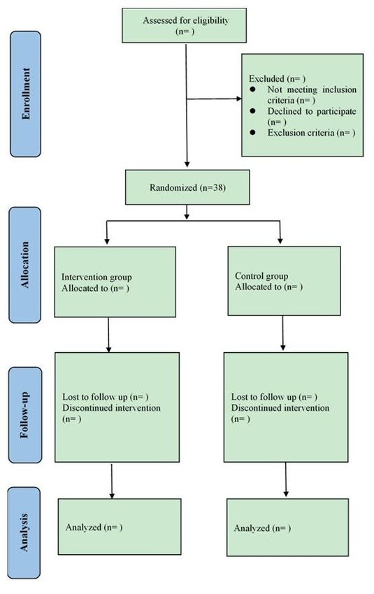
This study is a randomized, parallel-group clinical trial (Fig. 1). The 1:1 allocation to either receive radial
ESWT or sham-ESWT, after surgery is conducted using computer-generated random allocation sequences
and sealed envelopes. The patients of placebo group will also experience ESWT’s procedure, while the
apparatus’ therapeutic head will be placed a support, which will block the propagation of shock wave
without changing equipment’s appearance, noise and using process. Furthermore, the patients will feel
the vibration of the machine, but the insertional region won’t be directly stimulated. The physiotherapists
will not take any part in the posttreatment assessment of the patients. The outcome data will be collected
Page 3/16by research fellows who are blinded to the treatment procedures to reduce the risk of assessment bias.
The SPIRIT Figure shows in detail the schedule of enrollment and assessments (Fig. 2).
Characteristics of participants
Patients referred to Department of Sports Medicine, Huashan Hospital, Fudan University with insertional
Achilles tendinopathy who had no symptom relief after 3 to 6 months of conservative therapy31 other
than ESWT are assessed for eligibility. The diagnosis is based on a medical history and clinical
examination including persistent pain at the calcaneus tendon insertion region, and ultrasound or MRI
results con rm it.
Inclusion criteria
The inclusion criteria are as follows:
(1) Medical history, clinical examination and ultrasound or MRI results conform to the diagnosis of IAT.
(2) Age 18–65 years, both genders.
(3) The patient has decided to accept percutaneous radiofreqency coblation and radial ESWT.
(4) The patient must be expected to be able to attend post-operation examinations.
(5) The patient must be able to give informed consent.
Exclusion criteria
The exclusion criteria are as follows:
(1) Previous surgery on the affected limb.
(2) The patient would not be able to strictly follow trial procedures or complete questionnaires.
(3) Non-insertional or mixed tendinopathy (insertional and non-insertional).
(4) Pregnancy or intention to be pregnant.
(5) Currently undergoing follow-up and treatment in a psychiatry department or by other mental health
professionals.
Withdrawals
Participants may decline to continue from the trial at any time without prejudice and still encouraged to
keep in touch and welcomed to inquiry and asking for necessary help.
Other reasons for discontinuation include:
(1) The patient develops Achilles tendon rupture.
Page 4/16(2) The patient develops complex or increasing regional pain.
(3) Other unexpected and unpredictable circumstances when the research team judges that the risk of
continuing the protocol treatment is greater than the bene t.
Sample Size
The primary outcome for this study is the VISA-A score. McCormack J et al.32 has identi ed the minimum
clinically important difference (MCID) of 6.5 points for the VISA-A. The study of Wei M et al.33 indicates
an expected standard deviation (SD) of 6.9 points. We have referred to the sample size calculation
method of Kvalvaag, E et al.34 To detect a difference in results between the radical ESWT and the sham-
ESWT participants at a level of 5% signi cance and the most likely scenario for 80% power, 15 patients
per group will be needed. Assuming a margin of 20% loss of primary outcome data, we propose to recruit
a minimum of 38 patients in total.
Procedures
A patient is considered to be included in the protocol if he/she meets all of the inclusion criteria and none
of the exclusion criteria. Then the patient will be verbally informed of the study and given the opportunity
to decide whether he/she wants to participate in the trial on an informed basis without any pressure.
Then a written informed consent will be obtained from each participant. Once included, the VISA-A Score,
MRI UTE T2* value, FAOS Score, VAS Score, and Tenger Score will be assessed. Within one week after
being enrolled, the percutaneous radiofreqency coblation will be performed by an experienced surgeon.
After the surgery, each patient will randomly take an unmarked, opaque, sealed envelope containing a
piece of paper with a unique number from 1 to 38. Then the numbers will be divided into two sets via
computing software (https://www.randomizer.org/). All patients respectively entered the intervention
group and the placebo group according to their own numbers. Patients of intervention group will receive
ESWT at 6 months post-operatively. At the same time, the patients of placebo group will also experience
sham ESWT’s procedure. Either the radial ESWT or sham ESWT will be administered 4 times at weekly
intervals. In all, the treatment protocols for the two groups are similar except for the intervention: radial
ESWT. All the outcome measurement will be assessed again at 6 months, 1 year, and 2 year after the
surgical procedure.
Percutaneous radiofreqency coblation
The symptomatic region will be identi ed and marked before the anesthesia. Then, the patient would
accept general anesthetic and be placed in a prone position. A grid with 5 mm interval will be mapped out
over the marked site.35 A 1.5 mm Kirschner wire will be used to puncture the skin and make eyelet-sized
holes. The radiofrequency probe (Topaz, ArthroCare, Sunnyvale, California) will be inserted and
subsequently activated for 1 second with minimum power, hole by hole. If there exists other concomitant
injury such as Haglund deformity, it will be recorded and addressed by endoscopic surgery at the same
time. After completion of the procedure, a standard sterile dressing would be applied to minimize tension
across the wounds.
Page 5/16Rehabilitation
Crutch will be recommended during the rst 4 weeks after surgery. No immobilization is needed. In
addition, all the patients will be encouraged to begin range of motion and isometric strength exercises
immediately after surgery, and eccentric training will begin 6 weeks after surgery.36 At 4-week follow-up,
the patients will be advised to wear regular shoe gear and continue the exercises. Patients shall be able to
walk normally without any assistive device since the seventh week.
Extracorporeal shockwave therapy
Patient will be lying on the stretcher in the prone position with barefoot, and the feet will be positioned
towards the shock wave apparatus. The symptomatic area will be marked, then ultrasound gel will be
applied on this region which the radial shock waves with a Dolorclast equipment (EMS Electro Medical
Systems, Nyon, Switzerland), the intensity being 2000 pulses, 6–8 Hz of frequency, and 1.5–2.5 Bar of
pressure per application. Compared with those of radial ESWT group, patients of sham ESWT group will
go through the same process with the same equipment, in which the apparatus’ therapeutic head will be
installed a support. The support will block the propagation of shock wave without changing equipment’s
appearance, noise and using process. Furthermore, the patients will feel the vibration of the machine, but
the insertional region won’t be directly stimulated. Either the radial ESWT or sham ESWT will be
administered 4 times at weekly intervals.
MRI
Patients will be positioned feet rst-supine and both feet are placed simultaneously in the dorsal part of
the head coil then will be examined in a 3.0 T horizontal magnet (Discovery MR750, GE Medical System,
Milwaukee, WI). For morphological evaluation of the Achilles tendon, three-dimensional fat-saturated
proton-density-weighted turbo-spin echo (PDTSE) will be used. Other parameters were set as follows:
repetition time (TR) = 202.1 ms, eld of view (FOV) = 140 mm × 140 mm,slice thickness (ST) = 2 mm. Four
echo time images (TE = 0.032, 7.5, 20.5, and 28 ms) will be obtained. T2* value of Achilles tendon
insertion region is calculated by tting the acquired signal at different echo time to a single exponential
decay model.
Primary outcome
Victorian Institute of Sports Assessment Achilles (VISA-A) Score
The VISA-A score is a validated questionnaire for Achilles tendinopathy and contains 8 questions
assessing physical activity and pain (0, no activity/maximum pain; 100, maximum activity/no pain).34
And it is adapted to the local language (Attachment 1). The Chinese version has been used in clinical
trials37 38, proving its validity.
Secondary outcome
(1) MRI: UTE-T2* value
Page 6/16The MRI UTE-T2* has been reported to show the signi cant differences between Achilles tendinopathy
and normal tendon.27–29 Thus, the morphological information of tendon after treatment could be
monitored.
(2) Foot and Ankle Outcome Score (FAOS) scale
(3) Visual Analogue Scale (VAS)
(4) Tegner Score
(5) Complications
Data collection
All relevant data will be collected by research fellows who are blinded to the treatment procedures and
recorded in specially designed case report les. The patients will be identi ed by an assigned number. At
the completion of the study, all identi able data will be destroyed. The patients are informed, both
verbally and in writing, that data are stored and analyzed in a computer, that the patient ’s anonymity is
preserved, and that the data protection legislation is adhered to.
Adverse event management
Achilles tendon rupture is presumably a complication of both the percutaneous radiofreqency coblation
and the ESWT treatment, therefore the number of Achilles tendon ruptures will be registered. Furthermore,
an adverse event is de ned as any unintended, unfavorable nding, symptom, or disease that occurs,
whether it is considered to be related to the study or not. Adverse events will be recorded. Serious adverse
events (SAEs) is de ned as an event or reaction which will cause death, life-threatening situations,
hospitalization or prolongation of existing hospitalization, or permanent or severe disability. SAEs will be
reported to the ethics committee within 24 hours.
End of trial
The end of the trial will be de ned as the collection of 2 years outcome data from the last participant.
Statistical analysis
All continuous data from primary and secondary outcome measurements will be summarized by
descriptive statistics with the number of observations (n), mean, SD, minimum, median and maximum.
Uncontinuous data such as co-morbidities concomitant injuries will be summarized by descriptive
statistics with the number of observations, calculating the incidence separately and analyze whether it is
associated with prognosis. The primary point for analyses of e cacy will be 6 months after surgery, then
1 year and 2 years. A statistical software program (SPSS for Windows, version 23.0; SPSS Inc, Chicago,
Illinois) will be used for statistical analysis in this study. Group comparison will be statistically analyzed
by either the Student’s t test or the Mann–Whitney U test, depending on whether the data is normally
distributed. The signi cance level will be set at a p value < 0.05.
Page 7/16Patient and Public Involvement
Neither patients nor the public were involved in the design of the study, including conceiving the research
questions and deciding the outcomes. However, all participants will be asked whether they would like to
receive a copy of the trial results at the time of consent. Once completed the trial, a copy of the personal
results will be emailed to participants who said yes. Also, the burden of the intervention will be assessed
by participants themselves using a questionnaire39 40.
Discussion
Nonoperative management is the rst line of treatment for IAT, though the most effective standard
procedure is controversially debated.41 However, conservative therapy is a time-consuming procedure,
and approximately a quarter to half of all patients do not respond to it.20 42 Thus, as a minimal invasive
operation, percutaneous radiofreqency coblation has been brought into attention. This technique allows
patients to hasten their resolution of pain and shorten their return to activity26 43 though there is
insu cient evidence to prove its clinical e cacy of Achilles tendinopathy, especially when the BMI of the
patient is relatively high.33 On the other hand, isolated ESWT has been reported as an alternative option
for IAT by extensive studies.15 44 Rompe et al.45 has showed ESWT is more bene cial in combination with
eccentric exercises in non-insertional tendinopathy. Then we intend to seek for a combination method for
insertional tendinopathy as well. This study protocol describes the randomized controlled trial directly
comparing the effectiveness of percutaneous radiofreqency coblation associated with ESWT and
isolated percutaneous radiofreqency coblation.
To our best knowledge, most studies used isolated VISA-A score as the primary outcome measurement
for prognosis of Achilles tendinopathy46 47 because it is the validated assessment for this quite frequent
disorder. However, at the same time, there are few radiographic descriptions of the Achilles tendon.
Therefore, we introduce MRI UTE-T2* as our secondary outcome measurement to monitor the status of
Achilles tendon and to see the correlations with clinical scores besides comparison of the effectiveness
on function and pain between 2 interventions, which could be our secondary aim.
In conclusion, we aim to investigate if radiofreqency coblation associated with ESWT can provide more
encouraging imaging ndings, functional and clinical outcomes regarding the treatment of the IAT
comparing to the single radiofreqency coblation treatment.
Trial status
Protocol Amendment Number: 02. Issue Date: 1 Aug 2020. The randomized trial is currently enrolling
participants. Recruitment began in October 2018 and the approximate date when recruitment will be
completed in October 2021. The end of the trial will be de ned as the collection of 2 years outcome data
from the last participant.
Page 8/16Abbreviations
IAT: insertional Achilles tendinopathy
ESWT: extracorporeal shockwave therapy
VISA-A: Victorian Institute of Sports Assessment Achilles
FAOS: Foot and Ankle Outcome Score
VAS: Visual Analogue Scale
UTE: ultra-short echo time
PRP: platelet-rich plasma
MRI: Magnetic Resonance Imaging
MCID: minimum clinically important difference
SD: standard deviation
TR: repetition time
FOV: eld of view
ST: slice thickness
SAEs: Serious adverse events
Declarations
Ethics approval and consent to participate:
The protocol was approved by the institutional review board of Huashan Hospital A liated to Fudan
University on 13 January 2017 (KY2016 - 315). And the project was registered in the Chinese Clinical Trial
Registry on 20 August 2018 (ChiCTR1800017898). Written consent will be obtained from all participants
by the investigator.
Consent for publication:
Not applicable.
Availability of data and materials:
Page 9/16Data sharing is not applicable to this article as no datasets were generated or analysed during the current
study.
Competing interests:
The authors declare that they have no competing interests.
Funding:
This study is supported by Shanghai Hospital Development Center (SHDC12018X26). The study funder
has no role in the study design, data collection and management, and manuscript writing publication.
Authors' contributions:
YJS drafted. WKX substantively revised the manuscript. YHH participated in the design of the study. All
authors read and approved the nal manuscript.
Acknowledgements:
Not applicable.
References
1. Hirschmuller A, Frey V, Deibert P, et al. Achilles Tendon Power Doppler Sonography in 953 Long
Distance Runners - A Cross Sectional Study. Ultraschall in Der Medizin 2010;31(4):387-93. doi:
10.1055/s-0029-1245189
2. Kujala UM, Sarna S, Kaprio J. Cumulative incidence of achilles tendon rupture and tendinopathy in
male former elite athletes. Clinical journal of sport medicine : o cial journal of the Canadian
Academy of Sport Medicine 2005;15(3):133-5.
3. Kvist M. Achilles tendon injuries in athletes. Annales chirurgiae et gynaecologiae 1991;80(2):188-
201.
4. Lysholm J, Wiklander J. Injuries in runners. The American journal of sports medicine 1987;15(2):168-
71. doi: 10.1177/036354658701500213
5. Li HY, Hua YH. Achilles Tendinopathy: Current Concepts about the Basic Science and Clinical
Treatments. BioMed research international 2016;2016:6492597. doi: 10.1155/2016/6492597
6. Caudell GM. Insertional Achilles Tendinopathy. Clinics in podiatric medicine and surgery
2017;34(2):195-205. doi: 10.1016/j.cpm.2016.10.007
7. Kedia M, Williams M, Jain L, et al. The effects of conventional physical therapy and eccentric
strengthening for insertional achilles tendinopathy. International journal of sports physical therapy
2014;9(4):488-97.
8. Rees JD, Wolman RL, Wilson A. Eccentric exercises; why do they work, what are the problems and
how can we improve them? British journal of sports medicine 2009;43(4):242-6. doi:
Page 10/1610.1136/bjsm.2008.052910
9. Maquirriain J, Kokalj A. Management of acute Achilles tendinopathy: effect of etoricoxib on pain
control and leg stiffness. Georgian medical news 2013(222):36-43.
10. Lee JY, Yoon K, Yi Y, et al. Long-Term Outcome and Factors Affecting Prognosis of Extracorporeal
Shockwave Therapy for Chronic Refractory Achilles Tendinopathy. Annals of rehabilitation medicine
2017;41(1):42-50. doi: 10.5535/arm.2017.41.1.42
11. Vulpiani MC, Trischitta D, Trovato P, et al. Extracorporeal shockwave therapy (ESWT) in Achilles
tendinopathy. A long-term follow-up observational study. The Journal of sports medicine and
physical tness 2009;49(2):171-6.
12. Rasmussen S, Christensen M, Mathiesen I, et al. Shockwave therapy for chronic Achilles
tendinopathy: a double-blind, randomized clinical trial of e cacy. Acta orthopaedica 2008;79(2):249-
56. doi: 10.1080/17453670710015058
13. Sadoghi P, Rosso C, Valderrabano V, et al. The role of platelets in the treatment of Achilles tendon
injuries. Journal of orthopaedic research : o cial publication of the Orthopaedic Research Society
2013;31(1):111-8. doi: 10.1002/jor.22199
14. Filardo G, Kon E, Di Matteo B, et al. Platelet-rich plasma injections for the treatment of refractory
Achilles tendinopathy: results at 4 years. Blood transfusion = Trasfusione del sangue
2014;12(4):533-40. doi: 10.2450/2014.0289-13
15. Traina F, Perna F, Ru lli A, et al. Surgical treatment of insertional Achilles tendinopathy: a systematic
review. Journal of biological regulators and homeostatic agents 2016;30(4 Suppl 1):131-38.
16. Kearney R, Costa ML. Insertional achilles tendinopathy management: a systematic review. Foot &
ankle international 2010;31(8):689-94. doi: 10.3113/fai.2010.0689
17. Roche AJ, Calder JD. Achilles tendinopathy: A review of the current concepts of treatment. The bone
& joint journal 2013;95-b(10):1299-307. doi: 10.1302/0301-620x.95b10.3188110.1302/0301-
620x.95b9.33025
18. Lee GP, Ogden JA, Cross GL. Effect of extracorporeal shock waves on calcaneal bone spurs. Foot &
ankle international 2003;24(12):927-30. doi: 10.1177/107110070302401210
19. Alfredson H, Lorentzon R. Chronic Achilles tendinosis: recommendations for treatment and
prevention. Sports medicine (Auckland, NZ) 2000;29(2):135-46. doi: 10.2165/00007256-200029020-
00005
20. Johnston E, Scranton P, Jr., Pfeffer GB. Chronic disorders of the Achilles tendon: results of
conservative and surgical treatments. Foot & ankle international 1997;18(9):570-4. doi:
10.1177/107110079701800907
21. Meknas K, Odden-Miland A, Mercer JB, et al. Radiofrequency microtenotomy: a promising method for
treatment of recalcitrant lateral epicondylitis. The American journal of sports medicine
2008;36(10):1960-5. doi: 10.1177/0363546508318045
22. Tasto JP, Cummings J, Medlock V, et al. Microtenotomy using a radiofrequency probe to treat lateral
epicondylitis. Arthroscopy : the journal of arthroscopic & related surgery : o cial publication of the
Page 11/16Arthroscopy Association of North America and the International Arthroscopy Association
2005;21(7):851-60. doi: 10.1016/j.arthro.2005.03.019
23. Taverna E, Battistella F, Sansone V, et al. Radiofrequency-based plasma microtenotomy compared
with arthroscopic subacromial decompression yields equivalent outcomes for rotator cuff
tendinosis. Arthroscopy : the journal of arthroscopic & related surgery : o cial publication of the
Arthroscopy Association of North America and the International Arthroscopy Association
2007;23(10):1042-51. doi: 10.1016/j.arthro.2007.04.018
24. Yeap EJ, Chong KW, Yeo W, et al. Radiofrequency coblation for chronic foot and ankle tendinosis.
Journal of orthopaedic surgery (Hong Kong) 2009;17(3):325-30. doi: 10.1177/230949900901700317
25. Smith WB, Melton W, Davies J. Midsubstance Tendinopathy, Percutaneous Techniques (Platelet-Rich
Plasma, Extracorporeal Shock Wave Therapy, Prolotherapy, Radiofrequency Ablation). Clinics in
podiatric medicine and surgery 2017;34(2):161-74. doi: 10.1016/j.cpm.2016.10.005
26. Takahashi N, Tasto JP, Ritter M, et al. Pain relief through an antinociceptive effect after
radiofrequency application. The American journal of sports medicine 2007;35(5):805-10. doi:
10.1177/0363546506297085
27. Grosse U, Syha R, Hein T, et al. Diagnostic value of T1 and T2 * relaxation times and off-resonance
saturation effects in the evaluation of Achilles tendinopathy by MRI at 3T. Journal of magnetic
resonance imaging : JMRI 2015;41(4):964-73. doi: 10.1002/jmri.24657
28. Silbernagel KG, Gustavsson A, et al. Evaluation of lower leg function in patients with Achilles
tendinopathy.Knee Surg Sports Traumatol Arthrosc. 2006 Nov;14(11):1207-17. doi: 10.1007/s00167-
006-0150-6
29. Juras V, Zbyn S, Pressl C, et al. Regional variations of T(2)* in healthy and pathologic achilles tendon
in vivo at 7 Tesla: preliminary results. Magnetic resonance in medicine 2012;68(5):1607-13. doi:
10.1002/mrm.24136
30. Robinson JM, Cook JL, Purdam C, et al. The VISA-A questionnaire: a valid and reliable index of the
clinical severity of Achilles tendinopathy. British journal of sports medicine 2001;35(5):335-41.
31. Chinese Society of Sports Medicine, Xu H, Li H, et al. Chinese Consensus on Insertional Achilles
Tendinopathy. Orthop J Sports Med. 2019;7(10):2325967119879052.
doi:10.1177/2325967119879052
32. McCormack J, Underwood F, Slaven E, et al. THE MINIMUM CLINICALLY IMPORTANT DIFFERENCE
ON THE VISA-A AND LEFS FOR PATIENTS WITH INSERTIONAL ACHILLES TENDINOPATHY. Int J
Sports Phys Ther. 2015;10(5):639–644
33. Wei M, Liu Y, Li Z, et al. Comparison of Clinical E cacy Among Endoscopy-Assisted Radio-Frequency
Ablation, Extracorporeal Shockwaves, and Eccentric Exercises in Treatment of Insertional Achilles
Tendinosis. J Am Podiatr Med Assoc. 2017;107(1):11–16. doi:10.7547/14-146
34. Kvalvaag E, Brox JI, Engebretsen KB, et al. Is radial Extracorporeal Shock Wave Therapy (rEWST)
combined with supervised exercises (SE) more effective than sham rESWT and SE in patients with
Page 12/16subacromial shoulder pain? Study protocol for a double-blind randomised, sham-controlled
trial. BMC Musculoskelet Disord. 2015;16:248. doi:10.1186/s12891-015-0712-1
35. Deol P, Philbin T. Bipolar Radiofrequency Microtenotomy for Chronic Achilles Tendinosis. Operative
Techniques in Orthopaedics 2008;18(4):254-58. doi: 10.1053/j.oto.2009.02.004
36. Shibuya N, Thorud JC, Humphers JM, et al. Is percutaneous radiofrequency coblation for treatment
of Achilles tendinosis safe and effective? The Journal of foot and ankle surgery : o cial publication
of the American College of Foot and Ankle Surgeons 2012;51(6):767-71. doi:
10.1053/j.jfas.2012.08.011
37. Song YJ, Chen G, Jia SH, et al. Good outcomes at mid-term following the reconstruction of chronic
Achilles tendon rupture with semitendinosus allograft. Knee Surg Sports Traumatol Arthrosc.
2018;10.1007/s00167-018-5113-1. doi:10.1007/s00167-018-5113-1
38. Zhang S, Li H, Yao W, et al. Therapeutic Response of Extracorporeal Shock Wave Therapy for
Insertional Achilles Tendinopathy Between Sports-Active and Nonsports-Active Patients With 5-Year
Follow-up.Orthop J Sports Med. 2020;8(1):2325967119898118. doi:10.1177/2325967119898118
39. Tran VT, Montori VM, Eton DT, et al. Development and description of measurement properties of an
instrument to assess Treatment Burden among patients with multiple chronic conditions. BMC Med
2012, 10:68.
40. Eton DT, Oliveira DR, Egginton J, et al. Understanding the burden of treatment in patients with
multiple chronic conditions: Evidence from exploratory interviews. Qual Life Res 2010, 19:ab. 1673.
41. Alfredson H, Cook J. A treatment algorithm for managing Achilles tendinopathy: new treatment
options. British journal of sports medicine 2007;41(4):211-6. doi: 10.1136/bjsm.2007.035543
42. Paavola M, Kannus P, Paakkala T, et al. Long-term prognosis of patients with achilles tendinopathy.
An observational 8-year follow-up study. The American journal of sports medicine 2000;28(5):634-42.
43. Ochiai N, Tasto JP, Ohtori S, et al. Nerve regeneration after radiofrequency application. The American
journal of sports medicine 2007;35(11):1940-4. doi: 10.1177/0363546507304175
44. Gerdesmeyer L, Mittermayr R, Fuerst M, et al. Current evidence of extracorporeal shock wave therapy
in chronic Achilles tendinopathy. International journal of surgery (London, England) 2015;24(Pt
B):154-9. doi: 10.1016/j.ijsu.2015.07.718
45. Rompe JD, Furia J, Maffulli N. Eccentric loading versus eccentric loading plus shock-wave treatment
for midportion achilles tendinopathy: a randomized controlled trial. The American journal of sports
medicine 2009;37(3):463-70. doi: 10.1177/0363546508326983
46. Habets B, van Cingel REH, Backx FJG, et al. Alfredson versus Silbernagel exercise therapy in chronic
midportion Achilles tendinopathy: study protocol for a randomized controlled trial. BMC
musculoskeletal disorders 2017;18(1):296. doi: 10.1186/s12891-017-1656-4
47. McCormack JR, Underwood FB, Slaven EJ, et al. Eccentric Exercise Versus Eccentric Exercise and
Soft Tissue Treatment (Astym) in the Management of Insertional Achilles Tendinopathy. Sports
health 2016;8(3):230-37. doi: 10.1177/1941738116631498
Page 13/16Figures
Figure 1
Participant ow diagram.
Page 14/16Figure 2
Standard Protocol Items: Recommendations for Interventional Trials (SPIRIT) Figure.
Supplementary Files
This is a list of supplementary les associated with this preprint. Click to download.
Page 15/16SPIRITChecklist.doc
Page 16/16You can also read