Intra abdominal resection of the umbilical vein and urachus of bovine fetuses using laparoscopy and celiotomy: surgical time and feasibility ...
←
→
Page content transcription
If your browser does not render page correctly, please read the page content below
www.nature.com/scientificreports
OPEN Intra‑abdominal resection
of the umbilical vein and urachus
of bovine fetuses using laparoscopy
and celiotomy: surgical time
and feasibility (cadaveric study)
Francisco Décio de Oliveira Monteiro1*, Heytor Jales Gurgel1,4, Simon Silva de Sousa1,4,
João Pedro Monteiro Barroso1,4, Gabrielle Patrizi Braga Vasconcelos1,4,
Daniele Lira dos Santos1,4, Luiz Henrique Vilela Araújo1,4, Loise Araújo de Sousa1,4,
Gabriela Melo Alves dos Santos1,4, Kayan da Cunha Rossy1,4, Verena Siqueira da Silva1,4,
Camila do Espirito Santo Fernandes1,4, Barbara da Conceição Guilherme1,4,
Helaine Freitas Miranda1,4, Carla Rozilene Guimarães Silva1,4,
Rodrigo dos Santos Albuquerque1,4, Luisa Pucci Bueno Borges1,4, Gilson Ferreira de Araújo2,4,
Renata Sitta Mariano Landers3,4 & Pedro Paulo Maia Teixeira1,4
Surgical intervention for umbilical diseases in calves, when indicated, is a complementary and
indispensable therapeutic resource for the treatment of umbilical conditions and is commonly
performed using celiotomy. However, laparoscopy has demonstrated feasibility in many diagnostic
and therapeutic procedures. The aim of this study was to assess the feasibility of the techniques and
the surgical time of laparoscopy and celiotomy used in intra-abdominal resection of the umbilical
vein and urachus of bovine fetuses (cadavers). Resection of the umbilical vein and urachus using
laparoscopy and celiotomy was performed in 26 anatomical specimens (bovine fetuses obtained
from an official slaughterhouse). Resection of umbilical structures was feasible with both techniques,
but shorter surgical time and minimal tissue damage were achieved using laparoscopy. Laparoscopy
requires specialized training and appropriate instruments and is an important tool for diagnostic and
therapeutic exploration of the umbilical structures, liver, bladder, and associated/adjacent structures.
Umbilical disorders are often diagnosed in newborn bovine calves, and their etiologies may be associated with
infections of the umbilical vessels and urachus, persistent urachus, and umbilical h ernias1. In many cases, drug
therapy alone is not sufficient to treat these conditions, and surgical intervention is a complementary and indis-
pensable therapeutic resource for effective management of these d isorders2–4.
For the satisfactory and effective treatment of umbilical infections, reduction of bacterial load and removal of
infected structures likely to have low antimicrobial penetration are indispensable as they significantly improve
the prognosis of these d iseases5. Surgical resection of umbilical structures is indicated for the treatment of calves
with infected umbilical components and patent urachus and is usually performed using c eliotomy6,7.
Surgical resection of umbilical components using celiotomy has been shown to be effective for the treatment
of omphalitis in calves, and it is easily performed by the surgeon using conventional surgical instruments, unlike
1
Institute of Veterinary Medicine, Federal University of Pará (UFPA), Castanhal Campus, Castanhal, Pará,
Brazil. 2Agency of Agricultural Defense of Pará (ADEPARA), Belém, Pará, Brazil. 3Physiology of Reproduction,
Texas A&M University Departmente of Animal Science, College Station, USA. 4These authors contributed equally:
Heytor Jales Gurgel, Simon Silva de Sousa, João Pedro Monteiro Barroso, Gabrielle Patrizi Braga Vasconcelos,
Daniele Lira dos Santos, Luiz Henrique Vilela Araújo, Loise Araújo de Sousa, Gabriela Melo Alves dos Santos,
Kayan da Cunha Rossy, Verena Siqueira da Silva, Camila do Espirito Santo Fernandes, Barbara da Conceição
Guilherme, Helaine Freitas Miranda, Carla Rozilene Guimarães Silva, Rodrigo dos Santos Albuquerque, Luisa Pucci
Bueno Borges, Gilson Ferreira de Araújo, Renata Sitta Mariano Landers and Pedro Paulo Maia Teixeira. *email:
deciomonteiro@ifto.edu.br
Scientific Reports | (2021) 11:5328 | https://doi.org/10.1038/s41598-021-84621-y 1
Vol.:(0123456789)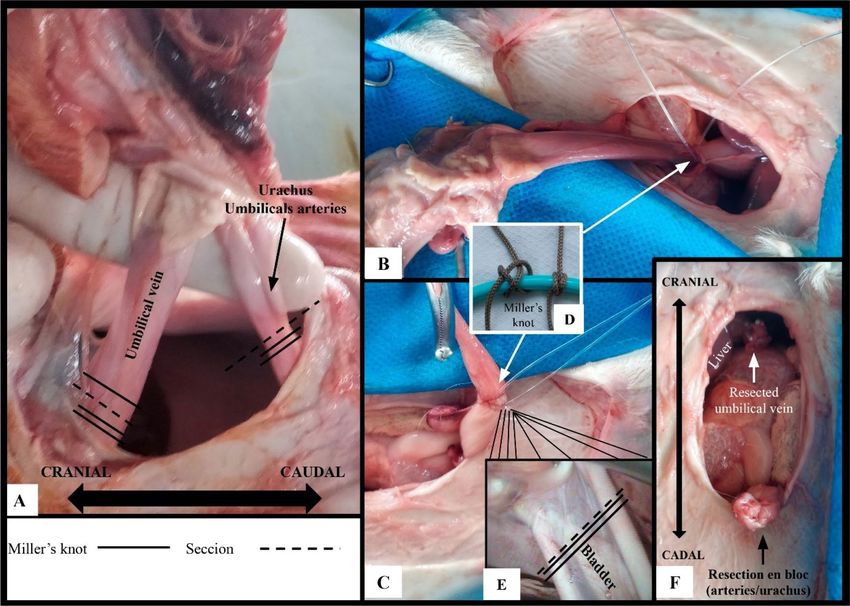
www.nature.com/scientificreports/
laparoscopy, which requires special equipment and instruments that are usually more expensive than conven-
tional ones, a fact that greatly influences their application in veterinary m edicine8–10. In addition, performing
laparoscopy requires the surgeon to have specific skills that can only be acquired with specialized t raining11–13.
Therefore, the advantages of celiotomy make it the most used surgical technique in cases of umbilical conditions
requiring surgical t reatment3,7.
Laparoscopy is a minimally invasive technique and an important alternative to celiotomy that allows, in
addition to the observation of abdominal organs and structures, surgical techniques and exploratory procedures
to be performed with less pain and better recovery for the patient than conventional techniques13. The use of
laparoscopy in cattle is promising; there are many potential applications, and, because it is a safe and effective
technique, it is an important alternative to conventional m ethods11,14.
Surgical indication for the treatment of omphalitis should be preceded by a complete clinical examination,
in particular an examination of the umbilical structures, with palpation of the umbilical region associated with
ultrasound and/or laparoscopy, thereby allowing the surgeon to determine the severity of the abnormality and
establish an appropriate surgical plan for the management of the specific c ase10,15–17.
Data on the feasibility of the techniques and surgical time of procedures performed for anatomical specimens
provide important information for the execution of these procedures in live animals. Thus, the objective of the
present study was to assess the feasibility and the surgical time of laparoscopy and celiotomy for intra-abdominal
resection of the umbilical vein and urachus of bovine fetuses (cadavers).
Materials and methods
Study site and groups. This study was carried out in accordance with the recommendations of the
National Council for Experimentation Control in Brazil (CONCEA). This research was approved by the Animal
Ethics and Welfare Committee of the Federal University of Pará (protocol N ° 4848261017). As the study cor-
responds to a new experimental technique, all surgical procedures were performed on cadavers from a local
slaughterhouse in accordance with inspection requirements. Thus, the procedures did not cause pain or suffer-
ing in animals, as they were performed on bovine fetuses from the slaughtered of pregnant cows.
The experiment was conducted at the Institute of Veterinary Medicine (IMV) in Campus II of the Federal
University of Pará (UFPA), located in the municipality of Castanhal, Pará, Brazil, and involved the resection of
the umbilical vein and urachus using laparoscopy and celiotomy in 26 anatomical specimens (weighing between
30 and 40 kg, bovine fetuses from cows slaughtered in the last third of gestation.
The anatomical specimens were classified into two study groups: one group with 13 anatomical specimens
that underwent resection of the umbilical vein and urachus using laparoscopy, i.e., laparoscopic surgery (LG,
n = 13) and the other group with 13 anatomical specimens that underwent resection of the umbilical vein and
urachus using celiotomy, i.e., open surgery (OG, n = 13).
All the steps of the procedures in both groups were performed by the same surgeon in a standardized and
systematic manner throughout the study.
The feasibility of the techniques and the surgical time (both the total time and that of the steps of abdominal
cavity entry, resection of the umbilical vein, resection of the urachus, and closure of the abdominal cavity) were
analyzed in all the anatomical specimens in both groups. A descriptive comparison of both techniques was
performed, in which the feasibility of performing the surgical procedures was assessed.
Instruments and equipment used in the study. The experimental simulation of the surgical proce-
dures took into consideration all the surgical principles applicable to laparoscopy and conventional open sur-
gery, and the necessary equipment and instruments were used to perform the techniques. We used a 10-mm
laparoscope, 10-mm or 5-mm Babcock forceps, 5-mm laparoscopic scissors, a set of gas insufflator/light source/
monitor, and basic surgical instruments for conventional surgery.
Access to the umbilical vein and urachus using laparoscopic surgery with three access
ports. The anatomical specimens of the LG group were placed in the left lateral recumbency position and
underwent laparoscopy using three laparoscopic access ports in the right flank, with two 10-mm cannulas in the
first and second access ports and a 5-mm cannula in the third port for access to the umbilical vein and allantoic
duct, or urachus. The access ports were established in the right flank near the paralumbar fossa, caudal to the
ribs, using the modified hasson technique (Fig. 1A). Skin incisions of approximately 8 to 10 mm for 10-mm
ports and 3 to 5 mm for 5-mm ports were made using a scalpel to insert the trocars transmurally into the
abdominal cavity, maintaining the triangulation of the access doors (Fig. 1B).
The first 10-mm laparoscopic access port with an insufflation valve was inserted, through which a carbon
dioxide (CO2)-induced pneumoperitoneum of 8 mmHg was established, and the abdominal cavity was inspected
by viewing the image on the monitor. The inspected elements included the umbilical vein from the umbilical
ring to its insertion in the liver, the umbilical arteries from the umbilical ring to the vicinity of the bladder, and
the urachus from the umbilical ring to the bladder.
The second laparoscopic access port was established using a 10-mm trocar to insert the Babcock forceps,
through which manipulation of the umbilical structures was performed, simulating the clipping of these com-
ponents and maintaining an appropriate position to perform the resections in the insertion regions and vicinity
of the umbilical ring.
The third 5-mm port was established to perform the umbilical vein and urachal resection, in the insertion
regions and near the umbilical ring, through which the laparoscopic scissors were introduced and resection of
these components was performed. Subsequently, the components were removed through the second access port
using the Babcock forceps.
Scientific Reports | (2021) 11:5328 | https://doi.org/10.1038/s41598-021-84621-y 2
Vol:.(1234567890)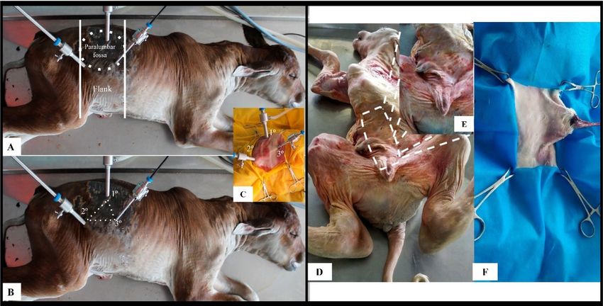
www.nature.com/scientificreports/

                                  Figure 1.  Positions of bovine fetuses that underwent laparoscopy and celiotomy for umbilical vein and urachal
                                  resection. (A, B) show the flank region and triangulation of laparoscopic ports. (C) shows the umbilical and
                                  preputial regions and surgical field.

                                  Resection of the umbilical vein using laparoscopic surgery with three access ports. For resec-
                                  tion of the umbilical vein, the assistant operated the optics placed on the first port. The surgeon manipulated
                                  the umbilical vein, performed the clipping simulation, positioned the vein for resection, and resected it using
                                  laparoscopic scissors inserted in the third port. First, the surgeon placed the Babcock forceps in the second port
                                  and manipulated the umbilical vein for a better inspection of the regions to be resected, later removing the for-
                                  ceps. Subsequently, the Babcock forceps were reinserted to mimic a laparoscopic clipper used for hemostasis of
                                  large vessels, thereby simulating the application of four hemostatic clips in the umbilical vein: two in the vicinity
                                  of the umbilical ring and two in the vicinity of the liver, at the vein insertion site, and the Babcock forceps were
                                  again removed from the port. Finally, the Babcock forceps were reinserted to position and secure the umbilical
                                  vein. The surgeon placed the laparoscopic scissors through the third portal and resected the umbilical vein that
                                  was secured by the Babcock forceps at the “clipped” sites, and the vein was removed through the second access
                                  port using the Babcock forceps.

                                  Resection of the urachus using laparoscopic surgery with three access ports. The procedures
                                  for urachal resection followed the same sequence as those performed for umbilical vein resection described in
                                  the previous paragraph; however, separation of the tissues surrounding the urachus and bladder was performed
                                  using laparoscopic scissors introduced in the third port, for better identification of the “clipping” sites and resec-
                                  tion. Simulation of clipping and urachal resection were performed near the umbilical ring and anterior to the
                                  bladder, following the same procedure used for simulation of clipping and resection of the umbilical vein.

                                  Closure of the abdominal cavity in laparoscopic surgery with three access ports. After resection
                                  of the urachus, the pneumoperitoneum was undone, the trocar cannulas and laparoscopic ports were removed,
                                  and the skin incisions were sutured with one stitch (U suture) using 0.6-mm polyamide thread.
                                      Access to the umbilical vein and urachus using celiotomy (open surgery grup—OG).
                                      OG procedures were performed according to Marchionatti et al.7 adapted, working with the anatomical
                                  examples in the dolsal recumbency position, making elliptical access at the umbilical base, accessing as struc-
                                  tures that make up the umbilical cord. After access was established, an umbilical vein was inspected, connected
                                  with a Miller knot in the region adjacent to the liver, without marsupialization maneuver. The tissues between
                                  the urachus and the artery were separated for better bladder identification inspection. Two block ligatures were
                                  made in the region close to the bladder using the Miller knots. Subsequently, the entire region of the umbilical
                                  ring was removed and the access reversed. Myorrhaphy was performed with superimposed suture, reduction of
                                  the subcutaneous space in a continuous suture and demorraphy using U suture.

                                  Statistical analyses. The Shapiro–Wilk test was used to confirm that surgical time data were distributed
                                  normally. Data on total surgical time and duration of each surgical step were grouped for the GV and GA groups.
                                  Student’s t-test was used to compare normally distributed data on surgical time, and the Mann–Whitney test
                                  (Wilcoxon) was used for non-normally distributed data.
                                     Descriptive statistics were processed using the R statistical program, version 3.0.2. The confidence interval
                                  was 0.95, and when p ≤ 0.05, the difference was considered significant and the null hypothesis was rejected.

Scientific Reports |   (2021) 11:5328 |                https://doi.org/10.1038/s41598-021-84621-y                                                     3

                                                                                                                                                 Vol.:(0123456789)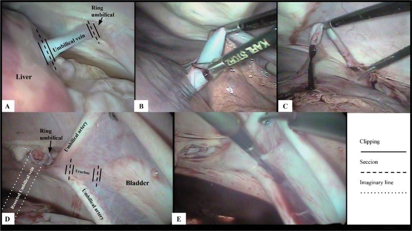
www.nature.com/scientificreports/
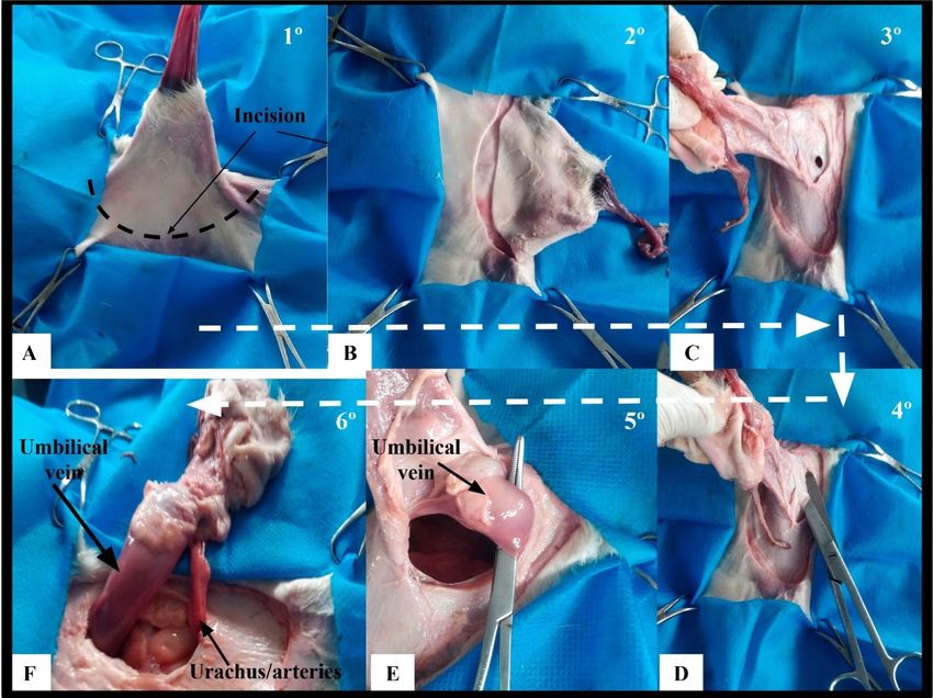
Figure 2. Steps for intra-abdominal access to the umbilical vein and urachus in celiotomy.
Results
All fetuses of bovine calves in the final third of gestation had well defined umbilical structures, including the
components of interest, i.e., the umbilical vein and the allantoic duct, or urachus. These components had a
cylindrical shape, defined walls, and flaccid texture, and it was possible to distinguish the umbilical veins and
arteries as separate units. The liver and bladder were also well formed and defined. The bladder was empty, and
the allantoic duct was cranially inserted in the anterior apex.
In the LG, with the fetuses positioned in left lateral recumbency, it was possible to perform resection of the
umbilical vein and urachus using laparoscopy with three access ports established in the right flank, in the par-
alumbar fossa region (Fig. 1A,B). In the OG group, the most appropriate position for performing the proposed
procedures was dorsal recumbency with abducted legs (Fig. 1C).
The pneumoperitoneum established using an intra-abdominal pressure of 8 mmHg allowed satisfactory
separation of the abdominal wall from the visceral structures, as well as excellent intra-abdominal visualization
of the umbilical structures and associated/adjacent organs such as the liver and bladder.
Access to the abdominal cavity and target structures to perform the procedures was achieved through three
laparoscopic ports in laparoscopy and through a paramedian and circumscribed incision in the abdominal
wall (subcutaneous tissue, muscle, and peritoneum) in celiotomy (Figs. 1B and 2). In laparoscopy, the surgical
wounds consisted of three openings of approximately 3 to 5 mm in length, and in celiotomy the openings in the
abdominal cavity ranged from 5.5 to 18.5 cm, with an average of 9.7 cm (Fig. 2). Access to the abdominal cav-
ity, down to the target structures, was secured in 5.52 (± 1.82) and 4.78 (± 0.74) min in the LG and OG groups,
respectively (Table 1).
During access to the abdominal cavity using laparoscopy with three ports, the umbilical vein was the first
component inspected because the laparoscope focused directly on the structure, thereby enabling its complete
inspection (Fig. 3A). It was possible to simulate the clipping of the umbilical vein with Babcock forceps in
the resection sites and extract it through the second access port using 10-mm Babcock forceps (Fig. 3A–C).
Resection of the umbilical vein using celiotomy was achieved, and Miller’s knot was made to contain bleeding
(Fig. 4B). The duration of umbilical vein resection was 2.27 (± 1.33) and 2.89 (± 0.87) min in the LG and OG
groups, respectively (Table 1).
With regard to the urachus, it was possible to simulate clipping with the Babcock forceps and perform its
resection using laparoscopy (Fig. 3E). Resection of the urachus using celiotomy was also achieved, and Miller’s
knot in the duct was made in this case (Fig. 4C). The duration of urachal resection was 7.09 (± 1.27) and 3.13
(± 0.91) min in the LG and OG groups, respectively (Table 1).
Scientific Reports | (2021) 11:5328 | https://doi.org/10.1038/s41598-021-84621-y 4
Vol:.(1234567890)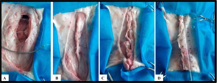
www.nature.com/scientificreports/
LEG LG OG P value
Access to the abdominal cavity 5.52 ± 1.82 4.78 ± 0.74 0.1946
Resection of the umbilical vein 2.27 ± 1.33 2.89 ± 0.87 0.1734
Resection of the urachus 7.09 ± 1.27 3.13 ± 0.91 < 0.0001
Closure of the abdominal cavity 0.92 ± 0.09 28.64 ± 7.32 < 0.0001
Total surgical time 15.81 ± 3.26 39.44 ± 8.46 < 0.0001
Table 1. Results of surgical time for each step of laparoscopy and celiotomy performed for resection of
umbilical vein and urachus in bovine fetuses.
Figure 3. Intra-abdominal resection of the umbilical vein and urachus of bovine fetuses using laparoscopy.
(A–C) show the steps of umbilical vein resection. (D, E) show the steps of urachal resection.
In the LG group, closure of the abdominal cavity was performed by placing a stitch on the skin (U suture) that
created a post-surgical wound of approximately 5 mm in length. In the OG group, peritoneum/muscle dieresis
was performed with overlapping suture, subcutaneous tissue dieresis was performed with simple continuous
suture, and dead space was reduced by anchoring the subcutaneous tissue to the musculature. Skin closure was
performed with a U suture that created a post-surgical wound ranging from 10 to 20 cm, with an average of
13.6 cm (Fig. 5). The surgical time for abdominal cavity closure was 0.92 (± 0.09) min in the GV group and 28.64
(± 7.32) min in the OG group (Table 1).
The total surgical time in the LG and OG groups was 15.81 (± 3.26) and 39.44 (± 8.46) min, respectively
(Table 1).
Discussion
The treatments indicated for umbilical vein and urachal diseases in newborn calves are based on clinical and/or
surgical therapy, with conventional open surgery commonly performed in cases requiring surgical treatment.
The aim of the present study was to assess the use of laparoscopy in umbilical vein and urachal interventions
when surgical treatment is indicated. The surgical management of umbilical conditions usually has a good
prognosis. The use of laparoscopy improves prognosis, with better postoperative results including less pain and
better recovery for patients3,18.
In OG, a satisfactory procedure involving the umbilical structures was possible, with adequate inspection
and palpation of the umbilical structures, which allowed the surgeon to examine and resect each component.
However, a more comprehensive exploration of the abdominal cavity was achieved using laparoscopy, with good
intervention in the umbilical structures and adjacent tissues/organs because the visual field projected on the
monitor enlarged the structures and improved the visualization of smaller areas. Using laparoscopy, intervention
in tissues present in small areas and that are difficult to access is possible. This is a minimally invasive procedure
that, when associated with advanced multimedia resources, enables new approaches and procedures that are
more precise and objective, thereby improving the outcomes and reducing c omplications11,14.
Scientific Reports | (2021) 11:5328 | https://doi.org/10.1038/s41598-021-84621-y 5
Vol.:(0123456789)www.nature.com/scientificreports/
Figure 4. Intra-abdominal resection of the umbilical vein and urachus of bovine fetuses using celiotomy.
Figure 5. Abdominal tissue and skin closure of the abdominal cavity. (A, B) Show peritoneum/muscle dieresis
with overlapping suture. (C) Shows reduction of subcutaneous dead space with muscle anchoring. (D) Shows
skin closure with U suture.
In the first step, there was no statistically significant difference in the time required to access the umbilical
vein and urachus between the two groups. The duration of access in the LG was longer because the time to
insufflate the abdominal cavity was included in this step. In both groups, caution as well as the establishment of
standardized access ports allowed the procedures to be completed without complications, both morphological
complications and macroscopic lesions of abdominal structures. In laparoscopy, a skin incision smaller than
the diameter of the trocar allowed the latter to be introduced slowly and carefully, thereby preventing potential
injuries to the abdominal organs and CO2 losses from the cannulas of the laparoscopic ports during pneumop-
eritoneum. The trocars were inserted directly without injuring the abdominal organs and structures adjacent to
the umbilical components and allowed working with instruments sufficiently far from the umbilical structures.
Standardizing abdominal access techniques with safer and reliable methods reduces the likelihood of periopera-
tive complications14,19,20.
Scientific Reports | (2021) 11:5328 | https://doi.org/10.1038/s41598-021-84621-y 6
Vol:.(1234567890)www.nature.com/scientificreports/
Although preventive measures were not adopted during CO2 insufflation in laparoscopy, it may be important
to adopt them in live patients, especially in those with infected umbilical structures and with metabolic impair-
ment, because pneumoperitoneum can induce hypothermia and respiratory and cardiovascular c hanges2,21,22.
The difference in the surgical times of umbilical vein resection (second step) between the LG and OG groups
was not statistically significant. In laparoscopy, the time required for simulation of the clipping and umbilical
vein resection was shorter than that for the execution of Miller’s knot followed by vein resection in conventional
open surgery; however, the surgeon’s skill may have been a factor that influenced the surgical time of this step.
A surgeon’s skill is directly related to specialized training that ensures the ability to perform procedures quickly
and efficiently11,13. In laparoscopy, umbilical vein resection was performed with ease, and the positioning of
laparoscopic ports, in particular, was very important to guarantee direct access to the structure, avoiding organs
or tissues that could prevent or hinder the procedures and allowing resection to be performed safely and without
complications2,15. Animals with omphalophlebitis would have swollen structures due to the condition; however,
there are different sizes of clips as well as other options for electrosurgical vessel sealing23–25. Thus, there are
several possibilities for in vivo procedures, requiring few adaptations of the model described. Another question
to investigate is the exteriorization of the sectioned structure (segment of umbilical vein and portion of the
urachus). The removal of intra-abdominal structures is common in laparoscopic procedures, requiring small
extensions in the portal access or the use of laparoscopic specimen retrieval b ags26,27.
The surgical time for urachal resection in laparoscopy was longer than that in conventional open surgery
because access to the urachus in laparoscopy was not direct, similar to the access to the umbilical vein. The
urachus lies in the ventral region of the abdomen, between the umbilical arteries, and is enveloped by tissues
that make its identification and manipulation difficult (not detected during laparoscopic visualization). For
satisfactory intervention in the urachus, it is important to perform tissue divulsion around the duct, a proce-
dure that requires a skilled laparoscopist and may have influenced the surgical time of this stage, although not
greatly and not affecting the final surgical time. In general, urachal resection was achieved in all the animals in
laparoscopy, which was shown to be an efficient technique for urachal interventions, both from a therapeutic
and a diagnostic perspective. Considerable advances have already been achieved in the use of laparoscopy in
urachal conditions15,28.
In convencional open surgery, the umbilical artery is seccioned and disconected from the abdominal wall.
The same procedure could be performed in the laparoscopy. However, the authors believe that the techinique
connecting the urachus to the bladder could be the best option. This procedure avoids potential trauma to adja-
cent structures, minimizing the formation of intra-abdominal adhesions, besides considering that the umbilical
artery atrophy with the development of the c alf29,30.
In the final step, i.e., closure of the abdominal cavity, the surgical time in the GA group was longer than that
in the GV group. This was expected because the opening of the abdominal wall in conventional open surgery was
multiple times larger than that in laparoscopy, and additional time was required to perform abdominal tissue clo-
sure. In laparoscopy, a simple stitch ensured closure of the cavity, whereas in conventional open surgery, several
stitches were required to perform peritoneum/muscle and subcutaneous dieresis and skin closure. Abdominal
tissue closure in laparoscopy is satisfactorily performed with one or two simple stitches in each created wound
and has lower rates of complications compared with abdominal tissue closure performed in conventional open
surgeries15,19.
The total surgical time of laparoscopy was significantly shorter than that of conventional open surgery (exactly
15.81 ± 3.26 min), which shows that resection using endosurgery of the umbilical vein and urachus is faster than
resection using conventional open surgery. The step of abdominal cavity closure in conventional open surgery
significantly affected the total surgical time in the GA group because access wounds in the abdominal wall were
significantly larger than those caused by laparoscopy. This increased the time of abdominal cavity closure and,
consequently, the total surgical time of conventional open surgery. The total surgical time of laparoscopy in the
present study was significantly shorter than that in other similar studies in which the laparoscopy technique was
used in diagnostic and surgical interventions of umbilical structures with times ranging from 36 to 160 m in2,15,31.
Abdominal wall injuries were less common in laparoscopy than in conventional open surgery; laparoscopy
caused only three lesions of approximately 10 mm, as well as less tissue damage. This allows us to infer that
laparoscopy causes less surgical trauma in living patients and provides better postoperative recovery of newborn
calves. Because it is a minimally invasive technique, it is important to determine whether it is possible to perform
it under regional anesthesia and sedation, a suitable alternative for treating calves with umbilical disorders asso-
ciated with concomitant diseases and with reduced ability to adapt to general a nesthesia15. Other studies have
shown that laparoscopic techniques are less traumatic than conventional open techniques32–35.
The results of laparoscopy allowed the identification of additional benefits of its use in production animals
because the technique enabled a large visual field for the diagnostic exploration of the abdominal cavity, thereby
providing diagnostic advantages from a macroscopic and morphological perspective, including the accurate
detection of macroscopic disorders in the umbilical structures, liver, bladder, and other adjacent structures, with
results that can be correlated with ultrasound and laparatomy. Laparoscopy is a potentially viable diagnostic alter-
native that allows diagnosing multifocal abscesses in the liver, changes in umbilical arteries near their branching
from the internal iliac artery, and focal thickenings and a dhesions15,20,36. The use of laparoscopy in production
animals is still incipient, but its use provides better results in semiological and clinical/surgical procedures in
bovine veterinary medicine, with advances in sanitary/production methods and animal w elfare18,37–39.
Conclusions
The laparoscopic technique proposed in our study allowed to perform the ressection of the umbilical vein and
urachus. The effectiveness in the evaluation of the structures is highlighted, allowing an excellent diagnosis.
Scientific Reports | (2021) 11:5328 | https://doi.org/10.1038/s41598-021-84621-y 7
Vol.:(0123456789)www.nature.com/scientificreports/
Even though this research was performed in an experimental model, the study supports the attempt to per-
form the technique in patients in vivo, based on its minimally invasive quality and the short surgical time in the
proposed assessment.
More studies in the clinical routine are required to verify each operative step in the face of altered structures,
however, the team allows few adaptations in live animals.
Received: 8 October 2020; Accepted: 18 February 2021
References
1. Hopker, A. Umbilical swellings in calves: A continuing challenge. Vet. Rec. 174, 219–220. https://doi.org/10.1136/vr.g1790 (2014).
2. Bouré, L., Foster, R. A., Palmer, M. & Hathway, A. Use of an endoscopic suturing device for laparoscopic resection of the apex of
the bladder and umbilical structures in normal neonatal calves. Vet. Surg. 30, 319–326 (2001).
3. Williams, H. J., Gillespie, A. V., Oultram, J. W., Cripps, P. J. & Holman, A. N. Outcome of surgical treatment for umbilical swellings
in bovine youngstock. Vet. Rec. 174(9), 221–221. https://doi.org/10.1136/vr.101736 (2014).
4. Rodrigues, C. A. et al. Correlação entre os métodos de concepção, ocorrência e formas de tratamento das onfalopatias em bovinos:
Estudo retrospectivo. Pesqui Vet. Bras. 30(8), 618–622. https://doi.org/10.1590/S0100-736X2010000800002 (2010).
5. Reig Cordina, L., Were, S. R. & Brown, J. A. Short-term outcome and risk factors for post-operative complications following
umbilical resection in 82 Foals (2004–2016). Equine Vet. J. Suppl. https://doi.org/10.1111/evj.13021 (2018).
6. Baird, A. N. Umbilical surgery in calves. Vet. Clin. N. Am. Food Anim. Pract. 24(3), 467–477. https://doi.org/10.1016/j.
cvfa.2008.06.005 (2008).
7. Marchionatti, E. et al. Surgical management of omphalophlebitis and long term outcome in calves: 39 cases (2008–2013). Vet. Surg.
45, 194. https://doi.org/10.1111/vsu.12433 (2016).
8. Mulon, P. Y. & Desrochers, A. Surgical abdomen on the calf. Vet. Clin. N. Am. Food Anim. Pract. 21(1), 101–132. https://doi.
org/10.1016/j.cvfa.2004.12.004 (2005).
9. Yanmaz, L. E., Okumus, Z. & Dogan, E. Laparoscopic surgery in veterinary medicine. Medwell J. Vet. Res. 1(1), 23–39 (2007).
10. Nichols, S. Umbilical surgery in calves. in: ProcMich Vet Conf. https: //pdfs.semant icsch olar. org/7a17/77e92d
e2ec1 d10f4 23f0f 0809
49d9a6948f5.pdf. (2014).
11. Buia, A., Stockhausen, F. & Hanisch, E. Laparoscopic surgery: A qualified systematic review. World J. Methodol. 5(4), 238–254.
https://doi.org/10.5662/wjm.v5.i4.238 (2015).
12. Ferrufino, F. L. et al. Simulation in laparoscopic surgery. Circ. Esp. 93(1), 4–11. https: //doi.org/10.1016/j.cireng .2014.02.022 (2015).
13. Saber, A., Bayumi, E. K., Sophia, L. & Hoek, L. S. V. D. Minimal access and minimally invasive surgery in veterinary practice. J.
Surg. 5(3–1), 39–42. https://doi.org/10.11648/j.js.s.2017050301.18 (2017).
14. Babkine, M. & Desrochers, A. Laparoscopic surgery in adult cattle. Vet. Clin. N. Am. Food Anim. Pract. 21(1), 251–279. https://
doi.org/10.1016/j.cvfa.2004.12.003 (2005).
15. Robert, M. et al. Laparoscopic evaluation of umbilical disorders in calves. Vet. Surg. 45(8), 1041–1048. https://doi.org/10.1111/
vsu.12559(2016).
16. Seino, C. H. et al. Avaliação ultrassonográfica de componentes umbilicais inflamados em bezerros da raça Holandesa com até 30
dias de vida. Pesqui Vet. Bras. 36(6), 492–502. https://doi.org/10.1590/S0100-736X2016000600006 (2016).
17. Sato, R., Yamada, K., Shinozuka, Y., Ochiai, H. & Onda, K. Gas-filled urachal abcess with a pinging sound in a heifer calf. Vet. Med.
64(8), 362–366. https://doi.org/10.17221/61/2019-VETMED (2019).
18. Patel, A. M., Parikh, P. V. & Patil, D. B. Laparoscopy in veterinary practice. Vet. Res. Int. 2(1), 01–07 (2014).
19. Bianchi, G. et al. Laparoscopic access overview: Is there a safest entry method?. Actas Urol. Esp. 40(6), 386–392 (2016).
20. Oztoprak, M. Y. & Karaca, I. Abdominal access techniques used in laparoscopic surgery. Int. J. Exp. Clin. Anat. 13(2), 129 (2019).
21. Rezende, M., Prado, O., Bandeira, C., Petri, A. & Montero, E. Body temperature evaluation during induced pneumoperitoneum
with CO2: An experimental study in pigs. Surg. Endosc. 26(6), 1724–1729. https://doi.org/10.1007/s00464-011-2099-x (2012).
22. Radlinsky, M. G. Complications and conversion from endoscopic to open surgery veterinary. Vet. Clin. N. Am. Food Anim. Pract.
46(1), 137–145. https://doi.org/10.1016/j.cvsm.2015.07.004 (2016).
23. Nuss, K. Erkrankungen der inneren Nabelstrukturen beim Rind. Tierarzti Prax. 35(G), 149–156. https : //doi.
org/10.1055/s-0038-1624018 (2007).
24. Yip, S. K. et al. Routine vascular control using the Hem-o-lok clip in laparoscopic nephrectomy: Animal study and clinical applica-
tion. J. Endourol. 18(1), 77–81. https://doi.org/10.1089/089277904322836730 (2004).
25. Barnes, R. F., Greenfield, C. L., Schaeffer, D. J., Landolfi, J. & Andrews, J. Comparison of biopsy samples obtained using standard
endoscopic instruments and the harmonic scalpel during laparoscopic and laparoscopic assisted surgery in normal dogs. Vet. Surg.
35, 243–251. https://doi.org/10.1111/j.1532-950X.2006.00134.x (2006).
26. Stavroulis, A., Memtsa, M. & Yoong, W. Methods for specimen removal from the peritoneal cavity after laparoscopic excision.
Obstet. Gynaecol. 15(1), 26–30. https://doi.org/10.1111/j.1744-4667.2012.00148.x (2013).
27. Baird, A. N. Surgery of the umbilicus and related structures. Vet. Clin. Food. Anim. 32, 673–685. https://doi.org/10.1016/j.
cvfa.2016.05.008 (2016).
28. Marques, L. C., Marques, J. A., Marques, I. C. S. & Teixeira, M. C. A. Dilatação cística do úraco e uroperitônio em touros: Relato
de cinco casos. Arq. Bras. Med. Vet. Zootec. 62(6), 1320–1324. https://doi.org/10.1590/S0102-09352010000600004 (2010).
29. Araújo, S. E. A., Caravatto, P. P. P., Chang, A. J. B. A., Campos, F. G. C. M. & Sousa, M. Impacto da vídeocirurgia na prevenção de
aderências. Rev. Bras. Colo-proctol. https://doi.org/10.1590/S0101-98802006000200014 (2006).
30. Guerri, G., Vignoli, M., Palombi, C., Monaci, M. & Petrizzi, L. Ultrasonographic evaluation of umbilical structures in Holstein
calves: A comparison between healthy calves and calves affected by umbilical disorders. J. Dairy Sci. 103(3), 2578–2590. https://
doi.org/10.3168/jds.2019-16737(2019).
31. Fisher, A. T. Jr. Laparoscopically assisted resection of umbilical structures in foals. J. Am. Vet. Med. Assoc. 214(12), 1813–1816
(1999).
32. Easley, J. T., Garofolo, S. Q., Ruehlman, D. & Hackett, E. S. A 3-portal laparoscopic ovariectomy technique in ewes. Small Ruminant.
Res. 121(2–3), 336–339. https://doi.org/10.1016/j.smallrumres.2014.08.001 (2014).
33. Zhang, S., Hao, M. & Ma, Y. Comparasion of laparoscopic and traditional abomasal cannulation in sheep. J. Vet. 60, 113–117. https
://doi.org/10.1515/jvetres-2016-0016 (2016).
34. Tripathi, S. D., Khandekar, G. S., Datir, A. A., Ambore, G. S. & Pawar, A. Comparative evaluation of laparoscopic and open vasec-
tomy techniques in teaser bulls. Intas. Polivet. 18(2), 415 (2017).
35. Santos, G. M. A. et al. Minimally invasive video-assisted ruminostomy in sheep. Small Ruminant. Res. 167, 78–81. https://doi.
org/10.1016/j.smallrumres.2018.07.023 (2018).
36. Milovancev, M. & Townsend, K. L. Current concepts in minimally invasive surgery of the abdomen. Vet. Clin. N. Am. Small Anim.
Pract. 45(3), 507–522. https://doi.org/10.1016/j.cvsm.2015.01.00.4 (2015).
37. Corrêa, R. R. et al. Evaluation of ventral laparoscopic abomasopexy using surgical staples associated with suture material in dairy
cattle. Acta Sci. Vet. 46, 1545. https://doi.org/10.22456/1679-9216.81834 (2018).
Scientific Reports | (2021) 11:5328 | https://doi.org/10.1038/s41598-021-84621-y 8
Vol:.(1234567890)www.nature.com/scientificreports/
38. Gnus, M., Ratajczak, K. & Antosik, P. Application of a laparoscopic spieker of own design in reposition and fixation of the left
displacement of the abomasum (LDA) in cattle. Med. Water. 74(2), 99–103. https://doi.org/10.21521/mw.6059 (2018).
39. Sofi, K. A., Singh, M., Kumar, P. & Sharma, A. Laparoscopic chromopertubation for the evaluation of tubal patency in dairy cattle.
Indian J. Anim. Reprod. 39(1), 1 (2018).
Acknowledgements
We would like to thank the members of the VOR research team (UFPA) for their collaboration and contribution
to this research work.
Author contributions
F.D.O.M (Conceptualization, Investigation, Formal Analysis, Project administration, Validation, Writing—
Review & Editing), H.J.G. (Investigation, Data Curation), S.S.S (Investigation, Data Curation), J.P.M.B. (Inves-
tigation, Data Curation), G.P.B.V. (Investigation, Data Curation), D.L.S. (Investigation, Data Curation), L.H.V.A.
(Investigation, Data Curation), L.A.S. (Investigation, Data Curation), G.M.A.S. (Investigation, Data Curation),
K.C.R. (Investigation, Writing—Original Draft Preparation), V.S.S. (Investigation, Data Curation), C.E.S.F.
(Investigation, Data Curation), B.C.G. (Investigation, Data Curation), H.F.M. (Investigation, Data Curation),
C.R.G.S. (Investigation, Data Curation), R.S.A. (Investigation, Data Curation), L.P.B.B. (Investigation, Writing—
Original Draft Preparation), G.F.A. (Resources), R.S.M.L. (Investigation, Writing—Original Draft Preparation)
and P.P.M.T. (Conceptualization, Writing—Review & Editing, Supervision, Project administration, Funding
acquisition, Validation, Methodology). All authors reviewed the manuscript.
Competing interests
The authors declare no competing interests.
Additional information
Correspondence and requests for materials should be addressed to F.D.O.M.
Reprints and permissions information is available at www.nature.com/reprints.
Publisher’s note Springer Nature remains neutral with regard to jurisdictional claims in published maps and
institutional affiliations.
Open Access This article is licensed under a Creative Commons Attribution 4.0 International
License, which permits use, sharing, adaptation, distribution and reproduction in any medium or
format, as long as you give appropriate credit to the original author(s) and the source, provide a link to the
Creative Commons licence, and indicate if changes were made. The images or other third party material in this
article are included in the article’s Creative Commons licence, unless indicated otherwise in a credit line to the
material. If material is not included in the article’s Creative Commons licence and your intended use is not
permitted by statutory regulation or exceeds the permitted use, you will need to obtain permission directly from
the copyright holder. To view a copy of this licence, visit http://creativecommons.org/licenses/by/4.0/.
© The Author(s) 2021
Scientific Reports | (2021) 11:5328 | https://doi.org/10.1038/s41598-021-84621-y 9
Vol.:(0123456789)You can also read