Exosomal-mediated transfer of OIP5-AS1 enhanced cell chemoresistance to trastuzumab in breast cancer via up-regulating HMGB3 by sponging miR-381-3p
←
→
Page content transcription
If your browser does not render page correctly, please read the page content below
Open Medicine 2021; 16: 512–525
Research Article
Qiang Yu#, Yinmou Li#, Shijun Peng, Jing Li, Xianxiong Qin*
Exosomal-mediated transfer of OIP5-AS1 enhanced cell
chemoresistance to trastuzumab in breast cancer via
up-regulating HMGB3 by sponging miR-381-3p
https://doi.org/10.1515/med-2021-0249 besides, miR-381-3p targeted HMGB3. Murine xenograft
received September 26, 2020; accepted February 12, 2021 analysis showed exosomal OIP5-AS1 induced trastuzumab
Abstract resistance in vivo. Exosomal OIP5-AS1 was dysregulated in
Background ‒ Long noncoding RNA OPA-interacting the serum of breast cancer patients and might be a pro-
protein 5 antisense transcript 1 (OIP5-AS1) was confirmed mising diagnostic biomarker in trastuzumab resistance.
to involve in the malignancy of breast cancer. However, Conclusion ‒ Intercellular transfer of OIP5-AS1 by exo-
whether exosomal OIP5-AS1 is implicated in trastuzumab somes enhanced trastuzumab resistance in breast cancer
resistance remains unclear. via miR-381-3p/HMGB3 axis, indicating a potential ther-
Methods ‒ The IC50 value of cells to trastuzumab, cell apeutic strategy to boost the effectiveness of trastuzumab
proliferation, migration, and apoptosis was analyzed by in resistant breast cancer patients.
cell counting kit-8 assay, colony formation assay, trans- Keywords: OIP5-AS1, miR-381-3p, HMGB3, breast cancer,
well assay, or flow cytometry, respectively. The expres- exosomes, chemoresistance
sion of OIP5-AS1 and microRNA (miR)-381-3p was detected
using quantitative real-time polymerase chain reaction.
Exosomes were isolated by ultracentrifugation and qua-
lified by nanoparticle tracking analysis software. Western 1 Introduction
blot was used to detect the protein levels of tumor sus-
ceptibility gene 101 (TSG101), CD81, CD63, or high-mobi- Breast cancer is the most commonly diagnosed female
lity group protein B3 (HMGB3). The interaction between malignancy on a global scale, which is a serious threat
miR-381-3p and OIP5-AS1 or HMGB3 was confirmed to the health of women worldwide [1]. A cancer cell has
by dual-luciferase reporter assay and pull-down assay. a variety of receptors on its surface, which can be bound
In vivo experiments were conducted using murine xeno- by chemicals to result in changes within cancer cell.
graft models. HER2 is a member of the epidermal growth factor receptor
Results ‒ OIP5-AS1 was elevated in trastuzumab-resis- family that occurs in about one-third of all breast cancers
tant breast cancer cells, and OIP5-AS1 knockdown rescued [2]; importantly, HER2 amplification causes aggressive
trastuzumab sensitivity. Extracellular OIP5-AS1 was pack- cancer phenotype and poor clinical outcome [3,4]. Tras-
aged into exosomes, which were secreted by trastuzumab- tuzumab, a humanized antibody of HER2, is thought to be
resistant cells, and could be absorbed by trastuzumab- a successful strategy to block HER2 receptors when there
sensitive cells in breast cancer. Importantly, intercellular is overexpression, thereby blocking growth of cancer and
transfer of OIP5-AS1 via exosomes enhanced trastuzumab prolonging the overall survival of HER2+ breast cancer
resistance in vitro. OIP5-AS1 was a sponge of miR-381-3p; patients in adjuvant and metastatic settings [5,6]. Never-
theless, the response rate to trastuzumab-containing therapies
still needs to be improved because of the development
# Contributed equally. of acquired trastuzumab resistance [7]. Therefore, a
better understanding on the mechanism of trastuzumab
resistance is essential for developing new approaches to
* Corresponding author: Xianxiong Qin, Department of Breast overcome trastuzumab resistance in breast cancer.
Surgery, The Central Hospital of Enshi Tujia and Miao Autonomous Recently, it has been documented that cells can com-
Prefecture, No. 158 Wuyang Street, Enshi 445000, Hubei, China,
municate with each other via microvesicles [8]. Exosomes
e-mail: rvv3sll@163.com, tel: +86-0718-8263359
Qiang Yu, Yinmou Li, Shijun Peng, Jing Li: Department of Breast
are one of the extracellular vesicles, contain lipids, protein,
Surgery, The Central Hospital of Enshi Tujia and Miao Autonomous noncoding, or coding RNAs, can be secreted by numerous
Prefecture, No. 158 Wuyang Street, Enshi 445000, Hubei, China cell types, like cancer cells, and can be absorbed by other
Open Access. © 2021 Qiang Yu et al., published by De Gruyter. This work is licensed under the Creative Commons Attribution 4.0
International License.OIP5-AS1 enhances trastuzumab resistance in BC 513
cells to transfer and exchange the cargo [9,10]; thus, exo- China) and cultured in RPMI-1640 medium (Gibco, Carlsbad,
somes are considered as potential modes of intercellular CA, USA) harboring with 15% fetal bovine serum (FBS) and
communication [11]. Recently, emerging evidence has 0.1 IU/mL insulin at 37°C with 5% CO2. Trastuzumab-resis-
revealed that exosomes potentially impact the thera- tant breast cancer cells, named SKBR3-TR and BT474-TR,
peutic response of the recipient cells via the transfer of were established by continuously exposing parental cells to
proteins and lncRNAs [12]. Long noncoding RNA OPA- increasing concentration of trastuzumab (Sigma, St. Louis,
interacting protein 5 antisense transcript 1 (OIP5-AS1), MO, USA) for more than 6 months until cells displayed
mapped on chromosome 15q15.1, is a novel identified and resistance to trastuzumab. Trastuzumab-resistant cells
promising tumor-associated lncRNA. It has been demon- were maintained in the same media supplemented with
strated that OIP5-AS1 plays various roles in multiple cancers 3 μg/mL trastuzumab.
and contributes to deterioration of malignant tumors [13]. In
breast cancer, OIP5-AS1 was found significantly up-regu-
lated and functioned as an oncogene via regulating cell
malignant phenotypes [14], whereas the role of OIP5-AS1 2.3 Cell viability assay
in trastuzumab resistance in breast cancer still needs to be
explored. Parental or resistant cells (5,000 cells/well) were seeded
Thus, we attempted to elaborate the functions of OIP5- in 96-well plates overnight. Following transfection or
AS1 in trastuzumab resistance in breast cancer, explored trastuzumab treatment (0, 0.3125, 0.625, 1.25, 2.5, 5, or
whether the exosome-transmitted OIP5-AS1 conferred drug 10 μg/mL) for additional 48 h, cells in 96-well plates
resistance to recipient cells, as well as the potential mole- were incubated with 10 μL cell counting kit-8 solution
cular mechanism underlying OIP5-AS1 effects on breast (Sigma) for 4 h at 37°C. The optical density at 450 nm
cancer. was determined by a microplate reader, and the half-
maximal inhibitory concentration (IC50) value of trastu-
zumab was assessed on the basis of the relative survival
curve.
2 Materials and methods
2.1 Patients and specimens
2.4 Cell transfection
Blood samples were collected from 57 breast cancer patients
The miR-381-3p mimic (miR-381-3p), small interfering RNA
diagnosed by histopathological examination at The Central
(siRNA) targeting OIP5-AS1 (si-OIP5-AS1#1, si-OIP5-AS1#2,
Hospital of Enshi Tujia and Miao Autonomous Prefecture.
si-OIP5-AS1#3), pcDNA3.1 OIP5-AS1 overexpression vector
All blood samples were centrifuged at 3,000 g for 10 min
(oe-OIP5-AS1), pcDNA3.1 HMGB3 overexpression vector
after collecting for 1 h, and the supernatant serum was col-
(HMGB3), and their corresponding negative control (miR-
lected using RNase-free tubes and stored at −80°C until used.
NC, si-NC, Vector) were synthesized by Genepharma
All patients only received trastuzumab-based neo-adjuvant
(Shanghai, China). The transfection of cells was performed
chemotherapy and were classified into trastuzumab-resistant
using Lipofectamine™ 2000 transfection reagent (Invitrogen,
(non-response, N = 30) and trastuzumab-sensitive (response,
Carlsbad, CA, USA).
N = 27) depending on the sensitivity to trastuzumab. This
research was authorized by the Ethics Committee of The
Central Hospital of Enshi Tujia and Miao Autonomous
Prefecture and was carried out according to the guidelines 2.5 RT-PCR and real-time quantitative
of Declaration of Helsinki. Written informed consents had
PCR (qPCR)
been collected from all subjects.
Whole-RNA extracts from parental or resistant cells were
prepared using TRIzol reagent (Invitrogen), and exo-
2.2 Cell culture somal RNAs were isolated with the exoRNeasy Midi Kit
(Qiagen, Valencia, CA, USA) according to the standard
Human breast cancer cell lines SKBR3 and BT474 were pur- procedure. Complementary DNA (cDNA) was synthesized
chased from Shanghai Academy of life Science (Shanghai, using SuperScript III® (Qiagen), and quantitative PCR514 Qiang Yu et al.
was performed using SYBR Premix Ex Taq (Qiagen) on 2.9 Exosome (exo) isolation
the Bio-Rad CFX96 Sequence Detection system (Bio-Rad,
Hercules, CA, USA). The expression levels were detected Exosomes were isolated from serum samples or cells
by 2−ΔΔCt method with glyceraldehyde 3-phosphate dehy- using ultracentrifuge method. Cell culture fluid from
drogenase (GADPH) or U6 small nuclear B noncoding exosome-depleted medium or serum was centrifuged at
RNA (U6) serving as an internal reference. The primer 3,000 g for 30 min at 4°C to remove cell fragments. Then,
sequences were listed as follows: OIP5-AS1: F, 5′-TGCGA the resulting supernatant was further centrifuged at
AGATGGCGGAGTAAG-3′ and R, 5′-TAGTTCCTCTCCTCTGG 100,000 g for 70 min at 4°C and filtered using 0.22 μm
CCG-3′; miR-381-3p: F, 5′-TAATCTGACTATACAAGGGCAA filtration. Subsequently, pelleted exosomes were washed
GCT-3′ and R, 5′-TATGGTTGTTCTGCTCTCTGTCTC-3′; with PBS and centrifuged at 100,000 g for 70 min again.
GADPH: F 5′-GAGAAACCTGCCAAGTATGATGAC-3′ and R Finally, purified exosomes were resuspended in PBS for
5′-GGAGTTGCTGTTGAAGTCAC-3′, U6: F, 5′-CTCGCTTCGG the detection of the size and quality of exosomes using
CAGCACA-3′ and R, 5′-AACGCTTCACGAATTTGCGT-3′. nanoparticle tracking analysis (NTA) software or for func-
tional assays. Exosomes pellets were interacted with Trizol
reagent to isolate RNA and were lysed with RIPA lysis
buffer used for protein detection. For blocking of exosome
2.6 Colony formation assay release, parental or resistant breast cancer cells were
treated with GW4869 (10 μM) or vehicle (as control) for
Transfected parental or resistant cells (5,000 per well) 48 h. For exosome co-cultures, exosomes (50 μg/mL) were
suspended in RPMI-1640 medium with 0.5 μg/mL trastu- incubated with parental SKBR3 and BT474 cells (5 × 10⁵) in
zumab were seeded in 6-well plates. After 21 days of cul- a 6-well plate with 10% FBS exosome-depleted culture
tures at 37°C with 5% CO2, cell colonies were fixed with medium for 48 h.
methanol and stained with 0.1% crystal violet. Finally,
the number of visible colonies (≥50 cells) was counted.
2.10 Transmission electron
microscopy (TEM)
2.7 Cells migration assay
Purified exosomes were dropped on the carbon-coated
The migratory capacity of cells was performed by a 24-well copper grid and allowed to absorb for 5 min at 37°C and
transwell chamber (8 μm; Corning Costar, Cambridge, then stained with 2% phosphotungstic acid solution for
MA). Transfected cells suspended in serum-free RPMI- 2 min, followed by washing with PBS thrice. After air-
1640 medium filled the top chambers. Then, 500 μL RPMI- dried, the grid was visualized using a transmission elec-
1640 medium mixed with 10% FBS was added into the tron microscope (TEM) (JEOL, Akishima, Japan).
lower chambers. After 24 h, cells on the lower face of the
membranes were fixed and stained. Finally, migrated cells
in five random fields were counted with a microscope.
2.11 Western blot
Proteins were extracted from cells or exosomes using
2.8 Flow cytometry RIPA lysis buffer (Beyotime, Beijing, China) and quanti-
fied and determined using a bicinchoninic acid Protein
Annexin V-fluorescein isothiocyanate (FITC)/propidium Assay Kit (Beyotime). Extractive protein was loaded on
iodide (PI) apoptosis detection kit (BD Biosciences, sodium dodecyl sulfate polyacrylamide gel electrophor-
San Jose, CA, USA) was used to detect cell apoptosis. In esis for separation and then shifted onto polyvinylidene
brief, after transfection with the designed vector for 48 h, fluoride membranes. Later, membranes were interacted
cells were interacted with 5 μL FITC annexin V and 10 μL with CD81 (ab79559, 1:1,000, Abcam, Cambridge, MA, USA),
PI. Finally, the apoptotic rate was measured by a flow CD63 (1:2,000, ab68418, Abcam), TSG101 (ab125011, 1:5,000,
cytometer. Abcam), HMGB3 (1:1,000, #6893, Cell Signaling Technology,OIP5-AS1 enhances trastuzumab resistance in BC 515
Beverly, MA, USA), and the secondary HRP-conjugated anti- performed in accordance with the guidelines of the National
body (1:1,000, ab9482, Abcam). The β-actin (1:1,000, #4970, Animal Care and Ethics Institution.
Cell Signaling Technology) was used as an internal refer-
ence. The protein bands were visualized using the Image J
software.
2.15 Statistical analysis
Numerical results from three independent experiments
were manifested as the mean ± standard deviation. The
2.12 Dual-luciferase reporter assay
statistical difference between each group was analyzed
by Student’s t-test or one-way analysis of variance with
The predicted potential binding sequences of miR-381-3p
GraphPad Prism 7 software. Receiver operating characteris-
in OIP5-AS1 and HMGB3 3′-untranslated (3′UTR) regions
tic (ROC) curves were plotted to analyze the diagnostic
and their mutated sequence were separately cloned into
value of exosomal OIP5-AS1. P-values less than 0.05 were
pmirGLO Dual-luciferase vectors (Promega, Madison,
considered as statistically significant.
WI, USA). Subsequently, these constructed vectors were
co-transfected into SKBR3 and BT474 with miR-381-3p
mimics or miR-NC using Lipofectamine™ 2000 (Invitrogen).
The luciferase activities were detected using a dual luci- 3 Results
ferase assay kit (Promega).
3.1 OIP5-AS1 is up-regulated in
trastuzumab-resistant breast cancer
2.13 Pull-down assay cells
Biotin (bio)-miR-381-3p and bio-NC synthesized by
To explore the role of OIP5-AS1 in trastuzumab resis-
Genepharma Company were transfected into SKBR3 and
tance, trastuzumab-resistant breast cancer cells, named
BT474 for 48 h. Then, cells were lysed, and the lysates
SKBR3-TR and BT474-TR, were established using the par-
were incubated with M-280 streptavidin magnetic beads
ental SKBR3 and BT474 cells to continuously expose with
(Invitrogen). After elution, the bead-bound RNA complex
increasing concentration of trastuzumab for more than
was purified and subjected to qPCR analysis.
6 months. Then, cell viability was detected in parental and
resistant cells. Results showed the viability of parental
SKBR3 and BT474 cells was significantly inhibited by
trastuzumab in 0.3125–10 μg/mL compared with SKBR3-
2.14 Xenograft experiments in vivo TR and BT474-TR cells, and the IC50 values of SKBR3-TR
and BT474-TR cells to trastuzumab were markedly higher
Female BALB/c mice (5-week-old) from Jinan Pengyue
than that in SKBR3 and BT474 cells (Figure 1a and b);
Animal Center (Jinan, China) were randomly divided
thus, SKBR3-TR and BT474-TR cells were defined as resis-
into four groups (N = 5 each). Each group was injected
tance. Later, the level of OIP5-AS1 was analyzed in breast
with BT474 cells (1 × 10⁶) in the flank region of mice.
cancer cells, and we found OIP5-AS1 was markedly higher
When tumors grew to 100 mm3, groups 2, 3, and 4 were
in SKBR3-TR and BT474-TR cells than that in corresponding
intratumorally injected with trastuzumab (3 mg/kg) every
parental SKBR3 and BT474 cells (Figure 1c and d). These
2 days, and the negative control of group 1 was injected
data indicated that the increase in OIP5-AS1 might be
with PBS; besides, isolated exosomes (10 μg) from BT474-
related to trastuzumab resistance in breast cancer.
TR cells loaded with si-OIP5-AS1 lentiviral vector (si-OIP5-
AS1#1) or si-NC lentiviral vector (si-NC) were injected into
the center of tumor of groups 4 and 3 every two days,
respectively. Tumor volume was calculated every 4 days. 3.2 OIP5-AS1 knockdown restores
At day 32, all mice were killed and tumor masses were trastuzumab sensitivity in trastuzumab-
weighed and harvested for further molecular analysis. resistant breast cancer cells
Animal experimental protocols were permitted by the
Animal Care and Use Committee of The Central Hospital To investigate the detailed functions of OIP5-AS1 in tras-
of Enshi Tujia and Miao Autonomous Prefecture and tuzumab resistance, we knocked down OIP5-AS1 by516 Qiang Yu et al.
Figure 1: OIP5-AS1 is up-regulated in trastuzumab-resistant breast cancer cells. (a and b) CCK-8 Analysis of the viability of parental and
resistant cell in combination with increasing concentrations of trastuzumab (0.3125–10 μg/mL) as well as the IC50 values of cells to
trastuzumab. (c and d) Analysis of levels of OIP5-AS1 in SKBR3 and SKBR3-TR cells as well as in BT474 and BT474-TR cells. *P < 0.05.
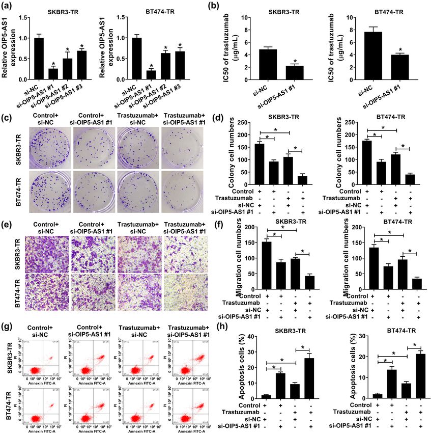
transfecting with constructed si-OIP5-AS1 plasmid into 3.3 Exosomal OIP5-AS1 derived from
SKBR3-TR and BT474-TR cells. As shown in Figure 2a, trastuzumab-resistant cells can be
three forms of si-OIP5-AS1 all notably reduced OIP5-AS1 absorbed by trastuzumab-sensitive cells
expression compared with the si-NC group, and then
in breast cancer
si-OIP5-AS1#1 was selected for subsequent analyses
because of the efficient interference efficiency, which Exosomes can be actively secreted by most cell types, and
was reflected by the lowest OIP5-AS1 expression after exosome-containing lncRNAs can be transmitted and
si-OIP5-AS1 transfection in cells. By contrast with si-NC exchanged into culture medium. To explore the impact
group, OIP5-AS1 down-regulation significantly reduced of exosome transfer on trastuzumab resistance in breast
the IC50 values of SKBR3-TR and BT474-TR cells to tras- cancer, we first isolated exosomes from the conditioned
tuzumab (Figure 2b); in addition, colony formation ana- medium supernatant of SKBR3-TR and BT474-TR cells.
lysis demonstrated that OIP5-AS1 knockdown combined The vesicles displayed a round shape with bilayered
with trastuzumab markedly decreased the number of membranes, and the diameter from 40 to 250 nm under
colonies formed of SKBR3-TR and BT474-TR cells a TEM, and NTA analysis further confirmed that the pre-
(Figure 2c and d). Meanwhile, transwell assay displayed dominant size of the vesicles was 100 nm (Figure 3a and b);
the number of migrated and invaded SKBR3-TR and BT474- in addition, as exhibited by western blot analysis, the
TR cells was significantly suppressed by the treatment of exosomal markers CD63, CD81, and TSG101 were detect-
trastuzumab, and this reduction was markedly enhanced by able in exosomes but not in cell lysates (Figure 3c).
the down-regulation of OIP5-AS1 (Figure 2e and f). Besides In addition, the expression change of OIP5-AS1 in the
that, it was also proved that OIP5-AS1 depletion reinforced culture medium of SKBR3-TR and BT474-TR cells was
trastuzumab-induced elevation of the apoptosis of SKBR3- detected after treatment with RNase. Results exhibited
TR and BT474-TR cells (Figure 2g and h). Taken together, that OIP5-AS1 expression in culture medium was little
OIP5-AS1 knockdown impeded breast cancer cells to trastu- affected upon RNase treatment but was greatly reduced
zumab resistance. when treated with RNase and Triton X-100 simultaneouslyOIP5-AS1 enhances trastuzumab resistance in BC 517 Figure 2: OIP5-AS1 knockdown restores trastuzumab sensitivity in trastuzumab resistant breast cancer cells. SKBR3-TR and BT474-TR cells were transfected with si-OIP5-AS1 or si-NC. After transfection, (a) qPCR analysis of OIP5-AS1 expression; (b) CCK-8 analysis of IC50 values of resistant cells to trastuzumab; (c and d) colony formation analysis of resistant cell proliferation; (e and f) transwell analysis of resistant cell migration; (g and h) resistant cell apoptosis analysis with flow cytometry. *P < 0.05. (Figure 3d), indicating that OIP5-AS1 presented in exo- Next, exosomes from the culture supernatant of SKBR3 somes. The treatment of SKBR3-TR and BT474-TR cells and BT474 cells were isolated, and qPCR analysis with GW4869, an inhibitor of the secretion of exosomes revealed OIP5-AS1 expression in SKBR3-TR and BT474- from cells, led to a reduction in the quantity of exosomes TR secreted exosomes was significantly higher than that (Figure 3e) as well as OIP5-AS1 expression (Figure 3f). All in exosomes from parental cells (SKBR3 and BT474) these results indicated that extracellular OIP5-AS1 was (Figure 3g). Importantly, SKBR3 and BT474 cells co-cul- packaged into exosomes, which were also secreted by tured with exosomes derived from SKBR3-TR and BT474- SKBR3-TR and BT474-TR cells. TR cells were treated with GW4869 or vehicle, and we
518 Qiang Yu et al. Figure 3: Exosomal OIP5-AS1 derived from trastuzumab resistant cells can be absorbed by trastuzumab-sensitive cells in breast cancer. (a and b) The image of purified exosomes derived from SKBR3-TR and BT474-TR cells captured by TEM; size distribution of purified exosomes was analyzed by NTA. (c) Western blot analysis of exosomal marker CD81, CD63, and TSG101 in isolated exosomes. (d) qPCR analysis of OIP5-AS1 expression in SKBR3-TR and BT474-TR cells following treatment with RNase alone or in combination with 0.1% Triton X-100. (e) The effect detection of GW4869 on the concentration of exosomes with a BCA protein assay. (f) qPCR analysis of OIP5-AS1 expression in exosomes treated with Vehicle and GW4869. (g) Analysis of levels of OIP5-AS1 in exosomes from SKBR3-TR and BT474-TR cells as well as parental SKBR3 and BT474 cells. (h) qPCR analysis of OIP5-AS1 expression in SKBR3 and BT474 cells co-cultured with exosomes, derived from SKBR3-TR and BT474-TR cells, and GW4869 or Vehicle. *P < 0.05. found the levels of OIP5-AS1 were increased by exosomes (Figure 3h), revealing OIP5-AS1 could be transmitted incubation but were decreased by GW4869 treatment into trastuzumab-sensitive cells via exosome transfer.
OIP5-AS1 enhances trastuzumab resistance in BC 519
3.4 Intercellular transfer of OIP5-AS1 by (Figure 4b). Subsequently, the two recipient cell lines
exosomes enhances trastuzumab exhibited increased IC50 values (Figure 4c), elevated cell
resistance in vitro viability (Figure 4d), migration (Figure 4e), and decreased
apoptosis (Figure 4f) following treatment with si-NC-exo
To identify whether OIP5-AS1 conferred trastuzumab compared with PBS group. By contrast with the si-NC-exo
resistance through the delivery of exosomes, first, exo- group, proliferation and migration of cells declined and apop-
somes were isolated from SKBR3-TR and BT474-TR cells tosis raised in the si-OIP5-AS1#1-exo group (Figure 4c–f).
transfected with si-OIP5-AS1#1 or si-NC, and qPCR Altogether, these data revealed that SKBR3 and BT474 cells
revealed a high expression of OIP5-AS1 in exosomes with exosomal OIP5-AS1 exhibited resistance to trastuzumab
from donor SKBR3-TR and BT474-TR cells transfected resistance.
with si-NC (si-NC-exo) compared with si-OIP5-AS1#1 (si-
OIP5-AS1#1-exo) (Figure 4a). Second, SKBR3 and BT474
cells were co-cultured with exosome-depletion PBS, si-
OIP5-AS1#1-exo or si-NC-exo, and we found OIP5-AS1 3.5 OIP5-AS1 is a sponge of miR-381-3p
expression was elevated with si-NC-exo incubation rela-
tive to PBS treatment, while si-OIP5-AS1#1-exo incuba- To explore the underlying mechanism of OIP5-AS1, miRNA
tion reduced the level of OIP5-AS1 in recipient cells targets were searched through starBase program, and
Figure 4: Intercellular transfer of OIP5-AS1 by exosomes enhances trastuzumab resistance in vitro. (a) qPCR analysis of OIP5-AS1 expression
in exosomes isolated from SKBR3-TR and BT474-TR cells transfected with si-OIP5-AS1#1 or si-NC. SKBR3 and BT474 cells were co-cultured
with exosome-depletion PBS, si-OIP5-AS1#1-exo, or si-NC-exo. After incubation; (b) levels of OIP5-AS1 were detected with qPCR; (c) CCK-8
analysis of IC50 values of parental cells to trastuzumab; (d) colony formation analysis of parental cell proliferation; (e) migration analysis of
parental cells using transwell assay; (f) flow cytometry assay for parental cell apoptosis. *P < 0.05.520 Qiang Yu et al. miR-381-3p was found that might be a target of OIP5-AS1 Then, a dual luciferase reporter assay was conducted (Figure 5a). To confirm this prediction, first, SKBR3 and and results showed that the luciferase activity of OIP5- BT474 cells were transfected with miR-NC or miR-381-3p, AS1 wt of both forms was decreased in SKBR3 and BT474 and miR-381-3p expression was significantly elevated by cells treated with miR-381-3p; however, there was no sig- miR-381-3p transfection compared with miR-NC (Figure 5b). nificant difference in luciferase activity on the two forms Figure 5: OIP5-AS1 is a sponge of miR-381-3p. (a) The potential binding sites of OIP5-AS1 and miR-381-3p. (b) qPCR analysis of miR-381-3p in SKBR3 and BT474 cells transfected with miR-NC or miR-381-3p. (c and d) Dual-luciferase reporter assay in SKBR3 and BT474 cells co- transfected with the reporter plasmid and the indicated miRNAs. (e) qPCR analysis of the enrichment level of OIP5-AS1 pulled down by bio- miR-381-3p or bio-NC in SKBR3 and BT474 cells. (f) qPCR analysis of OIP5-AS1 in SKBR3 and BT474 cells transfected with oe-OIP5-AS1 or Vector. (g) qPCR analysis of miR-381-3p expression in SKBR3 and BT474 cells transfected with oe-OIP5-AS1, Vector, si-NC, or si-OIP5-AS1#1. (h) Detection of levels of miR-381-3p in parental and resistant breast cancer cells with qPCR. *P < 0.05.
OIP5-AS1 enhances trastuzumab resistance in BC 521 of OIP5-AS1 mut (Figure 5c and d). Besides that, the 3.6 HMGB is a target of miR-381-3p results of pull-down assay revealed that the enrichment level of OIP5-AS1 in the bio-miR-381-3p group was mark- According to the prediction of starBase program, miR- edly higher than that in the bio-NC group (Figure 5e). All 381-3p was found to have the binding sites on HMGB these results suggested OIP5-AS1 specifically bound miR- (Figure 6a). The results of dual luciferase reporter assay 381-3p. Next, SKBR3 and BT474 cells were transfected displayed that miR-381-3p overexpression reduced the with oe-OIP5-AS1 to elevate OIP5-AS1 (Figure 5f), and luciferase activity of HMGB wt of both forms but not mutant qPCR analysis exhibited that miR-381-3p expression was reporter vector in SKBR3 and BT474 cells (Figure 6b and c), decreased by OIP5-AS1 overexpression, but was increased indicating that miR-381-3p directly targeted to HMGB. by OIP5-AS1 down-regulation in SKBR3 and BT474 cells Meanwhile, western blot analysis exhibited that miR- (Figure 5g). Thus, OIP5-AS1 targetedly repressed miR- 381-3p restoration suppressed HMGB expression, while 381-3p expression in breast cancer cells. In addition, this inhibition was reversed by OIP5-AS1 overexpression miR-381-3p was found to be decreased in SKBR3-TR and in SKBR3 and BT474 cells (Figure 6d). Therefore, miR- BT474-TR cells relative to parental SKBR3 and BT474 cells 381-3p targetedly suppressed HMGB expression and OIP5- (Figure 5h), indicating miR-381-3p was associated with AS1 positively regulated HMGB via miR-381-3p. Interest- trastuzumab resistance in breast cancer. ingly, HMGB was increased in SKBR3-TR and BT474-TR Figure 6: HMGB is a target of miR-381-3p. (a) Schematic representation of the predicted binding sites of miR-381-3p on HMGB. (b and c) Dual-luciferase reporter assay in SKBR3 and BT474 cells co-transfected with the reporter plasmid and the indicated miRNAs. (d) Western blot analysis of HMGB expression in SKBR3 and BT474 cells transfected with miR-NC, miR-381-3p, miR-381-3p + Vector, or miR- 381-3p + OIP5-AS1. (e) Western blot analysis of HMGB expression in parental and resistant breast cancer cells. *P < 0.05.
522 Qiang Yu et al.
cells compared with their parental SKBR3 and BT474 cells OIP5-AS1 expression decreased miR-381-3p and elevated
(Figure 6e); thus, HMGB was also linked to trastuzumab HMGB in tumors, while these effects were antipodal in si-
resistance in breast cancer. OIP5-AS1#1-exo (Figure 7d and e). Collectively, exosomal
OIP5-AS1 might induce trastuzumab resistance and promote
tumor growth via regulating miR-381-3p and HMGB in vivo.
3.7 Exosomal OIP5-AS1 induces trastuzumab
resistance and promotes tumor growth
in vivo 3.8 Serum exosomal OIP5-AS1 level is
associated with trastuzumab resistant
The effect of OIP5-AS1 on trastuzumab resistance in vivo in breast cancer patients
was determined. As shown in Figure 7a and b, trastu-
zumab treatment significantly inhibited tumor growth We further attempted to analyze the expression level of
in nude mice compared with control groups (group 1 vs exosomal OIP5-AS1 in 57 serum samples from breast cancer
group 2). More importantly, with trastuzumab treatment, patients receiving trastuzumab treatment. Results implied
tumor cells in si-NC-exo group grew faster than trastu- that the serum exosomal OIP5-AS1 remarkably higher in
zumab or si-OIP5-AS1#1-exo group (group 3 vs group 2 or patients who did not respond to treatment than in those
group 4, respectively) (Figure 7a and b), suggesting that who responded to trastuzumab (Figure 8a). Later, the diag-
exosome-mediated transfer of OIP5-AS1 suppressed the nostic potential of exosomal OIP5-AS1 in serum was calcu-
cytotoxicity induced by trastuzumab treatment in vivo. lated. As revealed by ROC analysis, an area under curve of
In addition, molecule analysis showed si-NC-exo had high 0.764 with a diagnostic sensitivity and specificity reaching
levels of OIP5-AS1 compared with si-OIP5-AS1#1-exo, and 59.26 and 93.33%, respectively (95% CI = 0.6326–0.8958),
attenuated trastuzumab treatment-induced OIP5-AS1 reduc- was observed (Figure 8b). In addition, patients were classi-
tion in tumors (Figure 7c). Besides, si-NC-exo with high fied into a low and high exosomal OIP5-AS1 expression
Figure 7: Exosomal OIP5-AS1 induces trastuzumab resistance and promotes tumor growth in vivo. (a) Tumor volumes were calculated every
4 days. (b) Tumor masses were collected and weighed on day 32. (c and d) Detection of levels of OIP5-AS1 and miR-381-3p in the tumor
messes of each group with qPCR. (e) Western blot analysis of HMGB protein in the tumor messes of each group. *P < 0.05.OIP5-AS1 enhances trastuzumab resistance in BC 523
groups according to the cut-offs (1.355) established by ROC, cancer. For example, OIP5-AS1 accelerated the progres-
and we found the proportion of patients not responding to sion of gastric cancer by regulating HMGA2 through
trastuzumab was greatly higher in the high exosomal OIP5- miR-367-3p [22]. OIP5-AS1 interacted with ROCK1 to pro-
AS1 expressing group than in the low expressing group mote cell carcinogenesis in cervical cancer via absorb-
(Figure 8c). These data indicated that exosomal OIP5-AS1 ing miR-143-3p. OIP5-AS1 induced cisplatin resistance
was also dysregulated in the serum of breast cancer patients in osteosarcoma through regulating the LPAATβ/PI3K/
and might be a promising diagnostic biomarker for trastu- AKT/mTOR signaling pathway via a mechanism invol-
zumab resistance in patients with breast cancer. ving miR-340-5p [23]. Importantly, OIP5-AS1 was con-
firmed to act as an oncogene via regulating cell malignant
phenotypes in breast cancer [14]. Thus, we thought OIP5-
AS1 might also involve in the regulation of drug resis-
4 Discussion tance and then confirmed that knockdown of OIP5-AS1
restored trastuzumab sensitivity in trastuzumab-resistant
Molecular targeted therapies are one of the major and breast cancer cells.
revolutionized modalities of medical treatment, which Exosomes are one of the extracellular vesicles and
interfere with specific molecules to block cancer growth, have been reported to involve in the modulation of chemo-
survival, and progression [15]. In breast cancer, some resistance of the recipient cells via the transfer of
molecular targeted therapies, including trastuzumab and lncRNAs in diverse cancers [24,25]. In breast cancer, the
lapatinib, have been approved and demonstrated remark- effects of exosomal lncRNA in drug resistance were also
able clinical success in the treatment of cancer. They direct demonstrated. For instance, exosome-mediated transfer
against HER2 and bevacizumab as well as vascular of AFAP1-AS1 induced trastuzumab resistance in breast
endothelial growth factor [16,17]. lncRNAs are a class of cancer via the interaction with AUF1 and the activation of
RNAs with lengths exceeding 200 nucleotides and have ERBB2 translation [26]. Intercellular transfer of lncRNA
been widely revealed to implicate in various physical H19 promoted doxorubicin resistance in breast cancer
and pathological processes, such as cell differentiation, [27]. Thus, whether exosomal OIP5-AS1 is implicated in
proliferation, apoptosis, and metabolism, thereby affecting trastuzumab resistance in breast cancer was investigated.
the development and progression of cancers [18–21]. They Results exhibited that extracellular OIP5-AS1 was packaged
are considered as candidate therapeutic targets. into exosomes, which were secreted by trastuzumab-resistant
OIP5-AS1 is a functional RNA and has been identified cells and could be taken up by trastuzumab sensitive cells
to sever as an oncogene to participate in the evolution via exosomes transfer, thereby disseminated trastuzumab
of tumorigenesis and drug resistance in many types of resistance to recipient cells in vitro as well as inhibited
Figure 8: Serum exosomal OIP5-AS1 level is associated with trastuzumab resistant in breast cancer patients. (a) qPCR analysis of OIP5-AS1
in exosomes isolated from the serum of breast cancer patients responding or not responding to trastuzumab treatment. (b) ROC curve
analysis of the diagnostic value of exosomal OIP5-AS1 in breast cancer patients receiving trastuzumab treatment. (c) qPCR suggested that
the proportion of patients not responding to trastuzumab was greatly higher in the high exosomal OIP5-AS1 expressing group than in the
low expressing group. *P < 0.05.524 Qiang Yu et al.
cytotoxicity induced by trastuzumab in tumor growth in vivo [3] Slamon DJ, Leyland-Jones B, Shak S, Fuchs H, Paton V,
in breast cancer. In addition, exosomal OIP5-AS1 was Bajamonde A, et al. Use of chemotherapy plus a monoclonal
also dysregulated in the serum of breast cancer patients antibody against HER2 for metastatic breast cancer that
overexpresses HER2. N Engl J Med. 2001;344(11):783–92.
and might act as a promising diagnostic biomarker for
[4] Ross JS, Slodkowska EA, Symmans WF, Pusztai L, Ravdin PM,
trastuzumab resistance in breast cancer patients. There- Hortobagyi GN. The HER-2 receptor and breast cancer:
fore, exosomal OIP5-AS1 may be useful for the treatment ten years of targeted anti-HER-2 therapy and personalized
of breast cancer and the prediction of trastuzumab resis- medicine. Oncologist. 2009;14(4):320–68.
tance. The potential underlying molecular mechanism of [5] Marty M, Cognetti F, Maraninchi D, Snyder R, Mauriac L,
Tubiana-Hulin M, et al. Randomized phase II trial of the efficacy
the action of OIP5-AS1 was further probed. In this study,
and safety of trastuzumab combined with docetaxel in patients
we found OIP5-AS1 directly bound to miR-381-3p and with human epidermal growth factor receptor 2-positive meta-
subsequently served as a miRNA sponge to up-regulate static breast cancer administered as first-line treatment: the
the expression of the miR-381-3p target gene HMGB3. M77001 study group. J Clin Oncol. 2005;23(19):4265–74.
miR-381-3p is a well-recognized anti-tumor miRNA, the [6] Vogel CL, Cobleigh MA, Tripathy D, Gutheil JC, Harris LN,
Fehrenbacher L, et al. Efficacy and safety of trastuzumab as a
overexpression of which was confirmed to suppress cell
single agent in first-line treatment of HER2-overexpressing
proliferation, cell cycle progression, and migration in
metastatic breast cancer. J Clin Oncol. 2002;20(3):719–26.
breast cancer [28]. HMGB3 belongs to HMGB family and [7] Liang K, Lu Y, Jin W, Ang KK, Milas L, Fan Z. Sensitization of
plays significant effects on DNA repair, recombination, breast cancer cells to radiation by trastuzumab. Mol Cancer
transcription, and replication [29]. Besides that, it has Ther. 2003;2(11):1113–20.
been demonstrated that HMGB3 silence could inhibit [8] Lee TH, D’Asti E, Magnus N, Al-Nedawi K, Meehan B, Rak J.
Microvesicles as mediators of intercellular communication in
cell growth and progression in breast cancer [30,31].
cancer–the emerging science of cellular ‘debris’. Semin
Thus, we considered that exosomal OIP5-AS1 may confer Immunopathol. 2011;33(5):455–67.
trastuzumab resistance in breast cancer cells via the reg- [9] Cocucci E, Meldolesi J. Ectosomes and exosomes: shedding
ulation of miR-381-3p/HMGB3 axis. the confusion between extracellular vesicles. Trends Cell Biol.
In conclusion, this study demonstrated that exosome- 2015;25(6):364–72.
mediated transfer of OIP5-AS1 partially induced trastu- [10] Valencia K, Luis-Ravelo D, Bovy N, Antón I, Martínez-
Canarias S, Zandueta C, et al. miRNA cargo within exosome-
zumab resistance in breast cancer through miR-381-3p/
like vesicle transfer influences metastatic bone colonization.
HMGB3 axis, indicating a therapeutic strategy for the tras- Mol Oncol. 2014;8(3):689–703.
tuzumab resistance in patients with breast cancer. [11] Abels ER, Breakefield XO. Introduction to extracellular vesi-
cles: biogenesis, RNA cargo selection, content, release, and
Author contributions: Q. X. X. conceived the project and uptake. Cell Mol Neurobiol. 2016;36(3):301–12.
[12] Pefanis E, Wang J, Rothschild G, Lim J, Kazadi D, Sun J, et al.
supervised all experiments. Y. Q. and L. Y. M. conducted
RNA exosome-regulated long non-coding RNA transcription
all experiments and analyzed the data. Y. Q., L. Y. M., controls super-enhancer activity. Cell. 2015;161(4):774–89.
P. S. J., and L. J. wrote the paper. All authors read and [13] Li Y, Han X, Feng H, Han J. Long noncoding RNA OIP5-AS1 in
approved the final manuscript. cancer. Clin Chim Acta. 2019;499:75–80.
[14] Zeng H, Wang J, Chen T, Zhang K, Chen J, Wang L, et al.
Conflict of interest: There are no conflicts of interest. Downregulation of long non-coding RNA Opa interacting pro-
tein 5-antisense RNA 1 inhibits breast cancer progression by
targeting sex-determining region Y-box 2 by microRNA-129-5p
Data availability statements: The datasets used during upregulation. Cancer Sci. 2019;110(1):289–302.
the current study are available from the corresponding [15] Lee YT, Tan YJ, Oon CE. Molecular targeted therapy: treating
author on reasonable request. cancer with specificity. Eur J Pharmacol. 2018;834:188–96.
[16] Schlotter CM, Vogt U, Allgayer H, Brandt B. Molecular targeted
therapies for breast cancer treatment. Breast Cancer Res.
2008;10(4):211.
[17] Munagala R, Aqil F, Gupta RC. Promising molecular targeted
therapies in breast cancer. Indian J Pharmacol.
References 2011;43(3):236–45.
[18] Bhan A, Soleimani M, Mandal SS. Long noncoding RNA and
[1] Siegel R, Ma J, Zou Z, Jemal A. Cancer statistics, 2014. CA cancer: a new paradigm. Cancer Res. 2017;77(15):3965–81.
Cancer J Clin. 2014;64(1):9–29. [19] Huang HW, Xie H, Ma X, Zhao F, Gao Y. Upregulation of lncRNA
[2] Gong C, Yao Y, Wang Y, Liu B, Wu W, Chen J, et al. Up-regula- PANDAR predicts poor prognosis and promotes cell prolifera-
tion of miR-21 mediates resistance to trastuzumab therapy for tion in cervical cancer. Eur Rev Med Pharmacol Sci.
breast cancer. J Biol Chem. 2011;286(21):19127–37. 2017;21(20):4529–35.OIP5-AS1 enhances trastuzumab resistance in BC 525
[20] Hung CL, Wang LY, Yu YL, Chen HW, Srivastava S, Petrovics G, binding with AUF1 and activating ERBB2 translation. Mol
et al. A long noncoding RNA connects c-Myc to tumor meta- Cancer. 2020;19(1):26.
bolism. Proc Natl Acad Sci USA. 2014;111(52):18697–702. [27] Wang X, Pei X, Guo G, Qian X, Dou D, Zhang Z, et al. Exosome-
[21] Zhang A, Xu M, Mo YY. Role of the lncRNA-p53 regulatory mediated transfer of long noncoding RNA H19 induces doxo-
network in cancer. J Mol Cell Biol. 2014;6(3):181–91. rubicin resistance in breast cancer. J Cell Physiol.
[22] Tao Y, Wan X, Fan Q, Wang Y, Sun H, Ma L, et al. Long non-coding 2020;235(10):6896–904.
RNA OIP5-AS1 promotes the growth of gastric cancer through [28] Wu M, Fan B, Guo Q, Li Y, Chen R, Lv N, et al. Knockdown of
the miR-367-3p/HMGA2 axis. Dig Liver Dis. 2020;52(7):773–9. SETDB1 inhibits breast cancer progression by miR-381-3p-
[23] Song L, Zhou Z, Gan Y, Li P, Xu Y, Zhang Z, et al. Long non- related regulation. Biol Res. 2018;51(1):39.
coding RNA OIP5-AS1 causes cisplatin resistance in osteosar- [29] Nemeth MJ, Curtis DJ, Kirby MR, Garrett-Beal LJ, Seidel NE,
coma through inducing the LPAATβ/PI3K/AKT/mTOR signaling Cline AP, et al. Hmgb3: an HMG-box family member expressed
pathway by sponging the miR-340-5p. J Cell Biochem. in primitive hematopoietic cells that inhibits myeloid and
2019;120(6):9656–66. B-cell differentiation. Blood. 2003;102(4):1298–306.
[24] Zhao W, Liu Y, Zhang C, Duan C. Multiple roles of exosomal [30] Gu J, Xu T, Zhang CM, Chen HY, Huang QH, Zhang Q. HMGB3
long noncoding RNAs in cancers. Biomed Res Int. small interfere RNA suppresses mammosphere formation of
2019;2019:1460572. MDA-MB-231 cells by down-regulating expression of HIF1α.
[25] Wang M, Zhou L, Yu F, Zhang Y, Li P, Wang K. The functional Eur Rev Med Pharmacol Sci. 2019;23(21):9506–16.
roles of exosomal long non-coding RNAs in cancer. Cell Mol [31] Gu J, Xu T, Huang QH, Zhang CM, Chen HY. HMGB3 silence
Life Sci. 2019;76(11):2059–76. inhibits breast cancer cell proliferation and tumor growth by
[26] Han M, Gu Y, Lu P, Li J, Cao H, Li X, et al. Exosome-mediated interacting with hypoxia-inducible factor 1α. Cancer Manag
lncRNA AFAP1-AS1 promotes trastuzumab resistance through Res. 2019;11:5075–89.You can also read