Expression of intercellular adhesion molecule 1 (ICAM-1) on the human oviductal epithelium and mediation of lymphoid cell adherence
←
→
Page content transcription
If your browser does not render page correctly, please read the page content below
Journal of Reproduction and Fertility (2000) 120, 115–123

            Expression of intercellular adhesion molecule 1 (ICAM-1)
              on the human oviductal epithelium and mediation of
                            lymphoid cell adherence
                    E. Utreras, P. Ossandon, C. Acuña-Castillo, L. Varela-Nallar, C. Müller,
                                   J. A. Arraztoa, H. Cardenas and M. Imarai*
             Universidad de Santiago de Chile, Facultad de Química y Biología, Laboratorio de Inmunología de la Reproducción,
                                                   Casilla 40 Correo 33, Santiago, Chile

                 The epithelium of the human oviduct expresses the major histocompatibility complex
                 (MHC) class II and shows endocytic properties towards luminal antigens. Therefore,
                 the epithelial cells might behave as antigen-presenting cells, inducing a local immune
                 response. The activation of antigen-specific T cells not only requires presentation of the
                 peptide antigen by MHC class II, but also the presence of co-stimulatory molecules in
                 the antigen-presenting cells. Therefore, the expression of the intercellular adhesion
                 molecule 1 (ICAM-1) was examined in the epithelium of the human oviduct. Most
                 oviducts showed epithelial ICAM-1 expression, as assessed by immunocytochemistry,
                 western blot analysis and RT–PCR assay, and the expression was restricted to the
                 luminal border of ciliated and secretory cells. Interferon γ, interleukin 1 and
                 lipopolysaccharide treatments increased the percentage of ICAM-1-positive cells in
                 primary cultures, indicating that the expression of ICAM-1 in the oviduct might be
                 upregulated in vivo by inflammatory cytokines or bacterial infections. Binding assays
                 between allogenic phytohaemagglutinin-activated lymphocytes and epithelial
                 monolayers expressing ICAM-1 demonstrated that this molecule stimulated
                 lymphocyte adherence. The presence of ICAM-1, in addition to MHC class II, supports
                 the putative role of the oviductal epithelium in antigen presentation. The exclusive
                 apical distribution of ICAM-1 indicates that T-cell activation would occur in a polarized
                 manner. Binding of lymphoid cells to the surface of the oviductal epithelium may help
                 to retain these immune cells that are required for the clearance of pathogens.

                                                                              The induction of an immune response requires processing
                            Introduction
                                                                           and presentation of the peptide antigen by MHC class II-
All the elements of the mucosal immune system have been                    positive antigen-presenting cells to antigen-specific T cells. It
identified in the human oviduct, including intraepithelial T               also requires cell–cell interactions that are not antigen
lymphocytes (CD4+ and CD8+), T cells clustered in follicles or             specific between the antigen-presenting cells and the T cells.
dispersed in the subepithelial tissue, B lymphocytes and                   Those non-antigen-specific interactions include the binding
macrophages (Cooper et al., 1987; Otsuki et al., 1989; Wollen              of the intercellular adhesion molecule 1 (ICAM-1) on the
et al., 1994; Givan et al., 1997; Cardenas et al., 1998). The              antigen-presenting cell to the lymphocyte function-
epithelium of the oviduct also expresses the major                         associated antigen molecules (LFA-1) on the T-cell surface.
histocompatibility complex (MHC) class II molecules                        The binding of ICAM-1 to LFA-1 provides an important co-
(Bulmer and Earl, 1987; Edelstam et al., 1992; Imarai et al.,              stimulatory signal for the T-cell receptor mediated activation
1998) and shows endocytic properties towards luminal                       (Dougherty et al., 1988; van Seventer et al., 1990), the absence
antigens (Murakami et al., 1985; Imarai et al., 1998). These               of which leads to functional inactivation of the antigen-
observations indicate that the oviductal epithelium might                  specific T cells (van Parijs and Abbas, 1998). In addition to its
participate in the processing of antigens and in the local                 role in lymphocyte activation, ICAM-1 mediates leucocyte
stimulation of lymphoid cells, as proposed for other mucosal               adherence and is involved in transendothelial and
epithelial cells (Bland and Warren, 1986; Tabibzadeh et al.,               transepithelial migration during inflammation (Devine et al.,
1986; Kaiserlian et al., 1989; Wira and Rossoll, 1995).                    1986; Springer, 1994; Taguchi et al., 1998).
                                                                              ICAM-1 is a membrane-bound glycoprotein expressed on
*Correspondence.                                                           non-haematopoietic cells such as vascular endothelial cells,
Received 13 January 2000.                                                  thymic cells, certain epithelial cells, fibroblasts, and on

                                                © 2000 Journals of Reproduction and Fertility Ltd
                                                                0022–4251/2000                      Downloaded from Bioscientifica.com at 10/16/2021 06:50:45AM
                                                                                                                                                  via free access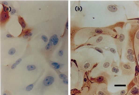
116                                                      E. Utreras et al.

haematopoietic cells such as tissue macrophages and mitogen-       conjugated anti-mouse IgG (1:500) at room temperature for
stimulated T lymphocytes (Dustin et al., 1986). Inflammatory       30 min. Incubation with horseradish peroxidase–biotin
mediators such as interferon γ (IFN-γ), tumour necrosis factor     complex (ABC-complex, Amersham Life Science Ltd; 1:100)
α (TNF-α) and interleukin 1 (IL-1) induce ICAM-1 expression        was also carried out at room temperature for 1 h followed by
on peripheral blood leucocytes and other cells (Dustin et al.,     diaminobenzidine (DAB) mixture (0.05% (w/v) DAB in
1986; Myers et al., 1992; Haraldsen et al., 1996). ICAM-1          0.05 mol Tris–HCl l–1, pH 8, plus hydrogen peroxide 0.003%
expression is also induced after bacterial infection and by        (v/v) final concentration) for 7 min. Positive controls were
bacterial products such as lipopolysaccharide (Elgavish, 1993;     the lymphoid cells present in the subepithelial tissue of most
Huang et al., 1996). Induction is dependent on protein and         samples. Negative controls, in which the primary antibodies
mRNA synthesis and is reversible (Dustin et al., 1986).            were omitted or replaced by non-immune serum, were run
   The present study investigated whether the epithelium of        routinely in all experiments.
the human oviduct expresses ICAM-1 and examined the role
of IFN-γ, IL-1 and lipopolysaccharide on the induction of
ICAM-1 expression. The study also investigated the
                                                                   Primary epithelial cell culture
potential role of ICAM-1 in leucocyte adherence to the
oviductal epithelium.                                                  After washing the organ using PBS, the lumen was
                                                                   exposed through a longitudinal cut and strips of mucosal
                                                                   folds were dissected. The strips were washed in TC199
                                                                   medium (Gibco BRL) and small pieces of about 1 mm in
                 Materials and Methods
                                                                   diameter were cut off and subsequently treated with 0.25%
                                                                   (w/v) trypsin (37⬚C, 10 min). The cell suspension was washed
Antibodies
                                                                   four times at 800 g for 8 min using Hank’s balanced salt
  The following antibodies were used: mouse monoclonal             solution (Gibco BRL). The final pellet was resuspended in
anti-human ICAM-1 (DAKO Co., Carpinteria, CA) and mouse            TC199 containing 10% (v/v) fetal calf serum (Gibco BRL),
monoclonal anti-cytokeratin (DAKO Co.), goat polyclonal IgG        insulin (5 mg ml–1), glutamine (1 mmol l–1), pyruvate (1 mmol
anti-human ICAM-1 (Santa Cruz Biotechnology Inc., Santa            l–1) and antibiotics (50 iu penicillin ml–1 and 50 µg
Cruz, CA), biotinylated goat anti-mouse IgG (Pierce Chemical       streptomycin ml–1). Cells were seeded in culture plates and
Co., Rockford, IL), affinity-purified mouse polyclonal anti-       incubated at 37⬚C in 5% CO2 in air to allow early attachment
goat IgG (Pierce Chemical Co.) and alkaline phosphatase-           of fibroblasts. After incubation for 1 h, the medium
conjugated anti-mouse IgG (Sigma Chemical Co., St Louis,           containing non-attached cells was removed and seeded in
MO).                                                               another plate. Cells were incubated for at least 3 days to
                                                                   obtain round colonies of epithelial cells which were identified
                                                                   routinely by cytokeratin immunostaining (Takeuchi et al.,
                                                                   1991).
Tissue samples
   Protocols were approved by the Ethics Committee of the
University of Santiago, Chile. Organs were obtained after
                                                                   Western blot analysis
informed consent from women aged 32–50 years that had
undergone hysterectomy because of myoma or other                      Pieces of mucosal tissue or cultured epithelial cells were
neoplasic conditions not affecting the oviduct. A blood            obtained from several oviducts. Cold RIPA buffer (1% (v/v)
sample was taken on the day of surgery and concentrations          Nonidet P-40, 0.5% (w/v) sodium deoxycholate, 0.1% (w/v)
of oestrogen and progesterone were measured by                     SDS and 0.1 mg phenylmethylsulfonyl fluoride (PMSF) l–1 in
radioimmunoassay. Organs were received in sterile                  PBS) and a mixture of antipain, chymostatin, leupeptin and
Dulbecco’s modified Eagle’s medium (DMEM; GIBCO BRL,               pepstatin (final concentration 5 µg ml–1) were added to the
Gaithersburg, MD), transported to the laboratory and               samples, which were homogenized using a potter and
processed within 2 h after surgery.                                incubated on ice for 30 min. Cell lysates were centrifuged at
                                                                   14 000 g for 20 min at 4⬚C. Protein concentration in the
                                                                   supernatant fluid was determined using the Bradford assay
                                                                   (Bradford, 1976). Protein samples (50 µg lane) and a
Immunohistochemistry
                                                                   molecular mass standard (BenchMark prestained protein
   Pieces of ampullary segments of 1 cm in length from each        ladder, GIBCO BRL) were subjected to 10% (w/v) SDS-PAGE
oviduct were snap-frozen in liquid nitrogen and stored at          under reducing conditions and transferred to nitrocellulose
–20⬚C until further processing. Tissue slices of 6 µm, obtained    (Sigma) by electroblotting (Harlow and Lane, 1988).
using a cryostat (Starlet Bright, Huntingdon), were fixed in       Membranes were incubated overnight in blocking solution at
cold 4% (w/v) paraformaldehyde for 20 min, followed by             4⬚C (1% BSA in PBS) and the goat polyclonal antibody
70% (v/v) cold ethanol treatment for 10 min. After 30 min of       against ICAM-1 was added (1:1000). After incubation for 2 h
incubation in 1% (v/v) BSA, sections were incubated                at room temperature, the blot was washed several times and
overnight at 4⬚C with the mouse primary antibody against           exposed to the alkaline phosphatase-conjugated antibody
human ICAM-1 or against cytokeratin (1:100). The sections          against goat IgG (1:2000). Detection was carried out using the
were washed several times and incubated with biotin-               substrates 5-bromo-4-chloro-3 indolyl phosphate (BCIP;

                                                                                             Downloaded from Bioscientifica.com at 10/16/2021 06:50:45AM
                                                                                                                                           via free access
ICAM-1 in the human oviduct 117
Amresco, Solon, OH) and nitro blue tetrazolium (NBT; 0.2 mmol dNTPs l–1, 3 mmol MgCl2 l–1 and reaction buffer
Amresco). (50 mmol KCl l–1, 10 mmol Tris–HCl l–1, pH 9.0, 0.1% (v/v)
Triton-X100) and 2.5 U Taq polymerase (Promega) in a final
volume of 25 µl. A total of 35 cycles of amplification were
performed using the following protocol: denaturing at 92⬚C
Cytokine and lipopolysaccharide treatment
for 1 min, annealing at 60⬚C for 1 min, extension at 72⬚C for
Epithelial cells (5 ⫻ 104) isolated from each organ were 1 min. Expected sizes of ICAM-1 and actin products were 447
seeded onto 12 mm round coverslips placed in 24-well plates and 448 base pairs (bp), respectively.
(Nalge Nunc, Naperville, IL) and incubated at 37ºC in an
atmosphere of 5% CO2 in air. After round cell colonies were
established, recombinant human IFN-γ (10 iu ml–1; Biosource
Activation and labelling of human lymphocytes
International SA, Camarillo, CA), IL-1 (50 and 1000 iu ml–1;
Biosource International SA) or lipopolysaccharide (5 and Blood was obtained from normal healthy volunteers and
10 µg ml–1) were added to the plate and cells were grown for 2 placed in heparinized tubes. Peripheral mononuclear cells
days (Haraldsen et al., 1996). For immunocytochemistry, were separated by centrifugation for 30 min at 900 g over
independent experiments using IFN-γ were performed with Ficoll Hypaque (Pharmacia, Uppsala). Interfacial cells were
epithelial cell cultures from five patients, whereas experiments recovered and washed in RPMI-1640 medium. Activation and
using IL-1 and lipopolysaccharide were performed using cell labelling of cells were performed according to Cunningham
cultures from four patients. All treatments and controls were and Kirby (1995). Briefly, cells were resuspended and
run in duplicate. Lipopolysaccharide was from E. coli 0122:38 cultured in RPMI supplemented with 10% (v/v) fetal calf
(Sigma). serum, 1 mmol glutamine l–1, 1 mmol pyruvate l–1 and
antibiotics, in the presence of 10 µg phytohaemaglutinin ml–1
for 2–3 days at 37⬚C in an atmosphere of 5% CO2 in air.
Activated lymphocytes were collected and separated from
Immunocytochemistry and cell counts
dead cells using Ficoll Hypaque. Lymphocytes were labelled
After cytokine and lipopolysaccharide treatment, cells on using 100 µCi Na251CrO4 (NEN, Boston, MA) for 90 min at
coverslips were fixed in cold 2% (w/v) paraformaldehyde for 37⬚C.
20 min and immunodetection of ICAM-1 and cytokeratine
was performed in duplicate as described for tissue slices.
Cells were counterstained with haematoxylin and coverslips
Assay of lymphocyte binding to the oviductal epithelium
were mounted in Kaiser’s glycerol gelatin (Merck,
Darmstadt). Samples were viewed under the ⫻ 40 objective Binding of lymphocytes to epithelial cells was examined
of an Olympus BX40 microscope. Positive cells were using the method of Ikuta et al. (1991). Confluent epithelial
searched for over the entire coverslip, which usually monolayers in 24 well-plates, with or without IFN-γ pre-
contained at least 500 cells. If there was positive ICAM-1 treatment (48 h), were incubated with 0.5 ml 51Cr-labelled
staining, at least 100 cells were classified as positive or lymphocytes (10 000 cells per well) for 1 h at 37⬚C. After
negative from three different fields selected randomly. Data washing to remove non-adherent lymphocytes, the
are expressed as percentage of ICAM-1-positive cells. remaining cells were lysed by the addition of 0.5 ml of 10%
(v/v) Triton-X100 (Sigma) for 10 min. The amount of 51Cr
released was measured using a Packard Cobra II gamma-
counter, and the percentage of adherent cells was calculated
RT–PCR assay
as follows: (c.p.m. in lysate ⫻ 100)/(c.p.m. in maximum
The cells were collected from culture dishes by control). A concentration of 10 µg ml–1 of the anti-ICAM-1
trypsin–EDTA (Gibco BRL) treatment. After centrifugation, monoclonal antibody (Dako) was added to epithelial cells
total RNA was prepared according to Chomczynski and 30 min before the addition of lymphocytes to investigate the
Sacchi (1987). For cDNA synthesis, 0.5 µg RNA, 50 pmol role of ICAM-1 in lymphocyte adherence to epithelial cells.
oligo dT (Promega, Madison, WI), 0.5 mmol dNTPs l–1 Experiments were performed in triplicate.
(Promega), 5 mmol (dithiothreitol) DTT l–1, 2 µl reaction
buffer (50 mmol KCl l–1, 10 mmol Tris–HCl l–1, pH 9.0, 0.1%
(v/v) Triton-X100) and 200 U M-MLV reverse transcriptase
Statistical analysis
(Promega) in 20 µl final volume were incubated for 1 h at
42⬚C. For PCR amplification, primers were designed The Mann–Whitney U test was used to compare the mean
according to sequences provided by the National Center for serum concentration of oestradiol and progesterone in
Biotechnology Information of the NIH. Primer sequences patients whose oviducts expressed ICAM-1 with those in
were: ICAM-1 sense sequence, 5’ GGG AGG CTC CGT GCT patients whose oviducts did not express ICAM-1. The paired
GGT GA 3’; ICAM-1 antisense sequence, 5’ TCA GTG CGG t test was used to examine cytokine and lipopolysaccharide
CAC GAG AAA TTG 3’; actin sense sequence, 5’ CTC ATC treatment effects. Data on ICAM-1-dependent adherence of
GTA CTC CTG CTT GCT G 3’; actin antisense sequence, 5’ lymphocytes to epithelial cells were analysed by multiple
GCT GTG CTA TGT TGC CCT AGA C 3’. PCR reaction comparisons using ANOVA followed by Duncan’s test. A
mixture included 5 µl cDNA, 50 pmol of each primer, P value of < 0.05 was considered significant.
Downloaded from Bioscientifica.com at 10/16/2021 06:50:45AM
via free access118 E. Utreras et al.
Results Table 1. Epithelial expression of intercellular adhesion
molecule 1 (ICAM-1) in the human oviduct
Expression of ICAM-1 in the human oviduct
Age of Serum
Immunohistochemical staining using a monoclonal
patient Uterine oestradiol Progesterone
antibody raised against ICAM-1 was performed in ampullary (years) pathology (pmol l–1) (nmol l–1) ICAM-1
segments. Eight of 14 specimens showed ICAM-1 expression
(Table 1) along the epithelium, which was always restricted to 35 IEN 71 3.4 +
the luminal border of both ciliated and secretory cells (Fig. 1). 36 IEN 92 1.0 +
ICAM-1 staining was also observed in a population of 40 Myoma 92 1.0 +
stromal cells probably consisting of mononuclear leucocytes. 50 Myoma 92 1.3 +
Sex steroid concentrations influence gene expression in 42 Myoma 106 2.0 +
receptor-bearing organs like the oviduct, thus the expression 46 Myoma 163 3.1 +
of ICAM-1 was correlated with the oestradiol and 38 Myoma 284 34.7 +
45 Myoma 596 4.2 +
progesterone serum concentration in the patients. Serum
Median 99a 2.6
oestradiol concentrations in the patients whose oviductal
epithelia expressed ICAM-1 were significantly lower (Table 1; 43 Myoma 108 9.3 –
median 99 pmol l–1, range 71–596) compared with those that 42 Myoma 138 5.9 –
did not (Table 1; median 508 pmol l–1, range 108–840) 46 Myoma 319 2.2 –
(P < 0.05). No difference was found in serum progesterone 44 Myoma 696 2.0 –
concentrations between the groups (Table 1). ICAM-1 32 IEN 710 2.4 –
expression was also determined in primary cultures of 48 Myoma 840 25.4 –
oviductal epithelial cells from ten patients. After 6 days of Median 508b 4.2
culture, epithelial cell monolayers contained more than 95% IEN: intraepithelial neoplasia.
of cells expressing cytokeratine 18 (that is, epithelial cells). a versus b, P < 0.05 (Mann–Whitney U test).
Nine of ten cell cultures expressed variable amounts of
ICAM-1 immunostaining, ranging from 0 to 80% of positive
cells (data not shown). epithelial cells (n = 3; Fig. 2). The signal was greatly reduced
Western blot analyses were also used to examine ICAM-1 when blots were incubated with a mixture of the polyclonal
expression in the oviductal mucosae and epithelial cells. A antibody and the ICAM-1 blocking peptide (Santa Cruz
specific protein band of approximately 70 kDa was present in Biotechnology Inc.) (Fig. 2), indicating that the band of
protein extracts prepared from mucosae (not shown) or 70 kDa was ICAM-1.
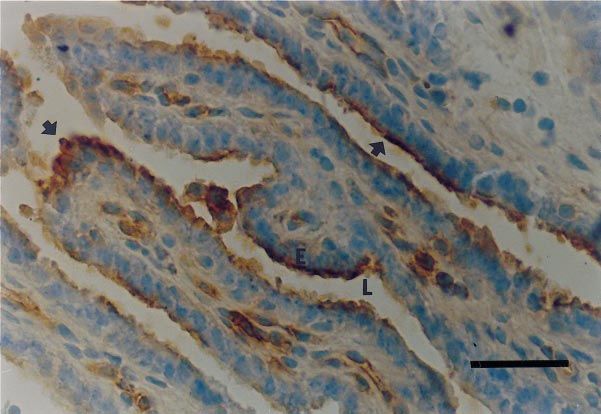
Fig. 1. Immunohistochemical detection of intercellular adhesion molecule 1 (ICAM-1) in the
epithelium of the human oviduct by a peroxidase-based staining system (brown) and haematoxylin
counterstaining (blue). Positive staining for ICAM-1 appeared along the epithelium and was restricted
to the luminal border of the cells (arrows). E: epithelium; L: lumen. Scale bar represents 50 µm.
Downloaded from Bioscientifica.com at 10/16/2021 06:50:45AM
via free accessICAM-1 in the human oviduct 119
kDa 1 2 3 Effect of IFN-γ, IL1 and lipopolysaccharide on the expression
of ICAM-1
220 Incubation of oviductal epithelial cells with 10 iu IFN-γ
ml–1 (n = 5) or with inflammatory mediators such as IL-1
(1000 iu ml–1; n = 4) and lipopolysaccharide (10 µg ml–1;
130 n = 4) for 48 h augmented the percentage of ICAM-1-positive
cells detected by immunocytochemistry (Figs 3 and 4). The
induction was observed in all cases studied. IFN-γ also
90 induced an increase in ICAM-1 mRNA in all cases examined
(n = 6; P < 0.03), as assessed by RT–PCR (Fig. 5).
70
Role of ICAM-1 in lymphocyte adherence to oviductal
epithelial cells
60
Binding assays between allogenic phytohaemagglutinin-
activated lymphocytes isolated from peripheral blood and
40 epithelial monolayers were performed to determine whether
epithelial ICAM-1 in the human oviduct promotes
lymphocyte adherence. The assays were performed twice
Fig. 2. Western blot analysis for detection of the intercellular and the results were similar on both occasions. Treatment of
adhesion molecule 1 (ICAM-1) protein in epithelial cells of the
epithelial cells with 10 iu IFN-γ ml–1 for 2 days, which
human oviduct. Lane 1: BenchMark prestained protein ladder
(GIBCO BRL). Lane 2: whole protein extract from cultured epithelial
induced the expression of ICAM-1, also produced a 17-fold
cells of a human oviduct. The blot was incubated with the ICAM-1- increase of lymphocyte binding to epithelial cells (Fig. 6).
specific polyclonal antibody. Lane 3: whole protein extract from Anti-ICAM-1 antibodies caused a 78% inhibition of the
cultured epithelial cells of a human oviduct. The blot was incubated lymphocyte binding to the epithelial cells (Fig. 6), indicating
with a mixture of the ICAM-1 peptide used to raise the polyclonal that this molecule mediated the adherence of lymphocytes to
antibody and the ICAM-1-specific antibody (5:1). the oviductal epithelium.
Fig. 3. Effect of interferon γ (IFN-γ) on intercellular adhesion molecule 1 (ICAM-1) expression in
cultured epithelial cells of the human oviduct. Immunohistochemical detection of the protein without
(a) and with (b) IFN-γ treatment. Interferon γ increased the number of ICAM-1-positive cells (brown).
Scale bar represents 10 µm.
Downloaded from Bioscientifica.com at 10/16/2021 06:50:45AM
via free access120 E. Utreras et al.
100 Treatment IFN-γ
(a)
Percentage of ICAM-1-positive cells
Time (h) 0 18 24 48
80 ICAM-1
β-actin
60
1.2 (b)
40
1.0
ICAM-1: actin ratio
20
0.8
0 0.6
0 10
IFN (iu ml–1)
0.4
100 (b)
Percentage of ICAM-1-positive cells
0.2
80
0.0
0 10
IFN-γ (iu ml–1)
60
Fig. 5. Effect of interferon γ (IFN-γ) treatment on the amount of
intercellular adhesion molecule 1 (ICAM-1) mRNA present in
40 cultured epithelial cells of the human oviduct. (a) ICAM-1 RT–PCR
products were separated by gel electrophoresis and stained with
ethidium bromide. The expected ICAM-1 band of 447 bp appeared
20 in the epithelial cultured cells after incubation with IFN-γ. The
actin RT–PCR product (448 bp) was used as a control. (b) Ratio of
ICAM-1:actin PCR signal after 48 h of IFN-γ treatment of cultured
0 epithelial cells from six oviducts.
0 1000
IL-1 (iu ml–1)
100 (c)
Percentage of ICAM-1-positive cells
Discussion
80
This is the first study to demonstrate the expression of
ICAM-1 in the epithelium of the human oviduct, both in
60 ciliated and secretory cells. The expression of ICAM-1 in the
human oviduct was confirmed by three independent
methods: immunocytochemistry, western blot analysis and
40 RT–PCR, which revealed the presence of the protein and the
mRNA. The oviductal ICAM-1 is a protein of approximately
70 kDa, which differs from the 85–114 kDa ICAM-1
20
described in other types of cell, probably due to differences
in the extent of the N-linked glycosylation of the 55 kDa core
0 protein (Dustin et al., 1986; Diamond et al., 1991). The
0 10 expression of ICAM-1 has been described in the human
Lipopolysaccharide (µg ml–1) endometrium (Tabibzadeh and Poubouris, 1990; Tawia et al.,
1993; Thomson et al., 1999) and, as was found in the present
study in the oviduct, ICAM-1 was localized in the apical
Fig. 4. Effect of interferon γ (IFN-γ), interleukin 1 (IL-1) and surface of the luminal epithelium but also in the glandular
lipopolysaccharide on the expression of intercellular adhesion
epithelium and stromal fibroblasts (Thomson et al., 1999).
molecule 1 (ICAM-1) in cultured epithelial cells of the human
oviduct. Immunohistochemical detection of the protein showed an
The relative molecular mass of the molecule in the
increase in the number of ICAM-1-positive cells after 48 h of endometrium has not been reported (Tabibzadeh and
treatment with (a) 10 iu IFN-γ ml–1 (P < 0.05; n = 5), (b) 1000 iu Poubouris, 1990; Tawia et al., 1993; Thomson et al., 1999). An
IL-1 ml–1 (P < 0.01; n = 4) and (c) 10 µg lipopolysaccharide ml–1 explanation for the lower level of protein glycosylation
(P < 0.01; n = 4). might be found in the study of Diamond et al. (1991), who
Downloaded from Bioscientifica.com at 10/16/2021 06:50:45AM
via free accessICAM-1 in the human oviduct 121
the transit of spermatozoa and embryos throughout the
30 c oviduct exposes the local immune cells to the challenge of
allogenic antigens, thus efficient regulatory mechanisms of
Percentage of lymphocyte adherence
the immune response must operate to avoid harmful
allogenic reactions. Induction of T-cell anergy might be one
of these mechanisms.
20 In addition to ICAM-1, B7 molecules (CD80/CD86)
binding to CD28 may provide accessory function for an
efficient T-cell response (Croft and Dubey, 1997). Expression
of the co-stimulatory molecule B7.2 was not detected in the
epithelium of oviductal tissue sections, although positive
10 stromal cells were frequently observed (Cardenas et al.,
d 1998). The presence of B7.1 and a number of other co-
stimulatory receptor–ligand pairs in the oviductal
epithelium requires further investigation.
a b The results of the present study demonstrate that ICAM-1
0 in oviductal epithelial cells meets the requirement for one of
C Anti-ICAM-1 IFN IFN + anti- the classical roles of this cell adhesion molecule, that is, the
Treatment ICAM-1 specific binding of leucocytes. The adhesive interactions
between lymphocyte ligands and ICAM-1 in the epithelium
Fig. 6. Intercellular adhesion molecule 1 (ICAM-1)-dependent
adherence of lymphocytes to epithelial cells from the human of the human oviduct might help to retain these immune
oviduct. 51Cr-labelled adherent cells were lysed and measured in a cells, which are required for clearance of luminal pathogens.
gamma counter. The treatment of epithelial cells with interferon γ To date, there are no reports of the presence of leucocytes
(IFN-γ) produced an increase in lymphocyte binding to epithelial bound to the epithelium in the lumen of the oviduct in vivo;
cells (a versus c, P < 0.001). The binding was inhibited by anti- however, migration of leucocytes into the lumen of different
ICAM-1 antibodies (c versus d, P < 0.001). Multiple comparisons organs appears to occur in healthy humans and other species
were carried out by ANOVA followed by Duncan’s test. (Heatley and Bienenstock, 1992; Kennedy et al., 1995;
Borgonovo et al., 1997). In the oviduct, leucocytes from the
stroma (Cooper et al., 1987; Otsuki et al., 1989; Wollen et al.,
1994; Givan et al., 1997; Cardenas et al., 1998) could migrate
into the lumen and be retained by the epithelium.
demonstrated that the size of N-linked oligosaccharide side Alternatively, because the human oviduct is open to the
chains affects the binding of ICAM-1 to Mac-1 (present in peritoneal cavity, it is possible that leucocytes present in this
myeloid and natural killer cells) but not to LFA-1 (present in compartment (Oosterlynck et al., 1994) migrate into the
myeloid cells and lymphocytes). Thus, a lower level or reproductive tissues and also bind to the oviductal and
different type of glycosylation in ICAM-1 of the epithelium uterine ICAM-1. Moreover, the binding of leucocytes to
of the reproductive tract may regulate biological interaction ICAM-1 could facilitate a putative leucocyte transepithelial
with leucocytes or other cells bearing different ICAM-1 migration in the oviduct, as has been described for T cells
ligands. bound to ICAM-1 in the epithelial cells of the retina and
The presence of MHC class II and ICAM-1 in this airway (Devine et al., 1986; Taguchi et al., 1998). Finally,
epithelium of the reproductive tract supports its putative binding of cells to ICAM-1 in the oviductal epithelium might
role in antigen presentation, as proposed by Cardenas et al. also include the embryo. Owing to the apical distribution of
(1998) and Imarai et al. (1998). However, the exclusive apical ICAM-1 in the epithelium of the endometrium (Thomson
distribution of ICAM-1 indicates that T-cell activation in this et al., 1999) some authors have suggested a possible role for
organ would occur in a highly polarized manner, that is, only ICAM-1 in embryo uterine implantation. Although there is
those T cells interacting with the luminal membrane of no direct evidence for such a role, it is possible that ICAM-1
epithelium will be activated after recognition of the has a role in ectopic implantation, which occurs most
peptide–MHC complex. Highly polarized functions for commonly in the oviduct.
antigen processing and presentation have been described for Several cytokines regulate the expression of ICAM-1 in
a human intestinal epithelial cell line (Hershberg et al., 1998). different cells (Dustin et al., 1986; Myers et al., 1992; Elgavish,
In this case, T-cell stimulation occurs only at the basolateral 1993; Haraldsen et al., 1996; Huang et al., 1996). In the human
surface, where MHC class II is expressed, whereas antigen oviduct, epithelial ICAM-1 was induced by IFN-γ and IL-1.
processing occurs only when antigen is endocytosed from These cytokines are produced by, and secreted in, the human
the epithelial apical surface. ICAM-1 is not detectable in the oviduct (Srivastava et al., 1996), indicating that they probably
basolateral surface of the oviductal epithelium, indicating play a role in regulating ICAM-1 synthesis in the epithelium,
that unless other co-stimulatory molecules are present in the therefore fostering the interaction between the epithelium and
basolateral and basal membrane of the epithelium, T-cell lymphoid cells. Lipopolysaccharide was also able to induce
anergy rather than activation will be the outcome of the expression of ICAM-1 in cultured oviductal epithelial cells,
interaction between intraepithelial or stromal T cells and the indicating that bacterial infection will also stimulate epithelial
epithelial peptide–MHC class II complex. In most mammals ICAM-1 expression in the reproductive tract. Invasion of
Downloaded from Bioscientifica.com at 10/16/2021 06:50:45AM
via free access122 E. Utreras et al.
human mucosal epithelial cell lines by Neisseria gonorrhoeae Cunningham AC and Kirby JA (1995) Regulation and function of adhesion
upregulates the expression of ICAM-1 (Jarvis et al., 1999). molecule expression by human alveolar epithelial cells Immunology 86
279–286
Since N. gonorrhoeae infects the human reproductive mucosa, it
Devine L, Lightman SL and Greenwood J (1986) Role of LFA-1, ICAM-1,
is possible that infection of the reproductive tract in vivo VLA-4 and VCAM-1 in lymphocyte migration across retinal pigment
induces upregulation of ICAM-1, which might function to epithelial monolayers in vitro. Immunology 88 456–462
recruit leucocytes at the site of infection. Differences in Diamond MS, Stauton DE, Marlin SD and Springer TA (1991) Binding of the
cytokine concentrations or subclinical or recent infections in integrin Mac-1 (CD11b/CD18) to the third immunoglobulin-like domain of
ICAM-1 (CD54) and its regulation by glycosylation Cell 65 961–971
the patients in the present study may explain the presence or Dickens GR, Matheny CJ, Morris PE, Clifton GD and Enson MH (1999) A
absence of ICAM-1 expression in the oviducts. Finally, not pilot study of estrogen’s effects on bronchial myocyte adhesion molecule
only cytokines and bacterial components can modulate expression Pharmacotherapy 19 1426–1431
ICAM-1 expression. Oestradiol also has modulatory effects Dougherty GJ, Murdoch S and Hogg N (1988) The function of human
intercellular adhesion molecule-1 (ICAM-1) in the generation of an immune
(down- and up-regulation) on endothelial expression of
response European Journal of Immunology 18 35–39
ICAM-1, which appear to depend upon cytokine induction of Dustin ML, Rothlein R, Bhan AK, Dinarello CA and Springer TA (1986)
the molecule (Cid et al., 1994; Aziz and Wakefield, 1996; Induction by IL1 and interferon, tissue distribution, biochemistry and
Dickens et al., 1999). Expression of ICAM-1 in the epithelium function of a natural adherence molecule (ICAM-1) Journal of Immunology
of the human oviduct appears to be related to oestradiol 137 245–254
Edelstam GAB, Lundkvist OE, Klareskog L and Karlsson-Parra A (1992)
serum concentrations. Therefore, the present study also Cyclic variation of major histocompatibility complex class II antigen
examined whether oestradiol modulates ICAM-1 expression expression in the human Fallopian tube epithelium Fertility and Sterility 57
in cultured oviductal epithelial cells. Preliminary results 1225–1229
indicated that oestradiol had no effect either on basal or IFN-γ- Elgavish A (1993) Effects of Escherichia coli and E. coli lipopolysaccharides on
the function of human ureteral epithelial cells cultured in serum-free
induced ICAM-1 expression (E. Utreras, unpublished).
medium Infection and Immunity 61 3304–3312
In summary, this study demonstrated apical ICAM-1 Givan AL, White HD, Stern JE, Colby E, Gosselin EJ, Guyre PM and Wira
expression in the epithelium of the human oviduct, which CR (1997) Flow cytometric analysis of leukocytes in the human female
was inducible by IFN-γ, IL-1 and lipopolysaccharide. The reproductive tract: comparison of Fallopian tube, uterus, cervix and vagina
results support a role for ICAM-1 in lymphocyte binding in American Journal of Reproductive Immunology 38 350–359
Haraldsen G, Kvale D, Lien B, Farstad IN and Brandtzaeg P (1996) Cytokine-
vitro, which may be important for mucosal immunity in vivo. regulated expression of E-selectin, intercellular adhesion molecule-1
(ICAM-1), and vascular cell adhesion molecule-1 (VCAM-1) in human
This study was supported by Fondecyt 1950272 and by Dicyt- microvascular endothelial cells Journal of Immunology 156 2558–2565
USACH. Harlow E and Lane D (1988) Antibodies – A Laboratory Manual Cold Spring
Harbor Laboratory, Cold Spring Harbor, New York
Heatley RV and Bienenstock J (1992) Luminal lymphoid cells in the rabbit
intestine Gastroenterology 82 268–275
Hershberg RM, Cho DH, Youakim A, Bradley B, Lee JS, Framson PE and
References
Nepom G (1998) Highly polarized HLA class II antigen processing and
Aziz KE and Wakefield D (1996) Modulation of endothelial cell expression of presentation by human intestinal epithelial cells Journal of Clinical
ICAM-1, E-selectin, and VCAM-1 by beta-estradiol, progesterone, and Investigation 102 792–803
dexamethasone Cellular Immunology 167 79–85 Huang GT, Eckmann L, Savidge TC and Kagnoff MF (1996) Infection of
Bland PW and Warren LG (1986) Antigen presentation by epithelial cells of human intestinal epithelial cells with invasive bacteria upregulates apical
the rat small intestine II. Selective induction of suppressor T cells intercellular adhesion molecule-1 (ICAM)-1) expression and neutrophil
Immunology 58 9–14 adhesion Journal of Clinical Investigation 98 572–583
Borgonovo B, Casorati G, Frittoli E, Gaffi D, Crimi E ad Burastero SE (1997) Ikuta S, Kirby JA, Shenton BK, Givan AL and Lennard TWJ (1991) Human
Recruitment of circulating allergen-specific T lymphocytes to the lung on endothelial cells: effect of TNF-α on peripheral blood mononuclear cell
allergen challenge in asthma Journal of Allergy and Clinical Immunology 100 adhesion Immunology 73 71–76
669–678 Imarai CM, Rocha A, Acuña C, Garrido J, Vargas R and Cardenas H
Bradford MM (1976) A rapid and sensitive method for the quantitation of (1998) Endocytosis and MHC class II expression by the epithelium of the
microgram quantities of protein utilizing the principle of protein–dye human oviduct according to the menstrual cycle Human Reproduction 13
binding Analytical Biochemistry 72 248–254 1163–1168
Bulmer JN and Earl U (1987) The expression of class II MHC gene products by Jarvis GA, Li J and Swanson KV (1999) Invasion of human epithelial cells by
Fallopian tube epithelium in pregnancy and throughout the menstrual cycle Neisseria gonorrhoeae upregulates expression of intercellular adhesion
Immunology 61 207–213 molecule 1 (ICAM-1) Infection and Immunity 67 1149–1156
Cardenas H, Corvalan L and Imarai M (1998) Is there a mucosal immune Kaiserlian D, Vidal K and Revillard JP (1989) Murine enterocytes can present
system associated with the mammalian oviduct? Biological Research 31 soluble antigen to specific class II-restricted CD4+ T cells European Journal of
329–338 Immunology 19 1513–1516
Chomczynski P and Sacchi M (1987) Single-step method of RNA isolation by Kennedy JD, Hatfield CA, Fidler SF, Winterrowd GE, Haas JV, Chin JE and
acid guanidium thiocyanate–phenol–chloroform extraction Analytical Richards IM (1995) Phenotypic characterization of T lymphocytes
Biochemistry 162 156–159 emigrating into lung tissue and the airway lumen after antigen inhalation in
Cid MC, Kleinman HK, Grant DS, Schnaper HW, Fauci AS and Hoffman GS sensitized mice American Journal of Respiratory Cell and Molecular Biology 12
(1994) Estradiol enhances leukocyte binding to tumor necrosis factor (TNF)- 613–623
stimulated endothelial cells via an increase in TNF-induced adhesion Murakami M, Nishida T, Shirimoto M and Iwanaga S (1985) Phagocytosis of
molecules E-selectin, intercellular adhesion molecule type 1 and vascular spermatozoa and latex beads by the epithelial cells of the cat oviduct:
cell adhesion molecule type 1 Clinical Investigation 93 17–25 combined SEM and TEM study Archives of Histology Japan 48 519–526
Cooper MD, Dever C, Tempel K, Moticka EJ, Hindman T and Stephens DS Myers CL, Wertheimer SJ, Schembri-King J, Parks T and Wallace RW (1992)
(1987) Characterization of lymphoid cells from the human Fallopian tube Induction of ICAM-1 by TNF-alpha, IL-1 beta, and LPS in human
mucosa Advances in Experimental Medicine and Biology 216 387–394 endothelial cells after downregulation of PKC American Journal of Physiology
Croft M and Dubey C (1997) Accessory molecule and costimulation 263 C767–772
requirements for CD4 T cell response Critical Reviews in Immunology 17 Oosterlynck DJ, Meuleman C, Lacquet FA, Waer M and Koninckx PR (1994)
89–118 Flow cytometry analysis of lymphocyte subpopulations in peritoneal fluid
Downloaded from Bioscientifica.com at 10/16/2021 06:50:45AM
via free accessICAM-1 in the human oviduct 123
of women with endometriosis American Journal of Reproductive Immunology Tawia SA, Beaton LA and Rogers PAW (1993) Immunolocalisation of the
31 25–31 cellular adhesion molecules, intercellular adhesion molecule-1 (ICAM-1)
Otsuki Y, Maeda Y, Magari S and Sigimoto O (1989) Lymphatics and and platelet endothelial cell adhesion molecule (PECAM) in the human
lymphoid tissue of the Fallopian tube: immunoelectronmicroscopic study endometrium throughout the menstrual cycle Human Reproduction 8
Anatomical Record 225 288–296 175–181
Springer TA (1994) Traffic signals for lymphocyte recirculation and leukocyte Thomson AJ, Greer MR, Young A, Boswell F, Telfer JF, Cameron IT, Noman
emigration: the multistep paradigm Cell 76 1–14 JE and Campbell S (1999) Expression of intracellular adhesion molecules
Srivastava MD, Lippes J and Srivastava S (1996) Cytokines of the human ICAM-1 and ICAM-2 in human endometrium: regulation by IFN-γ
reproductive tract American Journal of Reproductive Immunology 36 157–166 Molecular Human Reproduction 5 64–70
Tabibzadeh S and Poubouris D (1990) Expression of leukocyte adhesion van Parijs L and Abbas AK (1998) Homeostasis and self-tolerance in the
molecules in human endometrium American Journal of Clinical Pathology 93 immune system: turning lymphocytes off Science 280 243–248
183–189 van Seventer GA, Shimizu Y, Horgan K and Shaw S (1990) The LFA ligand
Tabibzadeh SS, Gerber MA and Satyaswaroop PG (1986) Induction of HLA- ICAM-1 provides an important costimulatory molecule signal for T cell
DR antigen expression in human endometrial epithelial cells in vitro by receptor-mediated activation of resting T cells Journal of Immunology 144
recombinant γ-interferon American Journal of Pathology 125 90–96 4579–4586
Taguchi M, Sampath D, Koga T, Castro M, Look DC, Nakajima S and Wira CR and Rossoll RM (1995) Antigen-presenting cells in the female
Holtzman MJ (1998) Patterns for RANTES secretion and intercellular reproductive tract: influence of the estrous cycle on antigen presentation by
adhesion molecule 1 expression mediate transepithelial T cell traffic based uterine epithelial and stromal cells Endocrinology 136 4526–4533
on analyses in vitro and in vivo. Journal of Experimental Medicine 187 1927–1940 Wollen AL, Sandvei R, Mork S, Marandon JL and Matre R (1994) In situ
Takeuchi K, Maruyama Y, Yamamoto S, Oki T and Nagata Y (1991) Isolation characterization of leukocytes in the Fallopian tube in women with or
and monolayer culture of human Fallopian tube epithelial cells in vitro. without an intrauterine contraceptive device Acta Obstetricia et Gynecologica
Cellular and Developmental Biology 27A 720–724 Scandinavica 73 103–112
Downloaded from Bioscientifica.com at 10/16/2021 06:50:45AM
via free accessYou can also read