LncRNA MALAT1 regulated ATAD2 to facilitate retinoblastoma progression via miR-655-3p
←
→
Page content transcription
If your browser does not render page correctly, please read the page content below
Open Medicine 2021; 16: 931–943
Research Article
Yuxin Zhao#, Zhaoxia Wang#, Meili Gao, Xuehong Wang, Hui Feng, Yuanyuan Cui, Xia Tian*
lncRNA MALAT1 regulated ATAD2 to facilitate
retinoblastoma progression via miR-655-3p
https://doi.org/10.1515/med-2021-0290
received August 24, 2020; accepted April 22, 2021
1 Introduction
Abstract: Long noncoding RNA (lncRNA) metastasis- Retinoblastoma (RB), originated from the retina, is the
associated lung adenocarcinoma transcript 1 (MALAT1) most aggressive intraocular cancer in children, which
was reported as an oncogene in many tumors including seriously threatened the infant vision and lives [1]. With
retinoblastoma (RB). This research mainly focused on the improvement of treatment, the survival of RB patients
the functions and mechanism of MALAT1 in RB. MALAT1 is almost 100% [2]. To some extent, the high cure rate of
was upregulated in RB tissues and cells, and it served as RB is based on the accurate diagnosis in the early stage
a competing endogenous RNA (ceRNA) and inhibited [3]. For example, the survival rate of RB in Africa was
miRNA-655-3p (miR-655-3p) expression, which even- only 20–46% because of the diagnosis at advanced stage
tually regulated the expression of miR-655-3p down- [4]. Thus, searching for effective treatment methods and
stream target ATPase Family AAA Domain Containing biomarkers is still essential for RB patients, especially for
2 (ATAD2). The level of ATAD2 significantly increased, patients who have suffered the spread of cancer.
while that of miR-655-3p remarkably decreased in RB Long noncoding RNAs (lncRNAs) are a category of
tissues and cells. MALAT1 depletion inhibited cell prolif- long RNAs (>200 nucleotides [nt]) with no translation
eration, metastasis, and epithelial–mesenchymal transi- capacity and could affect target gene expression at the
tion (EMT), but promoted apoptosis in vitro and blocked transcriptional stage [5]. Mounting evidence has indi-
xenograft tumor growth in vivo. MALAT1 exerted its onco- cated the enormous potential of lncRNAs as novel bio-
genic functions in RB by regulating miR-655-3p/ATAD2 markers and therapeutic targets for cancer [6]. Until
axis. now, several lncRNAs have been reported to be correlated
Keywords: MALAT1, miR-655-3p, ATAD2, tumor progres- with the progression of RB and exerted tumor promoter or
sion, retinoblastoma inhibitor role in RB. For example, long intergenic non-
protein coding RNA 202-1 (LINC00202) was highly expressed
in RB, and its silencing retarded cell growth and metastasis
in vitro [7]. lncRNA THOR (ENSG00000226856) was upre-
gulated in RB; the depletion of THOR resulted in the
decrease in cell growth and metastasis and the increase
in apoptosis [8]. Metastasis-associated lung adenocar-
cinoma transcript 1 (MALAT1) is a highly conserved
noncoding RNA. Amounting evidence indicated that
# The authors contributed equally to this work. the MALAT1 was highly expressed in various types of
cancer, including ovarian cancer [9], gastric cancer (GC)
[10], colorectal cancer [11], papillary thyroid cancer [12],
* Corresponding author: Xia Tian, Department of Ophthalmology,
Weihai Central Hospital, No. 3, Mishandongluxi, Wendeng District,
and non-small cell lung cancer [13]. A recent research
Weihai, 264400, Shandong, China, e-mail: chengcai1997@126. verified that MALAT1 was highly expressed in RB tis-
com, tel: +86-0631-3793358 sues, and MALAT1 depletion inhibited the proliferation
Yuxin Zhao, Meili Gao, Xuehong Wang, Hui Feng, Yuanyuan Cui: of RB cells, which indicated the pivotal role of MALAT1
Department of Ophthalmology, Weihai Central Hospital,
in RB [14]. However, the molecular mechanism of MALAT1
No. 3, Mishandongluxi, Wendeng District, Weihai, 264400,
Shandong, China
in RB was inadequately explained.
Zhaoxia Wang: Department of Pediatric, Weihai Central Hospital, MicroRNAs (miRNAs), a form of small RNAs (∼18–25 nt)
Weihai, Shandong, China without the ability of translation, were identified to
Open Access. © 2021 Yuxin Zhao et al., published by De Gruyter. This work is licensed under the Creative Commons Attribution 4.0
International License.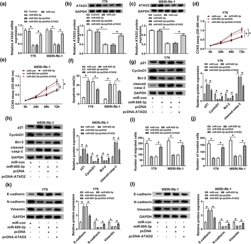
932 Yuxin Zhao et al.
influence the expression of the target gene by targeting In this research, we first detected the expression
messenger RNA (mRNA) [15]. The dysregulation of miRNAs of MALAT1 and miR-655-3p in RB. The function and
was documented in RB. For instance, the low expres- the correlation between MALAT1 and miR-655-3p were
sion of miR-125a-5p in RB promoted cell growth [16]. explored. Besides, we also detected the downstream target
Similarly, miR-3619-5p [7] and miR-758 [17] were down- of MALAT1/miR-655-3p axis in RB. Moreover, a mouse
regulated in RB and negatively associated with the RB xenograft model was employed to analyze the effect of
progression. However, miR-492 [18] was upregulated in MALAT1 knockdown on RB in vivo. These findings indicate
RB tissues and its depletion could impede the malig- the possibility of MALAT1 as a diagnostic biomarker or a
nant behavior of RB. miR-655-3p was encoded by a therapeutic target for RB.
polycistronic miRNA gene cluster in the human chro-
mosomal locus 14q32. This locus-encoded miRNAs par-
ticipated in the regulation of adhesion, invasion, and
motility pathways [19]. Not surprisingly, miR-655-3p 2 Materials and methods
was reported to function as a tumor suppressor, and its
overexpression decreased HCC cell proliferation, migra-
2.1 Tissue collection
tion, and invasion [20]. Besides, the effective delivery
of miR-655-3p using nanoscale coordination polymers
(NCPs) exhibit tumor-suppressive effects on advanced Thirty RB tissue samples and 18 matched normal globe
metastatic liver tumors [21]. Research also showed that tissue samples were collected from Weihai Central Hospital.
miR-655-3p impacted cell behaviors of tumor progres- Normal globe tissues were from patients who had suf-
sion in ovarian cancer [22], non-small cell lung cancer fered ruptured globes. All patients were not subjected
[23], and glioma [24]. These results indicating the enor- to radiation therapy and chemotherapy before enuclea-
mous potential of miR-655-3p as therapeutic candidate tion. All tissues were frozen at −80°C immediately after
target for cancer treatment. However, no study has resection until use. The research was permitted by
been published to show the effect and mechanism of the Ethics Committee of Weihai Central Hospital and
miR-655-3p in RB. carried out according to the Declaration of Helsinki
It is generally accepted that lncRNA as the compet- Principles. Written informed consents were provided
ing endogenous RNA (ceRNA) regulates miRNAs, thus by all patients.
affecting the transfection and translation of downstream
genes. ceRNAs are RNA transcripts which can function by
decreasing targeting concentration of miRNA and dere-
pressing other mRNAs containing the common miRNA 2.2 Cell culture and transfection
response elements (MREs) [25]. Sufficient research evi-
dence supported that this novel RNA cross talk exerted Human RB cell lines (Y79 and WERI-Rb-1) and adult
pivotal role in human health and disease. lncRNA highly retinal pigment epithelial cell line ARPE19 were pur-
upregulated in liver cancer (HULC) could reduce miR-372 chased from American Type Culture Collection (ATCC).
expression and activity and then reduce translational All cells were cultivated with RPMI-1640 medium (Biosun,
repression of its target transcript gene [26]. Nuclear Shanghai, China) containing 10% fetal bovine serum (FBS;
Enriched Abundant Transcript 1 (NEAT1) competed for Biosun) in 5% CO2 incubator at 37°C. Small interfering
miR-506 in GC and modulated the expression of signal RNA (siRNA) targeting MALAT1 (si-MALAT1, 5′-CACAGGG
transducer and activator of transcription 3 (STAT3), AAAGCGAGTGGTTGGTAA-3′) and its negative control
which ultimately caused an increase in growth, invasion, (si-con, 5′-UUCUCCGAACGUGUCACGUTT-3′), miR-655-3p
and migration [27]. In human RB, lncRNA X inactive-spe- mimics (miR-655-3p, 5′-AUAAUACAUGGUUAACCUC
cific transcript (XIST) [28], differentiation antagonizing UUU-3′) and its negative control (miR-con, 5′- UUCUCCG
nonprotein coding RNA (DANCR) [29], and homeobox AACGUGUCACGUTT-3′), miR-655-3p inhibitor (anti-miR-
A11 antisense RNA (HOXA11-AS) [30] were found that 655-3p, 5′-AAAGAGGUUAACCAUGUAUUAU-3′) and its
functioned as ceRNAs for its target miRNA and regulate negative control (anti-miR-con, 5′-UUGUACUACACAAAA
its endogenous targets and affect the progression of RB. GUACUG-3′), and ATAD2 overexpression vector (pcDNA-
Therefore, understanding ceRNA cross talk will expand ATAD2) and empty vector (pcDNA) were all obtained
the understanding in gene regulatory networks and pro- from Sangon Biotech (Shanghai, China). The increased
vide new treatment strategies and methods for RB. or decreased expression of miR-655-3p was achieved by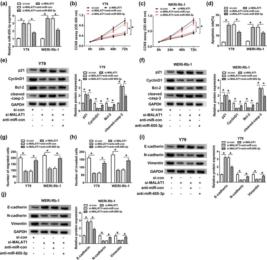
Role of MALAT1 in retinoblastoma progression 933
transfecting miR-655-3p mimics or miR-655-3p inhibitor. 2.5 Flow cytometry analysis of cell
Transfection of si-MALAT1 induced the decreased expres- apoptosis
sion of MALAT1, while ATAD2 overexpression was achieved
by transfecting pcDNA-ATAD2. Transfection was conducted An Annexin V-FITC/PI Apoptosis Detection Kit (Vazyme,
using Lipofectamine 2000 (Invitrogen, Carlsbad, CA, USA) Nanjing, China) was used to detect the apoptosis rate. In
according to the manufacturer’s instructions. Each group of brief, the transfected Y79 and WERI-Rb-1 cells were col-
cells was harvested for 24 or 48 h after transfection for lected and washed with phosphate buffer solution (PBS)
further assays. three times. Then cells were incubated with Annexin
V–fluorescein isothiocyanate (FITC) and propidium iodide
(PI) at 4°C for 15 min in dark condition. Cells were ana-
lyzed by flow cytometry (Agilent, Beijing, China).
2.3 Quantitative real time reverse
transcription polymerase chain reaction
(RT-qPCR)
2.6 Western blot assay
Total RNA was extracted from RB tissue samples or tumor
cells using TRIzol (Thermo Fisher Scientific, USA) reagent Total protein in Y79 and WERI-Rb-1 cells was extracted
according to the manufacturer’s instructions. To deter- using a RIPA Lysis buffer (Solarbio, Beijing, China) sup-
mine the expression of miR-655-3p, RT-qPCR was per- plemented with phenylmethanesulfonyl fluoride (1 mM,
formed using a Mir-X™ miRNA RT-qPCR TB Green® Kit Beyotime, China), and the concentration of protein sam-
(638314; TaKaRa, Japan) on a real time detection system ples was detected by a BCA Protein Assay Kit (Beyotime).
(Bio-Rad, Shanghai, China). To analyze the expression of Equal amount of protein samples were separated by
MALAT1 and ATAD2, a PrimeScript™ RT reagent Kit sodium dodecyl sulfonate-polyacrylamide gel electro-
(RR037A; TaKaRa) was used to synthesize the cDNA phoresis and then transferred onto the polyvinylidene
and TB Green® Premix Ex Taq™ II (RR820A; TaKaRa) fluoride membrane (Millipore, Billerica, MA, USA). The
was used to detect the expression levels of MALAT1 and membrane was blocked with 5% skim milk for 1 h and
ATAD2. The levels of MALAT1 and ATAD2 were normalized incubated with primary antibody for 12 h at 4°C. Then
by glyceraldehyde 3-phosphate dehydrogenase (GAPDH), the membrane was incubated with secondary antibody
while the level of miR-655-3p was normalized by small for 2 h at room temperature. The chemiluminescence
nuclear RNA U6 and processed by the 2−ΔΔCt method. intensity was detected using an ECL kit (Beyotime) at a
The primers were obtained from Songon (Shanghai, China) BIO-RAD imaging system (Bio-Rad, CA, USA). The primary
and presented as follows: MALAT1 (F: 5′-AATGTTAAGAGA antibodies p21 (1/1,500; ab218311), CyclinD1 (1/1,000;
AGCCCAGGG-3′, R: 5′-AAGGTCAAGAGAAGTGTCAGC-3′), ab40754), B-cell lymphoma-2 (Bcl-2; 1/1,000; ab32124),
miR-655-3p (F: 5′-CGCGCGATAATACATGGTTAAC-3′, R: cleaved caspase 3 (cleaved-casp-3, 1/500; ab32042), E-
5′-GTGTCTTAAGGCTAGGCCTA-3′), ATAD2 (F: 5′-GGAATC cadherin (ab40772, 1/2,000), N-cadherin (ab18203, 1/2,000),
CCAAACCACTGGACA-3′, R: 5′-GGTAGCGTCGTCGTAAAGC Vimentin (ab45939, 1/1,000), ATPase family AAA domain
ACA-3′), GAPDH (F: 5′-GGAAATGAATGGGCAGCCGT-3′, R: containing 2 (ATAD2, 1/500; ab176319), GAPDH (1/2,500;
5′-GTTAAAAGCAGCCCTGGTGAC-3′), and U6 (F: 5′-CTCGCT ab9485), and secondary antibody Goat Anti-Rabbit IgG
TCGGCAGCACA-3′, R: 5′-AACGCTTCACGAATTTGCGT-3′). H&L (HRP) (1/10,000; ab97051) were purchased from
Abcam (Cambridge, MA, USA).
2.4 Cell counting kit-8 (CCK-8) assay
2.7 Transwell assay
A CCK-8 kit (Beyotime, Shanghai, China) was utilized to
assess cell viability. The Y79 and WERI-Rb-1 cells (2 × 10⁴ Transwell chambers (Solarbio) were utilized to monitor
per well) were seeded into the 96-well plate and main- the migrated and invaded abilities of Y79 and WERI-Rb-1
tained for 24 h. At the end of the incubation time, CCK-8 cells. For the migration assay, RPMI-1640 medium contain-
solution was added into each well at 0, 24, 48, and 72 h, ing 10% FBS was supplemented into the lower chamber,
and the optical density at 450 nm was assessed by a while the upper chamber was added with suspended cells
Multiscan Spectrum (Tecan, Switzerland). in serum-free medium. After 24-h incubation, the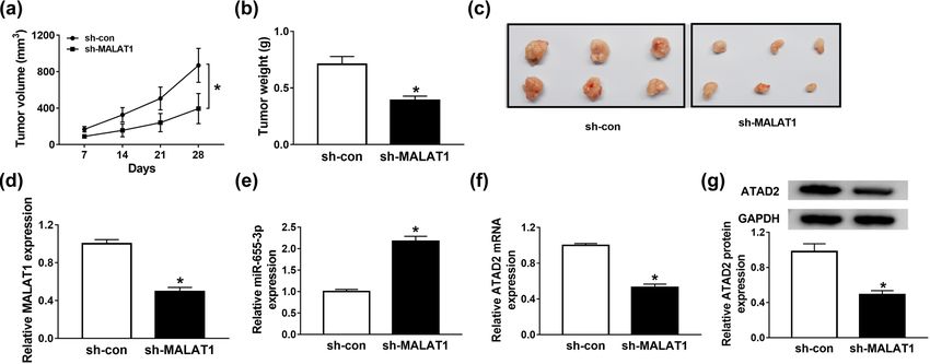
934 Yuxin Zhao et al.
migrated cells were stained with 0.1% crystal violet. 2.10 Statistical analysis
Cell numbers in ten random fields were counted using a
light microscope and calculated using Image Pro Plus. GraphPad Prism 7 (GraphPad Inc., La Jolla, CA, USA) was
The protocol of invasion assay was similar to that of the used to process the experimental data which were per-
migration assay, while the difference was that the upper formed in triplicate. The unpaired Student’s t-test was
chamber was covered with a matrigel matrix (BD, Franklin used to analyze the difference between the two groups,
Lakes, USA). and one-way analysis of variance (ANOVA) was utilized
to analyze the differences among multiple groups. Pearson
correlation analysis was used to test the association
among the expression of MALAT1, miR-655-3p, and
2.8 Dual-luciferase reporter assay ATAD2. P < 0.05 was considered statistically significant.
The wide-type and mutant complementary sequences of
MALAT1 or 3′-untranslated regions (3′-UTR) of ATAD2
were inserted into pGL3 vector (Promega, Madison, WI, 3 Results
USA) to construct the luciferase reporter WT-MALAT1,
MUT-MALAT1, WT-ATAD2, or MUT-ATAD2. Y79 and
3.1 MALAT1 was upregulated, while miR-
WERI-Rb-1 cells were co-transfected with luciferase
reporter and miR-655-3p or miR-con. The cells were har- 655-3p was downregulated in RB tissues
vested after 48 h and the luciferase activity was evaluated
by using a Dual-Lucy Assay Kit (Solarbio). Renilla luci- The expressions of long noncoding RNA MALAT1 and
ferase values were used to normalize the Firefly luciferase miRNA-655-3p in 30 RB tissue samples and 18 normal
values. globe tissue samples were detected first. As displayed
in Figure 1a and b, the expression of MALAT1 was appar-
ently elevated in RB tissues compared to that in normal
tissues. Conversely, the expression of miR-655-3p was
2.9 Mouse xenograft experiments distinctly decreased in RB tissues (Figure 1b). Besides,
Pearson’s correlation analysis exhibited that the expres-
Y79 cells stable expressing sh-MALAT1 or sh-con were sion of miR-655-3p in RB was negatively correlated with
produced by inserting sh-MALAT1 or sh-con into the lenti- MALAT1 expression (Figure 1c). These data indicated that
viral vector pLKO.1-puro lentiviral vectors (Sigma, MALAT1 was highly expressed and miR-655-3p was lowly
Merck KGaA, Darmstadt, Germany). Then the cells were expressed in RB tissues.
selected 24 h after transfection with 1 µg/mL puromycin
(Sigma) for 10–14 days.
For the RB tumor xenograft mouse model construc-
3.2 MALAT1 silencing constrained cell
tion, 4-week-old nude mice that were bought from
Shanghai Laboratory Animal Company (SLAC, Shanghai, proliferation, metastasis, and EMT but
China) was used. 4 × 10⁶ stable MALAT1 knockdown Y79 promoted apoptosis in Y79 and WERI-
cells with sh-MALAT1 or sh-con were injected into the Rb-1 cells
right lateral flanks of the nude mice. The mice were
divided into two groups (n = 6 per group): the sh- Consistent with its expression in RB tissues, MALAT1
MALAT1 group and the sh-con group. The tumor volume expression in RB cell lines (Y79 and WERI-Rb-1) was
was measured every 7 days to 28 days and tumor size obviously increased in contrast with the retinal pigment
was calculated according to the formula: volume = epithelial cell line ARPE19 (Figure 2a). To investigate the
width2 × length/2. After 28 days, the mouse was exe- functions of MALAT1 in RB, Y79 and WERI-Rb-1 cells were
cuted and xenograft tumors were resected and weighed. transfected with si-NC or si-MALAT1. As exhibited in
The tumor tissues were stored in a −80°C refrigerator for Figure 2b, transfection of si-MALAT1 successfully reduced
further exploration. All animal experiment procedures the expression of MALAT1 compared with the si-NC group.
were approved by the Animal Care Committee of Weihai Moreover, the transfection of si-MALAT1 resulted in a
Central Hospital. marked decline of cell viability in Y79 and WERI-Rb-1 cellsRole of MALAT1 in retinoblastoma progression 935
Figure 1: MALAT1 was upregulated, while miR-655-3p was downregulated in RB tumor tissues. (a and b) The expression of MALAT1 and miR-
655-3p in RB tumor tissues and normal globe tissues were tested by RT-qPCR. (c) The correlation between miR-655-3p and MALAT1 was
verified by Pearson’s correlation analysis. *P < 0.05.
in comparison with the si-con group (Figure 2c and d). 3.3 MALAT1 negatively interacted with miR-
Flow cytometry assay showed that the apoptosis rate of 655-3p in Y79 and WERI-Rb-1 cells
cells with MALAT1 depletion was notably facilitated in Y79
and WERI-Rb-1 cells transfected with si-MALAT1 (Figure 2e). As is widely known, lncRNAs could function as a ceRNA
Cyclin D1 phosphorylates the RB protein and drives to specific miRNAs and decrease miRNA expression. We
G1 to S phase progression by forming active complexes wondered if miR-655-3p was a target miRNA of MALAT1.
with cyclin-dependent kinases (CDKs), and its abnormal StarBase online database (http://starbase.sysu.edu.cn/
elevated expression in tumors promoted cell growth starbase2/) was used to predict the putative targets of
and ultimately accelerated neoplastic growth [31]. p21, MALAT1. The results showed that miR-655-3p contains
a member of Cip/Kip family of CKIs, exerted proliferation the complete binding sites in MALAT1 (Figure 3a). Subse-
inhibitor and apoptosis inhibitor roles in cancers [32]. quent dual-luciferase reporter assay confirmed this inter-
Since p21 and Cyclin D1 reflect the cell proliferation and action, as transfection of miR-655-3p induced a remarkable
Bcl-2 (anti-apoptotic protein) and cleaved-casp-3 were reduction of luciferase activity in the WT-MALAT1 group
apoptosis-associated proteins, we further confirmed their in contrast with the miR-con group, while it has little
protein levels in Y79 and WERI-Rb-1 cells. The western change in the luciferase activity of MUT-MALAT1 group
blot assay exhibited that the protein levels of p21 and (Figure 3b and c). Besides, the expression of miR-655-3p
cleaved-casp-3 were strikingly enhanced, but the pro- was evidently downregulated in Y79 and WERI-Rb-1 cells
tein levels of Cyclin D1 and Bcl-2 were conspicuously compared with the ARPE19 cells (Figure 3d). Moreover,
decreased in si-MALAT1-transfected Y79 and WERI-Rb-1 Y79 and WERI-Rb-1 cells were transfected with si-NC, si-
cells (Figure 2f and g). Besides, the introduction of si- MALAT1, pcDNA, or pcDNA-MALAT1, and the expressions
MALAT1 notably reduced the migration and invasion of MALAT1 and miR-655-3p were detected. As shown
abilities of Y79 and WERI-Rb-1 cells compared to the in Figure 3e–h, transfection of si-MALAT1 inhibited the
si-con group (Figure 2h and i). Epithelial–mesenchymal expression of MALAT1 but suppressed miR-655-3p expres-
transition (EMT) was the most common primary reason sion. Instead, MALAT1 expression was elevated and miR-
for tumor invasion and metastasis [33]. The hallmark of 655-3p was downregulated when cells were transfected
EMT is the upregulation of epithelial markers including with pcDNA-MALAT1. These data implicated that MALAT1
N-cadherin and Vimentin followed by the downregula- suppressed the expression of miR-655-3p by binding
tion of E-cadherin [34]; thus, we further detected the directly to the latter.
levels of these proteins. As exhibited in Figure 2j and k,
the protein levels of E-cadherin were apparently ele-
vated, while N-cadherin and Vimentin were strikingly 3.4 MALAT1 knockdown inhibited cell
decreased in the si-MALAT1 group in comparison with proliferation but facilitated apoptosis in
that in the si-con group. Taken together, the depletion RB by sponging miR-655-3p
of MALAT1 inhibited cell proliferation, migration, inva-
sion, and EMT but promoted apoptosis in Y79 and WERI- To investigate if miR-655-3p participates in the effect
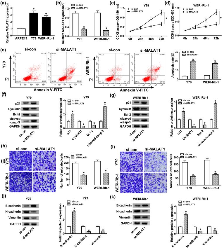
Rb-1 cells. of the functions of MALAT1 on RB, a loss-of-function936 Yuxin Zhao et al. Figure 2: MALAT1 depletion suppressed cell proliferation but induced apoptosis and EMT in Y79 and WERI-Rb-1 cells. (a) The expression of MALAT1 in RB cells (Y79 and WERI-Rb-1) and retinal pigment epithelial cell ARPE19 was detected by RT-qPCR. (b–k) Y79 and WERI-Rb-1 cells were transfected with si-NC or si-MALAT1. (b) The expression of MALAT1 in Y79 and WERI-Rb-1 cells was examined by RT-qPCR after transfection for 24 h. (c and d) The cell viability was detected by CCK-8 assay after transfection for 0, 24, 48, and 72 h. (e) The apoptosis rate was assessed by flow cytometry after transfection for 48 h. (f and g) The protein levels of p21, Cyclin D1, Bcl-2, and cleaved-casp-3 were detected by western blot after transfection for 48 h. (h and i) After transfection for 24 h, cell migration and invasion were detected by transwell assay. (j and k) The protein levels of E-cadherin, N-cadherin, and Vimentin were examined by western blot after transfection for 48 h. *P < 0.05.
Role of MALAT1 in retinoblastoma progression 937
Figure 3: MALAT1 negatively interacted with miR-655-3p in Y79 and WERI-Rb-1 cells. (a) The fragment of MALAT1 containing the putative
or mutant miR-655-3p binding sites. (b and c) WT-MALAT1 or MUT-MALAT1 reporter plasmids and miR-con or miR-655-3p were co-transfected
into Y79 and WERI-Rb-1 cells for 48 h. The luciferase activities were evaluated by dual-luciferase reporter assay. (d) The expression of
miR-655-3p in RB cells Y79 and WERI-Rb-1 and retinal pigment epithelial cell line ARPE19 was tested by RT-qPCR. (e–h) Y79 and WERI-Rb-1
cells were transfected with si-NC, si-MALAT1, pcDNA, or pcDNA-MALAT1 for 24 h. (e and f) The expression of MALAT1 in the cells upon
transfection was detected by RT-qPCR. (g and h) The expression of miR-655-3p in Y79 and WERI-Rb-1 cells upon transfection was
detected via RT-qPCR. *P < 0.05.
experiment was performed. Si-con, si-MALAT1, si-MALAT1 + putative target of miR-655-3p in RB. As shown in Figure 5a,
anti-miR-con, and si-MALAT1 + anti-miR-655-3p were ATAD2 3′-UTR had complementary binding sites with
transfected into Y79 and WERI-Rb-1 cells. As exhibited in miR-655-3p. This was further confirmed by dual-luci-
Figure 4a, the expression of miR-655-3p was increased in ferase assay. The luciferase activity of WT-ATAD2 trans-
si-MALAT1-transfected cells, but it was declined by intro- fected cells was markedly declined by transfection of
duction of anti-miR-655-3p. Moreover, the inhibitory effects miR-655-3p, while the luciferase activity of MUT-ATAD2
of si-MALAT1 on cell proliferation (Figure 4b and c) and group had changed little when transfected with miR-655-
migration and invasion abilities (Figure 4g and h) of Y79 3p (Figure 5b and c). Subsequently, ATAD2 expression in
and WERI-Rb-1 cells were reversed by anti-miR-655-3p. RB was uncovered. As displayed in Figure 5d–g, the
miR-655-3p inhibitor also attenuated the promotion impact mRNA and protein levels of ATAD2 in RB tissues and
on apoptosis rate in Y79 and WERI-Rb-1 cells induced by cells were higher than that in normal tissues and cells.
MALAT1 silencing (Figure 4d). In addition, miR-655-3p Besides, a negative correlation was discovered between
inhibitor counteracted the elevated protein levels of p21, ATAD2 and miR-655-3p expression (Figure 5h). To inves-
cleaved-casp-3, and E-cadherin as well as the declined pro- tigate their relationship thoroughly, the expression of
tein levels of Cyclin D1, Bcl-2, N-cadherin, and Vimentin in ATAD2 in cells transfected with miR-655-3p mimic or
Y79 and WERI-Rb-1 cells induced by si-MALAT1 (Figure 4e, f,
anti-miR-655-3p was detected. Transfection of miR-655-
i, and j). These data implicated that the silencing of MALAT1
3p mimic promoted the expression of miR-655-3p (Figure 5i)
suppressed RB progression by sponging miR-655-3p.
but inhibited ATAD2 mRNA and protein levels (Figure 5j–l).
Conversely, anti-miR-655-3p inhibited miR-655-3p expres-
sion, while the mRNA and protein levels of ATAD2 were
3.5 ATAD2 was a candidate target of miR- promoted in Y79 and WERI-Rb-1 cells (Figure 5i–l). These
655-3p in Y79 and WERI-Rb-1 cells results indicate that ATAD2 was a target of miR-655-3p and
it was inhibited by miR-655-3p.
To explore the underlying mechanism of miR-655-3p in To further explore the relationship among MALAT1,
RB, starBase online database was used to search the miR-655-3p, and ATAD2 in RB, we detected ATAD2938 Yuxin Zhao et al. Figure 4: MALAT1 regulated RB progression by sponging miR-655-3p. (a–j) The Y79 and WERI-Rb-1 cells were transfected with si-con, si-MALAT1, si-MALAT1 + anti-miR-con, or si-MALAT1 + anti-miR-655-3p. (a) The level of miR-655-3p was measured by RT-qPCR after transfection for 24 h. (b and c) The cell viability was examined by CCK8 assay after transfection for 0, 24, 48, and 72 h. (d) The apoptosis rate was evaluated through flow cytometry after transfection for 48 h. (e and f) The protein levels of p21, Cyclin D1, Bcl-2, and cleaved-casp-3 were tested via western blot assay after transfection for 48 h. (g and h) The migration and invasion abilities were estimated by transwell assay after transfection for 24 h. (i and j) After transfection for 48 h, the protein levels of E-cadherin, N-cadherin, and Vimentin were detected by western blot. *P < 0.05. expression in Y79 and WERI-Rb-1 cells transfected with whereas the levels were altered by introduction of anti- si-con, si-MALAT1, si-MALAT1 + anti-miR-con, or si-MALAT1 + miR-655-3p. Besides, the expression of MALAT1 was posi- anti-miR-655-3p. As presented in Figure 5m–o, the levels of tively correlated with ATAD2 (Figure 5p). These data demon- ATAD2 mRNA and protein were apparently downregulated strated that MALAT1 could regulate ATAD2 expression by in Y79 and WERI-Rb-1 cells transfected with si-MALAT1, targeting miR-665-3p in Y79 and WERI-Rb-1 cells.
Role of MALAT1 in retinoblastoma progression 939 Figure 5: MALAT1 upregulated ATAD2 expression by repressing miR-655-3p in Y79 and WERI-Rb-1 cells. (a) Putative or mutant binding sites of miR-655-3p in the ATAD2 3′-UTR were predicted by StarBase. (b and c) The luciferase activity of Y79 and WERI-Rb-1 cells co-transfected with WT-ATAD2 or MUT-ATAD2 and miR-655-3p or miR-con for 48 h was evaluated by dual-luciferase reporter assay. (d) The mRNA level of ATAD2 in RB and normal tissues was detected by RT-qPCR. (e) The protein levels of ATAD2 in 12 RB and normal tissues were exposed by western blot assay. (f and g) The mRNA and protein levels of ATAD2 in RB cells (Y79 and WERI-Rb-1) and retinal pigment epithelial cells ARPE19 were uncovered by RT-qPCR and western blot assay, respectively. (h) The correlation between miR-655-3p and ATAD2 was processed by Pearson’s correlation analysis. (i–l) Y79 and WERI-Rb-1 cells were co-transfected with miR-con, miR-655-3p, anti-miR-con, and anti-miR- 655-3p. (i and j) After transfection for 24 h, the expressions of miR-665-3p and ATAD2 mRNA were detected by RT-qPCR. (k and l) After transfection for 48 h, the protein level of ATAD2 was examined by western blot. (m–o) Y79 and WERI-Rb-1 cells were transfected with si-con, si-MALAT1, si-MALAT1 + anti-miR-con, or si-MALAT1 + anti-miR-655-3p. After transfection for 24 or 48 h, the mRNA and protein levels of ATAD2 were measured via RT-qPCR and western blot assay. (p) The correlation between MALAT and ATAD2 was validated by Pearson’s correlation analysis. *P < 0.05.
940 Yuxin Zhao et al.
3.6 miR-655-3p negatively regulated ATAD2 two critical problems for RB treatment. Convincing evi-
expression to impede cell proliferation dence indicated that MALAT1 was an oncogene in various
and impel apoptosis in Y79 and WERI- types of cancer. In the current study, the roles of MALAT1
in RB were mainly explored. The results suggested that
Rb-1 cells
MALAT1 promoted RB progression partially through miR-
655-3p/ATAD2 axis.
To investigate whether the effects of miR-655-3p on RB
Emerging evidence demonstrated that the dysregula-
progression were mediated by ATAD2, cells were trans-
tion of MALAT1 was implicated in tumor progression.
fected with Control, miR-con, miR-655-3p, miR-655-3p +
For instance, a recent study disclosed that MALAT1 was
pcDNA, or miR-655-3p + pcDNA-ATAD2. As shown in
strikingly elevated in ovarian cancer, and its depletion
Figure 6a–c, transfection of pcDNA-ATAD2 relieved the
retarded cell growth and metastasis in vitro [9]. Another
inhibition effect on ATAD2 mRNA and protein levels that
study in GC revealed that MALAT1 was conspicuously
were induced by miR-655-3p. Furthermore, the elevated
boosted in GC, and its depletion impeded cell growth,
ATAD2 partly reversed the repression impacts on cell via-
metastasis in vitro as well as induced apoptosis [10].
bility, migration and invasion, and the promotion effect
Similar results were reported in colorectal cancer [11],
on cell apoptosis retarded by miR-655-3p in Y79 and
papillary thyroid cancer [12], and non-small cell lung
WERI-Rb-1 cells (Figure 6d–f, i, and j). Finally, the trans-
cancer [13]. In this study, MALAT1 was highly expressed
fection of pcDNA-ATAD2 mitigated the promotion effect
in RB tumors and cells. Meanwhile, MALAT knockdown
on the protein levels of p21, cleaved-casp-3, E-cadherin
blocked cell growth, metastasis, and EMT but enhanced
as well as the restraint effect on the protein levels of
apoptosis. In addition, MALAT1 depletion restrained
Cyclin D1, Bcl-2, N-cadherin, and Vimentin in Y79 and
xenograft tumor growth in vivo. These data were con-
WERI-Rb-1 cells caused by miR-655-3p mimics (Figure 6g,
sistent with previous reports in RB [35,36], which
h, k, and l). These data implicated that miR-655-3p regu-
demonstrated that MALAT1 played a vital role in RB
lated cell proliferation, apoptosis, and migration and inva-
progression.
sion in Y79 and WERI-Rb-1 cells by modulating ATAD2.
miR-655-3p was encoded by 14q32 locus, which was
reported to associate with the behaviors of tumor cells.
For instance, Wang et al. reported that miR-655-3p was
3.7 MALAT1 depletion impeded xenograft lowly expressed in non-small cell lung cancer and miR-
tumor growth in vivo 655-3p mimics resulted in the impediment of cell metas-
tasis [23]. Another study in hepatocellular carcinoma
To verify the effects of MALAT1 in vivo, the nude mouse (HCC) demonstrated that the low expression of miR-
models of RB were established. The nude mice were 655-3p in HCC promoted cell growth and metastasis
injected with stable MALAT1 knockdown Y79 cells, and [37]. In our research, miR-655-3p was downregulated in
the tumor volume was measured every 7 days for 28 days. RB tumors and cells and exhibited a negative correlation
Twenty eight days upon injection, the mice were exe- with MALAT1. As lncRNAs exerted their biological func-
cuted, the tumor tissues were removed, and stored in tion by exerting as ceRNAs for miRNAs, we speculated
liquid nitrogen for further investigation. As shown in that miR-655-3p might be a target of MALAT1 in RB.
Figure 7a–c, MALAT1 depletion inhibited the tumor volume As predicted, miR-655-3p contained the complementary
and weight in contrast with the sh-NC group. The expres- binding sites of MALAT1, and it was further confirmed
sion of MALAT1, miR-655-3p, and ATAD2 in tumor tissue of by dual-luciferase assay. Besides, miR-655-3p depletion
nude mice were also analyzed. The levels of MALAT1, could curb the effect of si-MALAT1 on cell growth, mobi-
ATAD2 mRNA, and protein were decreased in the sh- lity, EMT, and apoptosis. These data implicated that
MALAT1 group. However, the level of miR-655-3p was sig- MALAT accelerated RB progression by sponging miR-
nificantly elevated in the sh-MALAT1 group (Figure 7d–g). 655-3p.
These results suggested that the silencing of MALAT1 inhi- miRNAs could regulate the transcription and transla-
bited xenograft tumor growth in vivo. tion of their target mRNAs, thus affecting numerous
biological processes [38]. To investigate the molecular
mechanism of MALAT1/miR-655-3p involved in RB pro-
gression, we further predicted the target mRNA of miR-
4 Discussion 655-3p. ATAD2 was predicted as a target gene of miR-655-3p.
ATAD2, located on human chromosome 8q24, is a member
RB is a rare type of intraocular cancer. At present, the of the ATPase family containing an AAA domain which is
early diagnosis and treatment of metastatic RB tumor are vital for ATPase activity and protein assembly [39].Role of MALAT1 in retinoblastoma progression 941 Figure 6: miR-655-3p negatively regulated ATAD2 expression to impede cell proliferation and impel apoptosis in Y79 and WERI-Rb-1 cells. (a–c) The Y79 and WERI-Rb-1 cells were transfected with Control, miR-con, miR-655-3p, miR-655-3p + pcDNA, or miR-655-3p + pcDNA- ATAD2. The levels of ATAD2 mRNA and protein were detected via RT-qPCR and western blot assay. (d–l) The Y79 and WERI-Rb-1 cells were transfected with miR-con, miR-655-3p, miR-655-3p + pcDNA, or miR-655-3p + pcDNA-ATAD2. (d and e) The cell viability was detected by CCK8 assay. (f) The apoptosis rate was measured through flow cytometry. (g and h) The protein levels of p21, Cyclin D1, Bcl-2, and cleaved- casp-3 were assessed via western blot assay. (i and j) The migration and invasion abilities were estimated by transwell assay. (k and l) The levels of E-cadherin, N-cadherin, and Vimentin protein were detected by western blot. *P < 0.05. ATAD2 was reported to affect many biological processes Another study in HCC illustrated that ATAD2 was enhanced in various types of tumors [40,41]. Accumulating evi- in HCC, and its knockdown regulated cell behaviors dence demonstrated that ATAD2 was involved in the pro- mediated by miR-372 [42]. In our research, ATAD2 gression of tumors. For example, a research in cervical was upregulated in RB tumors and cells and was nega- cancer reported that the mRNA and protein levels of tively regulated by miR-655-3p. These results mani- ATAD2 were elevated in cervical cancer, and the knock- fested that MALAT1 modulated ATAD2 expression to down of ATAD2 impeded cell growth and metastasis [41]. promote RB progression via miR-655-3p.
942 Yuxin Zhao et al.
Figure 7: MALAT1 depletion impeded xenograft tumor growth in vivo. (a–g) The nude mice were injected with stable Y79 cell line expressing
shRNA against MALAT1 or negative control. (a) The tumor volume was examined every 7 days for 28 days. (b) 28 days post-injection, the
mice were executed, and tumors were removed and weighed. (c) Images of mice with subcutaneous transplanted tumors. (d–f) The
expression levels of MALAT1, miR-655-3p, and ATAD2 in transplanted tumors were detected via RT-qPCR. (g) The protein level of ATAD2 in
transplanted tumors was detected via western blot assay. *P < 0.05.
5 Conclusion [4] Chantada GL, Qaddoumi I, Canturk S, Khetan V, Ma Z,
Kimani K, et al. Strategies to manage retinoblastoma in
developing countries. Pediatr Blood Cancer. 2011;56:341–8.
Taken together, we concluded that MALAT1 promoted
[5] Kung JT, Colognori D, Lee JT. Long noncoding RNAs: past,
ATAD2 expression to regulate cell proliferation, migra-
present, and future. Genetics. 2013;193:651–69.
tion, invasion, apoptosis, and EMT in RB by sponging [6] Bhan A, Soleimani M, Mandal SS. Long noncoding RNA and
miR-655-3p. The new regulatory network sheds light on cancer: a new paradigm. Cancer Res. 2017;77:3965–81.
the mechanism of RB progression, which indicates that doi: 10.1158/0008-5472.can-16-2634.
MALAT1 could be used as a diagnostic biomarker or a [7] Yan G, Su Y, Ma Z, Yu L, Chen N. Long noncoding RNA
LINC00202 promotes tumor progression by sponging miR-
therapeutic target for RB. A recent research disclosed
3619-5p in retinoblastoma. Cell Struct Funct. 2019;44:51–60.
that combining noncoding RNAs with conventional cyto- [8] Shang Y. lncRNA THOR acts as a retinoblastoma promoter
toxic chemotherapies for the treatment of cancers is a through enhancing the combination of c-myc mRNA and
potential therapeutic strategy [19,43]. In follow-up study, IGF2BP1 protein. Biomed Pharmacother. 2018;106:1243–9.
we will focus on whether the MALAT1/miR-655-3p/ATAD2 [9] Gordon MA, Babbs B, Cochrane DR, Bitler BG, Richer JK. The
axis affected the effectiveness of the chemotherapy for RB. long non-coding RNA MALAT1 promotes ovarian cancer pro-
gression by regulating RBFOX2-mediated alternative splicing.
Mol Carcinogen. 2019;58:196–205.
Funding information: No funding. [10] Lee NK, Lee JH, Ivan C, Ling H, Zhang X, Park CH, et al. MALAT1
promoted invasiveness of gastric adenocarcinoma. BMC
Conflict of interest: Authors state no conflict of interest. Cancer. 2017;17:46.
[11] Xie J, Li W, Li X, Ye W, Shao C. lncRNA MALAT1 promotes
colorectal cancer development by sponging miR-363-3p to
Data availability statements: All data generated or ana-
regulate EZH2 expression. J Biol Regul Homeost Agents.
lyzed during this study are included in this article. 2019;33:331–43.
[12] Liu J, Dong H, Yang Y, Qian Y, Liu J, Li Z, et al. Upregulation of
long noncoding RNA MALAT1 in papillary thyroid cancer and its
References diagnostic value. Future Oncol. 2018;14:3015–22.
[13] Chen W, Zhao W, Chen S, Zhang L, Guo Z, Wang L, et al.
[1] Dimaras H, Corson TW. Retinoblastoma, the visible CNS tumor: Expression and correlation of MALAT1 and SOX9 in non‐small
a review. J Neurosci Res. 2019;97:29–44. cell lung cancer. Clin Respir J. 2018;12:2284–91.
[2] Broaddus E, Topham A, Singh AD. Incidence of retinoblastoma [14] Wang L, Zhang Y, Xin X. Long non-coding RNA MALAT1 aggra-
in the USA: 1975–2004. Br J Ophthalmol. 2009;93:21–3. vates human retinoblastoma by sponging miR-20b-5p to
[3] Lin P, O’Brien JM. Frontiers in the management of retinoblas- upregulate STAT3. Pathol Res Pract. 2020;216:152977.
toma. Am J Ophthalmol. 2009;148:192–8. doi: 10.1016/j.prp.2020.152977.Role of MALAT1 in retinoblastoma progression 943
[15] Huang Y, Shen XJ, Zou Q, Wang SP, Tang SM, Zhang GZ. [29] Wang JX, Yang Y, Li K. Long noncoding RNA DANCR aggravates
Biological functions of microRNAs: a review. J Physiol retinoblastoma through miR-34c and miR-613 by targeting
Biochem. 2011;67:129–39. MMP-9. J Cell Physiol. 2018;233:6986–95. doi: 10.1002/
[16] Zhang Y, Xue C, Zhu X, Zhu X, Xian H, Huang Z. Suppression of jcp.26621.
microRNA-125a-5p upregulates the TAZ-EGFR signaling [30] Han N, Zuo L, Chen H, Zhang C, He P, Yan H. Long non-coding
pathway and promotes retinoblastoma proliferation. RNA homeobox A11 antisense RNA (HOXA11-AS) promotes
Cell Signal. 2016;28:850–60. retinoblastoma progression via sponging miR-506-3p. Oncol
[17] Li J, You X. MicroRNA‑758 inhibits malignant progression of Targets Ther. 2019;12:3509–17. doi: 10.2147/ott.s195404.
retinoblastoma by directly targeting PAX6. Oncol Rep. [31] Qie S, Diehl JA. Cyclin D1, cancer progression, and opportu-
2018;40:1777–86. nities in cancer treatment. J Mol Med (Berl). 2016;94:1313–26.
[18] Sun Z, Zhang A, Zhang L. Inhibition of microRNA‑492 attenu- doi: 10.1007/s00109-016-1475-3.
ates cell proliferation and invasion in retinoblastoma via [32] Gartel AL, Tyner AL. The role of the cyclin-dependent kinase
directly targeting LATS2. Mol Med Rep. 2019;19:1965–71. inhibitor p21 in apoptosis. Mol Cancer Ther. 2002;1:639–49.
[19] Uppal A, Wightman SC, Mallon S, Oshima G, Pitroda SP, [33] Pastushenko I, Blanpain C. EMT transition states during tumor
Zhang Q, et al. 14q32-encoded microRNAs mediate an oligo- progression and metastasis. Trends Cell Biol.
metastatic phenotype. Oncotarget. 2015;6:3540–52. 2019;29:212–26. doi: 10.1016/j.tcb.2018.12.001.
doi: 10.18632/oncotarget.2920. [34] Savagner P. The epithelial-mesenchymal transition (EMT)
[20] Wu G, Zheng K, Xia S, Wang Y, Meng X, Qin X, et al. MicroRNA- phenomenon. Ann Oncol. 2010;21(Suppl 7):vii89–92.
655-3p functions as a tumor suppressor by regulating ADAM10 doi: 10.1093/annonc/mdq292.
and β-catenin pathway in Hepatocellular Carcinoma. J Exp Clin [35] Liu S, Yan G, Zhang J, Yu L. Knockdown of long noncoding
Cancer Res. 2016;35:89. doi: 10.1186/s13046-016-0368-1. RNA (lncRNA) metastasis-associated lung adenocarcinoma
[21] Oshima G, Guo N, He C, Stack ME, Poon C, Uppal A, et al. transcript 1 (MALAT1) inhibits proliferation, migration, and
In vivo delivery and therapeutic effects of a MicroRNA on invasion and promotes apoptosis by targeting miR-124 in
colorectal liver metastases. Mol Ther. 2017;25:1588–95. retinoblastoma. Oncol Res Featur Preclin Clin Cancer Ther.
doi: 10.1016/j.ymthe.2017.04.005. 2018;26:581–91.
[22] Zha J, Chen D. miR-655-3p inhibited proliferation and migra- [36] Huang J, Yang Y, Fang F, Liu K. MALAT1 modulates the auto-
tion of ovarian cancer cells by targeting RAB1A. Eur Rev Med phagy of retinoblastoma cell through miR-124-mediated stx17
Pharmacol Sci. 2019;23:3627–34. regulation. J Cell Biochem. 2018;119:3853–63.
[23] Wang W, Cao R, Su W, Li Y, Yan H. miR-655-3p inhibits cell [37] Wu G, Zheng K, Xia S, Wang Y, Meng X, Qin X, et al. MicroRNA-
migration and invasion by targeting pituitary tumor-trans- 655-3p functions as a tumor suppressor by regulating ADAM10
forming 1 in non-small cell lung cancer. Biosci Biotechnol and β-catenin pathway in Hepatocellular Carcinoma. J Exp Clin
Biochem. 2019;83:1703–8. doi: 10.1080/ Cancer Res. 2016;35:89.
09168451.2019.1617109. [38] Salmena L, Poliseno L, Tay Y, Kats L, Pandolfi PP. A ceRNA
[24] Xin J, Zhao YH, Zhang XY, Tian LQ. lncRNA NFIA-AS2 promotes hypothesis: the Rosetta stone of a hidden RNA language? Cell.
glioma progression through modulating the miR-655-3p/ZFX axis. 2011;146:353–8. doi: 10.1016/j.cell.2011.07.014.
Hum Cell. 2020;33:1273–80. doi: 10.1007/s13577-020-00408-9. [39] Hussain M, Zhou Y, Song Y, Hameed HA, Jiang H, Tu Y, et al.
[25] Qi X, Zhang DH, Wu N, Xiao JH, Wang X, Ma W. ceRNA in cancer: ATAD2 in cancer: a pharmacologically challenging but tract-
possible functions and clinical implications. J Med Genet. able target. Expert Opin Ther Targets. 2018;22:85–96.
2015;52:710–8. doi: 10.1136/jmedgenet-2015-103334. [40] Wu S, Han M, Zhang C. Overexpression of microRNA-186
[26] Wang J, Liu X, Wu H, Ni P, Gu Z, Qiao Y, et al. CREB up-regulates inhibits angiogenesis in retinoblastoma via the Hedgehog
long non-coding RNA, HULC expression through interaction signaling pathway by targeting ATAD2. J Cell Physiol.
with microRNA-372 in liver cancer. Nucleic Acids Res. 2019;234:19059–72.
2010;38:5366–83. doi: 10.1093/nar/gkq285. [41] Zheng L, Li T, Zhang Y, Guo Y, Yao J, Dou L, et al. Oncogene
[27] Tan HY, Wang C, Liu G, Zhou X. Long noncoding RNA NEAT1- ATAD2 promotes cell proliferation, invasion and migration in
modulated miR-506 regulates gastric cancer development cervical cancer. Oncol Rep. 2015;33:2337–44.
through targeting STAT3. J Cell Biochem. 2019;120:4827–36. [42] Wu G, Liu H, He H, Wang Y, Lu X, Yu Y, et al. miR-372 down-
doi: 10.1002/jcb.26691. regulates the oncogene ATAD2 to influence hepatocellular
[28] Cheng Y, Chang Q, Zheng B, Xu J, Li H, Wang R. lncRNA XIST carcinoma proliferation and metastasis. BMC Cancer.
promotes the epithelial to mesenchymal transition of retino- 2014;14:107.
blastoma via sponging miR-101. Eur J Pharmacol. [43] Esteller M. Non-coding RNAs in human disease. Nat Rev Genet.
2019;843:210–6. doi: 10.1016/j.ejphar.2018.11.028. 2011;12:861–74. doi: 10.1038/nrg3074.You can also read


