Human Immunodeficiency Virus Type 1 Tat Regulates Endothelial Cell Actin Cytoskeletal Dynamics through PAK1 Activation and Oxidant Production
←
→
Page content transcription
If your browser does not render page correctly, please read the page content below
JOURNAL OF VIROLOGY, Jan. 2004, p. 779–789                                                                                                Vol. 78, No. 2
0022-538X/04/$08.00⫹0 DOI: 10.1128/JVI.78.2.779–789.2004
Copyright © 2004, American Society for Microbiology. All Rights Reserved.

                Human Immunodeficiency Virus Type 1 Tat Regulates
                   Endothelial Cell Actin Cytoskeletal Dynamics
                         through PAK1 Activation and
                               Oxidant Production
                             Ru Feng Wu,1 Ying Gu,1 You Cheng Xu,1 Stefania Mitola,2
                                   Federico Bussolino,2 and Lance S. Terada1*
                            The University of Texas Southwestern Medical Center and the Dallas Veterans
                             Administration Medical Center, Dallas, Texas,1 and The Institute for Cancer
                                   Research and Treatment and University of Turin, Turin, Italy2

                                                                                                                                                             Downloaded from http://jvi.asm.org/ on April 22, 2021 by guest
                                            Received 16 June 2003/Accepted 28 September 2003

             Human immunodeficiency virus type 1 Tat exerts prominent angiogenic effects which may lead to a variety
          of vasculopathic conditions in AIDS patients. Because endothelial cells undergo prominent cytoskeletal rear-
          rangement during angiogenesis, we investigated the specific effects of Tat on the endothelial cell actin cy-
          toskeleton. Glutathione S-transferase (GST)–Tat, at a level of 200 ng/ml (equivalent to 52 ng of Tat/ml), caused
          stress fiber disassembly, peripheral retraction, and ruffle formation in human umbilical vein endothelial cells
          (HUVEC) and human lung microvascular endothelial cells. At 600 ng of GST-Tat/ml (157 ng of Tat/ml), actin
          structures were lost, and severe cytoskeletal collapse occurred. In contrast, GST-Tat harboring mutations
          within either the cysteine-rich or basic domains exerted minimal effects on the endothelial cytoskeleton.
          HUVEC expressing a DsRed-Tat fusion protein displayed similar actin rearrangements, followed by actin
          collapse, whereas neighboring nontransfected cells retained normal actin structures. Because active mutants
          of p21-activated kinase 1 (PAK1) induce identical changes in actin dynamics, we hypothesized that Tat exerts
          its cytoskeletal effects through PAK1. GST-Tat activated PAK1 within 5 min, and adenovirus delivery of a
          kinase-dead PAK1 [PAK1(K298A)] completely prevented cytoskeletal collapse induced by GST-Tat or DsRed-
          Tat and also blocked downstream activation of c-Jun N-terminal kinase. Further, GST-Tat increased phos-
          phorylation of the NADPH oxidase subunit p47phox and caused its rapid redistribution to membrane ruffles.
          PAK1(K298A) blocked p47phox phosphorylation, and interference with NADPH oxidase function through
          superoxide scavenging or through expression of a transdominant inhibitor, p67(V204A), prevented GST-Tat-
          induced alterations in the actin cytoskeleton. We conclude that Tat induces actin cytoskeletal rearrangements
          through PAK1 and downstream activation of the endothelial NADPH oxidase.

   The vascular endothelium of human immunodeficiency virus                  angiogenic agents, such as vascular endothelial cell growth
(HIV)-infected patients displays a number of morphological                   factor (VEGF), dramatic alterations in the endothelial cell
and functional abnormalities that are thought to contribute to               actin cytoskeleton must occur to allow separation of interen-
the spectrum of AIDS-associated diseases. Chaotic endothelial                dothelial junctions and migration of cells to form vascular
cell morphology is seen in 94% of aortas of HIV type 1 (HIV-                 sprouts. Such actin cytoskeletal changes include ruffling of
1)-infected patients (31), and abnormalities seen in pulmonary               leading-edge membranes and depolymerization of trailing-
and coronary intima and in Kaposi’s sarcoma cells suggest                    edge stress fibers to allow treadmilling and locomotion to oc-
disorganized angiogenic behavior (18, 26). In this regard, the               cur. Angiogenic endothelium also becomes leaky, since periph-
viral accessory protein Tat presents a likely candidate vascu-               eral retraction occurs in conjunction with the breakdown of
lopathic factor. Tat is found circulating in the blood of AIDS               lateral intercellular junctions.
patients (28) and can enter uninfected cells through its protein                In the present study, we identify a signaling pathway through
transduction domain (24). In addition, Tat initiates outside-in              which HIV-1 Tat affects endothelial cell actin microfilament
signaling through receptor ligation, thus promoting endothelial              dynamics. Tat caused dramatic actin cytoskeletal rearrange-
cell migration and matrix invasion in vitro and angiogenesis in              ments such as membrane ruffling, peripheral retraction, and
vivo (3, 4, 19, 23).                                                         subsequent cytoskeletal disassembly, which we found to be
   Although the angiogenic behavior stimulated by Tat has                    driven by activation of the MAP kinase kinase kinase kinase
been well described, the signaling pathways underlying the                   p21-activated kinase 1 (PAK1). In addition, involvement of an
accompanying endothelial cell morphological changes are                      endothelial cell NADPH oxidase downstream of PAK1 ap-
poorly understood. Early during the response to other known                  peared necessary for these cytoskeletal changes.

                                                                                                    MATERIALS AND METHODS
  * Corresponding author. Mailing address: Dallas VAMC, MC151,                  Plasmid and adenovirus construction. All PCR amplifications were performed
4500 S. Lancaster Rd., Dallas, TX 75216. Phone: (214) 857-0405. Fax:         with either Pfu Turbo (Stratagene) or Pfx Platinum (Invitrogen), and all con-
(214) 857-0340. E-mail: lance.terada@med.va.gov.                             structs were confirmed by sequencing. DsRed-Tat was derived by BamHI exci-

                                                                       779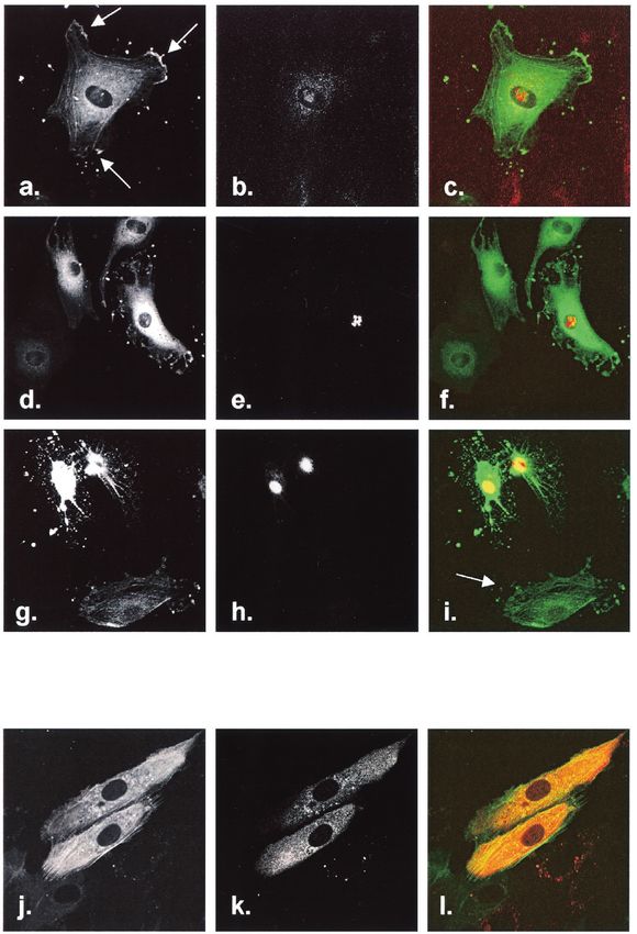
780 WU ET AL. J. VIROL.
Downloaded from http://jvi.asm.org/ on April 22, 2021 by guest
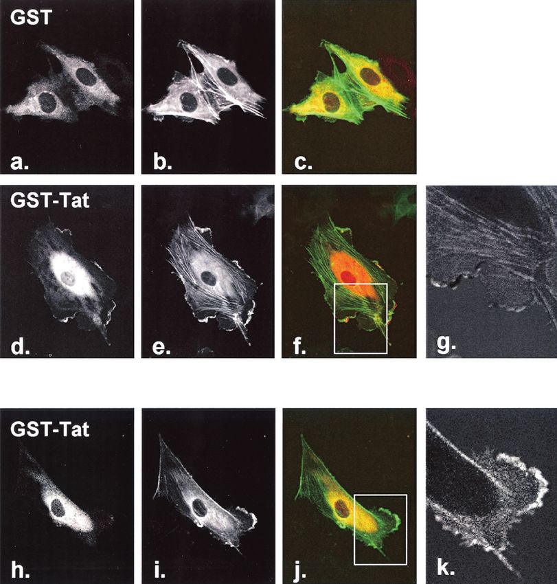
FIG. 1. Effect of GST-Tat on endothelial cell actin dynamics. (a) HUVEC were transfected with actin-GFP to visualize actin microfilament
structures. Cells were subsequently treated with GST-Tat or GST alone for the indicated times and observed live and unfixed. At a concentration
of 200 ng/ml, loss of stress fibers and peripheral retraction were seen within 5 min (b and c), and ruffling was observed within 5 min and peaking
at approximately 20 min (b to d, arrows). (e) GST alone had no effect on actin structures. (g) At a concentration of 600 ng/ml, ruffling occurred
early and was followed by the appearance of actin retraction fibers. (h) By 60 min, all cells showed marked collapse of the actin skeleton, with most
cells displaying a reticular or a stellate cytosolic remnant. (i) Treatment with GST alone (600 ng/ml) had no effect on actin dynamics. Collapse of
the actin cytoskeleton induced by cytochalasin D (10 M) is shown in panel j for comparison.
sion of HIV-1 Tat-86 from pGEX-Tat-86 (National Institutes of Health AIDS
Research and Reference Reagent Program) and ligation into pDsRed2-C1
(Clontech). The full-length p47phox coding region was excised from pBlue-
script-SK and ligated into the EcoRI site of pDsRed2-C1 to create DsRed-p47.
Human HA-JNK2 and pCINF-p47 (encoding Flag-p47) were constructed as
previously described (29). Mutations in the cysteine-rich [GST-Tat
C(22,25,27)A] or basic [GST-Tat R(49,52,53,55,56,57)A] domains of HIV-1 Tat
were introduced as described previously (19). pEGFP and actin-green fluores-
cent protein (GFP) were purchased from Clontech. pCMV5 M-PAK1(K298A)
was obtained from Melanie Cobb (University of Texas Southwestern), PCR
amplified with addition of terminal NheI and SalI sites, and ligated directionally
into the corresponding sites of the shuttle vector pDC315io (Microbix) contain-
ing a loxP site and the murine cytomegalovirus promoter upstream of a lac
operator site, to generate pDC315-PAK1(K298A). This plasmid was cotrans-
fected with the adenovirus backbone pBHGlox(del)E1,3Cre (Microbix) contain-
ing a Cre recombinase cassette. To avoid potential interference with growth and
survival signals in packaging cells due to overexpression of PAK1(K298A), trans-
gene expression was suppressed by constitutive lac repressor expression by the
packaging cell line 293IQ (Microbix). Viral plaques were cloned and titers were
determined by using 293IQ cells. The p67(V204A) mutation, previously con-
structed from p67phox (11), was excised and ligated into the EcoRI site of the
shuttle vector pDC316io (Microbix). Viruses were again produced after Cre-Lox
FIG. 2. Effect of GST-Tat on HLMVEC. (a and c) Unstimulated
HLMVEC or GST-treated HLMVEC transfected with actin-GFP dis-
played pronounced flattening and prominent stress fibers. (b) After
treatment with 200 ng of GST-Tat/ml, stress fibers resorbed and edge
ruffles appeared within 20 to 30 min, similar in character to changes
observed in HUVEC. (d) At 600 ng of GST-Tat/ml, actin collapse and
focal edge accumulation were seen.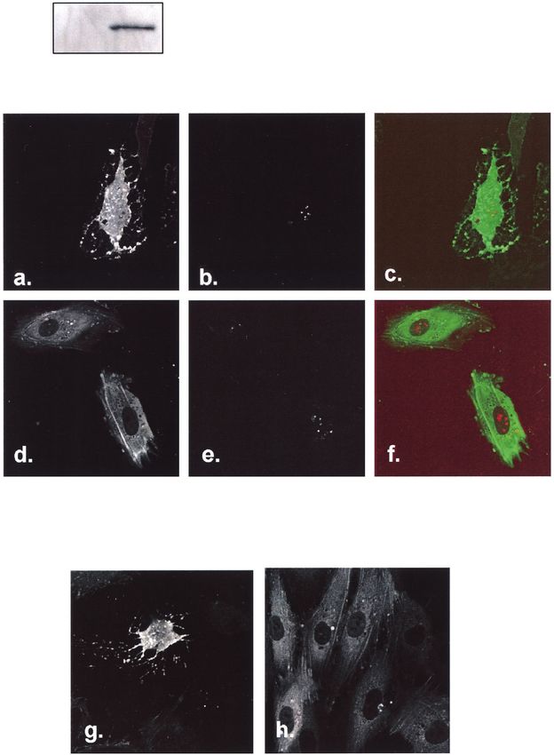
Downloaded from http://jvi.asm.org/ on April 22, 2021 by guest
781
HIV-1 Tat AND ENDOTHELIAL CYTOSKELETON

                                           FIG. 3. Effect of Tat basic and cysteine-rich domains. HUVEC were transfected with actin-GFP and exposed to 600 ng of GST/ml
                                         (a), wild-type GST-Tat (b), GST-Tat R(49,52,53,55,56,57)A (c), or GST-Tat C(22,25,27)A (d). Whereas all cells exposed to wild-type
                                         GST-Tat displayed marked cytoskeletal collapse, cells exposed to GST-Tat containing mutations within the basic domain remained
                                         identical to GST-treated cells. Rare cells treated with the cysteine-rich domain mutant GST-Tat developed retraction fibers.
VOL. 78, 2004
782 WU ET AL. J. VIROL.
Downloaded from http://jvi.asm.org/ on April 22, 2021 by guest
FIG. 4. Effect of DsRed-Tat on endothelial cell actin dynamics. HUVEC were cotransfected with actin-GFP and DsRed-Tat and observed at
the indicated times. Actin-GFP was imaged in the green channel (a, d, g, and j), and DsRed-Tat or DsRed was imaged in the red channel (b, e,
h, and k). At 15 h, frequent ruffle formation was observed (a, arrows), and DsRed-Tat was seen accumulating in nucleoli, with occasional
visualization in cytosol (b). Peripheral retraction started within 24 h (d), seen in DsRed-Tat-expressing cells (d to f). (g to i) By 48 h,
DsRed-Tat-expressing cells displayed actin collapse identical to that after GST-Tat treatment. Actin changes were visualized only in DsRed-Tat-
expressing cells and not in neighboring cells (i, arrow). Cells transfected with DsRed alone did not exhibit alterations in actin dynamics at 24 h (j
to l) or 48 h (not shown).VOL. 78, 2004 HIV-1 Tat AND ENDOTHELIAL CYTOSKELETON 783
RESULTS
GST-Tat induces prominent actin cytoskeletal rearrange-
ment. Actin structures were visualized in live cells by using
actin-GFP. After treatment of HUVEC with 200 ng of GST-
Tat/ml (corresponding to 52 ng of Tat)/ml, stress fibers were
seen to diminish within 5 to 10 min (Fig. 1b). Within 10 min
and peaking at 15 to 25 min, various amounts of peripheral
retraction (Fig. 1c) and prominent ruffling (Fig. 1d) were ob-
FIG. 5. Activation of PAK1 by GST-Tat. HUVEC were treated served. Membrane ruffles are vertical actin scaffolds that ac-
with GST-Tat or GST (600 ng/ml) for the indicated times. PAK1 company cell movement and were recognized by their charac-
activation was assessed by IP kinase and phosphorylation of myelin teristic morphology, rapid movement across cells, and dorsal
basic protein (MBP). TNF (100 ng/ml) was added for 15 min as a projection. Tat-induced ruffles were frequently disorganized,
positive control. The lower panel shows an immunoblot of immuno-
causing multiple leading edges to form and dissipate (e.g., Fig.
precipitated PAK1 with anti-PAK1. HC, immunoglobulin heavy chain.
1d). Ruffling diminished within 1 h. Cells treated with GST
Downloaded from http://jvi.asm.org/ on April 22, 2021 by guest
alone did not change within this time frame (Fig. 1e).
Treatment of HUVEC with 600 ng of GST-Tat/ml (corre-
sponding to 157 ng of Tat/ml) induced early ruffling but within
recombination in 293IQ cells. The Ad-lacZ virus was as previously described
(29). 30 min led to prominent actin retraction fibers as the cell
GST-Tat production. SURE Escherichia coli (Stratagene) was transformed periphery contracted (Fig. 1g). After 60 min, the HUVEC
with pGEX-2TK, pGEX-Tat-86, pGEX-Tat C(22,25,27)A, or pGEX-Tat actin skeleton collapsed in virtually all cells, leaving a trabec-
R(49,52,53,55,56,57)A and was induced with IPTG (isopropyl--D-thiogalacto- ulated, arborized cytosolic remnant (Fig. 1h). Collapse of the
pyranoside) for 3 to 4 h at 37°C, and the fusion proteins were extracted as
actin skeleton was confirmed by comparison with cytochalasin
previously described (10, 29), keeping all solutions degassed and at 4°C. The
yield, stability, and biological activity of wild-type Tat (assessed as activation of D-treated cells, which mimicked the Tat-induced arborized
endothelial cell c-Jun N-terminal kinase [JNK]) were highest when flash-frozen pattern (Fig. 1j). Despite the severe cytoskeletal disruption,
as a glutathione S-transferase (GST) fusion. however, the nuclear morphology was unchanged. Again,
Cell culture and transduction. Human umbilical vein endothelial cells treatment with 600 ng of GST/ml alone had no effect on actin
(HUVEC) were obtained from BioWhittaker, grown in EGM-2 media, and used
structures (Fig. 1i). The cytoskeletal effects of GST-Tat were
within five passages. Human lung microvascular endothelial cells (HLMVEC)
were obtained from BioWhittaker, grown in EGM-2-MV media, and used on the also observed in HLMVEC, suggesting that activation of these
first passage. Transfection was performed by electroporation after thymidine- pathways was not confined to HUVEC. Within 20 to 30 min of
induced cell cycle synchronization as previously described (29). Adenovirus GST-Tat treatment (200 ng/ml), prominent stress fibers within
transduction was achieved after infection of HUVEC at 70 to 90% confluence for HLMVEC were disassembled (Fig. 2a and b). At the higher
1 h at a multiplicity of infection (MOI) of 100:1, followed by a 24-h recovery
dose (600 ng/ml), GST-Tat caused both collapse of the normal
period in full media. In some experiments, cells were pretreated with the super-
oxide dismutase mimetic Mn(III) tetrakis(1-methyl-4-pyridil)porphyrin (MnT- actin skeleton and scattered lamellar formations (Fig. 2d).
MPyP; Alexis) at 100 M for 1 h. HIV-1 Tat possesses several known functional domains, in-
Microscopy. HUVEC were plated on fibronectin-coated coverslip chambers cluding a cysteine-rich putative dimerization motif and a poly-
(Nunc), and live cells were observed on a heated stage without fixation or basic domain involved in growth factor receptor recognition.
permeabilization. Confocal images were obtained with an inverted Zeiss Axio-
vert S100TV microscope and the LSM 410 laser-scanning system. Fluorescence
These latter two domains are both critical to Tat-induced au-
ratio imaging was performed by using Zeiss LSM (v3.98) software. tophosphorylation of VEGFR-2 and chemotactic migration of
Kinase activities. PAK1 activity was assessed by an immunoprecipitation ki- endothelial cells in response to Tat (19), a response that re-
nase method. After treatment, HUVEC were harvested in cold lysis buffer (20 quires active cytoskeletal reorganization. Thus, we tested the
mM Tris-HCl [pH 7.5], 150 mM NaCl, 1 mM disodium EDTA, 1 mM EGTA, 1% involvement of these two functional domains in altering actin
Triton X-100, 2.5 mM sodium pyrophosphate, 1 mM -glycerophosphate, 1 mM
Na3VO4, 1 g of leupeptin/ml, and 1 mM phenylmethylsulfonyl fluoride), son-
dynamics. We found that, whereas wild-type GST-Tat induced
icated briefly, and immunoprecipitated with rabbit anti-PAK1 (Santa Cruz). The marked actin changes within 60 min, GST-Tat R(49,52,
kinase reaction was performed with 5 g of myelin basic protein (Upstate) as a 53,55,56,57)A had no discernible effects on actin structure,
PAK1 substrate in the presence of [␥32P]ATP (Perkin-Elmer) at 30°C for 30 min. even at 600 ng/ml, in any cell observed (Fig. 3a to c). At 600
For JNK activity, HUVEC were cotransfected with HA-JNK2 and either ng/ml, GST-Tat C(22,25,27)A caused very little cytoskeletal
PAK1(K298A) or empty vector. The following day, an immunocomplex assay
was performed with anti-HA (Santa Cruz) and 2 g of GST-Jun as a substrate
movement, with rare cells displaying actin retraction fibers but
(29). Two-thirds of each sample was subjected to autoradiography, and one-third no membrane ruffling (Fig. 3d). Thus, both the basic and cys-
was subjected to immunoblot to assess capture of PAK1 or HA-JNK2. teine-rich domains appear necessary for Tat to initiate the
p47phox phosphorylation. HUVEC were cotransfected with Flag-p47 and ei- observed actin rearrangements.
ther PAK1(K298A) or empty vector, and the following day the medium was To further confirm the influence of Tat on actin dynamics,
replaced with phosphate-free Dulbecco modified Eagle medium (Sigma) con-
taining 0.5 mCi of [32P]orthophosphate/ml for 4 h. Cells were then stimulated
HUVEC were transfected with DsRed-Tat, allowing compar-
with GST or GST-Tat (600 ng/ml) for 15 min and lysed on ice for 15 min in lysis ison of Tat-expressing versus nonexpressing cells. We found
buffer containing 0.1% sodium dodecyl sulfate and 0.5% deoxycholate. After it early and prominent nucleolar localization of DsRed-Tat but
was sheared through a 23-gauge needle 10 times, the 13,000 ⫻ g supernatant was visualized cytosolic DsRed-Tat in occasional cells as well (Fig.
precleared and normalized for protein. Flag-p47 was immunoprecipitated with
4a to c). At 15 h, membrane ruffling was observed in DsRed-
anti-Flag and washed four times in lysis buffer with sodium dodecyl sulfate and
deoxycholate, twice in lysis buffer containing 0.5 M NaCl, and once in 50 mM Tris
Tat-expressing cells. By 24 h, DsRed-Tat expression was gen-
(pH 7.0). One-third of the immunoprecipitate was immunoblotted for p47phox to erally higher, and cells exhibited loss of stress fibers and
assess capture and loading, and the remainder was analyzed by autoradiography. marked peripheral retraction (Fig. 4d to f). By 48 h, DsRed-784 WU ET AL. J. VIROL.
Downloaded from http://jvi.asm.org/ on April 22, 2021 by guest
FIG. 6. Effect of PAK1(K298A) on actin rearrangement. HUVEC were cotransfected with actin-GFP and DsRed-Tat and then infected with
either Ad-lacZ or Ad-PAK1(K298A) (MOI ⫽ 100:1, 1 h) the following day. Cells were examined after 48 h. The upper panel shows immunoblot
with anti-Myc, demonstrating expression of Myc-tagged PAK1(K298A) (DN-PAK1). Actin-GFP was imaged in the green channel (a and d), and
DsRed-Tat was imaged in the red channel (b and e). DsRed-Tat-expressing cells infected with Ad-lacZ displayed prominent actin collapse (a to
c), whereas DsRed-Tat-expressing cells infected with Ad-PAK1(K298A) retained normal morphology (d to f). (g and h) HUVEC were transfected
with actin-GFP, infected with the corresponding adenoviruses, and treated with GST-Tat (600 ng/ml for 60 min). GFP-actin is shown. Cytoskeletal
collapse seen in Ad-lacZ-infected cells was absent in Ad-PAK1(K298A)-infected cells.VOL. 78, 2004 HIV-1 Tat AND ENDOTHELIAL CYTOSKELETON 785
of 600 ng of GST-Tat/ml, which induced cell collapse in virtu-
ally all cells infected with Ad-lacZ, did not increase ruffle
formation or induce actin collapse in PAK1(K298A)-trans-
duced cells observed well beyond 60 min (Fig. 6g and h). As
further evidence that Tat initiates angiogenic signal pathways
through PAK1, we examined activation of the JNK mitogen-
activated protein kinase (MAPK), which is known to mediate
proliferative and angiogenic signaling downstream of VEGF
receptor ligation (22). As with the cytoskeletal changes,
PAK1(K298A) effectively abolished JNK2 activation by GST-
Tat (Fig. 7).
FIG. 7. Effect of PAK1(K298A) on JNK activation. HUVEC were An NADPH oxidase acts upstream of GST-Tat-induced cy-
cotransfected with either empty vector or PAK1(K298A) (DN-PAK1), toskeletal rearrangements. We previously demonstrated that
and with HA-JNK2. After 24 h, cells were treated with GST-Tat (600 HIV-1 Tat increases oxidant production in endothelial cells
ng/ml, 15 min), and HA-JNK2 activation was assessed by IP kinase. through activation of a phagocyte-like NADPH oxidase and
Downloaded from http://jvi.asm.org/ on April 22, 2021 by guest
Capture of HA-JNK2 was assessed by immunoblotting for JNK (lower
that Tat-induced activation of the JNK MAPK occurs through
panel). PAK1(K298A) blocked JNK activation by GST-Tat.
such oxidants (10). Further, the NADPH oxidase adapter
p47phox was found to be constitutively associated with the en-
dothelial cell cytoskeleton (12). We therefore sought to deter-
mine whether the endothelial oxidase participates in Tat-in-
Tat-expressing cells acquired a collapsed actin skeleton with duced actin cytoskeletal changes. We found first that treatment
dendritic cytosolic projections, identical to cells treated with of HUVEC with GST-Tat caused redistribution of p47phox to
600 ng of GST-Tat (Fig. 4g to i)/ml. Notably, cytoskeletal membrane ruffles within 15 to 20 min. DsRed-p47 localized to
actin-GFP-containing ruffles upon stimulation with GST-Tat
abnormalities were restricted to DsRed-Tat-expressing cells,
but not GST alone (Fig. 8a to f). Fluorescence ratio imaging
whereas neighboring cells displayed normal actin structures
revealed cancellation of red and green intensities within ruffles,
and no visible DsRed-Tat (Fig. 4g to i). Thus, we did not find
further demonstrating colocalization of the two chromophores
evidence that DsRed-Tat could be exported and internalized
(Fig. 8g). In contrast, DsRed itself did not concentrate within
by other cells diffusely. DsRed not fused to Tat was confined to
Tat-induced ruffles (Fig. 8h to j); green/red ratio images there-
the cytosol and did not affect actin dynamics (Fig. 4j to l).
fore displayed prominent enhancement of actin-GFP within
Hence, the ability of Tat to cause dramatic rearrangements in
ruffles relative to DsRed (Fig. 8k).
the actin cytoskeleton did not appear to be an artifact of of
In phagocytes, phosphorylation of p47phox initiates assembly
DsRed itself, or of the GST fusion tag or preparative contam-
and activation of the NADPH oxidase (2). We found that
inants.
GST-Tat increased phosphorylation of Flag-p47 within 15 min
PAK1 transduces GST-Tat-induced cytoskeletal rearrange-
by ⬃5-fold (average of two independent experiments), whereas
ments. The morphology of the collapsed actin structures pre- GST alone did not (Fig. 9), a finding consistent with our prior
cipitated by exogenous or expressed Tat is highly reminiscent observations that Tat activates an endothelial cell NADPH
of actin dynamics seen after microinjection of active PAK1 oxidase (10). Unlike the situation in phagocytes, detectable
mutants into HeLa and Ref52 cells (8, 16, 30). Accordingly, we basal phosphorylation of p47phox was observed. Expression of
hypothesized that Tat may exert its cytoskeletal effects through PAK1(K298A) effectively blocked both basal and Tat-stimu-
activation of endothelial PAK1. We found robust activation of lated p47phox phosphorylation, demonstrating that PAK1 acts
PAK1 in HUVEC within 5 min of exposure to GST-Tat, de- upstream of p47phox.
creasing by 30 min, whereas GST alone had no effect (Fig. 5). To test for involvement of the NADPH oxidase in actin
PAK1 activation by GST-Tat was at least as strong as with the rearrangements, we constructed a second adenovirus deliver-
known PAK1 agonist TNF (Fig. 5, right lane). ing the oxidase-activating subunit p67phox harboring a mutation
To test the involvement of PAK1 in Tat-induced cytoskeletal in the purported activation domain. This mutant, p67(V204A),
dynamics, we generated an adenovirus harboring the kinase- decreases oxidant production in the presence of wild-type
dead mutant PAK1(K298A), which possesses dominant-inter- p67phox, thus possessing dominant-negative activity (13). Ex-
fering activity (8). Efficiency of adenovirus-dependent trans- pression of p67(V204A), confirmed by immunoblotting (Fig.
duction in HUVEC, assessed by in situ -galactosidase staining 10 top panel), completely blocked actin cytoskeletal collapse in
after Ad-lacZ infection, was in excess of 95% (not shown). GST-Tat-treated HUVEC (Fig. 10a to d). Further, treatment
Transgene expression was demonstrated by immunoblot for of cells with the membrane permeant superoxide dismutase
the Myc-tagged PAK1(K298A) (Fig. 6, top panel). We found mimetic MnTMPyP also completely blocked cytoskeletal col-
that expression of PAK1(K298A) essentially abolished Tat- lapse (Fig. 10e to f), confirming a role for oxidants in the
induced cytoskeletal rearrangements. Whereas all DsRed-Tat- control of actin dynamics by Tat.
expressing cells displayed various degrees of actin cytoskeletal
disruption when infected with the control virus Ad-lacZ (Fig.
DISCUSSION
6a to c), Ad-PAK1(K298A)-infected cells had quiescent actin
skeletons, many with well-formed stress fibers, even while ex- Known angiogenic factors such as VEGF, basic FGF, and
pressing DsRed-Tat (Fig. 6d to f). More remarkably, addition angiopoietin-1 induce characteristic changes in actin dynamics786 WU ET AL. J. VIROL.
Downloaded from http://jvi.asm.org/ on April 22, 2021 by guest
FIG. 8. DsRed-p47 translocates to Tat-induced membrane ruffles. HUVEC were cotransfected with actin-GFP and DsRed-p47. After stimu-
lation of cells with GST (a to c) or GST-Tat (d to g) (200 ng/ml, 15 to 20 min), DsRed-p47 was imaged in the red channel (a and d), and actin-GFP
was imaged in the green channel (b and e). DsRed-p47 translocated avidly to actin ruffles upon stimulation with GST-Tat (d to f). The green/red
fluorescence ratio image of inset from panel f shows cancellation of actin-GFP signal by DsRed-p47 within ruffles (g), indicating colocalization of
the two proteins. DsRed itself, when cotransfected with actin-GFP, did not translocate to ruffles (h to j). The green/red ratio image of inset from
panel j shows relative enhancement of actin-GFP within ruffle relative to DsRed (k).VOL. 78, 2004 HIV-1 Tat AND ENDOTHELIAL CYTOSKELETON 787
strated continuous shuttling of GFP-Tat between the nucleus
and cytoplasm of HeLa cells.
Although the proximal molecular target of Tat responsible
for initiating cytoskeletal changes is unknown, our data suggest
early involvement of PAK1. PAK1, an MAP kinase kinase
kinase kinase involved in mitogenic stimulation, is known to
concentrate in membrane ruffles, and active mutants of PAK1
cause ruffle formation (7, 8). When expressed in extremely
high levels achieved by microinjection, active PAK1 mutants—
containing deletions or mutations in the autoinhibitory domain
or fusion with active Cdc42—stimulate peripheral retraction,
FIG. 9. Effect of PAK1(K298A) on p47phox phosphorylation. followed by actin depolymerization and collapse of the cy-
HUVEC were cotransfected with either empty vector or toskeleton (8, 16, 30), highly reminiscent of our findings with
PAK1(K298A) (DN-PAK1) and with Flag-p47 and then stimulated Tat. To our knowledge, such severe architectural derange-
with GST or GST-Tat (600 ng/ml). Flag-p47 was immunoprecipitated,
ments have not been previously observed without microinjec-
Downloaded from http://jvi.asm.org/ on April 22, 2021 by guest
and phosphorylation (upper panel) and protein capture (anti-Flag,
lower panel) were assessed. GST-Tat increased phosphorylation of tion of these PAK1 mutants or cytochalasin treatment.
p47phox, whereas PAK1(K298A) completely blocked basal and Tat- The relevance of PAK1 activation by Tat to HIV-1-infected
induced p47phox phosphorylation. patients may extend to tumor biology as well as AIDS vascu-
lopathy. In tumor cells, PAK1 promotes migration and invasive
behavior (1). Notably, Tat-expressing adenocarcinoma cells
injected into mice produce aggressive, highly metastatic tu-
mors (6), suggesting an active PAK1 phenotype and consistent
with the biologically aggressive malignancies AIDS patients
leading to membrane ruffling and dissolution of stress fibers
develop. In addition, during angiogenesis, endothelial cell be-
with redistribution of focal adhesions. These cytoskeletal
havior recapitulates that of tumor cells, becoming invasive,
changes are currently thought to facilitate migration and inva-
metastatic, and proliferative. Thus, angiogenic behavior may
sion of endothelial cells during sprout formation. In the
logically be linked to PAK1. Indeed, PAK1 has been linked to
present study, we found that HIV-1 Tat induced rapid and
endothelial cell migration and contractility in vitro and angio-
dramatic changes in the actin cytoskeleton consistent with its
genesis in vivo (15, 17). The ability of Tat to activate endothe-
known biological effect as a proangiogenic agent.
lial PAK1 is therefore consistent with the intense angiogenesis
Extracellular Tat delivered as a GST fusion increased ruffle
invoked by Tat-secreting tumors and also with the develop-
formation and peripheral retraction and at higher levels caused
ment of invasive Kaposi’s sarcoma-like vascular lesions in Tat-
prominent loss of stress fibers with visible depolymerization
transgenic mice (6, 27). Accordingly, in AIDS-related Kaposi’s
and collapse of the actin cytoskeleton. These dramatic effects
sarcoma, the malignant vascular cells migrate and invade much
on actin dynamics are consistent with physiologic and biochem- more aggressively than in non-HIV-related Kaposi’s sarcoma.
ical studies demonstrating an increase in paracellular albumin Recently, we demonstrated that Tat activates an NADPH
permeability (21) and in phosphorylation of focal adhesion oxidase upstream of the JNK MAPK in endothelial cells (10).
proteins such as FAK, Pyk2, paxillin, and p130Cas (9) after Tat In addition, p47phox, the adapter subunit of this oxidase, was
treatment. Interestingly, cellular expression of Tat as a DsRed found to be constitutively associated with the cytoskeleton, and
fusion produced identical morphological changes, raising ques- disruption of the actin cytoskeleton blocked Tat-induced sig-
tions about the site of the molecular target(s) of Tat involved naling (10, 12), suggesting a functional relationship between
in cytoskeletal alterations. The appearance of normal cells in the oxidase and the cytoskeleton. In the present study, our data
close proximity to collapsed, DsRed-Tat-expressing cells sug- suggest that activation of the endothelial NADPH oxidase
gests that Tat may bind intracellular target proteins to effect occurs upstream of Tat-induced actin rearrangements. Exoge-
such changes rather than being exported and acting in a para- nous oxidants are known to cause actin reorganization, and
crine fashion. This suggests other mechanisms for cytoskeletal endogenous oxidants appear to be necessary for spontaneous
signaling beyond ligation of known Tat receptors such as endothelial cell migration (20). Thus, PAK1 may in part exert
VEGFR2 and ␣V3/␣51 integrins (5, 23), although in the its cytoskeletal effects through NADPH oxidase activation, a
present study we have not excluded a receptor-mediated mech- premise supported by the dependence of p47phox phosphory-
anism. An additional curiosity is that an intracellular site of lation on PAK1 kinase activity. Notably, Tat induced translo-
action would be expected to reside within the cytosol or inter- cation of p47phox to membrane ruffles. These actin structures
nal membrane surface, whereas DsRed-Tat accumulated in are known to be enriched in active PAK1, and oxidant produc-
endothelial cell nucleoli. However, a cytosolic phase for tion is focally produced in the ruffles of motile endothelial cells
DsRed-Tat was clearly seen in a fraction of cells, suggesting an as well as phagocytes (14, 20). Finally, the demonstration that
equilibrium between nucleus and cytosol. This possibility is PAK1(K298A) blocked Tat-induced JNK activation is consis-
consistent with trafficking of a GFP-Tat protein, which also tent with our prior observation that the NADPH oxidase also
accumulates in nucleoli of HeLa cells (25). In this latter study, lies upstream of JNK activation by Tat (10). Thus, activation of
analysis of membrane fusion between transfected and non- PAK1 and the oxidase appear to control HIV-1 Tat signaling
transfected cells by using polyethylene glycol clearly demon- to both MAPK and cytoskeletal pathways.788 WU ET AL. J. VIROL.
Downloaded from http://jvi.asm.org/ on April 22, 2021 by guest
FIG. 10. Effect of p67(V204A) on actin rearrangement. HUVEC were transfected with actin-GFP and then infected with Ad-lacZ (a and b)
or Ad-p67(V204A) (c and d) (MOI ⫽ 100:1, 1 h). The upper panel shows immunoblot with anti-p67phox, demonstrating expression of p67(V204A)
(DN-p67). (b) Cells infected with Ad-lacZ displayed prominent retraction and actin collapse after treatment with GST-Tat (600 ng/ml). (c and d)
Expression of p67(V204A) completely suppressed actin cytoskeletal rearrangements. (e and f) HUVEC were transfected with actin-GFP and then
pretreated with the superoxide dismutase mimetic MnTMPyP (100 M, 1 h) prior to treatment with GST-Tat (600 ng/ml). MnTMPyP prevented
the actin cytoskeletal changes induced by GST-Tat (f).VOL. 78, 2004 HIV-1 Tat AND ENDOTHELIAL CYTOSKELETON 789
ACKNOWLEDGMENTS Cofilin undergoes rapid dephosphorylation in stimulated neutrophils and
translocates to ruffled membranes enriched in products of the NADPH
We acknowledge the excellent technical assistance of Ginny Poffen- oxidase complex. Evidence for a novel cycle of phosphorylation and dephos-
berger. phorylation. Histochem. Cell Biol. 108:221–233.
This study was supported by NIH grants R01-HL061897 and R01- 15. Kiosses, W. B., J. Hood, S. Yang, M. E. Gerritsen, D. A. Cheresh, N.
HL067256 and by the Veterans Administration. Alderson, and M. A. Schwartz. 2002. A dominant-negative p65 PAK peptide
inhibits angiogenesis. Circ. Res. 90:697–702.
16. Manser, E., H.-Y. Huang, T.-H. Loo, X.-Q. Chen, J.-M. Dong, T. Leung, and
REFERENCES
L. Lim. 1997. Expression of constitutively active ␣-PAK reveals effects of the
1. Adam, L., R. Vadlamudi, M. Mandal, J. Chernoff, and R. Kumar. 2000. kinase on actin and focal complexes. Mol. Cell. Biol. 17:1129–1143.
Regulation of microfilament reorganization and invasiveness of breast can- 17. Master, Z., N. Jones, J. Tran, J. Jones, R. S. Kerbel, and D. J. Dumont. 2001.
cer cells by kinase dead p21-activated kinase-1. J. Biol. Chem. 275:12041– Dok-R plays a pivotal role in angiopoietin-1-dependent cell migration
12050. through recruitment and activation of Pak. EMBO J. 20:5919–5928.
2. Ago, T., H. Nunoi, T. Ito, and H. Sumimoto. 1999. Mechanism for phospho- 18. McNutt, N. S., V. Fletcher, and M. A. Conant. 1983. Early lesions of Kaposi’s
rylation-induced activation of the phagocyte NADPH oxidase protein sarcoma in homosexual men. An ultrastructural comparison with other vas-
p47phox: triple replacement of serines 303, 304, and 328 with aspartates cular proliferations in skin. Am. J. Pathol. 111:62–77.
disrupts the SH3 domain-mediated intramolecular interaction in p47phox, 19. Mitola, S., R. Soldi, I. Zanon, L. Barra, M. I. Gutierrez, B. Berkhout, M.
thereby activating the oxidase. J. Biol. Chem. 274:33644–33653. Giacca, and F. Bussolino. 2000. Identification of specific molecular struc-
3. Albini, A., G. Barillari, R. Benelli, R. C. Gallo, and B. Ensoli. 1995. Angio- tures of human immunodeficiency virus type 1 Tat relevant for its biological
genic properties of human immunodeficiency virus type 1 Tat protein. Proc. effects on vascular endothelial cells. J. Virol. 74:344–353.
Downloaded from http://jvi.asm.org/ on April 22, 2021 by guest
Natl. Acad. Sci. USA 92:4838–4842. 20. Moldovan, L., N. I. Moldovan, R. H. Sohn, S. A. Parikh, and P. J. Gold-
4. Albini, A., R. Soldi, D. Giunciuglio, E. Giraudo, R. Benelli, L. Primo, D. schmidt-Clermont. 2000. Redox changes of cultured endothelial cells and
Noonan, M. Salio, G. Camussi, W. Rockl, and F. Bussolino. 1996. The actin dynamics. Circ. Res. 86:549–557.
angiogenesis induced by HIV-1 tat protein is mediated by the Flk-1/KDR 21. Oshima, T., S. C. Flores, G. Vaitaitis, L. L. Coe, T. Joh, J. H. Park, Y. Zhu,
receptor on vascular endothelial cells. Nat. Med. 2:1371–1375. B. Alexander, and J. S. Alexander. 2000. HIV-1 Tat increases endothelial
5. Barillari, G., C. Sgadari, V. Fiorelli, F. Samaniego, S. Colombini, V. Man- solute permeability through tyrosine kinase and mitogen-activated protein
zari, A. Modesti, B. C. Nair, A. Cafaro, M. Sturzl, and B. Ensoli. 1999. The kinase-dependent pathways. AIDS 14:475–482.
Tat protein of human immunodeficiency virus type-1 promotes vascular cell 22. Pedram, A., M. Razandi, and E. R. Levin. 1998. Extracellular signal-regu-
growth and locomotion by engaging the ␣51 and ␣v3 integrins and by lated protein kinase/Jun kinase cross-talk underlies vascular endothelial cell
mobilizing sequestered basic fibroblast growth factor. Blood 94:663–672. growth factor-induced endothelial cell proliferation. J. Biol. Chem. 273:
6. Corallini, A., D. Campioni, C. Rossi, A. Albini, L. Possati, M. Rusnati, G. 26722–26728.
Gazzanelli, R. Benelli, L. Masiello, V. Sparacciari, M. Presta, F. Mannello, 23. Scheidegger, P., W. Weiglhofer, S. Suarez, S. Console, J. Waltenberger, M. S.
G. Fontanini, and G. Barbanti-Brodano. 1996. Promotion of tumour metas- Pepper, R. Jaussi, and K. Ballmer-Hofer. 2001. Signalling properties of an
tases and induction of angiogenesis by native HIV-1 Tat protein from BK HIV-encoded angiogenic peptide mimicking vascular endothelial growth
virus/tat transgenic mice. AIDS 10:701–710. factor activity. Biochem. J. 353:569–578.
7. Dharmawardhane, S., A. Schurmann, M. A. Sells, J. Chernoff, S. L. Schmid, 24. Schwarze, S. R., and S. F. Dowdy. 2000. In vivo protein transduction: intra-
and G. M. Bokoch. 2000. Regulation of macropinocytosis by p21-activated cellular delivery of biologically active proteins, compounds and DNA.
kinase-1. Mol. Biol. Cell 11:3341–3352. Trends Pharmacol. Sci. 21:45–48.
8. Frost, J. A., A. Khokhlatchev, S. Stippec, M. A. White, and M. H. Cobb. 1998. 25. Stauber, R. H., and G. N. Pavlakis. 1998. Intracellular trafficking and inter-
Differential effects of PAK1-activating mutations reveal activity-dependent actions of the HIV-1 Tat protein. Virology 252:126–136.
and -independent effects on cytoskeletal regulation. J. Biol. Chem. 273: 26. Tabib, A., C. Leroux, J. F. Mornex, and R. Loire. 2000. Accelerated coronary
28191–28198. atherosclerosis and arteriosclerosis in young human-immunodeficiency-vi-
9. Ganju, R. K., N. Munshi, B. C. Nair, Z. Y. Liu, P. Gill, and J. E. Groopman. rus-positive patients. Coronary Artery Dis. 11:41–46.
1998. Human immunodeficiency virus tat modulates the Flk-1/KDR recep- 27. Vogel, J., S. H. Hinrichs, R. K. Reynolds, P. A. Luciw, and G. Jay. 1988. The
tor, mitogen-activated protein kinases, and components of focal adhesion in HIV tat gene induces dermal lesions resembling Kaposi’s sarcoma in trans-
Kaposi’s sarcoma cells. J. Virol. 72:6131–6137. genic mice. Nature 335:606–611.
10. Gu, Y., R. F. Wu, Y. C. Xu, S. C. Flores, and L. S. Terada. 2001. HIV Tat 28. Westendorp, M. O., R. Frank, C. Ochsenbauer, K. Stricker, J. Dhein, H.
activates c-Jun amino-terminal kinase through an oxidant-dependent mech- Walczak, K. M. Debatin, and P. H. Krammer. 1995. Sensitization of T cells
anism. Virology 286:62–71. to CD95-mediated apoptosis by HIV-1 Tat and gp120. Nature 375:497–500.
11. Gu, Y., Y. C. Xu, R. F. Wu, F. E. Nwariaku, R. F. Souza, S. C. Flores, and 29. Xu, Y. C., R. F. Wu, Y. Gu, Y. S. Yang, M. C. Yang, F. E. Nwariaku, and L. S.
L. S. Terada. 2003. p47phox participates in activation of RelA in endothelial Terada. 2002. Involvement of TRAF4 in oxidative activation of c-jun amino
cells. J. Biol. Chem. 278:17210–17217. terminal kinase. J. Biol. Chem. 277:28051–28057.
12. Gu, Y., Y. C. Xu, R. F. Wu, R. F. Souza, F. E. Nwariaku, and L. S. Terada. 30. Zhao, Z. S., E. Manser, X. Q. Chen, C. Chong, T. Leung, and L. Lim. 1998.
2002. TNF alpha activates c-Jun amino terminal kinase through p47phox. A conserved negative regulatory region in alphaPAK: inhibition of PAK
Exp. Cell Res. 272:62–74. kinases reveals their morphological roles downstream of Cdc42 and Rac1.
13. Han, C. H., J. L. Freeman, T. Lee, S. A. Motalebi, and J. D. Lambeth. 1998. Mol. Cell. Biol. 18:2153–2163.
Regulation of the neutrophil respiratory burst oxidase: identification of an 31. Zietz, C., B. Hotz, M. Sturzl, E. Rauch, R. Penning, and U. Lohrs. 1996.
activation domain in p67phox. J. Biol. Chem. 273:16663–16668. Aortic endothelium in HIV-1 infection: chronic injury, activation, and in-
14. Heyworth, P. G., J. M. Robinson, J. Ding, B. A. Ellis, and J. A. Badwey. 1997. creased leukocyte adherence. Am. J. Pathol. 149:1887–1898.You can also read