Immunomodulatory Effects of Canine Adipose Tissue Mesenchymal Stem Cell-Derived Extracellular Vesicles on Stimulated CD4+ T Cells Isolated from ...
←
→
Page content transcription
If your browser does not render page correctly, please read the page content below

Hindawi
Journal of Immunology Research
Volume 2021, Article ID 2993043, 20 pages
https://doi.org/10.1155/2021/2993043

Research Article
Immunomodulatory Effects of Canine Adipose Tissue
Mesenchymal Stem Cell-Derived Extracellular Vesicles on
Stimulated CD4+ T Cells Isolated from Peripheral Blood
Mononuclear Cells

 Takahiro Teshima ,1,2 Yunosuke Yuchi ,1 Ryohei Suzuki ,1 Hirotaka Matsumoto ,1
 and Hidekazu Koyama 1
 1
 Laboratory of Veterinary Internal Medicine, Department of Veterinary Clinical Medicine, School of Veterinary Medicine, Faculty of
 Veterinary Science, Nippon Veterinary and Life Science University, 1-7-1 Kyonan-cho, Musashino, Tokyo 180-8602, Japan
 2
 Research Center for Animal Life Science Nippon Veterinary and Life Science University, 1-7-1 Kyonan-cho, Musashino,
 Tokyo 180-8602, Japan

 Correspondence should be addressed to Takahiro Teshima; teshima63@nvlu.ac.jp

 Received 17 June 2021; Revised 21 July 2021; Accepted 29 July 2021; Published 17 August 2021

 Academic Editor: Baohui Xu

 Copyright © 2021 Takahiro Teshima et al. This is an open access article distributed under the Creative Commons Attribution
 License, which permits unrestricted use, distribution, and reproduction in any medium, provided the original work is
 properly cited.

 Adipose tissue-derived mesenchymal stem cells (ADSCs) have anti-inflammatory and immunomodulatory characteristics. Many
 studies have suggested that the immunomodulation of ADSCs is largely mediated by secreted paracrine factors. Various factors
 are secreted from ADSCs, among which extracellular vesicles are considered to play a major role in the communication between
 ADSCs and target cells. Several studies have reported the function of canine ADSC-derived extracellular vesicles (cADSC-EVs),
 but few studies have reported the immunomodulatory effects of cADSC-EVs on immune cells. The purpose of this study was to
 investigate the effects of cADSC-EVs on in vitro-stimulated CD4+ T cells isolated from peripheral blood mononuclear cells
 (PBMCs). cADSC-EVs were isolated from cADSCs under naive conditions or primed conditions by tumor necrosis factor-α
 (TNFα) and interferon-γ (IFNγ). The expression levels of several microRNAs in cADSC-EVs were altered by priming with
 TNFα and IFNγ. Culturing PBMCs stimulated with concanavalin A in the presence of naive or primed cADSC-EVs inhibited
 the differentiation of PBMCs and CD4+ T cells and promoted apoptosis of PBMCs. CD4+, CD8+, and CD4+CD8+ T cells were
 decreased, while CD3+CD4-CD8- T cells were increased. T helper (Th) 1, Th2, Th17, and regulatory T (Treg) cells were
 analyzed by flow cytometry. cADSC-EVs inhibited the proliferation of Th1 and Th17 cells and enhanced Th2 and Treg cell
 proliferation. However, CD4+ T cells that had incorporated labeled cADSC-EVs comprised only a few percent of all cells.
 Therefore, these responses of stimulated CD4+ T cells may be due to not only direct effects of cADSC-EVs but also to indirect
 effects through interactions between cADSC-EVs and other immune cells. In conclusion, cADSC-EVs exert immunosuppressive
 effects on stimulated CD4+ T cells in vitro. These findings may be useful for further studies of immune diseases.

1. Introduction immune responses in autoimmune diseases in vivo and it
 has been thought that these benefits are partly due to secreted
Mesenchymal stem cells have various biological characteristics factors from MSCs [5–7]. Moreover, the MSC immunomodu-
that include an immunomodulatory capacity [1–3]. Many latory ability is altered by inflammatory cytokine, such as
studies have demonstrated that MSCs suppress the differenti- those present in the inflammatory microenvironment. Stimu-
ation, proliferation, secretions, and migration of immune cells lation with interferon-γ (IFNγ) enhances the immunosup-
[4]. It has been documented that MSCs improve abnormal pressive effects of MSCs. Priming MSCs with IFNγ
2 Journal of Immunology Research

upregulates indoleamine 2,3-dioxygenase (IDO), secretes Adipose tissue was aseptically collected from falciform liga-
important immunomodulatory molecules, such as prostaglan- ment fat of three anaesthetized dogs. The tissue was washed
din E2 (PGE2), hepatocyte growth factor (HGF), transforming extensively in phosphate buffer solution (PBS), minced, and
growth factor-β (TGFβ), and chemokine ligand 2, and digested with collagenase type I (Sigma-Aldrich, Tokyo,
increases the expression of human leukocyte antigen class I Japan) at 37°C for 45 min with intermittent shaking. After
and II molecules and costimulatory molecules [8]. Priming washing with PBS and centrifuging, the pellets containing
MSCs with tumor necrosis factor-α (TNFα) also promotes the stromal vascular fraction were resuspended, filtered
upregulation of immunoregulatory factors, such as PGE2, through a 100-μm nylon mesh, and incubated overnight in
IDO, and HGF, but is much less pronounced compared with Dulbecco’s Modified Eagle’s medium (DMEM) supple-
IFNγ priming [9]. However, the combination of inflammatory mented with 10% fetal bovine serum (FBS) and a 1%
cytokines to stimulate MSCs may lead to additional effects. antibiotic-antimycotic solution (Thermo Fisher Scientific,
Priming MSCs with TNFα and IFNγ increase factor H pro- Tokyo, Japan) in a humidified atmosphere with 5% CO2 at
duction [10], which potently inhibits complement activation. 37°C. Unattached cells were removed by changing the
Factor H secreted by MSCs is significantly suppressed by the medium, and the attached cells were washed twice with
inhibition of PGE2 and IDO. Therefore, priming MSCs with PBS. Thereafter, the medium was replaced every 3-4 days.
inflammatory cytokines is more useful for the treatment of At 80-90% confluence, the cells were detached with trypsin-
immune-mediated diseases [11–13]. EDTA solution (Sigma-Aldrich) and passaged repeatedly.
 MSCs secrete large numbers of molecules, which include The expression of several markers, such as CD14-FITC,
cytokines and growth factors, as well as extracellular vesicles CD29-PE, CD34-PE, CD44-PE, CD45-FITC, and CD90-PE,
(EVs), and these secretomes contribute to MSC abilities [14, on these cells was determined by flow cytometry using a
15]. Studies have focused on the immunomodulatory capac- CytoFLEX (BECKMAN COULTER, Tokyo, Japan) [24].
ity of MSC secretomes for various types of immune cells and cADSCs at passage 3 were seeded in 150-mm dishes
MSC-derived EVs (MSC-EVs) may have similar functions to (4 × 10⁶ cells/dish) and cultured in high glucose DMEM with
parent cells in terms of immunomodulatory effects [4]. EVs 10% exosome-free FBS (Thermo Fisher Scientific) and a 1%
transfer signaling molecules from one cell to another cell antibiotic-antimycotic solution in a humidified atmosphere
through a paracrine mechanism, because EVs enclose various with 5% CO2 at 37°C. After 24 h, unattached cells were
active molecules, such as bioactive proteins, as well as lipids, removed by changing the medium, and the attached cells
mRNAs, and microRNAs [16], which contribute to regulat- were stimulated for 24 h with tumor necrosis factor-α (TNFα,
ing the gene expression and phenotypic transformation of 20 ng/ml) and interferon-γ (IFNγ, 20 ng/ml). Naive and
target cells. MSC-EVs are incorporated by target cells primed cADSCs were cultured for 72 h.
through direct membrane fusion, receptor-mediated phago-
cytosis, and several other internalization mechanisms, 2.2. cADSC-EV Isolation and Characterization. After 72 h of
which leads to subsequent activation of signal transduction culture, the medium was harvested, and then, EVs were iso-
pathways and involvement in various physiological and lated by a MagCapture Exosome Isolation Kit PS (FUJIFILM
pathological processes that include immune responses Wako Pure Chemical, Osaka, Japan). In brief, the medium
[17–19]. Therefore, MSC-EVs are attracting attention as was centrifuged at 300 g for 5 min and then 1,200 g for
immunomodulators. 20 min at 4°C to remove cells and debris. The supernatant
 Similar to humans, immune-mediated diseases also exist was added to 1/100 volumes of EV-Save Extracellular Vesi-
in veterinary medicine, such as atopic dermatitis, inflamma- cles Blocking Reagent (FUJIFILM Wako Pure Chemical,
tory bowel disease, and immune-mediated arthritis. Almost Osaka, Japan) and concentrated using an ultrafiltration unit
all of these diseases have unclear mechanisms and some cases (Vivaspin; SARTORIUS, Tokyo, Japan). After concentrating
become intractable with current treatments. Therefore, the supernatant, extracellular vesicles including exosomes
MSC-based therapy is also expected to be an alternative treat- were isolated in accordance with the manufacturer’s instruc-
ment method for immune-mediated diseases [11–13]. How- tions. Finally, the EVs were suspended in PBS.
ever, few studies have documented the functions of canine The concentration and size of EVs were determined by
ADSC- (cADSC-) EVs [20–22]. Furthermore, studies that nanoparticle tracking analysis (NTA) using a NanoSight
focus on the effects of cADSV-EVs on T cells have not been LM10 (Malvern Panalytical, Tokyo, Japan) with the follow-
reported. In this study, we investigated the immunomodula- ing parameters: camera level 12, threshold 8, 21.4°C, and five
tory properties of cADSC-EVs on in vitro-stimulated CD4+ T videos per analyzed sample. Visualization of EVs was
cells isolated from peripheral blood mononuclear cells assessed by transmission electron microscopy (H-7600;
(PBMCs). Moreover, we evaluated whether immunomodula- Hitachi High-Tech, Tokyo, Japan).
tory effects of cADSC-EVs on stimulated CD4+ T cells were The total protein concentration of naive and primed EVs
enhanced by parent cells primed with inflammatory cyto- was measured by a BCA assay kit. Each 0.5 μg protein sample
kines such as TNFα and IFNγ. in 20 μl with 2.5 μl NuPAGE LDS sample buffer was electro-
 phoresed in 4%-12% Bis-Tris Gels and then transferred to a
2. Materials and Methods 0.45-μm PVDF membrane. The membranes were blocked
 for nonspecific binding with Tris-buffered saline with 0.1%
2.1. Stimulation of cADSCs with TNFα and IFNγ. cADSCs Tween-20 and 5% dry nonfat milk overnight at 4°C. After
were isolated and used as described previously [23]. In brief, blocking, the membranes were incubated with a primary
Journal of Immunology Research 3

antibody for 1 h, and then with a secondary antibody for 1 h using a CFSE Cell Division Tracer Kit (BioLegend, Tokyo,
at room temperature. An anti-CD9 antibody (clone MM2/57; Japan) before seeding and stimulation with ConA. PBMCs
Bio-Rad, Tokyo, Japan) was used at a 1 : 200 dilution, an anti- were cultured with naive or primed EVs at various concen-
CD63 antibody (clone H5C6; Novus Biologicals, CO, USA) trations (1, 5, and 10 μg/ml). After 4 days, PBMCs were col-
was used at a 1 : 500 dilution, an anti-tumor susceptibility lected and washed with FACS buffer (PBS with 2% FBS).
gene (TSG) 101 antibody (sc-7964; Santa Cruz BIOTECH- To inhibit nonspecific binding, canine Fc receptor binding
NOLOGY, TX, USA) was used at a 1 : 200 dilution, and an inhibitor (Thermo Fisher Scientific) was added to cells,
HRP-conjugated anti-mouse IgG secondary antibody (sc- followed by incubation on ice for 20 min. After blocking,
516102; Santa Cruz BIOTECHNOLOGY) was used at a PBMCs were stained with anti-CD4-APC (clone: YKIX302.9,
1 : 5000 dilution. Protein bands were visualized using a eBioscience, Tokyo, Japan) or the isotype control. The prolif-
chemiluminescence detection kit. Images were captured eration of PBMCs and CD4+ T cells among PBMCs was mea-
using a CCD camera (ImageQuant LAS 500; Cytiva, Tokyo, sured by flow cytometry.
Japan).
 2.7. Apoptosis Assay. PBMCs stimulated with ConA were
2.3. EV RNA Isolation and miRNA PCR Array. RNAs in naive cocultured with naive or primed EVs at various concentra-
and primed EVs were extracted using a Total Exosome RNA tions (1, 5, and 10 μg/ml) for 3 days. Annexin V and propi-
& Protein Isolation Kit (Thermo Fisher Scientific) and then dium iodide (PI) staining was performed using a FITC
concentrated using an RNA Clean-Up and Concentration Annexin V Apoptosis Detection Kit with PI (BioLegend) in
Micro-Elute Kit (NORGEN BIOTEK, ON, Canada) in accor- accordance with the manufacturers’ instructions. After stain-
dance with the manufacturers’ instructions. Extracted ing, PBMC apoptosis was detected by flow cytometry.
miRNA quality and quantity were evaluated by a Qubit 4
fluorometer (Thermo Fisher Scientific). cDNA was synthe- 2.8. CD3/CD4/CD8 T Cell Subset. To analyze the differentia-
sized from 10 ng miRNA in a 20 μl reaction using a miScript tion behavior of stimulated T cells, PBMCs stimulated with
II RT Kit (Qiagen, Tokyo, Japan) and then diluted in 180 μl ConA were cocultured with naive or primed EVs at various
distilled water. The gene expression profile of canine miRNA concentrations (1, 5, and 10 μg/ml) for 3 days. After cocul-
was obtained by quantitative real-time PCR using a miScript ture, PBMCs were collected, washed with FACS buffer, and
miRNA PCR Array Dog miFinder (Qiagen). A PCR master incubated with canine Fc receptor binding inhibitor on ice
mix was prepared using a miScript SYBR Green PCR Kit for 20 min. Then, PBMCs were stained with anti-CD3-
(Qiagen) in accordance with the manufacturers’ instructions FITC (Clone: CA17.2A12, Bio-Rad), anti-CD4-RPE (Clone:
and added to a 96-well PCR array plate to be cycled as indi- YKIX302.9, Bio-Rad), and anti-CD8-Alexa Fluor 647 (Clone:
cated. Data analyses were performed with free data analysis YCATE55.9, Bio-Rad) or their respective isotype controls.
software (miScript miRNA PCR Array Data Analysis; Fluorescence was evaluated by flow cytometry.
Qiagen) using the ΔΔCT method.
 2.9. Intracellular Cytokine Assay. To determine the prolifera-
2.4. PBMC Isolation. Five healthy adult Beagles were used as tion behavior of T helper (Th) cells, PBMCs stimulated with
blood donors. The dogs were handled in accordance with the ConA were cocultured with naive or primed EVs at various
animal care guidelines of the Institute of Laboratory Animal concentrations (1 or 3 μg/ml) for 3 days. Then, PBMCs were
Resources, Nippon Veterinary and Life Science University, stimulated with phorbol 12-myristate 13-acetate (PMA;
Japan. The Institutional Animal Care and Use Committee 50 ng/ml, Sigma-Aldrich), and ionomycin (1 μg/ml, Sigma-
of Nippon Veterinary and Life Science University approved Aldrich) for 6 h and brefeldin A (10 μg/ml, Sigma-Aldrich)
the experimental design (approval No. 2019-S58). Blood for 4 h. After stimulation, PBMCs were collected, washed
was collected from the jugular vein of each dog into heparin- with FACS buffer, and incubated with canine Fc receptor
ized tubes. PBMCs were immediately isolated by density gra- binding inhibitor on ice for 20 min. PBMCs were stained
dient centrifugation using Histopaque-1077 and SepMate-15 with anti-CD3-FITC (Clone: CA17.2A12, Bio-Rad) and
(VERITAS, Tokyo, Japan). After isolation, PBMCs were anti-CD4-APC (Clone: YKIX302.9, eBioscience, Tokyo,
resuspended in RPMI 1640 medium with 10% FBS, 1% Japan) or their respective isotype controls. Then, PBMCs
antibiotic-antimycotic solution, 1% nonessential amino acid, were fixed and permeabilized using Cyto-Fast Fix/Perm
1% GlutaMAX (Thermo Fisher Scientific), and 50 μM 2- Buffer Set (BioLegend). Finally, PBMCs were stained with
mercaptoethanol. anti-IFNγ-RPE (clone: CC302, Bio-Rad), anti-IL-4-RPE
 (clone: CC303, Bio-Rad), and anti-IL-17A-RPE (clone:
2.5. Stimulation of PBMCs and Coculture with EVs. To deter- eBio64DEC17, Thermo Fisher Scientific) or their respective
mine the immunomodulatory effects of cADSC-derived EVs isotype controls. Analysis by flow cytometry was performed
on stimulated PBMCs, 1 × 10⁶ PBMCs were seeded in a 12- by measuring the frequency of IFNγ, IL-4, and IL-17A
well culture plate (1 ml per well). After 6 h culture, PBMCs expression on gated CD3+CD4+ cells.
were stimulated with 5 μg/ml concanavalin A (ConA;
Sigma-Aldrich) and cocultured with or without cADSC- 2.10. Regulatory T Cells. To analyze Treg cells, PBMCs stim-
derived EVs at various concentrations. ulated with ConA were cocultured with naive or primed EVs
 at various concentrations (1 or 3 μg/ml) for 3 days. After
2.6. Cell Proliferation Assay. PBMCs were prelabeled with a coculture, PBMCs were collected, washed with FACS buffer,
5 μM carboxyfluorescein succinimidyl ester (CFSE) solution and incubated with canine Fc receptor binding inhibitor on
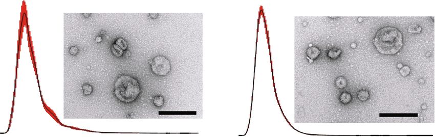
4 Journal of Immunology Research

 Naive Exo Primed Exo
 4.98
 6.56
 Concentration (E6 particles/ml)

 Concentration (E6 particles/ml)
 0 100 200 300 400 500 600 700 800 900 1000 0 100 200 300 400 500 600 700 800 900 1000
 Size (nm) Size (nm)
 (a)
 CD63 CD9 TSG101 CD63 CD9 TSG101
 kDa kDa kDa kDa kDa kDa
 75 20 50 75 20 50

 15 15

 50 25 50 25
 (b)

Figure 1: Characterization of cADSC-EVs. (a) Representative graph of nanoparticle tracking analysis and transmission electron microscopic
images. Bar = 200 nm. (b) Immunoblots of cADSC-EVs for CD63 and CD9.

ice for 20 min. PBMCs were stained with anti-CD3-FITC captured under a BZ-X700 multi-purpose microscope (KEY-
(Clone: CA17.2A12, Bio-Rad), anti-CD4-APC (Clone: ENCE, Osaka, Japan). The same samples without fixation
YKIX302.9, eBioscience), and anti-CD25-RPE (clone: were incubated with a canine Fc receptor binding inhibitor
P4A10, Thermo Fisher Scientific) or their respective isotype on ice for 20 min. Then, the cells were stained with anti-
controls. Then, PBMCs were fixed and permeabilized using CD3-FITC (Clone: CA17.2A12, Bio-Rad) and anti-CD4-
True-Nuclear Transcription Factor Buffer Set (BioLegend). APC (Clone: YKIX302.9, eBioscience) or their respective iso-
Finally, PBMCs were stained with anti-Foxp3-eFluor450 type controls. Fluorescence was evaluated by flow cytometry.
(clone: FJK-16 s, Thermo Fisher Scientific) or that of isotype
controls. Analysis by flow cytometry was performed by 2.12. Statistical Analysis. All data are presented as the mean
measuring the frequency of Foxp3 expression on gated ± standard deviation. Differences among multiple groups
CD3+CD4+CD25+ cells. were assessed by one- or two-way analysis of variance and
 differences were compared using the Tukey-Kramer post
2.11. Uptake of EVs by CD4+ T Cells. cADSC-derived naive hoc test. P < 0:05 was considered statistically significant. Sta-
and primed EVs were stained with Vybrant DiI Cell- tistical analyses were performed using Excel 2019 with add-in
labeling Solution (Thermo Fisher Scientific), and then, software Statcel 3.
unincorporated dye was removed from labeled EVs using
an Exosome Spin Column (Thermo Fisher Scientific) in 3. Results
accordance with the manufacturers’ instructions. PBMCs
were stimulated with ConA (5 μg/ml) for 3 days and then col- 3.1. Characterization of cADSC-Derived EVs. cADSCs were
lected, washed with PBS, and resuspended in RPMI-1640 pretreated with or without TNFα and IFNγ, and then, an
medium with 10% FBS, 1% antibiotic-antimycotic solution, enriched fraction of EVs was collected from the supernatant
1% nonessential amino acid, 1% GlutaMAX, and 50 μM 2- using the PS affinity method. The size of isolated naive EVs
mercaptoethanol. One million stimulated PBMCs were cul- was 166 ± 7:7 nm and that of primed EVs was 145 ± 1:5 nm.
tured in a 12-well plate with 1 μg/ml naive or primed The concentrations of EVs were 34:4 ± 3:1 and 42:8 ± 1:9 ×
labeled-EVs for 3 or 12 hours. Cells were fixed with 2% para- 109 particles/ml for naive and primed EVs, respectively
formaldehyde and then covered with a mounting medium (Figure 1(a)). Both naive and primed EVs expressed specific
with DAPI (VECTASHIELD: H-1200: VECTOR LABORA- exosomal markers CD9, CD63, and TSG101 [25]. The size
TORIES, CA, USA). Immunofluorescence images were of collected vesicles and marker expression indicated that
Journal of Immunology Research 5

 1 2 3 4 5 6 7 8 9 10 11 12

 cfa-let- cfa-let- cfa-let- cfa-let- cfa-let- cfa-miR- cfa-miR- cfa-miR- cfa-miR- cfa-miR- cfa-miR- cfa-miR-
 A 7a 7b 7c 7f 7g 1 101 103 106a 106b 10b 122

 cfa-miR- cfa-miR- cfa-miR- cfa-miR- cfa-miR- cfa-miR- cfa-miR- cfa-miR- cfa-miR- cfa-miR- cfa-miR- cfa-miR-
 B 124 125a 125b 126 130a 133a 133b 137 141 143 145 146a

 cfa-miR- cfa-miR- cfa-miR- cfa-miR- cfa-miR- cfa-miR- cfa-miR- cfa-miR- cfa-miR- cfa-miR- cfa-miR- cfa-miR-
 C 146b 148a 150 15a 15b 16 17 181a 181b 182 183 184

 cfa-miR- cfa-miR- cfa-miR- cfa-miR- cfa-miR- cfa-miR- cfa-miR- cfa-miR- cfa-miR- cfa-miR- cfa-miR- cfa-miR-
 D 18a 191 192 195 196a 19a 200a 200b 200c 203 204 205

 cfa-miR- cfa-miR- cfa-miR- cfa-miR- cfa-miR- cfa-miR- cfa-miR- cfa-miR- cfa-miR- cfa-miR- cfa-miR- cfa-miR-
 E 20a 21 210 214 218 22 222 223 224 23a 23b 24

 cfa-miR- cfa-miR- cfa-miR- cfa-miR- cfa-miR- cfa-miR- cfa-miR- cfa-miR- cfa-miR- cfa-miR- cfa-miR- cfa-miR-
 F
 25 26a 27a 27b 29b 29c 30b 30c 30d 31 335 342

 cfa-miR- cfa-miR- cfa-miR- cfa-miR- cfa-miR- cfa-miR- cfa-miR- cfa-miR- cfa-miR- cfa-miR- cfa-miR- cfa-miR-
 G
 34a 34b 34c 375 378 451 499 7 9 92a 93 96

 (a)

 cfa-miR-146a
 cfa-miR-31
 01 02 03 04 05 06 07 08 09 10 11 12 cfa-miR-124
 A cfa-miR-184
 B cfa-miR-375
 cfa-miR-7
 C
 cfa-miR-29c
 D cfa-miR-92a
 E cfa-miR-18a
 cfa-miR-25
 F
 cfa-miR-101
 G cfa-miR-34a
 cfa-miR-224
 Magnitude of log2 (fold change) cfa-miR-22
 −15 −10 −5 0 5 10 15 20 25 30
 −4.789 0 4.789 Fold change
 (b) (c)

Figure 2: Differential expression of microRNAs in naive and primed cADSC-EVs. (a) Layout for Qiagen’s canine miScript miRNA PCR
Array. (b) Heatmap of microRNA expression between naive and primed cADSC-EVs. Red an increase and green/black indicate a decrease
in relative expression in primed cADSC-EVs (n = 3 per group). Gray microRNAs represent not detected. (c) Significant differences
in expression levels of microRNAs. Red bar indicates an increase in primed cADSC-EVs, and blue bar indicates an increase in naive
cADSC-EVs.

cADSC-derived EVs isolated from the supernatant were an enhanced the proliferation of PBMCs and CD4+ T cells
enriched fraction of exosomes (Figure 1(b)). (unstimulated PBMCs: 40:0% ± 1:5%; ConA-stimulated
 Expression of exosome-associated microRNAs was PBMCs: 46:8% ± 1:5%; unstimulated CD4+ T cells: 43:8% ±
investigated by a miRNA PCR array. Seventy microRNAs 1:7%; ConA-stimulated CD4+ T cells: 57:0% ± 1:6%). The
were detected in both naive and primed EVs, and expression proliferation rate decreased gradually in both PBMCs
levels of 14 microRNAs in primed EVs were significantly dif- (Figure 3) and CD4+ T cells (Figure 4) when PBMCs were
ferent compared with that in naive EVs (Figure 2). All data of cocultured in the presence of EVs. The inhibitive effects on
fold regulation and P values of each microRNA are shown in PBMCs and CD4+ T cells were not different between naive
Supplemental Material 1. and primed EVs at same concentrations.

3.2. cADSC-Derived EVs Inhibit Proliferation of PBMCs and 3.3. cADSC-Derived EVs Induce Apoptosis of PBMCs. When
CD4+ T Cells. ConA (5 μg/ml) stimulation for 4 days PBMCs were stimulated with ConA and cultured without
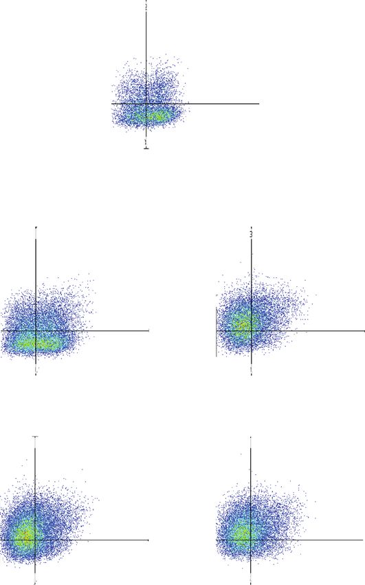
6 Journal of Immunology Research

 ConA(−) PBMC ConA(+) PBMC
 600
 (×101)
 P1(37.66%) P1(46.24%)

 100
 400
 Count

 Count
 50 200

 0 0
 101 102 103 104 104 106 107 101 102 103 104 104 106 107
 CFSE-FITC

 Naive EVs 1 g Naive EVs 5 g Naive EVs 10 g
 P1(41.78%) P1(32.68%) P1(30.02%)
 600
 600
 400
 400
 400
 Count

 Count

 Count
 200
 200 200

 0 0 0
 101 102 103 104 104 106 107 101 102 103 104 104 106 107 101 102 103 104 104 106 107

 Primed EVs 1 g Primed EVs 5 g Primed EVs 10 g
 P1(37.98%) P1(32.36%) P1(30.10%)
 600
 600
 400
 400
 400
 Count

 Count

 Count

 200
 200 200

 0 0 0
 101 102 103 104 104 106 107 101 102 103 104 104 106 107 101 102 103 104 104 106 107

 (a)
 60
 ⁎⁎ ⁎⁎
 Proliferation of PBMC (%)

 50
 ## ##
 40 ## ## ##
 ##

 30

 20

 10

 0
 ConA(−) ConA(+) 1 g 5 g 10 g 1 g 5 g 10 g
 Control Naive EVs Primed EVs
 (b)

Figure 3: Proliferative ability of PBMCs cocultured with cADSC-EVs. (a) Proliferation of PBMCs was assayed using the CFSE method by
flow cytometry. Segments represent the percentages of PBMC proliferation. (b) Comparison of proliferative PBMCs treated with various
concentrations (1, 5, and 10 μg/ml) of naive or primed cADSC-EVs. The inhibitive effects on PBMCs were not different between naive
and primed EVs at the same concentrations. The solid line indicates the average. ## P < 0:01 vs. PBMCs stimulated with ConA. ∗∗ P < 0:01,
between groups.
Journal of Immunology Research 7

 ConA(−) CD4+ cells ConA(+) CD4+ cells
 P2(42.72%) P2(56.98%)

 400

 50
 Count

 Count
 200

 0 0
 0 102 103 104 104 106 107 0 102 103 104 104 106 107
 CFSE-FITC

 Naive EVs 1 g Naive EVs 5 g Naive EVs 10 g
 P2(48.59%) P2(32.99%) P2(26.89%)
 100

 50 50

 Count
 Count

 Count

 50

 0 0 0
 0 102 103 104 104 106 107 0 102 103 104 104 106 107 0 102 103 104 104 106 107

 Primed EVs 1 g Primed EVs 5 g Primed EVs 10 g
 100 P2(46.60%) P2(30.63%) P2(29.69%)
 100
 Count

 Count
 Count

 50 50
 50

 0 0 0
 0 102 103 104 104 106 107 0 102 103 104 104 106 107 0 102 103 104 104 106 107
 (a)

 Figure 4: Continued.
8 Journal of Immunology Research

 70 ⁎⁎ ⁎⁎
 ⁎⁎

 Prolif eration of CD4+ cells (%)
 60 ##
 ⁎ ##
 50
 ##
 40 ## ##
 ##
 30

 20

 10

 0
 ConA(−) ConA(+) 1 g 5 g 10 g 1 g 5 g 10 g
 Control Naive EVs Primed EVs
 (b)

Figure 4: Proliferative ability of CD4+ T cells cocultured with cADSC-EVs. (a) Proliferation of CD4+ T cells was assayed using the CFSE
method by flow cytometry. Segments represent the percentages of CD4+ T cell proliferation. (b) Comparison of proliferative CD4+ T cells
treated with various concentrations (1, 5, and 10 μg/ml) of naive or primed cADSC-EVs. The inhibitive effects on CD4+ T cells were not
significantly different between naive and primed EVs at same concentrations. The solid line indicates the average. ## P < 0:01 vs. CD4+ T
cells stimulated with ConA. ∗ P < 0:05, between groups. ∗∗ P < 0:01, between groups.

cADSC-derived EVs, the apoptosis ratio was 20:0% ± 2:4%. EVs (7:2% ± 1:3%) (Figure 9). The ratio of Th1, Th2, and
However, PBMCs cocultured with various concentrations Th17 cells was not different between naive and primed EVs
of naive and primed EVs had increased apoptosis at the same concentrations.
(Figure 5). The induced ratio of PBMC apoptosis was not
significantly different between naive and primed EVs at the 3.6. cADSC-Derived EVs Enhance Treg Cells. The ratio of
same concentrations. Treg (CD3+CD4+CD25+Foxp3+) cells among PBMCs stimu-
 lated with ConA was 18:2% ± 1:7%. When PBMCs were cul-
3.4. cADSC-Derived EVs Affect the T Cell Subset Distribution. tured with EVs, the ratio of Treg cells in CD4+ T cells was
After stimulating PBMCs with ConA for 3 days, the ratios elevated significantly (1 μg/ml naive EVs: 23:0% ± 2:4%;
of CD4+ T, CD8+ T, CD4+CD8+ T, and CD4-CD8- T cells 3 μg/ml naive EVs: 30:1% ± 2:1%; 1 μg/ml primed EVs: 26:0
were 35:9% ± 2:4%, 29:1% ± 1:6%, 3:2% ± 0:5%, and 31:8% % ± 2:2%; 3 μg/ml primed EVs: 31:3% ± 3:5%) (Figure 10).
 ± 4:0%, respectively. When PBMCs cocultured with EVs The ratio of Treg cells was not different between naive and
at various concentrations, the ratios of CD4+ T, CD8+ T, primed EVs at the same concentrations.
and CD4+CD8+ T cells were decreased proportionately,
but that of CD4-CD8- T cells was increased (Figure 6). 3.7. cADSC-Derived EV Uptake by CD4+ T Cells. T cell-
The ratio of CD4-CD8- T cells cultured with 5 and enriched PBMCs were coculture with cADSC-derived EVs
10 μg/ml primed EVs was significantly decrease compared for 3 or 12 hours. Both naive and primed EVs were incor-
with naive EVs (Table 1), but the T cell subset distribution porated into T cells and CD4+ T cells treated with EVs
was not different between naive and primed EVs at the were observed by flow cytometry (Figure 11). The ratio of
same concentrations. CD4+ T cells with cADSC-derived EVs at 12 h was signifi-
 cantly elevated in both naive and primed EVs compared
3.5. cADSC-Derived EVs Alter Differentiation into T Helper to those at 3 h.
Cells. To examine Th1, Th2, and Th17 cells, intracellular
cytokines IFNr, IL-4, and IL-17 were detected by flow cytom- 4. Discussion
etry. When PBMCs were stimulated with PMA and ionomy-
cin for 6 h, intracellular expression of IFNr, IL-4, and IL-17 Mesenchymal stem cells (MSCs) have immunomodulatory
was increased compared with the unstimulated control. In and anti-inflammatory abilities by secreting numerous fac-
the presence of EVs, the ratio of Th1 cells (CD4+IFNr+) tors such as extracellular vesicles, cytokines, chemokines,
among CD3+ T cells was decreased (3 μg/ml naive EVs: 6:7 and growth factors. As research on MSC-based therapies
% ± 1:6%; 3 μg/ml primed EVs: 5:9% ± 1:5%) compared with has proceeded, exosomes have been focused on as important
no EVs (11:2% ± 2:0%) (Figure 7). Th2 cells (CD4+IL-4+) factors that exert immunomodulatory effects. Exosomes are
among CD3+ T cells were increased when PBMCs cocultured typically small membrane vesicles (30-150 nm) that include
with EVs (3 μg/ml naive EVs: 10:4% ± 1:3%; 3 μg/ml primed extracellular vesicles (30-1000 nm) secreted by various cell
EVs: 11:6% ± 1:6%) compared with no EVs (5:8% ± 1:0%) types. The classically used protocol to isolate exosomes is
(Figure 8). Th17 cells (CD4+IL-17+) among CD3+ T cells ultracentrifugation, but there are currently several exosome
after coculture with EVs were also suppressed (1 μg/ml naive isolation methods based on different principles [26, 27]. The
EVs: 5:5% ± 1:1%; 3 μg/ml naive EVs: 4:4% ± 0:9%; 3 μg/ml ultracentrifugation method is based on the principles of pre-
primed EVs: 3:3% ± 0:8%) compared with culture without cipitation and sedimentation. Therefore, ultracentrifugation

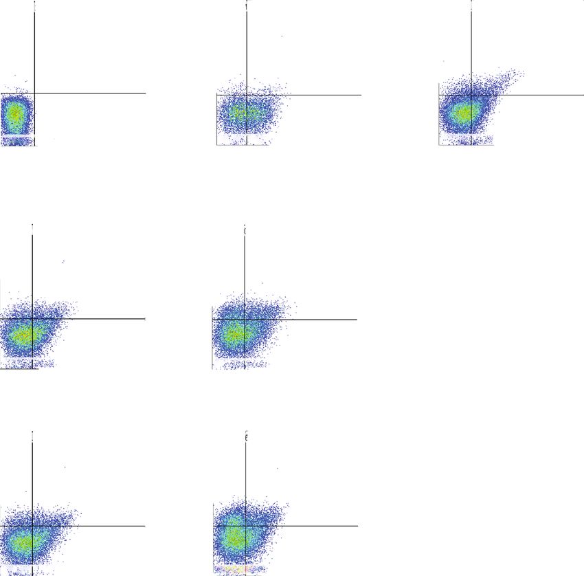
Journal of Immunology Research 9

 Control
 106
 Annexin v -FITC

 105

 104

 0

 0 104 105 106
 Propidium iodide

 Naive EVs1 g Naive EVs 5 g Naive EVs 10 g
 106 106 106

 105 105 105

 104 104 104

 0 0 0

 0 104 105 106 0 104 105 106 0 104 105 106

 Primed EVs 1 g Primed EVs 5 g Primed EVs 10 g
 106 106 106

 105 105 105

 104 104 104

 0 0 0

 0 104 105 106 0 104 105 106 0 104 105 106
 (a)

 Figure 5: Continued.
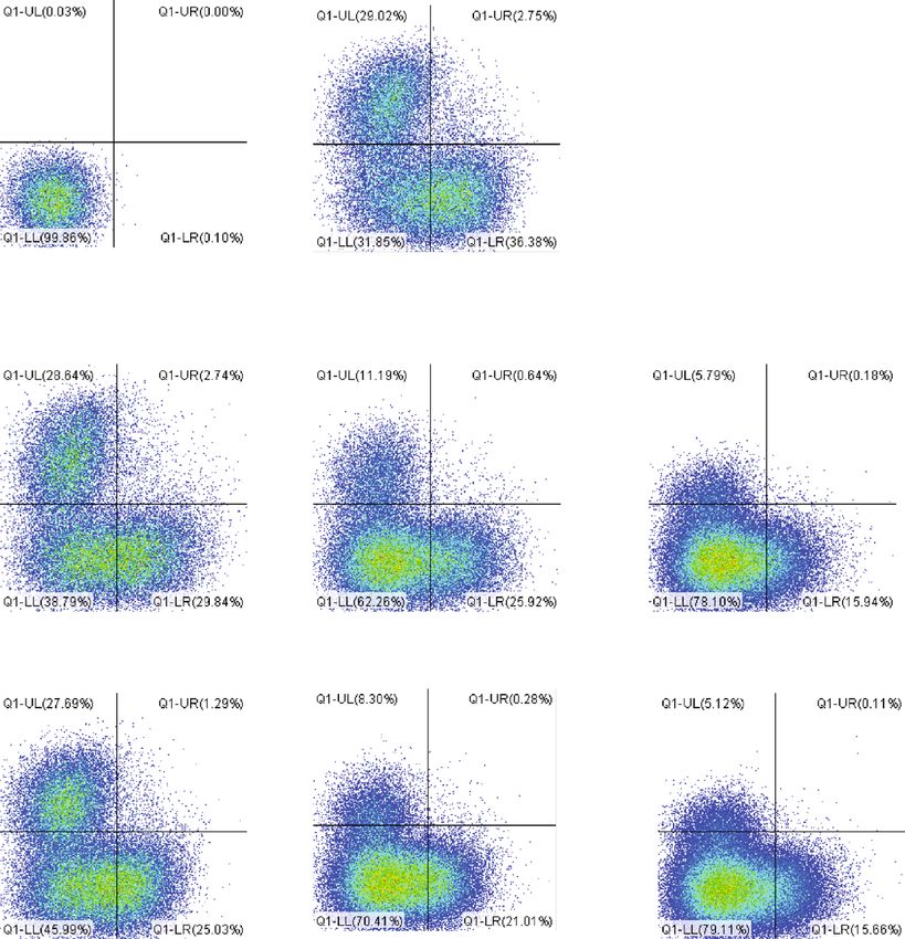
10 Journal of Immunology Research

 ⁎ ⁎⁎
 60 ##
 ⁎⁎ ## ##
 50 ##

 Apoptosis of PBMC (%)
 ## ##
 40

 30

 20

 10

 0
 Control 1 g 5 g 10 g 1 g 5 g 10 g
 Naive EVs Primed EVs
 (b)

Figure 5: Apoptosis of PBMC cocultured with cADSC-EVs. (a) PBMCs stimulated with ConA were cultured in the presence of naive or
primed cADSC-EVs at various concentrations (1, 5, and 10 μg/ml) for 3 days. After culture, PBMCs were stained with Annexin V and PI
and assayed by flow cytometry. (b) Comparison of apoptosis of PBMCs treated with various concentrations (1, 5, and 10 μg/ml) of naive
or primed cADSC-EVs. The induced ratio of PBMC apoptosis was not significantly different between naive and primed EVs at the same
concentrations. The solid line indicates the average. ## P < 0:01 vs. PBMC stimulated with ConA. ∗ P < 0:05, between groups. ∗∗ P < 0:01,
between groups.

 Isotype control Control

 104 104
 CD8-APC

 103 103

 0 0

 0 103 104 0 103 104
 CD4-PE

 Naive EVs 1 g Naive EVs 5 g Naive EVs 10 g

 4 4 4
 10 10 10

 103 103 103

 0 0 0

 0 103 104 0 103 104 0 103 104
 Primed EVs 1 g Primed EVs 5 g Primed EVs 10 g

 104 104 10 4

 103 103 103

 0 0 0

 0 103 104 0 103 104 0 103 104

Figure 6: Immunophenotypic analysis of CD3, CD4, and CD8 in PBMCs cocultured with naive or primed cADSC-EVs at various
concentrations (1, 5, and 10 μg/ml).
Journal of Immunology Research 11

 Table 1: Comparison of CD4+, CD8+, CD4+CD8+, and CD4-CD8- T cell populations after coculture of PBMCs with cADSC-EVs.

 Naive EVs Primed EVs
 Control
 1 μg/ml 5 μg/ml 10 μg/ml 1 μg/ml 5 μg/ml 10 μg/ml
 ∗∗ ∗∗ ∗∗ ∗∗ ∗∗
CD4 +
 35:9 ± 2:4 29:4 ± 1:9 26:1 ± 1:7 15:3 ± 1:9 25:7 ± 2:2 21:7 ± 1:7 15:5 ± 2:0∗∗
 ∗∗ ∗∗ ∗∗
CD8+ 29:1 ± 1:6 29:3 ± 2:0 11:8 ± 1:8 5:7 ± 1:3 27:1 ± 1:8 8:3 ± 1:6 5:4 ± 1:6∗∗
CD4+CD8+ 3:2 ± 0:5 3:0 ± 0:8 1:0 ± 0:5∗∗ 0:2 ± 0:1∗∗ 1:4 ± 0:5∗∗ 0:3 ± 0:1∗∗ 0:2 ± 0:1∗∗
CD4-CD8- 31:8 ± 4:0 38:3 ± 2:2∗ 61:1 ± 3:2∗∗ 78:8 ± 1:3∗∗ 45:7 ± 3:2∗ 69:7 ± 1:7∗∗ 78:9 ± 2:8∗∗
Data are shown as the mean ± S:D. The ratio of T cell subset was not significantly different between naive and primed EVs at the same concentrations. ∗∗ P
< 0:01 vs. control (PBMCs that were not cocultured with cADSC-EVs). ∗ P < 0:05 vs. control.

also concentrates nonspecifically sedimentable particles, and are known to enhance the immunosuppressive proper-
which include non-EV proteins, and is associated with low ties of MSCs [6]. For example, immunomodulatory effects
reproducibility because of the loss of unstable and invisible of T cells are attributed to the upregulation of IDO. MSC-
pellets after centrifugation. Conversely, the PS affinity method EVs also packaged various active proteins, which included
is based on the high affinity of T cell immunoglobulin domain IDO, but previous studies have demonstrated no significant
and mucin domain-containing protein 4 that strongly binds to change of IDO in PBMCs cocultured with or without MSC-
phosphatidylserine expressed on the surface of EVs. The PS EVs and thus suggested that the mechanism that underlies
affinity method only requires general laboratory equipment. the immunomodulatory capacities of MSC-EVs is different
Moreover, this method isolates EVs with higher quality and from that of MSC soluble factors [36]. Second, stimulation
reproducibility compared with ultracentrifugation [28]. In this methods of T cells may be related [35]. In this study, PBMCs
study, we collected cADSC-EVs using the PS affinity method. were stimulated with ConA. ConA is an antigen-independent
The results of NTA and analyses of exosome markers (CD9, mitogen and is widely used as a T cell stimulus. However, T
CD63, and TSG101) suggested that the cADSC-EVs isolated cell activation by an anti-CD3/28 antibody can be used in
from culture supernatants were an enriched exosome fraction. humans and mice as an antigen-dependent method.
 In this study, we examined the microRNA expression Antigen-presenting cells partially mediate T cell suppression
profile of naive and TNFα/IFNγ-primed cADSC-EVs. micro- induced by MSCs [37]. Therefore, another explanation might
RNAs, which are a class of noncoding RNAs of 20-22 base be a lack or small number of antigen-presenting cells in our
pairs, regulate the expression of multiple RNAs and play an experimental setting. In our study, we analyzed characteristic
important role in various biological processes that include features by only comparing the expression of microRNAs
the immune response. Several microRNAs affect the immune between naive and primed cADSC-EVs, but there are many
response in T cell development, proliferation, differentiation, other components such as proteins and mRNAs. Further-
and function [29, 30]. The microRNA profiles showed that more, several cytokines and growth factors have been
several microRNAs in cADSC-EVs varied after stimulation reported in MSCs, such as TGFβ, TNFα, IFNγ, IL-4, and
with TNFα and IFNγ. For example, miR-146a, which was IL-10 [6]. Further studies are required to clarify the alteration
overexpressed in primed cADSC-EVs, acts as a negative reg- of components in cADSC-EVs with or without priming and
ulator of T cells and promotes Treg cell functions [30]. MiR- using other stimulation methods.
34a, which was downregulated in primed cADSC-EVs, plays MSCs suppress T cell proliferation [38], B cell activity
an important role in T cell activation by targeting >30 genes [39], and NK cell proliferation [40] and interfere with the dif-
across different cellular pathways that control the immune ferentiation, maturation, and function of dendritic cells [41].
response [31]. The purpose of this study was to investigate the immuno-
 Several studies have reported that primed EVs increase modulatory capacity of cADSC-EVs for T cells. In this study,
immunomodulatory functions compared with naive EVs PBMCs were stimulated with ConA. The immunomodula-
[32, 33], whereas other reports have indicated that primed tory effects exerted by MSC-EVs on activated T cells remain
EVs do not enhance the immunomodulatory effects of a widely discussed topic. Primed and naive human MSC-EVs
MSC-EVs [34, 35]. In our study, primed cADSC-EVs did cocultured with PBMCs suppress the proliferation of T cells
not enhance any functions of PBMCs or stimulated CD4+ T but do not affect that of B and NK cells [42]. Other studies
cells. The reason that the immunomodulatory functions of have shown that MSC-EVs inhibit the proliferation of NK
primed cADSC-EVs were not enhanced remains unclear, and B cells, but their effects on the proliferation of T cells
but several hypotheses were proposed. First, packaged solu- remain unclear [32, 43]. Our results demonstrated that
ble factors inside EVs may be related [34]. The properties cADSC-EVs suppressed the proliferation of PBMCs and
of factors secreted from MSCs change depending on MSC CD4+ T cells and enhanced apoptosis of PBMCs. It has been
culture conditions [5]. There is evidence suggesting that demonstrated that MSC-EVs carry various active molecules
immunomodulatory effects of MSCs are enhanced in that may contribute to the MSC-EV capacity to inhibit T cell
response to inflammatory stimulation [5]. Inflammatory proliferation and activation and induce T cell apoptosis [4].
stimuli by TNFα and IFNγ have been commonly used to However, the mechanisms of suppressive proliferation and
research the immune and regenerative functions of MSCs induced apoptosis in T cells by MSC-EVs remain unclear.
12 Journal of Immunology Research

 Isotype control Non-stim control Stimulated control
 106 106 106
 Q2-UL(0.00%) Q2-UR(0.00%) Q1-UL(2.02%) Q1-UR(3.09%) Q1-UL(11.22%) Q1-UR(11.95%)

 Th1
 105 105 105
 IFN -PE

 104 104 104

 103 103 103

 0 0 0
 Q2-LL(99.99%) Q2-LR(0.01%) Q1-LL(54.30%) Q1-LR(40.60%) Q1-LL(60.76%) Q1-LR(16.07%)
 3 4 5 6 3 4 5 6
 0 10 10 10 10 0 10 10 10 10 0 103 104 105 106
 CD4-APC

 Naive EVs 1 g Naive EVs 3 g
 106 106
 Q1-UL(23.18%) Q1-UR(9.45%) Q1-UL(31.17%) Q1-UR(7.79%)

 105 105

 104 104

 103 103

 0 0
 Q1-LL(53.98%) Q1-LR(13.39%) Q1-LL(55.04%) Q1-LR(6.00%)
 3 4 5 6
 0 10 10 10 10 0 103 104 105 106

 Primed EVs 1 g Primed EVs 3 g
 106 106
 Q1-UL(23.73%) Q1-UR(8.81%) Q1-UL(29.36%) Q1-UR(6.86%)

 105 105

 104 104

 103 103

 0 0
 Q1-LL(54.68%) Q1-LR(12.77%) Q1-LL(54.37%) Q1-LR(9.41%)
 3 4 5 6 3 4
 0 10 10 10 10 0 10 10 105 106

 (a)
 16
 ⁎⁎
 14
 CD4+ IFN + cells (%)

 12
 ##
 10 ##
 8
 6
 4
 2
 0
 Non-stim stimulated 1 g 3 g 1 g 3 g
 Control Naive EVs Primed EVs
 (b)

Figure 7: Differentiation into Th1 cells after coculture of PBMCs with cADSC-EVs. (a) Flow cytometric detection of CD4+IFNγ+ cells among
CD3+ T cells (gated in red square). Non-stim control: PBMCs that were not stimulated with PMA and ionomycin. Stimulated control: PBMCs
after coculture with cADSC-EVs and stimulated by PMA and ionomycin. (b) Comparison of CD4+IFNγ+ cells among CD3+ T cells after
coculture of PBMCs with various concentrations (1 or 3 μg/ml) of naive or primed cADSC-EVs. The ratio of Th1 cells was not
significantly different between naive and primed EVs at the same concentrations. The solid line indicates the average. ## P < 0:01 vs.
stimulated control. ∗∗ P < 0:01, between groups.
Journal of Immunology Research 13

 Isotype control Non-stim control Stimulated control
 106 106 106
 Q1-UL(0.20%) Q1-UR(0.00%) Q1-UL(1.33%) Q1-UR(1.53%) Q1-UL(3.13%) Q1-UR(5.81%)

 105 105 105
 Th2
 IL-4-PE

 104 104 104

 103 103 103

 Q1-LL(99.76%) Q1-LR(0.04%) Q1-LL(52.80%) Q1-LR(44.34%) Q1-LL(65.12%) Q1-LR(25.93%)
 102 102 102
 0 103 104 105 106 0 103 104 105 106 0 103 104 105 106

 CD4-APC

 Naive EVs 1 g Naive EVs 3 g
 106 106
 Q1-UL(4.20 %) Q1-UR(5.88%) Q1-UL(10.13%) Q1-UR(10.01%)

 105 105

 104 104

 103 103

 Q1-LL(60.55%) Q1-LR(29.37%) Q1-LL(53.78%) Q1-LR(26.08%)
 102 102
 0 103 104 105 106 0 103 104 105 106

 Primed EVs 1 g Primed EVs 3 g
 106 106
 Q1-UL(4.40 %) Q1-UR(6.51%) Q1-UL(16.16%) Q1-UR(10.21%)

 105 105

 104 104

 103 103

 Q1-LL(58.00%) Q1-LR(31.09%) Q1-LL(52.25%) Q1-LR(21.37%)
 102 102
 0 103 104 105 106 0 103 104 105 106

 (a)
 ⁎⁎
 16 ⁎⁎
 ##
 14 ##
 12
 CD4+ IL-4+ cells (%)

 10
 ⁎⁎
 8
 6
 4
 2
 0
 Non-stim stimulated 1 g 3 g 1 g 3 g
 Control Naive EVs Primed EVs
 (b)

Figure 8: Differentiation into Th2 cells after coculture of PBMCs with cADSC-EVs. (a) Flow cytometric detection of CD4+IL-4+ cells among
CD3+ T cells (gated in red square). Non-stim control: PBMCs that were not stimulated with PMA and ionomycin. Stimulated control: PBMCs
after coculture with cADSC-EVs and stimulated by PMA and ionomycin. (b) Comparison of CD4+IL-4+ cells among CD3+ T cells after
coculture of PBMCs with various concentrations (1 or 3 μg/ml) of naive or primed cADSC-EVs. The ratio of Th2 cells was not different
between naive and primed EVs at the same concentrations. The solid line indicates the average. ## P < 0:01 vs. stimulated control.
∗∗
 P < 0:01, between groups.
14 Journal of Immunology Research

 Isotype control Non-stim control Stimulated control
 106 106 106
 Q1-UL(0.37%) Q1-UR(0.00%) Q1-UL(1.04%) Q1-UR(1.28%) Q1-UL(5.94%) Q1-UR(7.54%)

 Th17
 105 105 105

 104 104
 IL-17-PE

 104

 103 103 103

 0 0 0
 Q1-LL(99.63%) Q1-LR(0.01%) Q1-LL(55.87%) Q1-LR(41.81%) Q1-LL(69.68%) Q1-LR(16.83%)

 0 10 3
 10 4
 10 5 6
 10 0 103 104 105 106 0 103 104 105 106

 CD4 -APC

 Naive EVs 1 g Naive EVs 3 g
 106 106
 Q1-UL(8.37%) Q1-UR(6.18%) Q1-UL(21.76%) Q1-UR(4.44%)

 105 105

 104 104

 103 103

 0 0
 Q1-LL(65.82%) Q1-LR(19.63%) Q1-LL(62.90%) Q1-LR(10.90%)

 0 103 104 105 106 0 103 104 105 106

 Primed EVs 1 g Primed EVs 3 g
 106 106
 Q1-UL(9.55%) Q1-UR(4.83%) Q1-UL(15.87%) Q1-UR(3.63%)

 105 105

 104 104

 103 103

 0 0
 Q1-LL(67.48%) Q1-LR(18.14%) Q1-LL(62.90%) Q1-LR(10.90%)

 0 103 104 105 106 0 103 104 105 106

 (a)
 10 ⁎⁎

 8
 CD4+ IL-17+ cells (%)

 ##
 ##
 6
 ##

 4

 2

 0
 Non-stim stimulated 1 g 3 g 1 g 3 g
 Control Naive EVs Primed EVs
 (b)

Figure 9: Differentiation into Th17 cells after coculture of PBMCs with cADSC-EVs. (a) Flow cytometric detection of CD4+IL-17+ cells
among CD3+ T cells (gated in red square). Non-stim control: PBMCs that were not stimulated with PMA and ionomycin Stimulated
control: PBMCs after coculture with cADSC-EVs and stimulated by PMA and ionomycin. (b) Comparison of CD4+IL-17+ cells among
CD3+ T cells after coculture of PBMCs with various concentrations (1 or 3 μg/ml) of naive or primed cADSC-EVs. The ratio of Th17 cells
was not significantly different between naive and primed EVs at the same concentrations. The solid line indicates the average. ## P < 0:01
vs. stimulated control. ∗∗ P < 0:01, between groups.
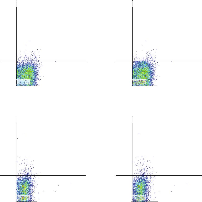
Journal of Immunology Research 15

 106
 Q3-UL(12.96%) Q3-UR(17.46%)

 Treg

 Foxp3-PB450
 104

 0
 Q3-LL(28.25%) Q3-LR(41.33%)
 3
 0 10 104 105
 CD4-APC/CD25-PE

 Naive EVs 1 g Naive EVs 3 g
 106 106
 Q3-UL(13.90%) Q3-UR(21.12%) Q3-UL(43.10%) Q3-UR(29.40%)

 104 104

 0 0
 Q3-LL(29.97%) Q3-LR(35.01%) Q3-LL(19.26%) Q3-LR(8.23%)

 0 103 104 105 0 103 104 105

 Primed EVs 1 g Primed EVs 3 g
 106 106
 Q3-UL(43.10%) Q3-UR(26.66%) Q3-UL(43.01%) Q3-UR(30.95%)

 104 104

 0 0

 Q3-LL(21.09%) Q3-LR(9.16%) Q3-LL(16.98%) Q3-LR(9.06%)
 3 4 5 3
 0 10 10 10 0 10 104 105
 (a)

 Figure 10: Continued.
16 Journal of Immunology Research

 ⁎⁎
 40
 ## ##
 35
 ##

 CD4+ CD25+ Foxp3+ cells (%)
 30

 25

 20

 15

 10

 5

 0
 Control 1 g 3 g 1 g 3 g
 Naive EVs Primed EVs
 (b)

Figure 10: Treg cells after coculture of PBMCs with cADSC-EVs. (a) Flow cytometric detection of CD3+CD4+CD25+Foxp3+ cells (gated in
red square). Control: PBMCs cultured without cADSC-EVs. (b) Comparison of the population of CD3+CD4+CD25+Foxp3+ cells after
coculture of PBMCs with various concentrations (1 or 3 μg/ml) of naive or primed cADSC-EVs. The ratio of Treg cells was not
significantly different between naive and primed EVs at the same concentrations. The solid line indicates the average. ## P < 0:01 vs.
control. ∗∗ P < 0:01, between concentrations of 1 and 3 μg/ml.

One report has demonstrated that TNFα/NF-κB signaling in cells because of the unique microRNA profiles of MSC-EVs
MSCs is required to inhibit T cell proliferation [44]. There- [49]. microRNAs are also thought to be related to changes
fore, MSC-EVs effects may also be related to activation of in the proliferation, apoptosis, and distribution of T cells
NF-κB. Another recent study has demonstrated that MSC- induced by cADSC-EVs.
EVs suppress the proliferation of T cells by inducing cell To clarify the alteration of the immune response of Th
cycle arrest through p27kip1/Cdk2 signaling [45]. P27kip1 cells induced by cADSC-EVs, intracellular cytokines were
is a representative factor that induces cell cycle arrest by detected by flow cytometry. Both naive and primed
downregulating Cdks, and MSC-EVs were associated with cADSC-EVs increased the ratio of Th2 cells but decreased
upregulation of p27kip1 and downregulation of Cdk2. It Th1 cells. Some studies of autoimmune diseases have
has been demonstrated that MSCs induce apoptosis of acti- reported that MSC-EVs drive a shift from Th1 toward
vated T cells through the FAS ligand-dependent FAS signal- Th2 cells and rebalances Th1/Th2 cells by downregulating
ing pathway but do not induce naive T cell apoptosis in vitro proinflammatory cytokines TNFα and IFNγ and upregulat-
[46]. However, there are no reports of a correlation between ing anti-inflammatory cytokines IL-10 or IL-4 [36, 50].
MSC-EVs and the FAS signaling pathway. The only report of Moreover, cADSC-EVs inhibited activated T cell differenti-
the mechanism of T cell apoptosis induction by MSC-EVs ation into Th17 cells and promoted differentiation into
assumed that MSC-EVs induce T cell apoptosis possibly Treg cells. Such regulation has also been observed in
through an MSC-EV mediated mechanism via the adenosine human MSC-EVs [4], but the mechanism is unclear. On
A2A receptor pathway [47]. After coculture of PBMCs stim- the basis of the immune balance effect exerted by
ulated with ConA in the presence of cADSC-EVs, CD4+, cADSC-EVs on Th and Treg cells, our results indicate that
CD8+, and CD4+CD8+ T cells were decreased, whereas cADSC-EVs may act as an ameliorating agent for autoim-
CD3+CD4-CD8- T cells were increased. A study has mune diseases.
evaluated the status of CD4+ and CD8+ T cells by In this study, we examined the effects of cADSC-EVs on
categorizing in accordance with CD45RA and CCR7 expres- T cells and focused on CD4+ T cells isolated from PBMCs.
sion, such as naive (CD45RA+CCR7+), central memory cADSC-EVs affected CD4+ T cell proliferation, apoptosis,
(CD45RA-CCR7+), effector memory (CD45RA-CCR7-), and and differentiation, but CD4+ T cells that took up cADSC-
terminally differentiated effector memory cells (CD45RA+- EVs were only a few percent of all cells. This result was sim-
CCR7-) [48]. This study demonstrated that human ADSC- ilar to previous studies of unfractionated PBMCs or purified
EVs inhibited the differentiation of CD4+ and CD8+ T cells T cells, and MSC-EVs were almost entirely incorporated by
into terminally differentiated effector memory cells. It is monocytes [32, 51]. MSC-EVs also act on monocytes/macro-
unclear how cADSC-EVs inhibited the differentiation of phages [4]. Thus, the influence of T cells was not only a direct
CD4+ and CD8+ T cells, but we hypothesize that cADSC- effect of cADSC-EVs but also included indirect effects of
EVs may suppress T cell differentiation toward immature monocytes affected by cADSC-EVs. Further studies of the
phenotypes. A study has examined the effects of MSC-EVs interactions between cADSC-EVs, T cells, and monocytes
on acute graft-versus-host disease and found that MSC-EVs are needed to clarify the effects of cADSC-EVs on the
are associated with the preservation of circulating naive T immune response.
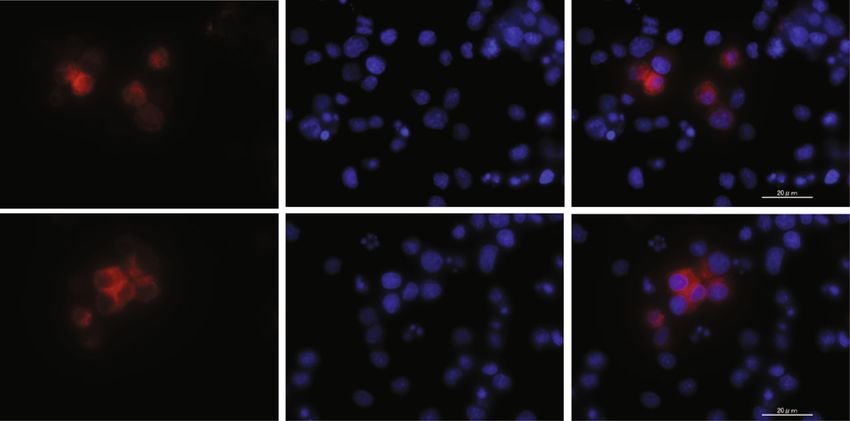
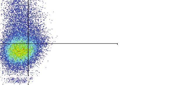
Journal of Immunology Research 17

 EVs DAPI Merge

 Naive
 EVs

 Primed
 EVs

 (a)
 Naive EVs Primed EVs
 106 106
 4h Q1-UL(0.00 %) Q1-UR(1.37 %) Q1-UL(0.00%) Q1-UR(1.49%)

 105 105
 CM-Dil-PE

 104 104

 103 103

 0 0
 Q1-LL(00.00%) Q1-LR(98.63%) Q1-LL(00.00%) Q1-LR(98.51%)
 3 4 5 6 3 4
 0 10 10 10 10 0 10 10 105 106

 CD4-APC

 106 106
 12 h Q2-UL(0.00 %) Q2-UR(2.37 %) Q2-UL(0.00%) Q2-UR(2.68%)

 105 105

 104 104

 103 103

 0 0
 Q2-LL(0.00%) Q2-LR(97.63%) Q2-LL(0.00%) Q2-LR(97.32%)
 4 5 6 4
 0 10 10 10 0 10 105 106
 (b)

 Figure 11: Continued.
18 Journal of Immunology Research

 3.5 ⁎⁎
 ⁎⁎
 3.0

 CD4+ T cells contained EVs (%)
 2.5

 2.0

 1.5

 1.0

 0.5

 0
 4h 12 h 4h 12 h
 Naive EVs Primed EVs
 (c)

Figure 11: Incorporation of cADSC-EVs into stimulated T cells. (a) Immunofluorescence staining of labeled EVs and T cells stimulated with
ConA. cADSC-EVs were labeled by Vybrant DiI (red) and nuclei were stained by DAPI (blue). (b) Flow cytometric detection of cADSC-EVs
incorporated into CD4+ T cells (gated in red square). (c) Comparison of the population of CD4+ T cells that contained cADSC-EVs at 4 and
12 hours after coculture of stimulated PBMCs with 1 μg/ml cADSC-EVs. The solid line indicates the average. ∗∗ P < 0:01, between 4 and 12 h.

5. Conclusions Supplementary Materials
Our study shows that cADSC-EVs have an immunoregula- Supplementary Material 1: comparison of the microRNAs
tory function by inducing PBMC apoptosis, suppressing the expression between naive and primed cADSC-EVs.
proliferation of PBMCs and stimulated CD4+ T cells as well (Supplementary Materials)
as the differentiation of CD4+ and CD8+ T cells, and chang-
ing Th1/Th2/Treg cell populations in vitro. To evaluate References
whether cADSC-EVs have beneficial effects on immune dis-
eases and are practical for use in treatments of immune dis- [1] A. R. R. Weiss and M. H. Dahlke, “Immunomodulation by
eases, further study is needed to analyze which components mesenchymal stem cells (MSCs): mechanisms of action of liv-
of cADSC-EVs exert the individual immunosuppressive ing, apoptotic, and dead MSCs,” Frontiers in Immunology,
effect and the pathways by which these immunosuppressive vol. 10, p. 1191, 2019.
effects occur. [2] X. L. Fan, Y. Zhang, X. Li, and Q. L. Fu, “Mechanisms under-
 lying the protective effects of mesenchymal stem cell-based
 therapy,” Cellular and Molecular Life Sciences, vol. 77, no. 14,
Data Availability pp. 2771–2794, 2020.
 [3] J. M. Kaplan, M. E. Youd, and T. A. Lodie, “Immunomodula-
The data used to support the findings of this study are avail- tory activity of mesenchymal stem cells,” Current Stem Cell
able from the corresponding authors upon reasonable Research & Therapy, vol. 6, no. 4, pp. 297–316, 2011.
request. [4] M. Xie, W. Xiong, Z. She et al., “Immunoregulatory effects of
 stem cell-derived extracellular vesicles on immune cells,” Fron-
 tiers in Immunology, vol. 11, p. 13, 2020.
Conflicts of Interest
 [5] M. Madrigal, K. S. Rao, and N. H. Riordan, “A review of ther-
The authors declare that no competing interest exists. apeutic effects of mesenchymal stem cell secretions and induc-
 tion of secretory modification by different culture methods,”
 Journal of Translational Medicine, vol. 12, no. 1, p. 260, 2014.
Acknowledgments [6] N. C. Noronha, A. Mizukami, C. Caliári-Oliveira et al., “Prim-
 ing approaches to improve the efficacy of mesenchymal stro-
This work was supported by JSPS KAKENHI [Grant number mal cell-based therapies,” Stem Cell Research & Therapy,
18K06005]. We thank Edanz (http://jp.edanz.com/ac) for vol. 10, no. 1, p. 131, 2019.
editing a draft of this manuscript, FUJIFILM Wako Bio Solu- [7] A. Leyendecker Jr, C. C. G. Pinheiro, M. T. Amano, and D. F.
tions Corporation for Nanoparticle Tracking Analysis of Bueno, “The use of human mesenchymal stem cells as thera-
extracellular vesicles, and Hanaichi UltraStructure Research peutic agents for the in vivo treatment of immune-related dis-
Institute for imaging of extracellular vesicles by transmission eases: a systematic review,” Frontiers in Immunology, vol. 9,
electron microscopy. 2018.
Journal of Immunology Research 19

 [8] S. F. de Witte, M. Franquesa, C. C. Baan, and M. J. Hoogduijn, [24] T. Teshima, K. Okamoto, K. Dairaku et al., “Generation of
 “Toward development of iMesenchymal stem cells for immu- insulin-producing cells from canine adipose tissue-derived
 nomodulatory therapy,” Frontiers in Immunology, vol. 6, 2016. mesenchymal stem cells,” Stem Cells International, vol. 2020,
 [9] S. J. Prasanna, D. Gopalakrishnan, S. R. Shankar, and A. B. Article ID 8841865, 10 pages, 2020.
 Vasandan, “Pro-inflammatory cytokines, IFNgamma and [25] C. Théry, K. W. Witwer, E. Aikawa et al., “Minimal
 TNFalpha, influence immune properties of human bone mar- information for studies of extracellular vesicles 2018
 row and Wharton jelly mesenchymal stem cells differentially,” (MISEV2018): a position statement of the International
 PLoS One, vol. 5, no. 2, p. e9016, 2010. Society for Extracellular Vesicles and update of the
[10] Z. Tu, Q. Li, H. Bu, and F. Lin, “Mesenchymal stem cells MISEV2014 guidelines,” Journal of Extracellular Vesicles,
 inhibit complement activation by secreting factor H,” Stem vol. 7, no. 1, 2018.
 Cells and Development, vol. 19, no. 11, pp. 1803–1809, 2010. [26] A. Rai, H. Fang, M. Fatmous et al., “A protocol for isolation,
[11] I. E. Dias, P. O. Pinto, L. C. Barros, C. A. Viegas, I. R. Dias, and purification, characterization, and functional dissection of
 P. P. Carvalho, “Mesenchymal stem cells therapy in compan- exosomes,” Methods in Molecular Biology, vol. 2261, pp. 105–
 ion animals: useful for immune-mediated diseases?,” BMC 149, 2021.
 Veterinary Research, vol. 15, no. 1, p. 358, 2019. [27] D. W. Greening, R. Xu, H. Ji, B. J. Tauro, and R. J. Simpson, “A
[12] M. B. Gugjoo, A. Amarpal, and G. T. Sharma, “Mesenchymal protocol for exosome isolation and characterization: evalua-
 stem cell basic research and applications in dog medicine,” tion of ultracentrifugation, density-gradient separation, and
 Journal of Cellular Physiology, vol. 234, no. 10, pp. 16779– immunoaffinity capture methods,” Methods in Molecular Biol-
 16811, 2019. ogy, vol. 1295, pp. 179–209, 2015.
[13] L. A. Fortier and A. J. Travis, “Stem cells in veterinary medi- [28] W. Nakai, T. Yoshida, D. Diez et al., “A novel affinity-based
 cine,” Stem Cell Research & Therapy, vol. 2, no. 1, p. 9, 2011. method for the isolation of highly purified extracellular vesi-
[14] J. Burrello, S. Monticone, C. Gai, Y. Gomez, S. Kholia, and cles,” Scientific Reports, vol. 6, no. 1, 2016.
 G. Camussi, “Stem cell-derived extracellular vesicles and [29] A. Rodríguez-Galán, L. Fernández-Messina, and F. Sánchez-
 immune-modulation,” Frontiers in Cell and Developmental Madrid, “Control of immunoregulatory molecules by miRNAs
 Biology, vol. 4, p. 83, 2016. in T cell activation,” Frontiers in Immunology, vol. 9, p. 2148,
[15] S. Eleuteri and A. Fierabracci, “Insights into the secretome of 2018.
 mesenchymal stem cells and its potential applications,” Inter- [30] L. T. Jeker and J. A. Bluestone, “MicroRNA regulation of T-cell
 national Journal of Molecular Sciences, vol. 20, no. 18, differentiation and function,” Immunological Reviews, vol. 253,
 p. 4597, 2019. no. 1, pp. 65–81, 2013.
[16] S. Gurunathan, M. H. Kang, M. Jeyaraj, M. Qasim, and J. H. [31] F. Taheri, S. O. Ebrahimi, S. Shareef, and S. Reiisi, “Regulatory
 Kim, “Review of the isolation, characterization, biological and immunomodulatory role of miR-34a in T cell immunity,”
 function, and multifarious therapeutic approaches of exo- Life Sciences, vol. 262, 2020.
 somes,” Cells, vol. 8, no. 4, p. 307, 2019. [32] M. di Trapani, G. Bassi, M. Midolo et al., “Differential and
[17] C. Cossetti, N. Iraci, T. R. Mercer et al., “Extracellular vesicles transferable modulatory effects of mesenchymal stromal cell-
 from neural stem cells transfer IFN-γ via Ifngr1 to activate derived extracellular vesicles on T, B and NK cell functions,”
 Stat1 signaling in target cells,” Molecular Cell, vol. 56, no. 2, Scientific Reports, vol. 6, no. 1, 2016.
 pp. 193–204, 2014. [33] R. Domenis, A. Cifù, S. Quaglia et al., “Pro inflammatory stim-
[18] L. A. Mulcahy, R. C. Pink, and D. R. Carter, “Routes and mech- uli enhance the immunosuppressive functions of adipose mes-
 anisms of extracellular vesicle uptake,” Journal of Extracellular enchymal stem cells-derived exosomes,” Scientific Reports,
 Vesicles, vol. 3, no. 1, 2014. vol. 8, no. 1, p. 13325, 2018.
[19] G. Raposo and W. Stoorvogel, “Extracellular vesicles: exo- [34] S. Cosenza, K. Toupet, M. Maumus et al., “Mesenchymal stem
 somes, microvesicles, and friends,” Journal of Cell Biology, cells-derived exosomes are more immunosuppressive than
 vol. 200, no. 4, pp. 373–383, 2013. microparticles in inflammatory arthritis,” Theranostics,
[20] J.-H. An, Q. Li, D.-H. Bhang, W.-J. Song, and H.-Y. Youn, vol. 8, no. 5, pp. 1399–1410, 2018.
 “TNF-α and INF-γ primed canine stem cell-derived extracel- [35] M. Monguió-Tortajada, S. Roura, C. Gálvez-Montón et al.,
 lular vesicles alleviate experimental murine colitis,” Scientific “Nanosized UCMSC-derived extracellular vesicles but not
 Reports, vol. 10, no. 1, p. 2115, 2020. conditioned medium exclusively inhibit the inflammatory
[21] A. J. Villatoro, C. Alcoholado, M. C. Martín-Astorga, response of stimulated T cells: implications for nanomedi-
 V. Fernández, M. Cifuentes, and J. Becerra, “Comparative cine,” Theranostics, vol. 7, no. 2, pp. 270–284, 2017.
 analysis and characterization of soluble factors and exosomes [36] W. Chen, Y. Huang, J. Han et al., “Immunomodulatory effects
 from cultured adipose tissue and bone marrow mesenchymal of mesenchymal stromal cells-derived exosome,” Immunologic
 stem cells in canine species,” Veterinary Immunology and Research, vol. 64, no. 4, pp. 831–840, 2016.
 Immunopathology, vol. 208, pp. 6–15, 2019. [37] M. Groh, B. Maitra, E. Szekely, and O. Koc, “Human mesen-
[22] Y. Kuwahara, K. Yoshizaki, H. Nishida et al., “Extracellular chymal stem cells require monocyte-mediated activation to
 vesicles derived from canine mesenchymal stromal cells in suppress alloreactive T cells,” Experimental Hematology,
 serum free culture medium have anti-inflammatory effect on vol. 33, no. 8, pp. 928–934, 2005.
 microglial cells,” Frontiers in Veterinary Science, vol. 8, 2021. [38] O. DelaRosa, E. Lombardo, A. Beraza et al., “Requirement of
[23] T. Teshima, A. Matsuoka, M. Shiba et al., “Comparison of IFN-gamma-mediated indoleamine 2,3-dioxygenase expres-
 properties of stem cells isolated from adipose tissue and lipo- sion in the modulation of lymphocyte proliferation by human
 mas in dogs,” Stem Cells International, vol. 2019, Article ID adipose-derived stem cells,” Tissue Engineering Part A, vol. 15,
 1609876, 15 pages, 2019. no. 10, pp. 2795–2806, 2009.
You can also read