Soft and plasmonic hydrogel optical probe for glucose monitoring
←
→
Page content transcription
If your browser does not render page correctly, please read the page content below
Nanophotonics 2021; 10(13): 3549–3558

Research article

Jingjing Guo*, Bingqian Zhou, Zhou Du, Changxi Yang, Lingjie Kong* and Lijun Xu

Soft and plasmonic hydrogel optical probe for
glucose monitoring
https://doi.org/10.1515/nanoph-2021-0360                                 wavelength differential approach is employed to endow
Received July 9, 2021; accepted August 21, 2021;                         the sensor with self calibration capability. We show that
published online September 2, 2021                                       the sensor is reversible and reusable for detecting physio-
                                                                         logical glucose levels with high linearity and negligible
Abstract: Glucose monitoring sensors with high softness
                                                                         hysteresis. The soft and flexible glucose sensor holds great
and flexibility are critical for the developments of wearable
                                                                         promises of serving as a minimally-invasive probe for
and implantable healthcare devices that enable diagnosis,
                                                                         point-of-care glucose monitoring in clinics.
prognosis, and management of diabetes. The design and
implementation of such sensors have been extensively                     Keywords: hydrogel optical fiber; nanocomposite; optical
exploited by electrochemical strategies, which, however,                 devices; optical glucose sensors.
suffer from poor reusability and complex modification
procedures, and necessitate frequent calibration or sensor
replacement due to enzymatic reaction instability. Here, a
soft and plasmonic hydrogel optical sensor is created for
                                                                         1 Introduction
quantitative and continuous glucose monitoring under
                                                                         Diabetes is a chronic and incurable disease indicated by
physiological conditions. The optical sensor consists of a
                                                                         elevated levels of blood glucose due to insulin deficiency
flexible optical fiber made from composites of gold nano-
                                                                         [1]. If left uncontrolled, high levels of blood glucose could
particles and glucose-responsive hydrogels. The reversible
                                                                         lead to serious diabetes complications including kidney
binding of glucose to the nanocomposite optical fiber
                                                                         failure, heart attack, nerve damage, and vision loss [2, 3].
results in dynamic volume expansion of the hydrogel
                                                                         Therefore, continuous monitoring and tight control of
matrix, which modulates the localized surface plasmon
                                                                         blood glucose level are of great significance in effectively
resonance effect, enabling glucose to be quantified from
                                                                         managing diabetes and reducing the risk of complications.
the light transmission. To achieve robust readout, a dual-
                                                                         In clinics, the concentration of blood glucose is generally
                                                                         monitored by performing finger-stick tests several times a
*Corresponding authors: Jingjing Guo, School of Instrumentation and      day, which are invasive and painful, resulting in low pa-
Optoelectronic Engineering, Beihang University, Beijing 100191,          tient compliance [4, 5]. To address the clinical issue,
China; and Beijing Advanced Innovation Center for Big Data-Based         implantable glucose sensors based on electrochemical
Precision Medicine, Interdisciplinary Innovation Institute of Medicine
                                                                         strategies have been developed for real-time and contin-
and Engineering, Beihang University, Beijing 100191, China; and
Lingjie Kong, State Key Laboratory of Precision Measurement
                                                                         uous glucose measurements, which can be combined with
Technology and Instruments, Department of Precision Instruments,         insulin pumps to automatically manage insulin infusion in
Tsinghua University, Beijing 100084, China,                              diabetes therapy [6–9]. However, these sensors often suffer
E-mail: guojj13@buaa.edu.cn (J. Guo), konglj@tsinghua.edu.cn             from the limitation of short lifetime and poor biocompati-
(L. Kong). https://orcid.org/0000-0002-3893-2319 (J. Guo),               bility, which hinder their applications for long-term in vivo
https://orcid.org/0000-0002-8250-7547 (L. Kong)
                                                                         glucose motoring [10]. Moreover, the instability of enzy-
Bingqian Zhou and Changxi Yang, State Key Laboratory of Precision
Measurement Technology and Instruments, Department of Precision          matic reactions could lead to serious signal drift of the
Instruments, Tsinghua University, Beijing 100084, China                  electrochemical sensors, which necessitate frequent cali-
Zhou Du, Department of Toxicology and Sanitary Chemistry, School of      bration or replacement [11, 12].
Public Health, Capital Medical University, Beijing 100069, China               To address above limitations, there have been tremen-
Lijun Xu, School of Instrumentation and Optoelectronic Engineering,
                                                                         dous attempts to quantify glucose concentration by using
Beihang University, Beijing 100191, China; and Beijing Advanced
Innovation Center for Big Data-Based Precision Medicine,
                                                                         minimally-invasive optical fiber probes, which provide real-
Interdisciplinary Innovation Institute of Medicine and Engineering,      time measurements over long periods with miniaturized size,
Beihang University, Beijing 100191, China                                built-in calibration, and electromagnetic interference (EMI)
  Open Access. © 2021 Jingjing Guo et al., published by De Gruyter.      This work is licensed under the Creative Commons Attribution 4.0
International License.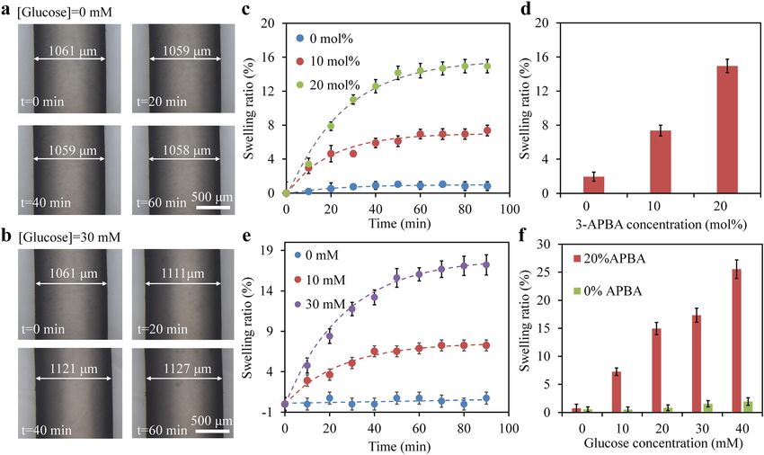
3550         J. Guo et al.: Soft and plasmonic hydrogel optical probe

immunity [13–21]. For glucose sensing, a common strategy is             quantitative and continuous glucose monitoring under
to incorporate the tip of optical fibers with functionalized             physiological conditions. The optical sensor was made of a
micro and nanostructures such as interferometric cavity                 biocompatible hydrogel optical fiber incorporating cova-
[14–16], fluorophores [17, 18], and plasmonic nanoparticles              lently immobilized gold nanoparticles (GNPs). To achieve
[19–21]. For example, Tierney et al. incorporated a glucose-            glucose responsivity, the nanocomposite hydrogel fiber
responsive coating layer at the end of an optical fiber to form          was functionalized with 3-(acrylamido)-phenylboronic
a Fabry–Perot cavity, which enabled sensitive glucose                   acid (3-APBA), which possesses a high binding affinity to
readout from the changes of the cavity length [15]. Liao et al.         glucose molecules. The complexation of 3-APBA with
demonstrated a percutaneous and disposable fiber-optic                   glucose molecules increased the boronate anions and
sensor by coating the fiber tip with a fluorophores-assembled             Donnan osmotic pressure in the hydrogel matrix, resulting
polymeric matrix for long-term glucose monitoring in vivo               in volume expansion of the fiber. This volumetric change
[17]. However, the practical applications of fiber-optic sen-            modulated the localized surface plasmon resonance
sors in clinics are hindered by poor biocompatibility of the            (LSPR) effect of the nanocomposite fiber, enabling quan-
fiber materials, and fragile nature of the fiber tip [22, 23].            titative glucose measurements from the transmitted light
Moreover, the high stiffness of conventional optical fibers              attenuation at the LSPR peak. To minimize the
(e.g., silica and plastics) may easily cause tissue lesions             LSPR-independent loss effects such as fiber bending, a
during implantation or body movements.                                  dual-wavelength differential approach was employed for
     Polymeric hydrogels have been intensively investi-                 the sensor readout, where a reference wavelength out of
gated as promising candidates for bio-optical sensing due               the plasmon resonance was introduced to provide an in-
to their exceptional optical and physico-mechanical                     ternal calibration. Glucose quantification tests were per-
properties [24–26]. Many synthetic hydrogels, such as                   formed, which demonstrated a reversible and linear
polyethylene glycol diacrylate (PEGDA), polyacrylamide                  response of the sensor with fast readout rate in physio-
(PAAm), and poly(vinyl alcohol) (PVA), have been utilized               logical ranges.
to fabricate optical waveguides with high softness and
biocompatibility for implantable and biomedical applica-
tions [27–34]. For example, hydrogel optical fibers                      2 Results and discussion
composed of a PEGDA core and an alginate cladding were
implanted in living mice for blood oxygenation sensing                  Glucose-responsive hydrogel optical fibers were fabricated
[29]. Low-modulus and stretchable alginate-PAAm hydro-                  from the 3-APBA functionalized PAAm hydrogel by mold-
gel optical fibers were demonstrated for optogenetic brain               ing and UV-induced photo-crosslinking (Figure 1a).
modulation in free-moving mice [33]. The hydrogel-based                 Hydrogel precursor was injected into a poly(vinyl chloride)
optical waveguide offers a versatile platform that enables              (PVC) tube mold, followed by UV irradiation for cross-
the incorporation of active recognition motif or function-              linking. Demolding of the hydrogel fiber from the PVC tube
alized nanostructures into the hydrogel matrix to meet the              was achieved by water pressure. For LSPR sensing, car-
demands in diverse biosensing and biomedical applica-                   boxylic acid-modified GNPs were covalently incorporated
tions [35–42]. Recently, hydrogel optical fibers involving               into the polymer matrix through EDC conjunction at pH 4.5.
phenylboronic acid (PBA) derivatives have been reported                 Figure 1b shows the photographs of the fabricated hydrogel
for continuous glucose monitoring, in which glucose con-                fibers with/without GNPs doping. In contrast to the trans-
centration was detected from the changes in transmitted                 parent undoped fiber, the GNPs-doped fiber showed up in
light intensities induced by the volumetric changes [43].               light red as result of the absorption and scattering of light
However, the detection of glucose directly from light in-               by the nanomaterials (Figure 1b). Transmission electron
tensity suffers from a variety of potential interferences such          microscope (TEM) image revealed uniform size (∼17.2 nm in
as the intensity fluctuation of light source, changes of the             average) and morphology of the GNPs (Figure 1c). To
surrounding environments, and light loss associated with                evaluate the optical performances, green laser at 532 nm
fiber deformation, which poses practical challenges for                  was coupled to the fiber through an objective lens. The fiber
precise quantification of glucose. The development of                    could efficiently guide light even when tied into knots or
minimally invasive hydrogel optical probe with robust and               transmitted through mediums of different refractive
continuous glucose readout can offer attractive building                indices, as confirmed from the bright light spot at the fiber
blocks for better glucose control in diabetes care.                     end (Figure 1d,e). The light-guiding capability of the
     Here, we present a soft and flexible optical glucose               hydrogel fiber suggests its potentials of serving as a flexible
sensor with self calibrated differential readout for                    optical implant to deliver light to target tissue for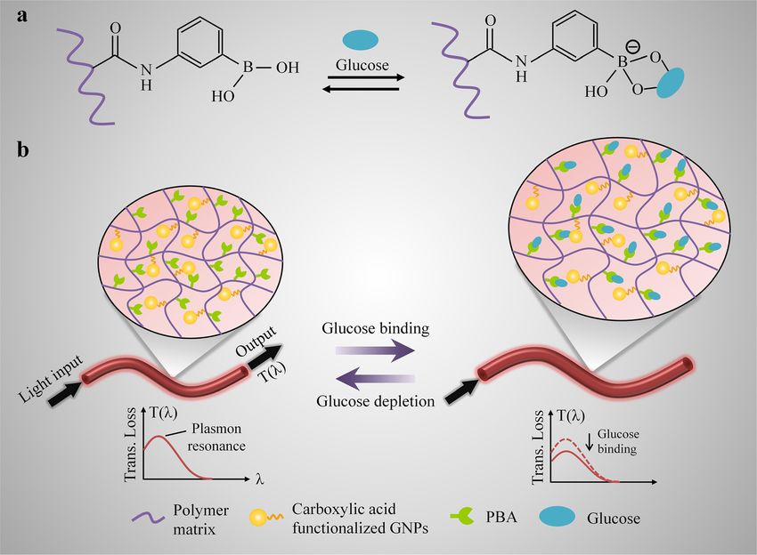
J. Guo et al.: Soft and plasmonic hydrogel optical probe 3551
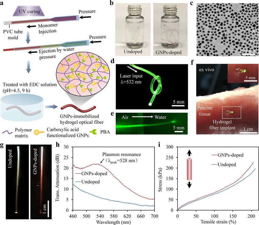
Figure 1: (a) Fabrication of the gold nanocomposite hydrogel optical fibers. (b) Photographs of the hydrogel fibers with/without gold
nanoparticles (GNPs) doping. (c) Transmission electron microscope (TEM) image of the GNPs. (d) A hydrogel fiber guides light at 532 nm when
tied into knot. (e) Light guiding from the air to water. (f) Implantation of the hydrogel fibers for deep-tissue light delivery. (g) Photographs of the
hydrogel fibers when illuminated by a white light source. Left: undoped; Right: GNPs-doped. (h) Transmission spectra. (i) Results of tensile
test.
biosensing and light-activated therapy applications. For deformations during body motions, and conformally
demonstrations, we implanted the hydrogel fiber inside a interact with the target tissues. The Young’s modulus of
porcine tissue at 4 cm depth with the aid of a gauge needle, human skin tissues is in the range of 0.1–2 MPa, and the
where the implanted fiber showed confined light through stretchability is 30–70% [44]. In contrast, a standard silica
tissue (Figure 1f). Transmission spectra of the fibers were optical fiber has a Young’s modulus of ∼70 GPa, five orders
further investigated by illuminating them with a white light of magnitudes higher than that of skin tissues [45]. Besides,
source (Figure 1g). The undoped fiber exhibited decreased the stretchability of silica fibers is less than 1%. Due to its
optical loss with the increasing wavelength due to the light significant mismatch in mechanical properties with soft
scattering from structural inhomogeneity and surface tissues, subcutaneously-implanted silica fiber could easily
roughness (Figure 1h). Incorporation of the GNPs within cause the host tissue injuries, which may subsequently
the hydrogel fiber resulted in a notable absorption peak at lead to chronic inflammation, swelling, and pain at the
528 nm, corresponding to the SPR of the GNPs. implant sites [46]. Hydrogel-based optical fibers are
For implantable and subcutaneous glucose moni- promising candidates to address the above limitations. We
toring, the soft and elastic nature of the biological tissues characterized the mechanical properties of the fabricated
required the sensing fibers to be highly flexible and hydrogel fibers by tensile testing (Figure 1i). The undoped
stretchable so that they could endure large mechanical fiber possessed a high stretchability of ∼210%, failure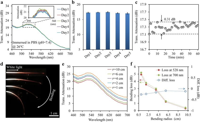
3552          J. Guo et al.: Soft and plasmonic hydrogel optical probe

stress of 196.3 kPa, and a low Young’s modulus of                        attenuation contributed by the LSPR effect, enabling
116.9 kPa, compatible with the soft skin tissues. Moreover,              quantitative glucose measurements (Figure 2b). As the
the incorporation of GNPs into the hydrogel matrix did not               hydrogel swells upon binding with glucose, the volume of
significantly influence its mechanical properties. The                     the fiber increased, but the amount of incorporated GNPs
GNPs-doped nanocomposite fiber showed a similar                           was kept unchanged due to the strong covalent attachment
stretchability (∼205%), but slightly increased mechanical                of GNPs to the hydrogel matrix. As a result, the concen-
strength (227.9 kPa) and Young’s Modulus (145.2 kPa) in                  tration of GNPs would decrease with the fiber expansion,
comparison with the undoped fiber. The high softness and                  leading to decreased light attenuation at the LSPR peak.
stretchability of the fibers make them particularly prom-                      The dynamic swelling behaviors of the nanocomposite
ising for potential clinical applications.                               fibers in response to glucose were investigated by optical
     We applied the gold nanocomposite hydrogel fiber for                microscopy. The fibers were fully swollen in phosphate-
glucose sensing through reversible complexation of the                   buffered saline (PBS) (pH = 7.4) prior to the test. Figure 3a
immobilized 3-APBA in the hydrogel matrix with the                       and b shows time-lapse microscope images of the nano-
glucose molecules (Figure 2a). Light transmitted through                 composite fibers in the absence and presence of glucose,
the nanocomposite fiber was scattered and absorbed by the                 respectively. As expected, no expansion of the fibers was
incorporated GNPs, causing light attenuation. Glucose                    observed in the absence of glucose. When exposed to
molecules could easily penetrate into the fiber through                   glucose solution (pH = 7.4, 30 mM), the fibers showed 6%
passive diffusion and bind with the boronic acid groups.                 expansion in diameter over 60 min due to the specific
The binding process increased the boronate anions and                    3-APBA-glucose binding. We further examined the influ-
Donnan osmotic pressure in the fiber, resulting in swelling               ence of 3-APBA concentrations on swelling kinetics of the
of the hydrogel matrix [47]. This glucose-dependent volu-                fibers, for which the swelling weight ratio of the fibers was
metric shift could be observed as a change in the light                  measured (Figure 3c). The weights of the fibers were

Figure 2: Schematic illustration of the gold nanocomposite hydrogel fiber for glucose detection.
(a) Complexation of 3-(acrylamido)-phenylboronic acid (3-APBA) with cis-diols in glucose molecules. (b) Sensing mechanism. Glucose binding
to the hydrogel fiber resulted in volume expansion of the hydrogel matrix, which could be monitored from the change in the light attenuation
contributed by the localized surface plasmon resonance (LSPR) effect.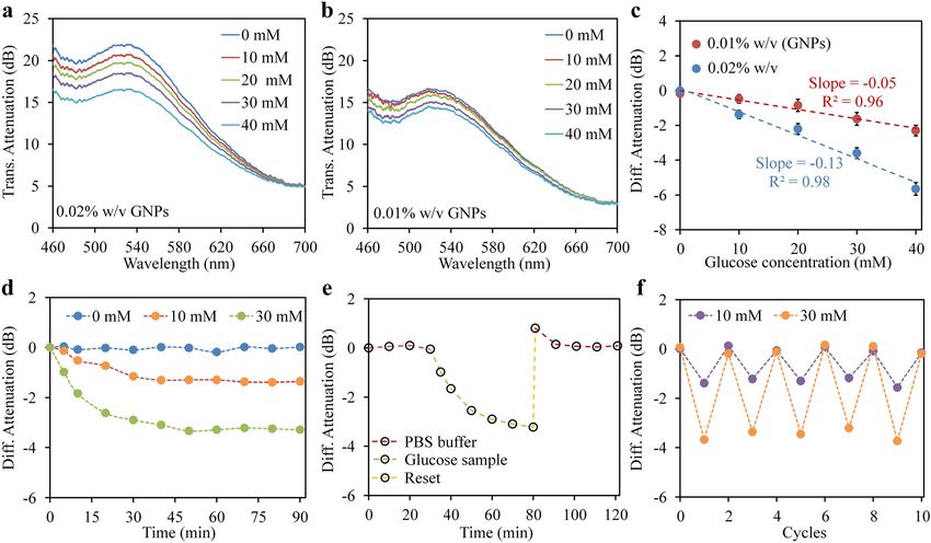
J. Guo et al.: Soft and plasmonic hydrogel optical probe          3553

Figure 3: (a, b) Microscope images of the nanocomposite fiber expansion over time under glucose concentrations of 0 mM (a) and 30 mM (b) at
pH = 7.4. The concentration of 3-(acrylamido)-phenylboronic acid (3-APBA) was set at 20 mol%. (c) Dependence of 3-APBA concentrations on
swelling kinetics of the fibers. (d) Swelling weight ratio of the fibers versus 3-APBA concentration. (e) Swelling kinetics of the fibers under
various glucose concentrations. (f) Swelling weight ratio versus glucose concentration for hydrogel fibers with/without 3-APBA
functionalization.

measured with time interval of 10 min and continuously                       To avoid the GNPs leakage, the GNPs were covalently
recorded for 90 min to reach an equilibrium state. As                   entrapped within the fiber by using EDC conjunction. The
shown in Figure 3d, the fibers showed increased volumetric               reaction of EDC with the surface carboxyl group on GNPs
shift with the increasing 3-APBA content, indicating a                  produced an amine-reactive O-acylisourea intermediate
higher glucose sensitivity with more 3-APBA. Considering                that spontaneously reacted with the amine groups of
the limited solubility of 3-APBA in the hydrogel precursor,             PAAm, forming a stable amide bond [48]. To confirm the
nanocomposite hydrogel fibers containing 20 mol%                         stability and immobilization of the GNPs, the transmission
3-APBA were found to be optimum for glucose detection.                  spectra of the nanocomposite fiber over time were recorded
The swelling responses of the fibers to various glucose                  in PBS buffer at constant temperature of 26 °C. The LSPR
concentrations were also investigated (Figure 3e). Increase             peak wavelength and intensity were indicative of the size
in the glucose concentration resulted in increased expan-               and concentration of the GNPs, respectively. As shown in
sion of the functionalized fibers, but did not induce                    Figure 4a, no shift in the LSPR peak of GNPs was observed
obvious volumetric changes for the fibers without 3-APBA                 in 5 days, suggesting no changes in the particle size. The
functionalization (Figure 3f). For a high glucose concen-               light attenuation at the resonance peak of the GNPs showed
tration of 40 mM, the functionalized fibers had a swelling               a maximum relative shift of about 0.42 and 0.31 dB over a
weight ratio as large as 25.5%, while the nonfunctionalized             long-term (5 days) and short-term (60 min) observation
fibers only swelled by 1.9%. The selectivity of the fibers was            period, respectively (Figure 4b,c). These results suggested
also tested with aqueous samples separately containing                  no leakage of the GNPs from the hydrogel matrix, allowing
CaCl2, KCl, NaCl, glycine, uric acid, ascorbic acid, and                highly stable optical readout based on LSPR. Due to its high
glucose (Figure S1, Supplementary information). The fibers               flexibility, the nanocomposite fiber could be bent or
showed high selectivity toward glucose due to the specific               twisted with the hosting tissues without mechanical fail-
binding of 3-APBA with cis-diol groups of glucose.                      ure, which, however, inevitably induces additional
3554          J. Guo et al.: Soft and plasmonic hydrogel optical probe

propagation losses in the fiber. The transmitted attenua-                 simulated physiological conditions (PBS buffer, pH = 7.4)
tion spectra of the fiber under various bending radius were               (Figure 5a,b). We defined the glucose sensitivity as the
further investigated (Figure 4d,e). The fiber showed                      magnitude in shift of the differential attenuation per unit
increased attenuation over the entire visible wavelength                 change of glucose concentration. As the glucose concen-
with the decreasing bending radius, which could greatly                  tration was increased from 0 to 40 mM, the sensor showed
influence the accuracy of glucose determination. To mini-                 linearly decreased attenuation with correlation coefficients
mize this effect, a dual-wavelength differential detection               of 0.96 and 0.98, and sensitivities of −0.05 dB/mM
method was employed for the sensor readout. Besides the                  and −0.13 dB/mM for low (0.01% w/v) and high (0.02%
LSPR peak, we chose another reference wavelength                         w/v) doping amounts of GNPs, respectively (Figure 5c). The
(700 nm) out of the plasmon resonance region to offer an                 higher sensitivity achieved with higher loading of GNPs
internal calibration. Figure 4f shows the bending-                       was attributed to the stronger LSPR effect. The detection
dependent light attenuation at 528 and 700 nm, respec-                   limit of the sensor at GNPs concentration of 0.02% w/v was
tively. As the bending radius was decreased to 1 cm, sig-                estimated to be 0.75 mM (S/N = 3). The temporal response
nificant light attenuation (>5 dB) were observed at both 528              of the fiber sensor for different glucose concentrations were
and 700 nm with similar trend. In contrast, the variation in             investigated by continuously recording its attenuation
the differential attenuation of the two wavelengths was                  spectra (Figure 5d). The sensor exhibited stable optical
below 0.2 dB due to the suppression of the bending-                      readout over time in the absence of glucose. Upon addition
dependent effects.                                                       of glucose, the sensor showed rapid response and reached
     To show the capability of the nanocomposite fiber                   binding equilibrium in 50 min, which provided a readout
sensor for quantifying glucose concentration, we charac-                 rate of 0.6 mM⋅min−1, much higher than the required speed
terized the spectral response of the sensor upon exposure                for diabetic patients (0.078 mM⋅min−1 [34]). The reusability
to glucose in a clinically relevant range (0–40 mM) under                of the sensor was evaluated by a cycling test of glucose

Figure 4: (a) Time-lapse transmission spectra of the nanocomposite fiber in phosphate-buffered saline (PBS) buffer at 26 °C. b, c) Changes of
light attenuation at the resonance peak (528 nm) of gold nanoparticles (GNPs) over a long-term (b) and short-term (c) observation period. (d)
Photograph showing a nanocomposite fiber applied with gradually decreased bending radius. (e) Transmission spectra of the fiber under
various bending radius. (f) Bending loss of the fiber at 528 and 700 nm (left axis), and the differential loss (right axis).
J. Guo et al.: Soft and plasmonic hydrogel optical probe 3555 Figure 5: (a, b) Spectral response of the fiber sensors in different glucose concentrations (0–40 mM). The doping amounts of gold nanoparticles (GNPs) in (a) and (b) were 0.02% and 0.01% w/v, respectively. (c) Differential light attenuation at 528 and 700 nm versus glucose concentration. (d) Temporal response of the sensor to various glucose concentration. (e) Reusability of the sensor. (f) Response of the sensor to repeated cycles of glucose binding and releasing under glucose concentrations of 10 and 30 mM. binding and releasing, where the sensor was first treated during the sensor operation (temperature changes:
3556           J. Guo et al.: Soft and plasmonic hydrogel optical probe

                                                                          exposure (365 nm) for 5 min. To extract the hydrogel fiber from the tube
3 Conclusions                                                             mold, water pressure was applied through syringe injection.
                                                                                The fiber glucose sensor was fabricated by using glucose-
In summary, we have demonstrated a soft and flexible                      responsive hydrogel fiber incorporating covalently immobilized
optical glucose sensor based on GNPs-incorporated                         GNPs. Carboxylic acid-modified GNPs were synthesized following
hydrogel optical fiber, which offers excellent optical and                previously reported procedures [50, 51], and the stock solution was
physio-mechanical properties suitable for implantable                     utilized for the sensor fabrication. The GNPs (0–0.02% w/v) were
                                                                          doped into the hydrogel fibers through precursor mixing, followed
glucose monitoring. To avoid the leakage of the GNPs from
                                                                          by UV polymerization for 5 min. Afterward, the gold nano-
the fiber, the GNPs were modified with carboxylic acid and                composite hydrogel fibers were treated with EDC for 9 h at pH 4.5.
covalently attached onto the hydrogel matrix through EDC                  The resulting fibers were rinsed with DI water and kept in PBS
conjugation. For glucose sensing, the fiber was function-                 buffer at pH 7.4.
alized with 3-APBA, which can bind with cis-diols in
glucose molecules, increasing the osmotic Donnan pres-
                                                                          4.2 Equipment and characterization
sure, and consequently resulting in volumetric changes of
the fiber. This glucose-dependent volumetric shift could be
                                                                          The microscopic image of the GNPs was taken by a 120 kV transmission
monitored from the change in light attenuation contributed                electron microscope (TEM, Tecnai Spirit). Mechanical characterization
by LSPR of the incorporated GNPs for glucose quantifica-                  of the hydrogels fibers was performed by using a tensile tester with a
tion. Readout of the optical sensor from light transmission               500 N load cell (Handpi Instrumnets). Transmission spectra of the
was susceptible to the loss effect associated with fiber                  hydrogel fibers were measured with a compact spectrometer (Ocean
                                                                          optics, Maya 2000) equipped with a halogen light source (Ocean op-
bending. This was minimized by a dual-wavelength dif-
                                                                          tics, HL-2000). The temperature-dependent effect of the fiber sensor
ferential approach, which employed a reference wave-                      was evaluated by using a thermocouple (resolution, 0.1 °C) and
length to offer an internal calibration. Quantitative                     heating tape. For pH titration experiments, Tris HCl and Tris base were
characterizations of the sensor showed a linear response in               used to prepare pH buffers (pH 4.6–10.5, ionic strength 150 mM). The
the clinically relevant range of 0–40 mM with high sensi-                 stability test was performed by immersing the fiber sensor in PBS
tivity (−0.13 dB/mM) and fast responsitivity (0.6 mM⋅min−1)               buffer at 26 °C, during which no agitation was applied.

under physiological conditions. Furthermore, the sensor
showed reversible and reproducible responses over                         4.3 Glucose sensing experiments
repeated testing cycles at different glucose levels with
negligible hysteresis. The presented optical sensor may                   The nanocomposite fiber sensor was integrated with silica multimode
find applications in wearable and implantable continuous                   fiber (MMF) for light coupling. PBS buffer solutions (pH 7.4, ionic
glucose monitoring at point-of-care settings. Harnessing                  strength 150 mM) were used to prepare glucose samples with con-
this sensor concept, it is also possible to realize biosensors            centrations ranging from 0 to 40 mM. Prior to the glucose testing, the
                                                                          fiber sensor was immersed in a Petri dish containing PBS buffer
for detection of other bioanalytes.
                                                                          (glucose-free) and allowed to be fully swollen at 26 °C. To investigate
                                                                          the glucose response of the sensor, the blank PBS buffer was replaced
                                                                          by glucose sample at a selected concentration, and the optical trans-
                                                                          missions of the nanocomposite fiber were continuously recorded by a
4 Methods                                                                 fiber-coupled spectrometer. To evaluate the reusability, the sensor
                                                                          was reset in acetate buffer (pH 4.6) for 2 min to release the bond
4.1 Fabrication of the nanocomposite fiber glucose                        glucose molecules and then immersed in PBS buffer for 30 min before
    sensor                                                                commencing the next glucose test.

Acrylamide (AAm), N,N′-methylenebis(acrylamide) (BIS), 3-(acryl-          Acknowledgments: J.G. acknowledges the funding from
amido) phenylboronic acid (3-APBA), diethoxyacetophenone (DEAP),
                                                                          the National Natural Science Foundation of China (No.
phosphate-buffered saline (PBS) buffer (pH 7.4, ionic strength
150 mM), dimethyl sulfoxide (DMSO), D-(+)-glucose, Tris HCl, Tris         61805126, 62175008). L.K. acknowledges the support from
base, and N-(3-dimethylaminopropyl)-N′-ethylcarbodiimide hydro-           the “Thousand Talents Plan” Youth Program of China.
chloride (EDC) were purchased from Sigma-Aldrich and used without         Author contribution: J.G. and L.K. conceived the idea. J.G.
further purification. Hydrogel precursor was prepared by mixing AAm        and B.Z. performed the experiments. J.G., L.K., and B.Z.
(78.5 mol%), BIS (1.5 mol %), 3-APBA (20 mol%) with DEAP (2% w/v)
                                                                          analyzed the data. All authors contributed to the editing of
in DMSO (1 mL), followed by monomer dilution with DI water (1 mL).
                                                                          the manuscript.
After being carefully degassed, the precursor solution was injected
into a polyvinyl chloride (PVC) tube mold (inner diameter: 1 mm) using    Research funding: None declared.
a syringe adapted with a 0.45 µm filter. Glucose-responsive hydrogel       Conflict of interest statement: The authors declare no
optical fibers were formed by curing the precursor solution under UV       conflicts of interest regarding this article.
J. Guo et al.: Soft and plasmonic hydrogel optical probe 3557
Data availability: The data that support the findings of this fiber-optic readout platform,” Biosens. Bioelectron., vol. 24,
study are available from the corresponding author upon p. 2034, 2009.
[16] S. W. Harun, A. A. Jasim, H. A. Rahman, M. Z. Muhammad, and
request.
H. Ahmad, “Micro-ball lensed fiber-based glucose sensor,” IEEE
Sensor. J., vol. 13, p. 348, 2012.
[17] K. C. Liao, T. Hogen-Esch, F. J. Richmond, L. Marcu, W. Clifton,
References and G. E. Loeb, “Percutaneous fiber-optic sensor for chronic
glucose monitoring in vivo,” Biosens. Bioelectron., vol. 23,
[1] C. Chen, Q. Xie, D. Yang, et al., “Recent advances in p. 1458, 2008.
electrochemical glucose biosensors: a review,” RSC Adv., vol. 3, [18] S. Yu, L. Ding, H. Lin, W. Wu, and J. Huang, “A novel optical fiber
p. 4473, 2013. glucose biosensor based on carbon quantum dots-glucose
[2] S. K. Vashist, D. Zheng, K. Al-Rubeaan, J. H. Luong, and oxidase/cellulose acetate complex sensitive film,” Biosens.
F. S. Sheu, “Technology behind commercial devices for blood Bioelectron., vol. 146, p. 111760, 2019.
glucose monitoring in diabetes management: a review,” Anal. [19] S. Singh and B. D. Gupta, “Fabrication and characterization of a
Chim. Acta, vol. 703, p. 124, 2011. surface plasmon resonance based fiber optic sensor using gel
[3] J. A. Usher-Smith, M. J. Thompson, S. J. Sharp, and F. M. Walter, entrapment technique for the detection of low glucose
“Factors associated with the presence of diabetic ketoacidosis concentration,” Sensor. Actuator. B Chem., vol. 177, p. 589,
at diagnosis of diabetes in children and young adults: a 2013.
systematic review,” BMJ, vol. 343, p. d4092, 2011. [20] H. Yuan, W. Ji, S. Chu, et al., “Fiber-optic surface plasmon
[4] L. Olansky and L. Kennedy, “Finger-stick glucose monitoring: resonance glucose sensor enhanced with phenylboronic acid
issues of accuracy and specificity,” Diabetes Care, vol. 33, modified Au nanoparticles,” Biosens. Bioelectron., vol. 117,
p. 948, 2010. p. 637, 2018.
[5] V. R. Kondepati and H. M. Heise, “Recent progress in analytical [21] Y. Yuan, X. Yang, D. Gong, et al., “Investigation for terminal
instrumentation for glycemic control in diabetic and critically ill reflection optical fiber SPR glucose sensor and glucose sensitive
patients,” Anal. Bioanal. Chem., vol. 388, p. 545, 2007. membrane with immobilized GODs,” Opt. Express, vol. 25,
[6] M. Vettoretti and A. Facchinetti, “Combining continuous glucose p. 3884, 2017.
monitoring and insulin pumps to automatically tune the basal [22] S. P. Nichols, A. Koh, W. L. Storm, J. H. Shin, and
insulin infusion in diabetes therapy: a review,” Biomed. Eng. M. H. Schoenfisch, “Biocompatible materials for continuous
Online, vol. 18, p. 37, 2019. glucose monitoring devices,” Chem. Rev., vol. 113, p. 2528, 2013.
[7] D. Chen, C. Wang, W. Chen, Y. Chen, and J. X. Zhang, [23] S. Vaddiraju, D. J. Burgess, I. Tomazos, F. C. Jain, and
“PVDF-Nafion nanomembranes coated microneedles for in vivo F. Papadimitrakopoulos, “Technologies for continuous glucose
transcutaneous implantable glucose sensing,” Biosens. monitoring: current problems and future promises,” J. Diabetes
Bioelectron., vol. 74, p. 1047, 2015. Sci. Technol., vol. 4, p. 1540, 2010.
[8] S. A. Zaidi and J. H. Shin, “Recent developments in nanostructure [24] J. Guo, C. Yang, Q. Dai, and L. Kong, “Soft and stretchable
based electrochemical glucose sensors,” Talanta, vol. 149, polymeric optical waveguide-based sensors for wearable and
p. 30, 2016. biomedical applications,” Sensors, vol. 19, p. 3771, 2019.
[9] J. Zhang, X. Yu, W. Guo, et al., “Construction of titanium dioxide [25] S. Shabahang, S. Kim, and S. H. Yun, “Light-guiding biomaterials
nanorod/graphite microfiber hybrid electrodes for a high for biomedical applications,” Adv. Funct. Mater., vol. 28,
performance electrochemical glucose biosensor,” Nanoscale, p. 1706635, 2018.
vol. 8, p. 9382, 2016. [26] R. Nazempour, Q. Zhang, R. Fu, and X. Sheng, “Biocompatible
[10] Y. J. Heo, H. Shibata, T. Okitsu, T. Kawanishi, and S. Takeuchi, and implantable optical fibers and waveguides for biomedicine,”
“Long-term in vivo glucose monitoring using fluorescent Materials, vol. 11, p. 1283, 2018.
hydrogel fibers,” Proc. Natl. Acad. Sci. USA, vol. 108, p. 13399, [27] J. Guo, X. Liu, N. Jiang, et al., “Highly stretchable, strain sensing
2011. hydrogel optical fibers,” Adv. Mater., vol. 28, p. 10244, 2016.
[11] D. Rodbard, “Continuous glucose monitoring: a review of [28] J. Feng, Y. Zheng, S. Bhusari, M. Villiou, S. Pearson, and
successes, challenges, and opportunities,” Diabetes Technol. A. del Campo, “Printed degradable optical waveguides for
Therapeut., vol. 18, p. S2, 2016. guiding light into tissue,” Adv. Funct. Mater., vol. 30, p. 2004327,
[12] M. Elsherif, M. U. Hassan, A. K. Yetisen, and H. Butt, “Wearable 2020.
contact lens biosensors for continuous glucose monitoring using [29] M. Choi, M. Humar, S. Kim, and S. H. Yun, “Step-index optical
smartphones,” ACS Nano, vol. 12, p. 5452, 2018. fiber made of biocompatible hydrogels,” Adv. Mater., vol. 27,
[13] M. J. Yin, B. Gu, Q. F. An, C. Yang, Y. L. Guan, and K. T. Yong, p. 4081, 2015.
“Recent development of fiber-optic chemical sensors and [30] J. Guo, H. Huang, M. Zhou, C. Yang, and L. Kong, “Quantum dots-
biosensors: mechanisms, materials, micro/nano-fabrications doped tapered hydrogel waveguide for ratiometric sensing of
and applications,” Coord. Chem. Rev., vol. 376, p. 348, metal ions,” Anal. Chem., vol. 90, p. 12292, 2018.
2018. [31] J. Guo, Y. Luo, C. Yang, and L. Kong, “In situ surface-enhanced
[14] S. Novais, C. I. Ferreira, M. S. Ferreira, and J. L. Pinto, “Optical Raman scattering sensing with soft and flexible polymer optical
fiber tip sensor for the measurement of glucose aqueous fiber probes,” Opt. Lett., vol. 43, p. 5443, 2018.
solutions,” IEEE Photonics J., vol. 10, p. 1, 2018. [32] R. Kumar, A. P. Singh, A. Kapoor, and K. N. Tripathi, “Effect of dye
[15] S. Tierney, S. Volden, and B. T. Stokke, “Glucose sensors based doping in poly (vinyl alcohol) waveguides,” J. Mod. Opt., vol. 52,
on a responsive gel incorporated as a Fabry-Perot cavity on a p. 1471, 2005.3558 J. Guo et al.: Soft and plasmonic hydrogel optical probe
[33] L. Wang, C. Zhong, D. Ke, et al., “Ultrasoft and highly stretchable [43] A. K. Yetisen, N. Jiang, A. Fallahi, et al., “Glucose-sensitive
hydrogel optical fibers for in vivo optogenetic modulations,” hydrogel optical fibers functionalized with phenylboronic acid,”
Adv. Opt. Mater., vol. 6, p. 1800427, 2018. Adv. Mater., vol. 29, p. 1606380, 2017.
[34] J. Feng, Q. Jiang, P. Rogin, P. W. de Oliveira, and A. Del Campo, [44] G. Chen, N. Matsuhisa, Z. Liu, et al., “Plasticizing silk protein for
“Printed soft optical waveguides of PLA copolymers for guiding on-skin stretchable electrodes,” Adv. Mater., vol. 30,
light into tissue,” ACS Appl. Mater. Interfaces, vol. 12, p. 20287, p. 1800129, 2018.
2020. [45] P. Antunes, H. Lima, J. Monteiro, and P. S. André, “Elastic
[35] C. Wang, X. Liu, V. Wulf, M. Vázquez-González, M. Fadeev, and constant measurement for standard and photosensitive single
I. Willner, “DNA-based hydrogels loaded with Au nanoparticles mode optical fibres,” Microw. Opt. Technol. Lett., vol. 50,
or Au nanorods: thermoresponsive plasmonic matrices for p. 2467, 2008.
shape-memory, self-healing, controlled release, and mechanical [46] U. Klueh, M. Kaur, D. C. Montrose, and D. L. Kreutzer,
applications,” ACS Nano, vol. 13, p. 3424, 2019. “Inflammation and glucose sensors: use of dexamethasone to
[36] E. Castellanos, B. Soberats, S. Bujosa, C. Rotger, R. de la Rica, extend glucose sensor function and life span in vivo,” J. Diabetes
and A. Costa, “Development of plasmonic Chitosan–Squarate Sci. Technol., vol. 1, p. 496, 2007.
hydrogels via bioinspired nanoparticle growth,” [47] M. Elsherif, M. U. Hassan, A. K. Yetisen, and H. Butt, “Glucose
Biomacromolecules, vol. 21, p. 966, 2019. sensing with phenylboronic acid functionalized hydrogel-based
[37] L. Moretti, A. Mazzanti, A. Rossetti, et al., “Plasmonic control of optical diffusers,” ACS Nano, vol. 12, p. 2283, 2018.
drug release efficiency in agarose gel loaded with gold [48] Y. Lei, H. Tang, C. Zhou, T. Zhang, M. Feng, and B. Zou,
nanoparticle assemblies,” Nanophotonics, vol. 10, p. 247, 2021. “Incorporating fluorescent quantum dots into water-soluble
[38] I. Vassalini, G. Ribaudo, A. Gianoncelli, M. F. Casula, and polymer,” J. Lumin., vol. 128, p. 277, 2008.
I. Alessandri, “Plasmonic hydrogels for capture, detection and [49] N. V. Gupta and H. G. Shivakumar, “Investigation of swelling
removal of organic pollutants,” Environ. Sci. Nano, vol. 7, behavior and mechanical properties of a pH-sensitive
p. 3888, 2020. superporous hydrogel composite, Iran,” J. Pharm. Res., vol. 11,
[39] S. C. Moorcroft, L. Roach, D. G. Jayne, Z. Y. Ong, and S. D. Evans, p. 481, 2012.
“Nanoparticle-loaded hydrogel for the light-activated release [50] S. Sabouri, H. Ghourchian, M. Shourian, and M. Boutorabi, “A
and photothermal enhancement of antimicrobial peptides,” ACS gold nanoparticle-based immunosensor for the
Appl. Mater. Interfaces, vol. 12, p. 24544, 2020. chemiluminescence detection of the hepatitis B surface
[40] C. L. Shen, Q. Lou, J. H. Zang, et al., “Near‐infrared antigen,” Anal. Methods., vol. 6, p. 5059, 2014.
chemiluminescent carbon nanodots and their application in [51] M. H. Jazayeri, H. Amani, A. A. Pourfatollah, H. Pazoki-Toroudi,
reactive oxygen species bioimaging,” Adv. Sci., vol. 7, and B. Sedighimoghaddam, “Various methods of gold
p. 1903525, 2020. nanoparticles (GNPs) conjugation to antibodies,” Sens.
[41] C. L. Shen, Q. Lou, K. K. Liu, L. Dong, and C. X. Shan, Biosensing Res., vol. 9, p. 17, 2016.
“Chemiluminescent carbon dots: synthesis, properties, and
applications,” Nano Today, vol. 35, p. 100954, 2020.
[42] C. L. Shen, G. S. Zheng, M. Y. Wu, et al., “Chemiluminescent Supplementary Material: The online version of this article offers
carbon nanodots as sensors for hydrogen peroxide and supplementary material (https://doi.org/10.1515/nanoph-2021-
glucose,” Nanophotonics, vol. 9, p. 3597, 2020. 0360).You can also read