Mechanism of tumor derived extracellular vesicles in regulating renal cell carcinoma progression by the delivery of MALAT1
←
→
Page content transcription
If your browser does not render page correctly, please read the page content below
ONCOLOGY REPORTS 46: 187, 2021

     Mechanism of tumor‑derived extracellular vesicles in regulating
      renal cell carcinoma progression by the delivery of MALAT1
                    CHENGLUO JIN1*, LINMEI SHI2*, KUNLUN LI1*, WEI LIU1, YU QIU1, YAKUN ZHAO1,
                               BAI ZHAO1, ZHEXUN LI1, YIFEI LI1 and QINGGUO ZHU1

        1
            Department of Urology, The Second Affiliated Hospital, Harbin Medical University; 2School of Health Management,
                            Harbin Medical University, Nangang, Harbin, Heilongjiang 150086, P.R. China

                                          Received August 12, 2020; Accepted April 28, 2021

                                                      DOI: 10.3892/or.2021.8138

Abstract. Renal cell carcinoma (RCC) is a major healthcare          enhanced the binding of ETS1 and the TFCP2L1 promoter and
burden globally. Tumor‑derived extracellular vesicles (EVs)         decreased TFCP2L1 expression. In vivo, 786‑O‑EVs promoted
contribute to the formation of a pro‑metastatic microenviron‑       tumor growth and RCC lung metastasis, which was suppressed
ment. In the present study, we explored the role and mechanism      following inhibition of MALAT1. Our findings indicated that
of RCC cell 786‑O‑derived EVs (786‑O‑EVs) in RCC. First,            786‑O‑EVs promoted RCC invasion and metastasis by trans‑
786‑O‑EVs were extracted and identified, and EV internaliza‑        porting MALAT1 to promote the binding of transcription
tion of RCC cells was observed. RCC cell malignant behaviors        factor ETS1 and TFCP2L1 promoter.
and long noncoding RNA (lncRNA) metastasis‑associated
lung adenocarcinoma transcript 1 (MALAT1) expression                Introduction
patterns were detected before and after 786‑O‑EV treatment.
MALAT1 was intervened to evaluate RCC cell behaviors. The           Renal cell carcinoma (RCC) is regarded as one of the most
downstream mechanism involving MALAT1 was predicted.                common malignancies of the genitourinary system, with an
In addition, the relationship among MALAT1, transcription           increasing incidence rate in the United States (1). RCC is
factor CP2 like 1 (TFCP2L1) and ETS proto‑oncogene 1,               generally rare in children and young adults as it primarily
transcription factor (ETS1) was analyzed. TFCP2L1 expres‑           manifested in the elderly (2,3). Some of the contributing factors
sion patterns were measured after 786‑O‑EV exposure. Tumor          of RCC include hypertension, smoking, familial syndromes,
xenograft formation assay and lung metastasis model were            with an unhealthy lifestyle and dietary habits (4). RCC has
adopted to verify the role of 786‑O‑EVs in vivo in RCC. It          multiple subtypes, with clear cell RCC (ccRCC) as the most
was found that 786‑O‑EVs could be internalized by RCC cells.        prevalent type with 85% proportion among other subtypes (5).
786‑O‑EVs promoted RCC cell malignant behaviors, accompa‑           Chest metastasis is a frequent finding in RCC, however lung
nied by elevated MALAT1 expression levels. The 786‑O‑EVs            metastasis is the most common form of distant metastasis (6).
with MALAT1 knockdown attenuated the promotive effect of            Additionally, RCC is habitually asymptomatic until the diag‑
sole 786‑O‑EVs on RCC cells. MALAT1 located ETS1 in the             nosis of metastasis by medical intervention. With expanding
TFCP2L1 promoter and negatively regulated TFCP2L1, and              alternative treatment options, surgical intervention persists as
ETS1 protein could specifically bind to MALAT1. 786‑O‑EVs           the gold standard for the treatment of RCC (7,8). Therefore, we
                                                                    sought to explore new and reliable protocols for RCC therapy.
                                                                        Extracellular vesicles (EVs), fundamentally recognized as
                                                                    the nanoscale tools composed of exosomes and microvesicles
                                                                    for intrinsic intercellular communication, are active contribu‑
Correspondence to: Dr Chengluo Jin, Department of Urology,
                                                                    tors in diverse physiological processes, including regulation
The Second Affiliated Hospital, Harbin Medical University,
148 Baojian Road, Nangang, Harbin, Heilongjiang 150086,
                                                                    of tumor development (9). A recent study ascertained the vital
P.R. China                                                          functionality of EVs in RCC initiation and progression (10).
E‑mail: jcl0114@163.com                                             Tumor‑derived EVs are the focus of increased research due
                                                                    to their ability to facilitate tumorigenesis and thus are being
Dr Linmei Shi, School of Health Management, Harbin Medical
                                                                    adopted as promising markers for tumor treatment, including
University, Nangang, Harbin, Heilongjiang 150086, P.R. China
E‑mail: is6434qytq8472@163.com
                                                                    RCC (11,12).
                                                                        Furthermore, the role of EVs in mediating cell‑to‑cell
*
    Contributed equally                                             communication and facilitating tumorigenesis is achieved
                                                                    via transfer of multiple RNAs, including long noncoding
Key words: renal cell carcinoma, extracellular vesicles, lncRNA     RNAs (lncRNAs) (13). Accumulating evidence has validated
MALAT1, ETS1, TFCP2L1, migration, invasion                          lncRNAs as crucial in regulating diverse cancers, including
                                                                    RCC (14,15). Metastasis‑associated lung adenocarcinoma
                                                                    transcript 1 (MALAT1) has been validated as a potent marker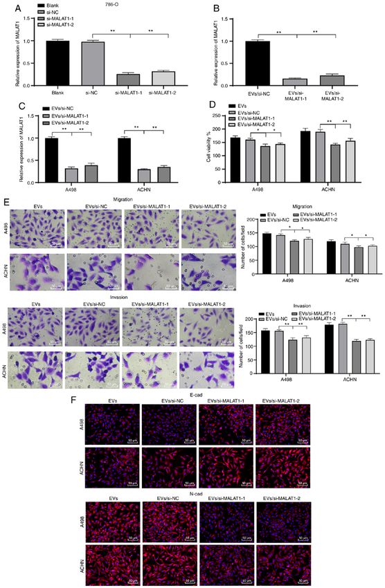
2                                    JIN et al: TUMOR-EV-SHUTTLED MALAT1/ETS1/TFCP2L1 IN RCC

for multiple human cancers accounting for its abnormal upreg‑          database (https://www.disgenet.org/). Gene interaction
ulation as an indicator of aggravated pathological alterations         network diagram was constructed using the STRING database
in cancerous organs (16). MALAT1 shows an aberrantly high              (https://string‑db.org/) and Cytoscape V3.7.1 software (www.
expression and promotes cell malignant biological behaviors            cytoscape.org). RCC microarray GSE16441 (20) including
in RCC (17). Furthermore, as previously evidenced, MALAT1              17 normal samples and 17 RCC samples was obtained from
can be diffused by the epithelial ovarian cancer cell‑secreted         the GEO database (https://www.ncbi.nlm.nih.gov/geo/). With
exosomes to the target recipient cells to promote cancer angio‑        the normal samples as the control, the R language ‘limma’
genesis (18). However, the mutual action of tumor‑derived EVs          package (21) was applied for differential analysis. TheP‑value
and MALAT1 in RCC has not yet been explored.                           was corrected using the false discovery rate (FDR) method, and
    Therefore, we hypothesized that tumor‑derived EVs may              |logFC|>2 and FDR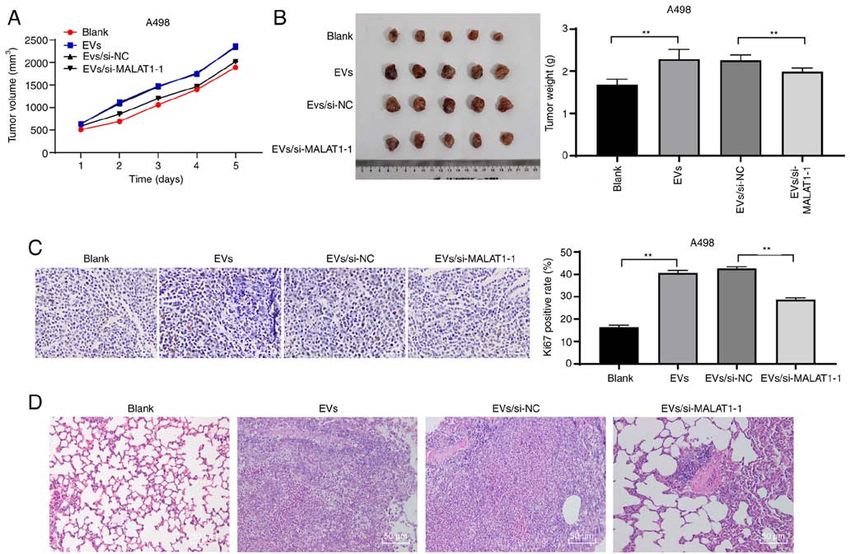
ONCOLOGY REPORTS 46: 187, 2021                                                     3

Table I. Primer sequences for qPCR.                                 photographed using a Nikon digital camera (magnification,
                                                                    x100). Three independent experiments were repeatedly set.
Gene                                  Primer                        The method for the migration experiment was the same as that
                                                                    of the invasion experiment, without utilization of Matrigel.
MALAT1             F: 5'‑GGGGCAGTAGTGTAGAGA‑3'
                   R: 5'‑CAGTGCGTGTCGTGGAGT‑3'                      Immunofluorescence. The cells were seeded on 24‑well plates
U6                 F: 5'‑CTCGCTTCGGCAGCACA‑3'                       and received different treatments. Next, the cells were fixed
                   R: 5'‑GTGTCGTGGAGTCGGCAA‑3'                      for 15 min in 4% paraformaldehyde, permeated for 10 min
TFCP2L1            F: 5'‑AGGTGCTGACCTCCTGAAGA‑3'                    with PBS containing 0.1% Triton X‑100, and sealed for 30 min
                   R: 5'‑GTTTTGCTCCAGCTCCTGAC‑3'                    using the 3% plasma blocking solution (all performed at room
ETS1               F: 5'‑CATGCTTTTCGTTTGACACCC‑3'                   temperature). Subsequently, the cells were subjected to over‑
                   R: 5'‑CTTTGCTTCCACCCGCCCCCC‑3'                   night incubation at 4˚C with the diluted primary antibodies
GAPDH              F: 5'‑ACCAGGTATCTGCTGGTTG‑3'                     E‑cadherin (dilution 1/200, ab1416, Abcam) and N‑cadherin
                                                                    (dilution 1/200, ab76057, Abcam), followed by a 1‑h incuba‑
                   R: 5'‑TAACCATGATGTCAGCGTGGT‑3'
                                                                    tion regimen with the secondary antibody goat anti‑mouse IgG
F, forward; R, reverse; MALAT1, metastasis‑associated lung adeno‑   H&L (Alexa Fluor ® 488) (dilution 1/200, ab150117, Abcam)
carcinoma transcript 1; TFCP2L1, transcription factor CP2 like 1;   in conditions devoid of light at room temperature. Finally,
ETS1, ETS proto‑oncogene 1; GAPDH, glyceraldehyde 3‑phosphate       4',6‑diamidino‑2‑phenylindole (DAPI) was added for nuclear
dehydrogenase.                                                      staining. After resting at room temperature for 1 min, the cells
                                                                    were observed under a fluorescence microscope (Olympus
                                                                    Optical Co., Ltd.).

primary antibodies [cluster of differentiation (CD)63 (dilution     Fluorescence in situ hybridization (FISH). MALAT1 subcel‑
1:1,000, ab134045), CD81 (dilution 1:1,000, ab109201) and           lular localization was observed using FISH assay in strict
TFCP2L1 (dilution 1:1,000, ab140197) (all from Abcam Inc.)]         accordance with the provided instructions of the Ribo™
at 4˚C. Following 3 rinses (10 min each) with PBS and 0.1%          lncRNA FISH Probe Mix (Red) (C10920, RiboBio Co., Ltd.).
Tween‑20 (PBST), the membranes underwent a 1‑h incuba‑              The cells (6x10⁴ cells/well) were seeded into 24‑well plates.
tion regimen with the horseradish peroxidase‑labeled goat           Upon achieving 60‑70% confluence, the cells were fixed
anti‑rabbit immunoglobulin G (IgG) H&L (dilution 1:2,000,           using 4% paraformaldehyde, rinsed and permeabilized. The
ab205718; Abcam Inc.), and rinsed 3 times with PBST (10 min         plates were sealed using the pre‑hybridization solution. After
each). An optical illuminator (General Electric) was utilized       elimination of the pre‑hybridization solution, the cells were
for membrane visualization. Image‑Pro Plus 6.0 (Media               subjected to overnight hybridization at 37˚C using the probe
Cybernetics) was applied for protein band gray value analysis.      hybridization solution supplemented with anti‑MALAT1
                                                                    nucleotide (Wuhan Genecreate Bioengineering Co., Ltd.) in
Cell Counting Kit‑8 (CCK‑8) assay. RCC cell prolifera‑              conditions devoid of light. Next, the cells were eluted, stained
tion was detected using the CCK‑8 kit (Dojindo Molecular            with DAPI, rinsed, and fixed using nail polish for observation
Technologies) in strict accordance with the provided instruc‑       under fluorescence microscopy (Olympus). Five different
tions. RCC cells were seeded into 96‑well plates and each           fields were selected, and the cells in these fields were observed
well was supplemented with EVs based on cell grouping and           and documented.
incubation for 24 h. Then approximately 10 µl CCK‑8 reagent
was added to each well for a 2‑h regimen of incubation at           Dual‑luciferase reporter gene assay. To explore the effect
37˚C. The optical density (OD) at the wavelength of 490 nm          of MALAT1 on TFCP2L1 promoter activity, overexpressed
was determined using a microplate reader (Thermo Fisher             (oe)‑NC, oe‑MALAT1, short hairpin (sh)‑NC, and sh‑MALAT1
Scientific, Inc.). Relative cell viability of each group was        were co‑transfected with the TFCP2L1‑2 kb luciferase reporter
determined.                                                         plasmid respectively into 293T cells. After 48 h of transfec‑
                                                                    tion, the cells were collected and lysed. The dual‑luciferase
Transwell assays. RCC cell invasion was assessed according          reporter gene assay was performed using a luciferase assay
to the passage of the number of transfected cells through           kit (K801‑200, BioVision Inc.) and a dual‑luciferase reporter
Transwell chambers (8‑micron chamber; Corning Inc., Life            gene analysis system (Promega Corp.). Renilla luciferase
Sciences). The Transwell chambers were pre‑coated with              was adopted for internal reference. The activation degree of
100 µl Matrigel (BD Biosciences) at 37˚C for 5 h until gelling      the target reporter gene was measured using the ratio of the
was visible. Next, 1x10⁵ A498 and ACHN cells that underwent         relative unit of firefly luciferase to that of Renilla luciferase.
a 24‑h starvation in 500 µl serum‑free MEM medium were
seeded in the apical chamber, respectively (3 technical repli‑      RNA immunoprecipitation (RIP). A RIP kit (Millipore Corp.)
cates for each group). The basolateral chamber was filled with      was utilized to assess the binding of MALAT1 to the ETS1
700 µl MEM with 10% FBS. Following a 48‑h incubation at             protein. The cells were lysed in an ice bath, and then subjected
37˚C, Matrigel and non‑invaded cells were removed from the          to centrifugation. Next, the supernatant was collected. The
upper surface of the filters. Cells that adhered to the lower       cell extract was incubated with the corresponding antibody for
surface of filters were fixed using 4% paraformaldehyde and         co‑precipitation. Following a rinse and re‑suspension in RIP
stained with crystal violet. Finally, the cells were counted and    wash buffer, the magnetic beads were supplemented with the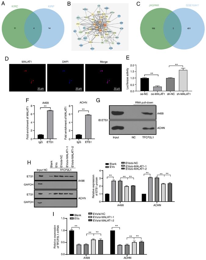
4                                 JIN et al: TUMOR-EV-SHUTTLED MALAT1/ETS1/TFCP2L1 IN RCC

appropriate antibody for binding. After a rinse, the magnetic      48 H&E staining. The extracted lung tissue was fixed using
bead‑antibody complex was resuspended in RIP wash buffer,          4% paraformaldehyde and embedded in paraffin. Then, the
incubated overnight at 4˚C with the cell extract, and harvested.   tissue was sectioned, dewaxed and hydrated independently.
The RNA content was extracted from the sample for subse‑           The sections were stained using the hematoxylin solution
quent PCR detection after detachment with proteinase K. The        for 10 min and decolorized in 70 and 90% ethanol after a
antibody used above was ETS1 (dilution 1:200, ab225868,            rinse with distilled water. Subsequently, the sections were
Abcam), with IgG (dilution 1:100, ab172730, Abcam) as the          stained with the eosin solution for 2‑3 min, dehydrated with
control.                                                           pure ethanol, cleared with xylene, sealed with neutral gum
                                                                   and then observed under a microscope (BX53M, Olympus;
RNA pull‑down. Biotinylated RNA sequence probe in the              magnification, x200).
TFCP2L1 promoter region and its negative control (NC) probe
were dissolved in washing/binding buffer at room tempera‑          Statistical analysis. SPSS 21.0 (IBM Corp.) software was
ture and incubated with the streptavidin‑coupled magnetic          used for data analysis. The experimental data were in normal
beads for 2 h. Then, the cell lysate was added and incubated       distribution as verified by Kolmogorov‑Smirnov test, and
for 2 h. The protein complex conjugated to magnetic beads          expressed as mean ± standard deviation. Comparison between
was washed. ETS1 content in the complex was determined by          two groups was conducted with the independent sample t‑test.
western blot analysis.                                             The comparison among groups was analyzed using one‑way
                                                                   analysis of variance (ANOVA), followed by the Tukey's
Ethics statement. The experimental procedures were approved        multiple comparisons test. The P‑value was obtained from a
by the Ethics Committee of The Second Affiliated Hospital of       two‑sided test and a value of P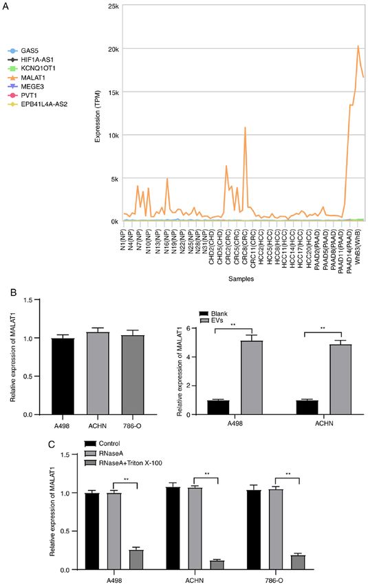
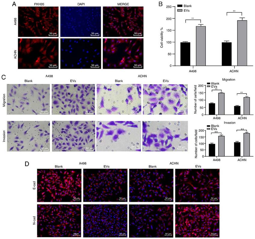
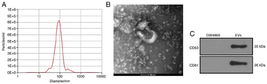
ONCOLOGY REPORTS 46: 187, 2021                                                                    5

Figure 1. 786‑O‑EVs are successfully extracted. (A) NTA was used to analyze the size of the EV population. (B) TEM images of purified 786‑O‑EVs. (C) WB was
used to detect the expression of EV symbolic markers. NTA, nanoparticle tracking analysis; TEM, transmission electron microscopy; EVs, extracellular vesicles.

Figure 2. 786‑O‑EVs promote RCC cell invasion and metastasis. 786‑O‑EVs were incubated with A498 and ACHN cells respectively for 24 h. (A) The inter‑
nalization of 786‑O‑EVs by A498 and ACHN cells was observed using PKH26 fluorescence labeling. (B) The viability of cells treated with 786‑O‑EVs was
evaluated using CCK‑8 assay. (C) The invasion and migration of cells were evaluated using Transwell assays. (D) The fluorescence expression of E‑cadherin
(E‑cad) and N‑cadherin (N‑cad) in A498 and ACHN cells was detected using immunofluorescence. Each experiment was repeated three times independently.
Data in panels B and C were analyzed using independent t-test. **P
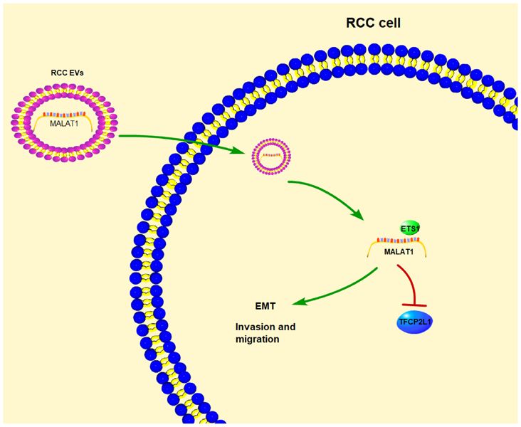
6                                 JIN et al: TUMOR-EV-SHUTTLED MALAT1/ETS1/TFCP2L1 IN RCC

explore the underlying mechanism, we referred to previous          retrieved known genes and the predicted four transcription
studies. For instance, EVs were found to release and carry         factors was analyzed, after which the gene interaction network
lncRNAs into the target cells to manipulate cellular func‑         diagram was constructed (Fig. 5B). It was evident that ETS1
tions (25). MALAT1 was also found to promote RCC cell              had the most interactions with known genes, suggesting the
malignant biological behaviors (26). Therefore, we searched        superior vitality of ETS1 relative to others. In a previous
RCC‑related lncRNAs through the lncDisease database                work, ETS1 served as an oncogene in RCC with intimate
(http://www.cuilab.cn/lncrnadisease), and found the involve‑       relations with a low survival rate in a large number of RCC
ment of lncRNAs such as MALAT1 in the regulation of RCC            samples (27). Meanwhile, a RCC microarray GSE16441 was
(Table SI). Furthermore, the expression of these candidate         obtained through the GEO database (https://www.ncbi.nlm.
lncRNAs in the EVs was searched through the exoRBase data‑         nih.gov/geo/), where 406 differentially downregulated genes
base (http://www.exorbase.org/) (Fig. 3A), which revealed that     were identified by differential analysis of the GSE16441 chip.
MALAT1 was expressed in various tumor‑EVs, suggesting              Concurrently, the ETS1 downstream regulatory factors were
that MALAT1 may also exist in RCC cell‑derived EVs.                predicted through the JASPAR database (http://jaspar.genereg.
Hence, we speculated that 786‑O‑EVs carried MALAT1 and             net/), where the intersection between the downregulated genes
perhaps were internalized by the A498 and ACHN cells, thus         in the chip and JASPAR prediction results was engaged,
inducing RCC cell malignant biological behaviors. MALAT1           and five genes were identified to be present in the respective
expression pattern in the A498, ACHN and 786‑O cells was           intersection (Fig. 5C). Their differential expression levels in
determined by qPCR with no distinctive difference indicated        the GSE16441 chip were further studied, where TFCP2L1
by the results. Subsequently, the MALAT1 expression pattern        was regarded as the most notably downregulated gene
in the A498 and ACHN cells before and after EV treatment           (Table II). As previously highlighted, TFCP2L1 is a critical
was initially detected, which revealed a significant increase      developmental transcription factor in normal kidney develop‑
after EV treatment (Fig. 3B) (both P
ONCOLOGY REPORTS 46: 187, 2021                                                                  7

Figure 3. 786‑O‑EVs are the communication mechanism of MALAT1 in RCC cells. (A) The expression of candidate lncRNAs in the EVs evaluated through
the exoRBase database (http://www.exorbase.org/); the abscissa represents the EVs from different tumors, and the ordinate represents the expression of
different lncRNAs; the left side shows the diagram. NP, normal person; CHD, coronary heart disease; CRC, colorectal cancer; HCC, hepatocellular carcinoma;
PAAD, pancreatic adenocarcinoma; WhB, whole blood. (B) Expression of MALAT1 in ACHN and A498 cells treated with 786‑O‑EVs was detected using
qPCR. (C) Expression of MALAT1 in 786‑O, ACHN and A498 cells after treatment of RNase and Triton X‑100 was detected using qPCR. Each experiment
was repeated three times independently. Data in panels B and C were analyzed using independent t‑test or one-way ANOVA, followed by Tukey's multiple
comparisons test. **P
8                                      JIN et al: TUMOR-EV-SHUTTLED MALAT1/ETS1/TFCP2L1 IN RCC

Figure 4. 786‑O‑EVs promote RCC cell invasion and migration via transferring MALAT1. EVs were extracted after si‑MALAT1 was transfected into 786‑O
cells. (A) siRNA transfection efficiency of MALAT1 in 786‑O cells was detected using qPCR. (B) MALAT1 expression in 786‑O‑EVs was detected using
qPCR. (C) MALAT1 expression in ACHN and A498 cells treated with EVs was detected using qPCR. (D) CCK‑8 assay was used to evaluate the viability
of ACHN and A498 cells. (E) Transwell assay was used to evaluate invasion and migration ability of cells. (F) Immunofluorescence was used to detect
EMT‑related protein fluorescence expression in each group. Each experiment was repeated three times independently. Data in panels A and B were analyzed
using one‑way ANOVA, and data in panels C‑E were analyzed using one‑way ANOVA, followed by Tukey's multiple comparisons test. *P
ONCOLOGY REPORTS 46: 187, 2021                                                                        9

Figure 5. 786‑O‑EVs carry MALAT1 to regulate transcription factor ETS1 and reduce TFCP2L1 activity. (A) Prediction of MALAT1 regulatory transcription
factors in KIRC and KIRP; the two circles in the figure represent the predicted transcription factors in KIRC and KIRP, and the middle part represents the intersec‑
tions of the two sets of data. (B) Network diagram of interaction analysis between candidate transcription factors and genes known related to RCC; the diamonds
represent the screened four candidate transcription factors, and the circles represent RCC‑related genes obtained from the DisGeNET database (https://www.
disgenet.org/); the darker color of the graph represents higher core degree of the gene in the network diagram and more interaction genes. (C) Prediction of ETS1
candidate target genes; the circle on the right side represents the downregulated genes in the GSE16441 chip, and the left side shows the prediction results of
JASPAR database (http://jaspar.genereg.net/) on ETS1 target gene, and the middle part represents the intersection of the two groups of data. (D) FISH was used
to detect subcellular localization of MALAT1. (E) Dual‑luciferase reporter gene assay was used to verify the effect of MALAT1 on TFCP2L1 promoter activity.
(F) RIP was used to detect the binding of MALAT1 to transcription factor ETS1. (G and H) RNA pull‑down was used to detect the binding relationship between
ETS1 and TFCP2L1 promoter. (I) qPCR was used to detect the mRNA expression of TFCP2L1. Each experiment was repeated three times independently. Data in
panel E were analyzed using one‑way ANOVA, and data in panel F were analyzed by independent t‑test and data in panels H and I were analyzed using two‑way
ANOVA, followed by Tukey's multiple comparisons test. **P
10                                 JIN et al: TUMOR-EV-SHUTTLED MALAT1/ETS1/TFCP2L1 IN RCC

Table II. Differential expression of candidate genes in the chip    induce cancer cell invasion and metastasis, and promote
GSE16441.                                                           EMT (32,33).
                                                                        Research has extensively established the ability of
Symbol              logFC            P‑value        adj.P‑value     lncRNAs to be packaged into EVs and then transferred to the
                                                                    recipient cells, possessing considerable aptitude for cancer
TFCP2L1         ‑5.698466882         6.02E‑17        3.09E‑14       treatment (34). LncRNA MALAT1 has been indicated to serve
ESRRB           ‑2.343714182         4.84E‑12        3.05E‑10       as an oncogenic mediator in RCC with an aberrant overexpres‑
PLAG1           ‑2.789086764         7.98E‑12        4.73E‑10       sion (35). Our study documented a notably elevated MALAT1
KLF5            ‑2.069653766         8.88E‑10        2.59E‑08       expression in A498 and ACHN cells after treatment of
TP63            ‑2.593375572         1.91E‑06        1.60E‑05       786‑O‑EVs, which elicited a profound reduction after silencing
                                                                    of MALAT1 in the 786‑O cells, with membrane‑encapsulated
TFCP2L1, transcription factor CP2 like 1; ESRRB, estrogen related   release of MALAT1 over direct release. Consistently, as
receptor β; PLAG1, pleomorphic adenoma gene 1; KLF5, Kruppel        evidenced by numerous studies, MALAT1 shows a predomi‑
like factor 5; TP63, tumor protein p63.                             nant expression in EVs secreted by multiple cancer cells,
                                                                    such as thyroid cancer and epithelial ovarian cancer, which is
                                                                    transported to the recipient cells to manipulate the progres‑
                                                                    sion of cancers (18,36). Altogether, 786‑O‑EVs can radically
(Fig. 6A). Five weeks after EV injection, the mice were eutha‑      mediate the intercellular communication in RCC via delivery
nized, and the xenografts were dissected and weighed. The           of MALAT1.
results revealed that 786‑O‑EV treatment apparently increased           To further validate the communicative ability of 786‑O‑EVs
the average tumor weight (Fig. 6B) (both P
ONCOLOGY REPORTS 46: 187, 2021 11 Figure 6. 786‑O‑EVs release MALAT1 to promote RCC cell growth and metastasis in vivo. (A) Tumor volume was measured. (B) Tumor weight was measured. (C) Immunohistochemical staining was used to detect the positive rate of Ki67. (D) H&E staining was used to evaluate the pathological conditions of lung tissues in each group. A‑D (n=6). Data were analyzed using one‑way ANOVA, followed by Tukey's multiple comparisons test. **P
12                                   JIN et al: TUMOR-EV-SHUTTLED MALAT1/ETS1/TFCP2L1 IN RCC

the inhibition of ETS1 transcription and evidently reduced             University (HM‑2018‑0524). Significant efforts were made
the TFCP2L1 level in RCC cells, which were reversed after              to minimize both the number of animals and their suffering.
knockdown of MALAT1 in the 786‑O cells. LncRNAs partici‑               All procedures were strictly conducted in accordance with the
pate in epigenetic modification via chromosome remodeling,             code of ethics.
transcriptional regulation via transcription factor modulation
and post‑transcriptional regulation via mRNA alternative               Patient consent for publication
splicing respectively (41). LncRNA CASC19, as a competitive
endogenous RNA, was found to upregulate ETS1 expression by             Not applicable.
sponging miR‑532 (39), a participant of post‑transcriptional regu‑
lation. The current study explored the direct interaction between      Competing interests
RNA and certain transcription factors. LncRNA MALAT1
localized to the nucleus, bound with the ETS1 protein and              The authors declared that they have no competing interests.
participate in transcriptional regulation by complex formation
and localization to specific gene sequences. In consistency with       References
the preceding finding, TFCP2L1 was proposed to be essential
in normal renal development (28), with abnormal downregula‑             1. Hanna KS: A review of checkpoint inhibitors in the manage‑
tion in ccRCC (42). However, the interplay between lncRNA                  ment of renal cell carcinoma. J Oncol Pharm Pract 26: 445‑458,
                                                                           2020.
MALAT1 and ETS1, and the relationship between ETS1 and                  2. Abdellah A, Selma K, Elamin M, Asmae T, Lamia R,
TFCP2L1 have not been elucidated yet, which, conversely,                   Abderrahmane M, Sanaa el M, Hanan E, Tayeb K and
validates the novelty of this study. Briefly, we concluded that            Noureddine B: Renal cell carcinoma in children: Case report and
                                                                           literature review. Pan Afr Med J 20: 84, 2015.
mechanically 786‑O‑EVs shuttled MALAT1 to downregulate                  3. Abdulfatah E, Kennedy JM, Hafez K, Davenport MS,
TFCP2L1 expression by promoting the binding of transcription               Xiao H, Weizer AZ, Palapattu GS, Morgan TM, Mannan R,
factor ETS1 and TFCP2L1 promoter in RCC.                                   Wang XM, et al: Clinicopathological characterisation of renal
                                                                           cell carcinoma in young adults: A contemporary update and
    Collectively, our study demonstrated that 786‑O‑EVs                    review of literature. Histopathology 76: 875‑887, 2020.
promoted RCC cell invasion and metastasis via transporting              4. Gray RE and Harris GT: Renal cell carcinoma: Diagnosis and
MALAT1 and regulating the ETS1/TFCP2L1 axis. These                         management. Am Fam Physician 99: 179‑184, 2019.
                                                                        5. Makhov P, Joshi S, Ghatalia P, Kutikov A, Uzzo RG and
results identified a novel tumor‑derived EV‑based therapy for              Kolenko VM: Resistance to systemic therapies in clear cell renal
RCC patients, where the development of a blockade of the                   cell carcinoma: Mechanisms and management strategies. Mol
MALAT1/ETS1/TFCP2L1 axis might serve as a promising                        Cancer Ther 17: 1355‑1364, 2018.
                                                                        6. Price M, Wu CC, Genshaft S, Sadow PM, Xie L, Shepard JO
therapeutic approach for RCC. Although the present study                   and McDermott S: Imaging and management of intrathoracic
provided therapeutic value for RCC treatment, the experimental             renal cell carcinoma metastases. AJR Am J Roentgenol 210:
results and clinical application need further verification.                1181‑1191, 2018.
                                                                        7. Adibi M, Thomas AZ, Borregales LD, Matin SF, Wood CG and
                                                                           Karam JA: Surgical considerations for patients with metastatic
Acknowledgements                                                           renal cell carcinoma. Urol Oncol 33: 528‑537, 2015.
                                                                        8. Liu X, Zhang M, Liu X, Sun H, Guo Z, Tang X, Wang Z,
                                                                           Li J, Li H, Sun W and Zhang Y: Urine metabolomics for
Not applicable.                                                            renal cell carcinoma (RCC) prediction: Tryptophan metabo‑
                                                                           lism as an important pathway in RCC. Front Oncol 9: 663,
Funding                                                                    2019.
                                                                        9. Watson DC, Bayik D, Srivatsan A, Bergamaschi C, Valentin A,
                                                                           Niu G, Bear J, Monninger M, Sun M, Morales‑Kastresana A, et al:
No funding was received.                                                   Efficient production and enhanced tumor delivery of engineered
                                                                           extracellular vesicles. Biomaterials 105: 195‑205, 2016.
                                                                       10. Qin Z, Xu Q, Hu H, Yu L and Zeng S: Extracellular vesicles in
Availability of data and materials                                         renal cell carcinoma: Multifaceted roles and potential applica‑
                                                                           tions identified by experimental and computational methods.
The data that support the findings of this study are available             Front Oncol 10: 724, 2020.
                                                                       11. Grange C, Brossa A and Bussolati B: Extracellular vesicles and
from the corresponding author upon reasonable request.                     carried miRNAs in the progression of renal cell carcinoma. Int J
                                                                           Mol Sci 20: 1832, 2019.
Authors' contributions                                                 12. Sheehan C and D'Souza‑Schorey C: Tumor‑derived extracellular
                                                                           vesicles: Molecular parcels that enable regulation of the immune
                                                                           response in cancer. J Cell Sci 132: jcs235085, 2019.
CJ, LS and KL conceived and designed the study. CJ, WL, YQ,            13. Ma P, Pan Y, Li W, Sun C, Liu J, Xu T and Shu Y: Extracellular
YZ, BZ, ZL, YL and QZ performed the experiments. CJ, LS and                vesicles‑mediated noncoding RNAs transfer in cancer. J Hematol
                                                                           Oncol 10: 57, 2017.
KL wrote the manuscript. CL and LS reviewed and edited the             14. Liu X, Hao Y, Yu W, Yang X, Luo X, Zhao J, Li J, Hu X and Li L:
manuscript. All authors read and approved the manuscript and               Long non‑coding RNA emergence during renal cell carcinoma
agree to be accountable for all aspects of the research in ensuring        tumorigenesis. Cell Physiol Biochem 47: 735‑746, 2018.
                                                                       15. Zhang Y and Tang L: The application of lncRNAs in cancer
that the accuracy or integrity of any part of the work are appropri‑       treatment and diagnosis. Recent Pat Anticancer Drug Discov 13:
ately investigated and resolved.                                           292‑301, 2018.
                                                                       16. Zhao M, Wang S, Li Q, Ji Q, Guo P and Liu X: MALAT1: A long
                                                                           non‑coding RNA highly associated with human cancers. Oncol
Ethics approval and consent to participate                                 Lett 16: 19‑26, 2018.
                                                                       17. Li Z, Ma Z and Xu X: Long non‑coding RNA MALAT1 corre‑
The present study received approval from the Ethics Committee              lates with cell viability and mobility by targeting miR‑22‑3p in
                                                                           renal cell carcinoma via the PI3K/Akt pathway. Oncol Rep 41:
of The Second Affiliated Hospital of Harbin Medical                        1113‑1121, 2019.
ONCOLOGY REPORTS 46: 187, 2021 13
18. Qiu JJ, Lin XJ, Tang XY, Zheng TT, Lin YY and Hua KQ: 31. Li YJ, Wu JY, Hu XB, Wang JM and Xiang DX: Autologous
Exosomal metastasis‑associated lung adenocarcinoma tran‑ cancer cell‑derived extracellular vesicles as drug‑delivery
script 1 promotes angiogenesis and predicts poor prognosis in systems: A systematic review of preclinical and clinical find‑
epithelial ovarian cancer. Int J Biol Sci 14: 1960‑1973, 2018. ings and translational implications. Nanomedicine (Lond) 14:
19. Essandoh K, Yang L, Wang X, Huang W, Qin D, Hao J, Wang Y, 493‑509, 2019.
Zingarelli B, Peng T and Fan GC: Blockade of exosome genera‑ 32. Chen J, Fei X, Wang J and Cai Z: Tumor‑derived extracellular
tion with GW4869 dampens the sepsis‑induced inflammation and vesicles: Regulators of tumor microenvironment and the enlight‑
cardiac dysfunction. Biochim Biophys Acta 1852: 2362‑2371, 2015. enment in tumor therapy. Pharmacol Res 159: 105041, 2020.
20. Liu H, Brannon AR, Reddy AR, Alexe G, Seiler MW, Arreola A, 33. Syn N, Wang L, Sethi G, Thier y J P and Goh BC:
Oza JH, Yao M, Juan D, Liou LS, et al: Identifying mRNA targets Exosome‑mediated metastasis: From epithelial‑mesenchymal
of microRNA dysregulated in cancer: With application to clear transition to escape from immunosurveillance. Trends Pharmacol
cell renal cell carcinoma. BMC Syst Biol 4: 51, 2010. Sci 37: 606‑617, 2016.
21. Ritchie ME, Phipson B, Wu D, Hu Y, Law CW, Shi W and 34. Li Z, Zhu X and Huang S: Extracellular vesicle long non‑coding
Smyth GK: limma powers differential expression analyses for RNAs and circular RNAs: Biology, functions and applications in
RNA‑sequencing and microarray studies. Nucleic Acids Res 43: cancer. Cancer Lett 489: 111‑120, 2020.
e47, 2015. 35. Hirata H, Hinoda Y, Shahryari V, Deng G, Nakajima K,
22. Wei JX, Lv LH, Wan YL, Cao Y, Li GL, Lin HM, Zhou R, Tabatabai ZL, Ishii N and Dahiya R: Long noncoding RNA
Shang CZ, Cao J, He H, et al: Vps4A functions as a tumor MALAT1 promotes aggressive renal cell carcinoma through
suppressor by regulating the secretion and uptake of exosomal Ezh2 and interacts with miR‑205. Cancer Res 75: 1322‑1331,
microRNAs in human hepatoma cells. Hepatology 61: 1284‑1294, 2015.
2015. 36. Hardin H, Helein H, Meyer K, Robertson S, Zhang R, Zhong W
23. Livak KJ and Schmittgen TD: Analysis of relative gene expres‑ and Lloyd RV: Thyroid cancer stem‑like cell exosomes:
sion data using real‑time quantitative PCR and the 2(‑Delta Delta Regulation of EMT via transfer of lncRNAs. Lab Invest 98:
C(T)) method. Methods 25: 402‑408, 2001. 1133‑1142, 2018.
24. Yeon JH, Jeong HE, Seo H, Cho S, Kim K, Na D, Chung S, 37. Jiang LT, Wan CH, Guo QH, Yang SJ, Wu JD and Cai J: Long
Park J, Choi N and Kang JY: Cancer‑derived exosomes trigger noncoding RNA metastasis‑associated lung adenocarcinoma tran‑
endothelial to mesenchymal transition followed by the induction script 1 (MALAT1) promotes renal cell carcinoma progression via
of cancer‑associated fibroblasts. Acta Biomater 76: 146‑153, 2018. sponging miRNA‑429. Med Sci Monit 24: 1794‑1801, 2018.
25. Pan L, Liang W, Fu M, Huang ZH, Li X, Zhang W, Zhang P, 38. Chen S, Ma P, Zhao Y, Li B, Jiang S, Xiong H, Wang Z,
Qian H, Jiang PC, Xu WR and Zhang X: Exosomes‑mediated Wang H, Jin X and Liu C: Biological function and mechanism
transfer of long noncoding RNA ZFAS1 promotes gastric cancer of MALAT‑1 in renal cell carcinoma proliferation and apoptosis:
progression. J Cancer Res Clin Oncol 143: 991‑1004, 2017. role of the MALAT‑1‑Livin protein interaction. J Physiol Sci 67:
26. Zhang H, Li W, Gu W, Yan Y, Yao X and Zheng J: MALAT1 577‑585, 2017.
accelerates the development and progression of renal cell carci‑ 39. Luo Y, Liu F, Yan C, Qu W, Zhu L, Guo Z, Zhou F and Zhang W: Long
noma by decreasing the expression of miR‑203 and promoting non‑coding RNA CASC19 sponges microRNA‑532 and promotes
the expression of BIRC5. Cell Prolif 52: e12640, 2019. oncogenicity of clear cell renal cell carcinoma by increasing ETS1
27. Zhai W, Ma J, Zhu R, Xu C, Zhang J, Chen Y, Chen Z, Gong D, expression. Cancer Manag Res 12: 2195‑2207, 2020.
Zheng J, Chen C, et al: MiR‑532‑5p suppresses renal cancer cell 40. Kotarba G, Krzywinska E, Grabowska AI, Taracha A and
proliferation by disrupting the ETS1‑mediated positive feedback Wilanowski T: TFCP2/TFCP2L1/UBP1 transcription factors in
loop with the KRAS‑NAP1L1/P‑ERK axis. Br J Cancer 119: cancer. Cancer Lett 420: 72‑79, 2018.
591‑604, 2018. 41. Fanelli GN, Gasparini P, Coati I, Cui R, Pakula H, Chowdhury B,
28. Tun HW, Marlow LA, von Roemeling CA, Cooper SJ, Kreinest P, Valeri N, Loupakis F, Kupcinskas J, Cappellesso R and Fassan M:
Wu K, Luxon BA, Sinha M, Anastasiadis PZ and Copland JA: long‑noncoding RNAs in gastroesophageal cancers. Noncoding
Pathway signature and cellular differentiation in clear cell renal RNA Res 3: 195‑212, 2018.
cell carcinoma. PLoS One 5: e10696, 2010. 42. Zaravinos A, Lambrou GI, Mourmouras N, Katafygiotis P,
29. Xu R, Rai A, Chen M, Suwakulsiri W, Greening DW and Papagregoriou G, Giannikou K, Delakas D and Deltas C: New
Simpson RJ: Extracellular vesicles in cancer‑implications for miRNA profiles accurately distinguish renal cell carcinomas and
future improvements in cancer care. Nat Rev Clin Oncol 15: upper tract urothelial carcinomas from the normal kidney. PLoS
617‑638, 2018. One 9: e91646, 2014.
30. Becker A, Thakur BK, Weiss JM, Kim HS, Peinado H and
Lyden D: Extracellular vesicles in cancer: Cell‑to‑cell mediators This work is licensed under a Creative Commons
of metastasis. Cancer Cell 30: 836‑848, 2016. Attribution-NonCommercial-NoDerivatives 4.0
International (CC BY-NC-ND 4.0) License.You can also read


