Numerical Analysis of the Influence of Porosity and Pore Geometry on Functionality of Scaffolds Designated for Orthopedic Regenerative Medicine - MDPI
←
→
Page content transcription
If your browser does not render page correctly, please read the page content below
materials
Article
Numerical Analysis of the Influence of Porosity and Pore
Geometry on Functionality of Scaffolds Designated for
Orthopedic Regenerative Medicine
Piotr Prochor *            and Anita Gryko

                                           Faculty of Mechanical Engineering, Institute of Biomedical Engineering, Bialystok University of Technology,
                                           Wiejska 45C Street, 15-351 Bialystok, Poland; grykoanita@gmail.com
                                           * Correspondence: p.prochor@pb.edu.pl; Tel.: +48-571-443-048

                                           Abstract: Background: Scaffolds are vital for orthopedic regenerative medicine. Therefore, compre-
                                           hensive studies evaluating their functionality with consideration of variable parameters are needed.
                                           The research aim was to evaluate pore geometry and scaffold porosity influence on first, cell cul-
                                           ture efficiency in a perfusion bioreactor and second, osteogenic cell diffusion after its implantation.
                                           Methods: For the studies, five pore geometries were selected (triangular prism with a rounded and a
                                           flat profile, cube, octagonal prism, sphere) and seven porosities (up to 80%), on the basis of which
                                           70 models were created for finite element analyses. First, scaffolds were placed inside a flow channel
                                           to estimate growth medium velocity and wall shear stress. Secondly, scaffolds were placed in a bone
                                           to evaluate osteogenic cell diffusion. Results: In terms of fluid minimal velocity (0.005 m/s) and
                                           maximal wall shear stress (100 mPa), only cubic and octagonal pores with 30% porosity and spherical
                                           pores with 20% porosity fulfilled the requirements. Spherical pores had the highest osteogenic cell
                                           diffusion efficiency for porosities up to 30%. For higher porosities, the octagonal prism’s pores gave
                                           the best results up to 80%, where no differences were noted. Conclusions: The data obtained allows
                                 for the appropriate selection of pore geometry and scaffold porosity for orthopedic regenerative

                                           medicine.
Citation: Prochor, P.; Gryko, A.
Numerical Analysis of the Influence        Keywords: scaffold; pores; geometry; orthopedics; numerical methods
of Porosity and Pore Geometry on
Functionality of Scaffolds Designated
for Orthopedic Regenerative
Medicine. Materials 2021, 14, 109.
                                           1. Introduction
https://doi.org/10.3390/ma14010109
                                                The scaffold is a porous structure whose key role is to provide temporary or permanent
Received: 23 November 2020                 mechanical integrity at the site of tissue damage until it is repaired or regenerated and its
Accepted: 24 December 2020                 normal biomechanical function is restored [1–3]. In this case, the possibility of recovering
Published: 29 December 2020                the mechanical integrity of a bone can be understood as enabling the load transfer through
                                           bone that is divided into at least two segments due to several reasons. Currently, scaffolds
Publisher’s Note: MDPI stays neu-          are also used in orthopedic regenerative medicine to rebuild a bone fragment that has been
tral with regard to jurisdictional clai-   excised due to neoplastic changes, crushed by excessive mechanical loads, or has been
ms in published maps and institutio-       subject to other factors causing the need to remove it and create bone discontinuity [4–10].
nal affiliations.                          A properly designed structure of the scaffold allows the restoration of human efficiency to a
                                           degree that is impossible to achieve with the use of conventional treatment methods [4,11].
                                                Scaffolds are structures that are in continuous development. Currently, many re-
                                           searchers from various scientific environments (i.e., materials science, bioengineering,
Copyright: © 2020 by the authors. Li-
censee MDPI, Basel, Switzerland.
                                           manufacturing engineering, computational science, and engineering) are trying to further
This article is an open access article
                                           develop this specific type of implant with the use of various experimental and numerical
distributed under the terms and con-
                                           methods. The main goal is to determine the optimal construction and material parameters
ditions of the Creative Commons At-        that allow the highest possible functionality of the scaffold to be obtained.
tribution (CC BY) license (https://             Melchels et al. used experimental and numerical methods to investigate the effect of
creativecommons.org/licenses/by/           two selected scaffold structures on the cell distribution [12]. They used scaffolds synthe-
4.0/).                                     sized by a ring opening polymerization of photo-polymerizable poly (D,l-lactide) (PDLLA)

Materials 2021, 14, 109. https://doi.org/10.3390/ma14010109                                                  https://www.mdpi.com/journal/materials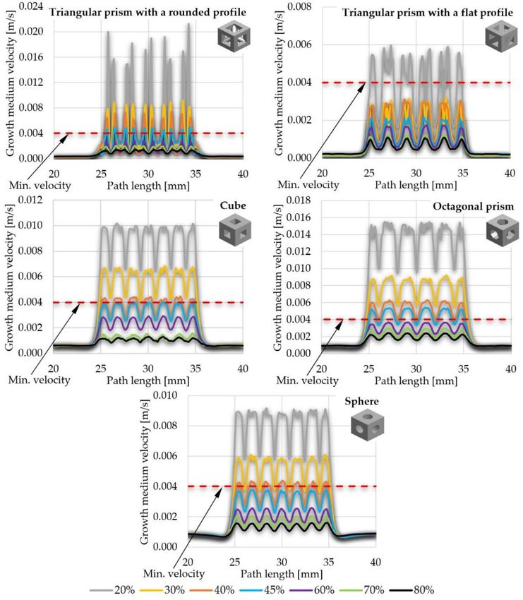
Materials 2021, 14, 109                                                                                             2 of 17

                          and further treatment in methacrylic anhydride. The first variant was an isotropic model
                          with a nearly constant pore size of 412 ± 13 µm and a porosity of 62 ± 1%, whereas the
                          second one had a variable pore size in the range of 250–500 µm and porosity of 35% to
                          85%. Computational fluid dynamics (CFD) models presented uniform distribution of flow
                          velocity and wall shear stress (WSS) for isotropic architecture and variable flow velocity
                          and wall shear stress for the second analyzed structure. The highest cell density was
                          correlated with the scaffold areas where pores were large and fluid velocity as well as wall
                          shear stresses were highest. This means that cell deposition depends mainly on the local
                          stress values, which are dependent on the pore size and type [12].
                                Zhao et al. used CFD to examine the functionality of scaffolds with highly irregular
                          pore geometry, created by micro-computed tomography (micro-CT) scanning of a silk
                          fibroin (SF) scaffold [13]. The simulation was carried out in a simulated channel of a
                          perfusion bioreactor, where the analyzed scaffold geometry was placed. By assigning
                          appropriate material properties and boundary conditions, they determined the value of the
                          wall shear stress and the velocity of the growth medium inside the scaffold. The stresses
                          obtained on the walls of the scaffolds ranged from 0 mPa to 50 mPa, whereas for velocity
                          the values were 0 mm/s to 2 mm/s [13].
                                Byrne et al. presented a fully three-dimensional approach to the computer simulation
                          of tissue differentiation and bone regeneration in a scaffold with regular pore geometry
                          as a function of porosity, Young’s modulus, and material dissolution rate (without consid-
                          ering any specific material type) under low and high load conditions [14]. They used a
                          mechanoregulation algorithm, which allowed the evaluation of the tissue differentiation
                          process both in terms of the dominant biophysical stimulus and the number of precursor
                          cells. Their research showed that initially, osteogenic cells were practically absent in the
                          granulation tissue filling the pores of the scaffold. As the simulation progressed, osteogenic
                          cells proliferated to form small clusters that eventually differentiated according to the
                          intensity of the mechanical stimulus, in the end creating a tissue with new material proper-
                          ties. The applied load produced a stimulus that promoted osteogenesis, leading to bone
                          formation in the center of the scaffold and soft tissue on its external sides, where high stress
                          concentrations occurred. Their simulations confirmed that all three analyzed variables had
                          an effect on the amount of bone formed, but not in an intuitive way. In a low-load environ-
                          ment, higher porosity and higher stiffness, as well as the average dissolution rate, give the
                          greatest amount of bone, whereas in an environment with high load, the dissolution rate
                          should be lower, otherwise the scaffold would collapse. However, at lower porosity, higher
                          dissolution rates can be used [14].
                                In general, studies can be divided into two main groups: studies with the use of
                          experimental or numerical methods. All of them present the importance of scaffolds not
                          only in orthopedic regenerative medicine to repair long bone defects [15,16], but also to
                          restore osteochondral [17,18], maxillofacial [19,20], or even spinal [21] damage. Moreover,
                          a properly designed scaffold can also be a drug delivery structure [22].
                                Despite the existence of a significant amount of research presenting comprehensive
                          observations, it is still impossible to clearly define the optimal parameters of scaffolds
                          designated for orthopedic regenerative medicine. This suggests that there is a strong
                          need for further research to understand as many factors as possible that have an impact
                          on their functionality. For this reason, the aim of the study was to fill the current gap
                          with the analyses of the influence of porosity and pore geometry on the functionality of
                          selected scaffolds.

                          2. Materials and Methods
                               Numerical studies were conducted within modules of ANSYS Workbench v19.2 soft-
                          ware: fluid flow (Fluent), which is a module for CFD calculations, and transient structural,
                          which allows for the evaluation of the process of osteogenic cell diffusion in the granulation
                          tissue formed at the fracture site. Computer Aided Design (CAD) models of analyzed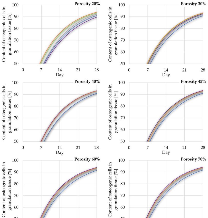
Materials
Materials2021, 14,14,x xFOR
2021, FORPEER
PEERREVIEW
REVIEW 3 of 319of 19
Materials 2021, 14, 109 3 of 17
2.2.Materials
Materials and Methods
Methods
Numerical studies
Numerical studieswerewereconducted
conductedwithinwithinmodules
modules ofof
ANSYS
ANSYS Workbench
Workbench v19.2 soft-
v19.2 soft-
ware: fluid
ware: fluid flow (Fluent),
(Fluent),which
whichisisaamodule
modulefor forCFD
CFD calculations,
calculations, andandtransient structural,
transient structural,
which
which allows
scaffolds
allows for
for the
were the evaluation
designed
evaluation ofofthe
theprocess
in SolidWorks process ofofosteogenic
2019 software.
osteogenic cell diffusion
Scaffold
cell diffusion in the
models granula-
in the were created with
granula-
tion
tion tissue
thetissue formed at
consideration
formed at the fracture
of two
the site.
site.Computer
variables:
fracture Aided
pore geometry
Computer Aided Design
and(CAD)
Design models
porosity.
(CAD) models of analyzed
of analyzed
scaffolds
scaffolds werefirst
In were
the designed
designed in
variable,inSolidWorks
SolidWorks 2019
2019software.
i.e., the geometry software. Scaffold
of pores, Scaffold
fivemodels
models
shapes were
were
werecreated with
created
selected:with
a triangular
the
the consideration
consideration of two variables:
of two variables: pore
pore geometry
geometry and porosity.
and porosity.
prism with a rounded (Figure 1a) and a flat profile (Figure 1b), a cube (Figure 1c), an octag-
In the
In the first
first variable,
variable, i.e., the
thegeometry
i.e.,and geometry ofofpores,
pores, five
fiveshapes
shapes werewereselected: a triangular
selected: a triangular
onal
prism prism
with (Figure
a rounded
rounded 1d),
(Figure 1a) aand
sphere
aaflat (Figure
profile 1e).
(Figure The
1b), pore
a cube geometries
(Figure used
1c),1c),
an oc- in conducted
prism with
analyses a
were (Figure
selected 1a)
on the and flat
basis(Figure profile
the data (Figure 1b), a cube (Figure an oc-
tagonal prism (Figure 1d), and a sphere 1e).ofThe
thepore
reference
geometriespresenting
used in con-commonly used
tagonal prism (Figure 1d), and a sphere (Figure 1e). The pore geometries used in con-
and evaluated
ducted analyses were shapes
selectedof scaffolds
on the basis[23,24].
the dataItofwas decidedpresenting
the reference to consider simple geometries
commonly
ducted analyses were selected on the basis the data of the reference presenting commonly
that and
used in practice
evaluatedcan be used
shapes to create
of scaffolds nearly
[23,24]. all decided
It was more complex
to consider shapes.
simpleMoreover,
geome- the use of
used and evaluated shapes of scaffolds [23,24]. It was decided to consider simple geome-
tries
pores that in practice
with defined can be used toallowed
geometry create nearly
for aall more complex
reduction in theshapes. Moreover,
influence of the
notches, randomly
tries that in practice can be used to create nearly all more complex shapes. Moreover, the
use of pores
created with with
thedefined
use ofgeometry
complex,allowed randomizedfor a reduction
geometries.in the influence of notches,
use of pores
randomly with
created defined
with geometry
the use allowed
of complex, for a reduction
randomized in the influence of notches,
geometries.
randomly created with the use of complex, randomized geometries.
(a) (b) (c) (d) (e)
Figure(a)
1. 1.
Analyzed (b)
geometries of pores (c) (d) (e)
Figure Analyzed geometries ofonpores
the example
on theofexample
a unit cell:
of(a)
a triangular
unit cell: prism with a
(a) triangular prism with a
rounded profile, (b) triangular prism with a flat profile, (c) cube, (d) octagonal prism, and (e)
Figure 1. Analyzed
rounded profile, geometries
(b) of pores
triangular prismonwith
the example
a flat of a unit
profile, (c)cell:
cube,(a) (d)
triangular prism
octagonal with aand (e) sphere.
prism,
sphere.
rounded profile, (b) triangular prism with a flat profile, (c) cube, (d) octagonal prism, and (e)
sphere. The seconds variable, i.e., porosity, was obtained by changing the size
The seconds variable, i.e., porosity, was obtained by changing the size of the pores, of the pores,
keeping
keeping their their number
number constant
constant in theinscaffold
the scaffold
volume. volume.
Despite Despite
the changestheinchanges
the geome- in the geometry
The seconds variable, i.e., porosity, was obtained by changing the size of the pores,
of of
try thethepore,
pore, the proportion
the proportion of the
of the material
material to empty to empty
space wasspace wasfor
the same theallsame for all analyzed
analyzed
keeping their number constant in the scaffold volume. Despite the changes in the geome-
structures
structures in in case
case of selected
of selected porosities.
porosities. Seven typesSeven of types ofwere
porosity porosity
chosen:were20%,chosen:
30%, 20%, 30%,
try of the pore, the proportion of the material to empty space was the same for all analyzed
40%,
40%, 45%,
45%, 60%,
60%,70%, and and
70%, 80%, 80%,
whichwhich
so far have
so been
far have used in experimental
been used in conditions conditions
experimental
structures in case of selected porosities. Seven types of porosity were chosen: 20%, 30%,
and
andcomputational
computational simulations
simulations[25,26]. In regenerative
[25,26]. medicine,medicine,
In regenerative porosities higher than higher than
porosities
40%,
40% are45%, 60%,
often 70%, and
considered. 80%, which
However, with sothe
farincrease
have been used in mechanical
in porosity, experimental conditions
properties
40% are often considered.
and However, In with the increase in porosity, mechanical thanproperties
of a computational
scaffold decrease. simulations [25,26].
For this reason, lower regenerative
porosities must medicine, porosities
be also evaluated inhigher
order to
of
40% a scaffold
are often
estimate
decrease.
considered. of
the functionality
For this
However, reason,
scaffoldswiththat the
lower
canincrease
porosities
provide in
must
porosity,
better support
be also
mechanical
in terms of
evaluated
properties
high
in order to
ofestimate
a scaffold the functionality
decrease. For this of scaffolds
reason, lower that can
porositiesprovide
must better
be also
load transfer that occurs in long bones. The consideration of porosities used in both prac-support
evaluated ininterms
order of
to high load
estimate
transfer the functionality
that occurs in of
longscaffolds
bones. that
The can provide
consideration better
of support
porosities
tical and numerical cases was crucial, as the research goal was to estimate the efficiency in terms
used of
in high
both practical
load
and
of transfer
most numericalthat occurs in
casesinwas
variables used long bones.
crucial,
current The
as the
studies. consideration
Theresearch
influence goal of porosities used
was toonestimate
of porosity the shapethein both prac-
of aefficiency
cell of most
tical
unit, and
variables numerical
on the example
used incases
of wasstudies.
crucial,
a triangular
current prism as
Thetheinfluence
with research
a rounded goal was to
ofprofile,
porosity estimate
is shown
on the the efficiency
in shape
Figure 2. a cell unit, on
of
ofthe
most variables
example of used in current
a triangular studies.
prism withThea influence
roundedof porosity
profile, on the shape
is shown of a cell
in Figure 2.
unit, on the example of a triangular prism with a rounded profile, is shown in Figure 2.
(a) (b) (c) (d) (e) (f) (g)
(a) (b) (c) (d) (e) (f) (g)
Figure 2. Analyzed porosities of scaffolds on the example of a selected unit cell (triangular prism
with a rounded profile): (a) 20%, (b) 30%, (c) 40%, (d) 45%, (e) 60%, (f) 70%, and (g) 80%.
2.1. Growth Medium Velocity and Wall Shear Stress inside Scaffolds
In the first part of the study the process of a cell culture within a scaffold placed in
a perfusion bioreactor was evaluated in order to estimate growth medium velocity and
wall shear stress inside scaffolds. It was simulated by modelling a bioreactor channel
(60 mm length and 10 mm diameter) where the cylindrical scaffolds, created from previ-
ously described cell units, with a height of 10 mm and diameter of 10 mm, were placed in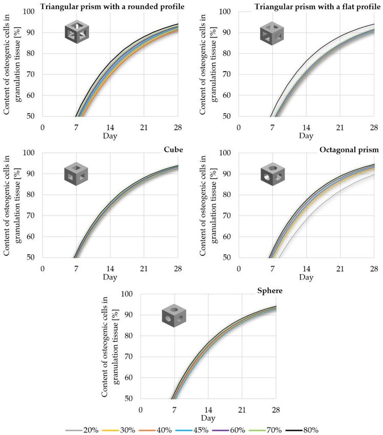
Materials 2021, 14, x FOR PEER REVIEW 4 of 19
Figure 2. Analyzed porosities of scaffolds on the example of a selected unit cell (triangular prism
with a rounded profile): (a) 20%, (b) 30%, (c) 40%, (d) 45%, (e) 60%, (f) 70%, and (g) 80%.
Figure 2. Analyzed porosities of scaffolds on the example of a selected unit cell (triangular prism
with a rounded profile): (a) 20%, (b) 30%, (c) 40%, (d) 45%, (e) 60%, (f) 70%, and (g) 80%.
2.1. Growth Medium Velocity and Wall Shear Stress inside Scaffolds
Materials 2021, 14, 109 In the first part of the study the process of a cell culture within a scaffold placed in a 4 of 17
2.1. Growth Medium Velocity and Wall Shear Stress inside Scaffolds
perfusion bioreactor was evaluated in order to estimate growth medium velocity and wall
In the first part of the study the process of a cell culture within a scaffold placed in a
shear stress inside scaffolds. It was simulated by modelling a bioreactor channel (60 mm
perfusion bioreactor was evaluated in order to estimate growth medium velocity and wall
length and 10 mm diameter) where the cylindrical scaffolds, created from previously de-
shear
the stress inside
middle scaffolds.
of it.with
A total ofIt35
was simulated by modelling a made
bioreactor channel (60 mm of a flow of
scribed cell units, a height ofscaffold
10 mm andstructures
diameter were
of 10 mm, and
weretested
placedininterms
the
length and 10 mm diameter) where the cylindrical scaffolds, created from previously de-
growth
middle of medium
it. A total through the structures
of 35 scaffold scaffold. were
Figuremade3 presents
and tested aninexample
terms of aof evaluated
flow of scaffold
scribed cell units, with a height of 10 mm and diameter of 10 mm, were placed in the
growth medium
geometries through
with a the scaffold.
porosity of 45%Figure
and 3all
presents
shapes an tested.
example of evaluated scaffold
middle of it. A total of 35 scaffold structures were made and tested in terms of a flow of
geometries with a porosity of 45% and all shapes tested.
growth medium through the scaffold. Figure 3 presents an example of evaluated scaffold
geometries with a porosity of 45% and all shapes tested.
(a) (b) (c) (d) (e)
Figure
Figure 3.
(a)Exemplary
3. Exemplary CAD models
(b)
CAD prepared
models (c)for analyses
prepared of (d)
growth of
for analyses medium
growth velocity
(e)medium and wall
velocity and wall shear
shear stress inside scaffolds: (a) triangular prism with a rounded profile, (b) triangular prism with
astress
Figure inside
3. (c) scaffolds:
Exemplary
flat profile, cube, CAD (a) triangular
models
(d) octagonal prepared prism
prism, andfor(e) with of
analyses
sphere. a growth
rounded profile,
medium (b) triangular
velocity and wall prism with a flat
shear stress
profile, (c) inside
cube,scaffolds: (a) triangular
(d) octagonal prism,prism
and (e)withsphere.
a rounded profile, (b) triangular prism with
a flat profile, (c) cube, (d) octagonal prism, and (e) sphere.
Channel models were adjusted by subtracting the scaffold geometry from the vol-
Channel models
ume. Subsequently, meshwere adjusted
was created withbythe
subtracting the finite
use of 4-node scaffold geometry
elements and a from
5% the volume.
Channel models were adjusted by subtracting the scaffold geometry from the vol-
convergence
Subsequently, test for both growth medium velocity and WSS. The number of finite
mesh was created with the use of 4-node finite elements and a 5% conver- ele-
ume. Subsequently, mesh was created with the use of 4-node finite elements and a 5%
ments
gence intest
the for
models was approximately 1,300,000 ± 200,000. Figure 4 number
presents an example
convergence testboth growth
for both medium
growth medium velocity
velocityand
andWSS.
WSS. The
The number of offinite
finiteele-
elements in the
of a model obtained after discretization.
models wasmodels
ments in the approximately 1,300,000
was approximately ± 200,000.
1,300,000 Figure
± 200,000. Figure4 4presents anexample
presents an example of a model
obtained
of a model after
obtaineddiscretization.
after discretization.
Figure 4. Finite element model obtained after discretization with the consideration of a 5% mesh
refinement test (example on a scaffold with cubic pores and a porosity of 45%).
Figure 4.4.Finite
Figure element
Finite model
element obtained
model after discretization
obtained with the consideration
after discretization of a 5% mesh
with the consideration of a 5% mesh
refinement test (example on a scaffold with cubic pores and a porosity of 45%).
Appropriate
refinement parameters
test (example on were set during
a scaffold the research
with cubic in order
pores and to simulate
a porosity the cell
of 45%).
culture process in the channel of the perfusion bioreactor. The density and dynamic vis-
Appropriate parameters were set during the research in order to simulate the cell
cosity Appropriate
of the growth medium as well as its
setinitial velocity and temperature were toused
culture process in theparameters
channel of thewere
perfusion during theThe
bioreactor. research
density in order
and dynamic simulate
vis- the cell
(Table 1). A non-slip feature was assumed for the scaffold wall. For this reason, the con-
culture
cosity ofprocess in the
the growth channel
medium of the
as well asperfusion bioreactor.
its initial velocity and The densitywere
temperature and dynamic
used viscosity
of the 1).
(Table growth medium
A non-slip featureaswas
well as its initial
assumed velocitywall.
for the scaffold andFor
temperature were
this reason, the used (Table 1).
con-
A non-slip feature was assumed for the scaffold wall. For this reason, the consideration of
scaffold material was not required as the analyzed results were not dependent upon the
scaffold’s material properties. This assumption is widely used in the analyses of fluid flow
through scaffolds in a perfusion bioreactor [27].
Table 1. Values of parameters used in the study to evaluate the growth medium velocity and wall
shear stress inside the pores of analyzed scaffolds.
Parameter Value Reference
Density [kg/m3 ] 1040 [28]
Dynamic viscosity [Pa·s] 0.00081 [29]
Initial velocity [m/s] 0.0005 [29]
Temperature [K] 310.15 [29]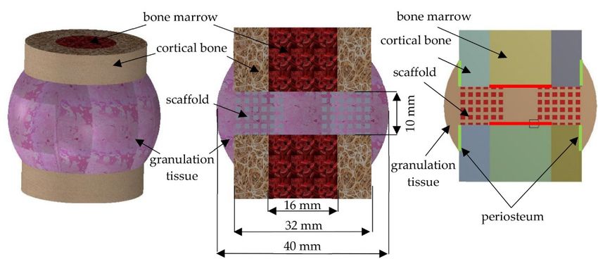
shear stress inside the pores of analyzed scaffolds.
Parameter Value Reference
Density [kg/m3] 1040 [28]
Dynamic viscosity [Pa·s]0.00081 [29]
Materials 2021, 14, 109 5 of 17
Initial velocity [m/s] 0.0005 [29]
Temperature [K] 310.15 [29]
As stated before, the velocity of the growth medium and WSS were calculated during
As stated before, the velocity of the growth medium and WSS were calculated during
simulations with the use of a double precision, pressure-based solver. It was proven that
simulations with the use of a double precision, pressure-based solver. It was proven
low velocities can disturb the appropriate washing away of metabolic waste or even pre-
that low velocities can disturb the appropriate washing away of metabolic waste or even
vent osteogenic differentiation [29–32]. At the same time, a too-high velocity can lead to
prevent osteogenic differentiation [29–32]. At the same time, a too-high velocity can lead to
apoptosis of cells [29,30,33]. Achieving appropriate velocity can enhance the deposition of
apoptosis of cells [29,30,33]. Achieving appropriate velocity can enhance the deposition
the mineralized matrix, which positively affects the cell culture process [34–37]. In the case
of the mineralized matrix, which positively affects the cell culture process [34–37]. In the
of WSS,
case it stimulates
of WSS, the growth
it stimulates of cells;
the growth however,
of cells; if it isiftoo
however, it ishigh, it canitdetach
too high, cells from
can detach cells
the scaffold’s
from internal
the scaffold’s wallswalls
internal [13,28]. For this
[13,28]. For reason, the considered
this reason, the considered obtained values
obtained of the
values of
analyzed
the analyzed parameters
parameters create an upper
create an upper andand
lower threshold
lower threshold thatthat
cancan
be used
be usedto evaluate the
to evaluate
functionality
the functionality of any
of anyscaffold. The
scaffold. Theupper
upper limit
limitisisthe
themaximum
maximumallowableallowable value
value of WSS,
of WSS,
whereas the lower limit is the minimal velocity of the
whereas the lower limit is the minimal velocity of the growth medium. growth medium.
Althoughthe
Although themaximal
maximalvalues
valuesofofWSS
WSS were
were taken
taken fromfrom all all internal
internal walls
walls of scaffold,
of the the scaf-
fold,velocity
the the velocity
results results
were were
takentaken
from from the path
the path passingpassing
through through the pores
the pores exactly
exactly in thein
the center of the scaffolds (Figure 5). An impermeable wall was
center of the scaffolds (Figure 5). An impermeable wall was modeled to include the area ofmodeled to include the
area of growth
growth mediummedium flow limited
flow limited by the
by the inner inner diameter
diameter of the bioreactor
of the bioreactor channel.channel.
No slip Noas
slip as the shear condition as well as no wall motion
the shear condition as well as no wall motion were considered. were considered.
Figure 5. Scheme of the
Figure 5. the analyzed
analyzed models
modelswith
withboundary
boundaryconditions
conditionsand
anda apath
pathfor
foranalyzing
analyzing the
the
growth medium
growth medium velocity.
velocity.
2.2.
2.2. Diffusion
Diffusion of
of Osteogenic
Osteogenic Cells
Cells to
to Granulation
Granulation Tissue
Tissue inside
inside Scaffolds
Scaffolds
In
In the second part of the study, a case of the direct usea scaffold
the second part of the study, a case of the direct use of to restore
of a scaffold long bone
to restore long
function, without an initial cell culturing process in the perfusion bioreactor,
bone function, without an initial cell culturing process in the perfusion bioreactor, was was analyzed.
Although
analyzed. the use of athe
Although bioreactor
use of acan hasten restoration
bioreactor can hastenpossibilities, a clean scaffold
restoration possibilities, can
a clean
be used immediately after its manufacturing, which can shorten hospitalization.
scaffold can be used immediately after its manufacturing, which can shorten hospitaliza- For this
reason,
tion. Forboth
thismethods are commonly
reason, both methods are used in clinical
commonly practice.
used in clinical practice.
In this part, a simulation of the diffusion of osteogenic cells in the granulation tissue
In this part, a simulation of the diffusion of osteogenic cells in the granulation tissue
occurred between bone fragments at the site of the scaffold placement. Determination of
occurred between bone fragments at the site of the scaffold placement. Determination of
the diffusion efficiency allowed for the estimation of when the scaffold could be subjected
the diffusion efficiency allowed for the estimation of when the scaffold could be subjected
to loading. For the research purposes, scaffolds with an outer diameter of 32 mm, an inner
to loading. For the research purposes, scaffolds with an outer diameter of 32 mm, an inner
diameter of 10 mm, and a height of 10 mm were modeled. The outside diameter was
diameter of 10 mm, and a height of 10 mm were modeled. The outside diameter was in-
intended to simulate the outside diameter of the long bone diaphysis, in this case the
tended to simulate the outside diameter of the long bone diaphysis, in this case the femur,
femur, whereas the inner diameter simulated the medullary cavity, a consideration that
also allowed for the facilitation of the propagation of osteogenic cells into the pores of the
scaffold. Besides the enhancement of the osteogenic cell diffusion process, the inner hole
must be considered to improve further load transfer after obtaining secondary osteointe-
gration (similar to the one of natural bone). As in the previous part of the presented study,
35 scaffold models were again prepared. Figure 6 presents an example of the prepared
models of all analyzed shapes with a porosity of 45%.
The scaffold is placed between bone fragments to restore continuity of the bone
and its basic function of transferring loads [38]. For this reason, in order to simulate the
bone tissue environment and its healing process, all analyzed scaffolds were placed in
a simplified bone model along with a bone marrow and a granulation tissue (Figure 7).
The anatomical dimensions of the femur diaphysis and the resulting granulation tissue
were used to create the model [39]. Due to computational complicity, three-dimensional
models were simplified to two-dimensional ones, created by a cross-section through the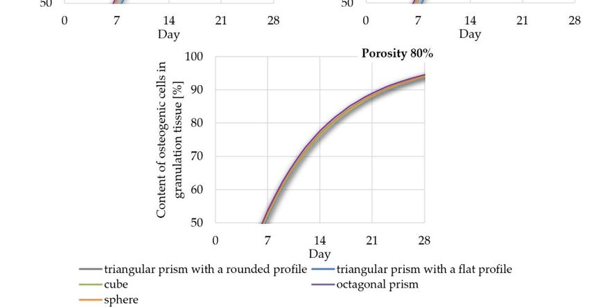
els of all analyzed shapes with a porosity of 45%.
Materials 2021, 14, x FOR PEER REVIEW 6 of 19
Materials 2021, 14, 109 6 of 17
whereas the inner diameter simulated the medullary cavity, a consideration that also al-
lowed for the facilitation of the propagation of osteogenic cells into the pores of the scaf-
center
fold. of the
Besides themodel.
enhancementPeriosteum and bone
of the osteogenic cell marrow were considered
diffusion process, as must
the inner hole the source of
osteogenic cells. The models’ simplification was possible as the considered
be considered to improve further load transfer after obtaining secondary osteointegration bone-implant
geometries
(similar to thewere
one characterized byAs
of natural bone). appropriate symmetries.
in the previous This,
part of the as wellstudy,
presented as the35
previously
scaffold models were again prepared. Figure 6 presents an example of the prepared mod- done in
described features, allowed for an analysis of the two-dimensional models, as was
related
els research shapes
of all analyzed [40–42].with a porosity of 45%.
(a) (b) (c) (d) (e)
Figure 6. Exemplary CAD models prepared for analyses of the diffusion of osteogenic cells to
granulation tissue inside scaffolds: (a) triangular prism with a rounded profile, (b) triangular
prism with a flat profile, (c) cube, (d) octagonal prism, and (e) sphere.
The scaffold is placed between bone fragments to restore continuity of the bone and
its basic function of transferring loads [38]. For this reason, in order to simulate the bone
tissue environment and its healing process, all analyzed scaffolds were placed in a sim-
plified bone model along with a bone marrow and a granulation tissue (Figure 7). The
anatomical dimensions of the femur diaphysis and the resulting granulation tissue were
used to create the model [39]. Due to computational complicity, three-dimensional models
were simplified to two-dimensional ones, created by a cross-section through the center of
the model. Periosteum and bone marrow were considered as the source of osteogenic
(a) (b) (c) (d) (e)
cells. The models’ simplification was possible as the considered bone-implant geometries
Figure
were
Figure 6. 6.
Exemplary
characterized
ExemplaryCAD
by models
appropriate
CAD prepared
models for analyses
symmetries.
prepared for of
This, thewell
as diffusion
analyses as
of theofpreviously
the osteogenic of
diffusion cells to
described
osteogenic cells to
granulation
features, tissue inside
allowed for an scaffolds:
analysis (a)
of triangular
the prism with a rounded
two-dimensional models, profile,
as was (b) triangular
done in related
granulation tissue inside scaffolds: (a) triangular prism with a rounded profile, (b) triangular prism
prism with
research a flat profile, (c) cube, (d) octagonal prism, and (e) sphere.
[40–42].
with a flat profile, (c) cube, (d) octagonal prism, and (e) sphere.
The scaffold is placed between bone fragments to restore continuity of the bone and
its basic function of transferring loads [38]. For this reason, in order to simulate the bone
tissue environment and its healing process, all analyzed scaffolds were placed in a sim-
plified bone model along with a bone marrow and a granulation tissue (Figure 7). The
anatomical dimensions of the femur diaphysis and the resulting granulation tissue were
used to create the model [39]. Due to computational complicity, three-dimensional models
were simplified to two-dimensional ones, created by a cross-section through the center of
the model. Periosteum and bone marrow were considered as the source of osteogenic
cells. The models’ simplification was possible as the considered bone-implant geometries
were characterized by appropriate symmetries. This, as well as the previously described
features, allowed for an analysis of the two-dimensional models, as was done in related
research [40–42].
(a) (b) (c)
Figure 7. Components of a model for diffusion studies: (a) isometric view of three-dimensional
model, (b) a cross-section through a three-dimensional model, and (c) two-dimensional model used
for analyses.
Mesh was created with the use of 8-node Plane223 elements and with a consideration
of a 5% convergence test for the analyzed results (content of osteogenic cells in granulation
tissue). The applied elements allowed the diffusive degree of freedom in the nodes to be
unlocked. Moreover, the node-sharing method was included to bond each part of tested
models, which consisted of approximately 35,000 ± 20,000 elements (Figure 8).
In simulations of the process of osteogenic cell diffusion, the Fick’s law was applied,
which is presented
(a) in the equation [43]:(b) (c)
J = −D × (δØ/δx), (1)
where J is the diffusion flux [kg/m2 × s], D is the diffusion coefficient [m2 /s], Ø is the
concentration [kg/m3 ], and x is the distance from the source of the diffusing substance [m].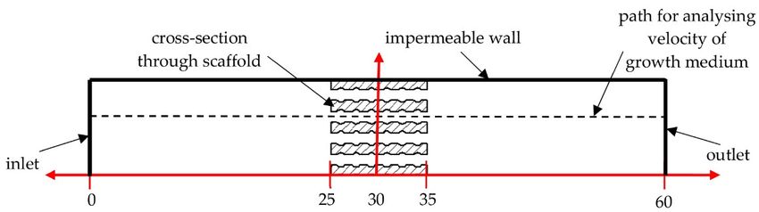
model, (b) a cross-section through a three-dimensional model, and (c) two-dimensional model
used for analyses.
Mesh was created with the use of 8-node Plane223 elements and with a consideration
of a 5% convergence test for the analyzed results (content of osteogenic cells in granulation
Materials 2021, 14, 109 tissue). The applied elements allowed the diffusive degree of freedom in the nodes to be 7 of 17
unlocked. Moreover, the node-sharing method was included to bond each part of tested
models, which consisted of approximately 35,000 ± 20,000 elements (Figure 8).
Figure 8. Model for analyses of osteogenic cell diffusion after discretization (example on a scaffold
Figure 8. Model for analyses of osteogenic cell diffusion after discretization (example on a scaffold
with cubic pores and a porosity of 45%).
with cubic pores and a porosity of 45%).
In simulations of the process of osteogenic cell diffusion, the Fick’s law was applied,
To include Fick’s law, a subroutine was created with the use of Ansys Parametric De-
which is presented in the equation [43]:
sign Language (APDL). A single iteration was considered as equal to one day of osteogenic
cell diffusion. This allowed the changes J = −D ×in(δØ/δx),
content of osteogenic cells in granulation (1) tissue
to be estimated
where over days
J is the diffusion of the2 healing
flux [kg/m × s], D isprocess. The coefficient
the diffusion adapted simulation
[m2/s], Ø ismethod
the con- of the dif-
fusion process
centration [kg/m3simplifies phenomena
], and x is the distance from occurring
the sourcein real conditions.
of the However,
diffusing substance the literature
[m].
To include
presents that itsFick’s
use law,
allowsa subroutine
the data thatwas approximates
created with thethe useactual
of Ansys Parametric
diffusion of osteogenic
Design Language
cells within (APDL).
the callus toAbesingle iteration
obtained was
[44]. Theconsidered
material as equal to one
properties useddayduring
of osteo-
analyses of
genic cell diffusion. This allowed the changes in content of osteogenic cells
this phenomenon are presented in Table 2. Titanium alloy (Ti6Al4V) was considered as the in granulation
tissue to bethe
material estimated
scaffoldsover daysmade
were of theofhealing process. The adapted
as its biocompatibility withsimulation method
appropriate mechanical
of the diffusion process simplifies phenomena occurring in real conditions. However, the
strength can fulfil the requirements of scaffolds designated for orthopedic regenerative
literature presents that its use allows the data that approximates the actual diffusion of
medicine, especially in terms of recovering long bone damage as presented in the data
osteogenic cells within the callus to be obtained [44]. The material properties used during
of the references [45,46]. Evaluated structures can be manufactured with currently know
analyses of this phenomenon are presented in Table 2. Titanium alloy (Ti6Al4V) was con-
support-free
sidered 3D-printing
as the material methods,
the scaffolds weresuch
madeasofselective laser sintering
as its biocompatibility with (SLS), which makes it
appropriate
possible to use them in experimental conditions [47,48].
mechanical strength can fulfil the requirements of scaffolds designated for orthopedic re-
generative medicine, especially in terms of recovering long bone damage as presented in
Table
the 2. Material
data properties[45,46].
of the references used inEvaluated
the study to evaluate the
structures can influence of pore geometry
be manufactured and scaffold
with cur-
rently know
porosity support-free
on osteogenic cell3D-printing
diffusion in methods,
granulation such as selective laser sintering (SLS),
tissue.
which makes it possible to use them in experimental conditions [47,48].
Young’s Diffusion
Tissue Type/ Density Poisson’s
Table 2. Material properties used in the study to evaluate the influence
3 Modulus
of pore geometry and Coefficient
scaffold porosity on osteo- Reference
Implant Type [kg/cm ] Ratio
genic cell diffusion in granulation tissue. [GPa] [m2 /Day]
Cortical bone 1740 0.30 20 Coefficient
Diffusion - [49]
Tissue Type/Implant Type Density [kg/cm3] Poisson’s Ratio Young’s Modulus [GPa] Reference
Bone marrow 1100 0.17 [m2/Day]
0.002 - [50]
Cortical bone 1740
Granulation tissue 0.30 910 20 0.17 0.0001- [49]
2.5 [50]
Bone marrow 1100
Scaffold (Ti6Al4V) 0.17 4500 0.002 0.33 110 - [50]
- [51]
Granulation tissue 910 0.17 0.0001 2.5 [50]
Scaffold (Ti6Al4V) 4500 0.33 110 - [51]
3. Results
3.1. The Influence of Pores Geometry and Scaffold Porosity on Growth Medium Velocity and Wall
Shear Stress inside Scaffolds
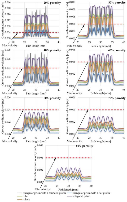
The results of the velocity of the growth medium through the scaffold pores are
presented in the form of comparative graphs. In order to improve the readability of the
results, various combinations were created, namely, a comparison of the efficiency of pore
geometry for selected scaffold porosities (Figure 9) and a comparison of the efficiency of
scaffold porosity for selected pore geometries (Figure 10). All graphs are shown for the
path length (Figure 5) in the range of x (20; 40). The value of the minimum velocity
of growth medium, which ensures the proper exchange of nutrients between it and the
cultured cells, was set to 0.004 m/s, which was determined on the basis of the data of the
reference [12]. Lower values suggest that the appropriate exchange of nutrients would be
disturbed and, at the same time, waste products could not be removed from the inside of
the scaffold. A minimal velocity of 0.004 m/s was a lower threshold for further evaluation
of the influence of selected parameters on the scaffold efficiency. To present the resultsMaterials 2021, 14, 109 8 of 17
more comprehensively, additional views of streamlines of the growth medium velocity
inside scaffolds are presented in supplementary materials (Figures S1–S5).
The upper threshold was set on the basis of the maximal allowable WSS value inside
a scaffold obtained during fluid flow through its pores. It was also determined based on
the literature of the reference and set equal to 100 mPa [52]. Higher values can cause call
detachment of scaffold walls, which disturbs appropriate cell growth. The results of the
obtained WSS are compressively presented in the form of a graph in Figure 11, comparing
maximal stresses obtained in the scaffolds of selected pore geometries and porosities. Due
to the significant number of obtained stress maps, they are presented in Figures
Materials 2021, 14, x FOR PEER REVIEW 9 of 19S6–S15 of
supplementary materials in order to not disrupt the flow of the main text.
Figure9.9. The
Figure The influence
influenceofofpore
poregeometry on the
geometry on growth medium
the growth velocityvelocity
medium inside a inside
scaffold.
a scaffold.Materials 2021, 14, 109 9 of 17
Materials 2021, 14, x FOR PEER REVIEW 10 of 19
Materials 2021, 14, x FOR PEER REVIEW 10 of 19
Figure
Figure10.10.
The influence
The of scaffold
influence porosity
of scaffold on the
porosity ongrowth medium
the growth velocity
medium insideinside
velocity a scaffold.
a scaffold.
Figure 10. The influence of scaffold porosity on the growth medium velocity inside a scaffold.
Figure
Figure11.11.
The influence
The of scaffold
influence porosity
of scaffold and the geometry
porosity of pores on of
and the geometry thepores
maximal
onWSS value
the maximal WSS value
generated inside scaffold: (a) triangular prism with a rounded profile, (b) triangular prism with a
Figure 11. inside
generated The influence of scaffold
scaffold: porosity
(a) triangular and the
prism geometry
with of pores
a rounded on the
profile, (b)maximal WSS
triangular valuewith a flat
prism
flat profile, (c) cube, (d) octagonal prism, and (e) sphere.
generated
profile, (c) inside
cube, scaffold: (a) triangular
(d) octagonal prism, prism with
and (e) a rounded profile, (b) triangular prism with a
sphere.
flat profile, (c) cube, (d) octagonal prism, and (e) sphere.
3.2. The Influence of Pore Geometry and Scaffold Porosity on the Diffusion of Osteogenic Cells into
the Scaffold Pores
For the sake of clarity, the data obtained was compiled in two comparisons. Figure 12
shows the influence of pore geometry on the percentage of osteogenic cells in granula-
tion tissue, whereas Figure 13 presents the influence of scaffold porosity on the sameMaterials 2021, 14, 109 10 of 17
phenomenon. Figure 12 shows the influence of the scaffold porosity on the studied phe-
nomenon, whereas Figure 13 shows the influence of pore shapes. The simulations were
carried out for 50 days after the start of the healing process, but to increase the readability
of the results, it was decided to present the obtained data for 28 days, because at that time,
for most scaffolds, the cell content was already over 90%. Moreover, the graphs present
changes in the percentage of osteogenic cells in a range from 50% to 100%, as under 50%
there were no noticeable differences between the results. To increase the readability of the
results, an additional figure was included (Figure 14) that presents the time needed to fulfil
the scaffold with osteogenic cells at 90%. The healing process was analyzed only in terms
of osteogenic cell diffusion. In further research, tissue differentiation within pores can also
be included by the use of appropriate mechanoregulation principles and methods. This is
especially important in the evaluation of long-term efficiency and secondary osteointe-
gration of orthopedic scaffolds [40]. Due to the significant number of maps showing the
process of the propagation of osteogenic cells in granulation tissue, they are presented in
the Figures S16–S20 of supplementary materials, in order to not disrupt the flow of the
Materials 2021, 14, x FOR PEER REVIEW 12 of 19
main text.
Figure 12. The influence of scaffold geometry on the content of osteogenic cells in granulation tis-
Figure 12.
sue within The
28 of influence
the healing of scaffold geometry on the content of osteogenic cells in granulation tissue
process.
within 28 of the healing process.Materials 2021, 14, 109 11 of 17
Materials2021,
Materials 2021,14,
14,x xFOR
FORPEER
PEERREVIEW
REVIEW 1313ofof1919
Figure13.
Figure
Figure 13.The
13. Theinfluence
The influenceof
influence ofofscaffold
scaffoldporosity
scaffold porosityon
porosity onthe
on thecontent
the contentof
content ofofosteogenic
osteogeniccells
osteogenic cellsin
cells iningranulation
granulationtissue
granulation tissue
tissue
within
within 28
within 28
28 of of the
of the healing
the healing process.
healing process.
process.
Figure 14.Time
Figure Time neededtotofulfilfulfil the scaffold withosteogenic
osteogeniccells
cellsatat90%:
90%:(a)
(a)triangular
triangularprism
prismwith
with
Figure14.
14. Time needed
needed to fulfil the scaffold with osteogenic cells at 90%: (a) triangular prism with a
a rounded
arounded profile,
roundedprofile,
profile,(b) (b) triangular
(b)triangular
triangular prism
prism with
with a flat
a flat profile,
profile, (c) cube, (d) octagonal prism, and (e)
prism with a flat profile, (c)(c) cube,
cube, (d)(d) octagonal
octagonal prism,
prism, andand
(e) (e)
sphere.
sphere.
sphere.Materials 2021, 14, 109 12 of 17
4. Discussion
4.1. Efficiency of Pore Geometry and Scaffold Porosity in Terms of Cell Culture Process Conducted
in Perfusion Bioreactor
While conducting the overall analyses of all the obtained results, it could be noted
that growth medium flowed steadily with a velocity of approximately 0.005 m/s (0–25 mm
of a path) before reaching the location of a scaffold (25–35 mm of a path). Then the fluid
was pressed into pores of the analyzed structures, which caused a sudden increase of
its velocity, which dropped fast when reaching internal part of the pore. This process
repeated while pressing the fluid from pore to pore until the end of the scaffold, where the
velocity reached again the initial value of approximately 0.005 m/s (35–60 mm of a path).
The same phenomenon could be noted for all analyzed structures and pore geometries,
but at a different intensity. Its highest intensity was achieved with the use of a triangular
prism with a rounded profile as a pore geometry, whereas the lowest was in the case of
spherical pores.
There was also another noticeable dependency—the smaller the porosity, the higher
the fluid velocity values achieved inside the scaffolds. Moreover, with the increase of poros-
ity, the differences between the maximal and minimal growth medium velocity values
obtained inside pores became smaller. The highest flow fluctuations were obtained in the
scaffolds with triangular prisms, while other analyzed structural fluctuations were compa-
rable. On this basis it could be concluded that after achieving a certain level of porosity
(described in detail in the following bulleted list) that was different for the analyzed ge-
ometries, appropriate nutrient exchange between cell cultures inside a scaffold and growth
medium may be disturbed. Moreover, at the same time, sufficient gas exchange would
also be handicapped. In the end, this could result in creating a non-uniform distribution of
developing cells, as mentioned in the data of the reference [12,53,54]. Scaffolds that did not
meet the first considered criterium (min. velocity of growth medium inside pores) were:
• Scaffolds with a pore geometry of a triangular prism with a rounded profile and a
porosity equal to or higher than 60%;
• Scaffolds with a pore geometry of a triangular prism with a flat profile and a porosity
equal to or higher than 30%;
• Scaffolds with a pore geometry of a cube and a porosity equal to or higher than 45%;
• Scaffolds with a pore geometry of an octagonal prism and a porosity equal to or higher
than 60%; and
• Scaffolds with a pore geometry of a sphere and a porosity equal to or higher than 45%.
In some cases, the growth medium minimal velocity was not maintained during the
flow through a scaffold. In such cases, there were noticeable areas where the growth
medium velocity was lower than the required. This velocity vastly increased when fluid
was being pressed to the next pore and decreased right after. This may also suggest that in
those cases, metabolic waste would not be appropriately removed from the scaffold and
the cell culture process would be disturbed [53,54]. From the analyzed structures, only a
few were distinguished that fulfilled this requirement:
• Scaffolds with a pore geometry of a cube and a porosity equal to or lower than 30%;
• Scaffolds with a pore geometry of an octagonal prism and a porosity equal to or lower
than 30%; and
• Scaffolds with a pore geometry of a sphere and a porosity equal to 20%.
In all the presented models, the highest stresses were obtained in the center of the
implants, and the lowest on the outer walls (for detailed insight see Figures S6–S15 in
supplementary materials). What is more, the higher the porosity, the lower WSS values
obtained. This phenomenon must be taken into consideration while designing the cell
culture process individually for the considered internal geometry of a scaffold, as a too-high
WSS leads to cell detachment. In extreme cases, WSS can even lead to cell death [55,56].
However, appropriate intensity and distribution of WSS must be maintained, as it stim-Materials 2021, 14, 109 13 of 17
ulates cells to produce extracellular matrix [56]. Scaffolds that did not meet the second
considered criterium (max. WSS) were:
• Scaffolds with a pore geometry of a triangular prism with a rounded profile and a
porosity equal to or lower than 40%;
• Scaffolds with a pore geometry of a triangular prism with a flat profile and a porosity
equal to or lower than 30%;
• Scaffolds with a pore geometry of a cube and a porosity equal to 20%; and
• Scaffolds with a pore geometry of an octagonal prism and a porosity equal to 20%.
All the analyzed types of scaffold with spherical pores and every evaluated porosity
fulfilled the second criterium.
Considering both criteria (min. velocity of growth medium inside pores and max.
WSS), which could ensure appropriate cell culture process in orthopedic scaffolds inside
a perfusion bioreactor (in terms of allowing for appropriate nutrient exchange and cell
stimulation), there were single structures that met the assumed requirements, such as:
• Scaffolds with a pore geometry of a cube and a porosity equal to 30%;
• Scaffolds with a pore geometry of an octagonal prism and a porosity equal to 30%;
and
• Scaffolds with a pore geometry of a sphere and a porosity equal to 20%.
All findings are summarized in Table 3, which presents the criteria that were fulfilled
or failed for each analyzed scaffold geometry and porosity.
Table 3. Summary of the efficiency of pore geometry and scaffold porosity in terms of the selected criteria.
Scaffold Geometry
Scaffold Feature
(a) (b) (c) (d) (e)
− − + + +
20%
− − − − +
− − + + −
30%
− − + + +
− − − − −
40%
− + + + +
Scaffold Porosity
− − − − −
45%
+ + + + +
− − − − −
60%
+ + + + +
− − − − −
70%
+ + + + +
− − − − −
80%
+ + + + +
Min. Velocity Criterium Max. WSS Criterium
+ −
+ Both Criteria Fulfilled − Both Criteria Failed
− +
Upper Criteria Failed/Lower Fulfilled Upper Criteria Fulfilled/Lower Failed
+ −
Note: (a) triangular prism with a rounded profile, (b) triangular prism with a flat profile, (c) cube, (d)
octagonal prism, and (e) sphere.Materials 2021, 14, 109 14 of 17
4.2. Efficiency of Pore Geometry and Scaffold Porosity in Terms of Osteogenic Cell Diffusion after
Implantation of a Scaffold
The obtained data suggest that osteogenic cells nearly completely fill granulation
tissue within scaffolds in 28 days after initiating the healing process. This is especially
important, as they are required for further bridging of bone segments. What is more,
appropriate bone healing occurs only when the appropriate content of osteogenic cells is
obtained within the callus, as presented by Lacroix et al. [41]. The diffusion starts from
the sources of the cells, that is, bone marrow and periosteum (for detailed insight see
Figures S16–S20 in supplementary materials). However, the intensity of this phenomenon
can be influenced by the analyzed factors of pore geometry and scaffold porosity. Moreover,
the higher the porosity, the faster the diffusion process occurs. This phenomenon can also
be observed in the data of the reference, which suggest the correctness of the obtained
results [39].
Pore geometry had a noticeable impact in the case of lower porosities but only for
three out of five analyzed cases. In the case of cubical and spherical pores, the osteogenic
cell diffusion efficiency was similar for all considered scaffold porosities. Spherical pore
geometry allowed for the highest content of osteogenic cells in granulation tissue to be
obtained for porosities equal to or lower than 30%. The pores of the octagonal prism had
the lowest efficiency in the porosity case of 20%, but with a porosity equal to or higher than
40%. This pore geometry allowed the highest content of osteogenic cells to be obtained.
The differences in efficiency between pore geometries were the highest with the use of a
porosity of 20% and decreased with the increase of porosity up to 80%, where no significant
differences were noted. The lowest efficiency for most of the analyzed porosities (30–70%)
had the pores of a triangular prism with a flat profile.
5. Conclusions
The first part of the study allowed for the estimation of growth medium flow through
scaffolds of different porosities and pore geometry, whereas the second part approximated
the osteogenic cell diffusion process into granulation tissue within scaffolds. The presented
comprehensive results allowed for the appropriate selection of scaffold properties to obtain
the highest possible functionality in terms of its use in orthopedic regenerative medicine.
All of the presented scaffolds can be manufactured with the currently known support-free
3D-printing methods, such as selective laser sintering (SLS), which makes it possible to
use them in experimental conditions. The data presented can also be a starting point for
further analyses in order to increase the quality of the analyzed structures and in the end
to increase the efficiency of regenerative medicine.
Supplementary Materials: The following are available online at https://www.mdpi.com/1996-194
4/14/1/109/s1, Figure S1: Streamlines of growth medium velocity in a scaffold with pores geometry
of triangular prism with a rounded profile and various porosity: (a) 20% porosity; (b) 30% porosity;
(c) 40% porosity; (d) 45% porosity; (e) 60% porosity; (f) 70% porosity; (g) 80% porosity; Figure S2:
Streamlines of growth medium velocity in a scaffold with pores geometry of triangular prism with
a flat profile and various porosity: (a) 20% porosity; (b) 30% porosity; (c) 40% porosity; (d) 45%
porosity; (e) 60% porosity; (f) 70% porosity; (g) 80% porosity; Figure S3: Streamlines of growth
medium velocity in a scaffold with pores geometry of cube and various porosity: (a) 20% porosity;
(b) 30% porosity; (c) 40% porosity; (d) 45% porosity; (e) 60% porosity; (f) 70% porosity; (g) 80%
porosity; Figure S4: Streamlines of growth medium velocity in a scaffold with pores geometry of
octagonal prism and various porosity: (a) 20% porosity; (b) 30% porosity; (c) 40% porosity; (d) 45%
porosity; (e) 60% porosity; (f) 70% porosity; (g) 80% porosity; Figure S5: Streamlines of growth
medium velocity in a scaffold with pores geometry of sphere and various porosity: (a) 20% porosity;
(b) 30% porosity; (c) 40% porosity; (d) 45% porosity; (e) 60% porosity; (f) 70% porosity; (g) 80%
porosity; Figure S6: WSS distribution in isometric and cross-section views in a scaffold with pores
geometry of triangular prism with a rounded profile and various porosity: (a) 20% porosity; (b) 30%
porosity; (c) 40% porosity; (d) 45% porosity; (e) 60% porosity; (f) 70% porosity; (g) 80% porosity;
Figure S7: WSS distribution in isometric double cross-section view in a scaffold with pores geometryYou can also read