Ketamine promotes the neural differentiation of mouse embryonic stem cells by activating mTOR
←
→
Page content transcription
If your browser does not render page correctly, please read the page content below
Molecular Medicine REPORTS 21: 2443-2451, 2020

                    Ketamine promotes the neural differentiation of
                    mouse embryonic stem cells by activating mTOR
         XUHUI ZHOU*, XIANG LV*, LEI ZHANG, JIA YAN, RONG HU, YU SUN, SIWEI XI and HONG JIANG

                                 Department of Anesthesiology, Shanghai Ninth People's Hospital,
                   Shanghai Jiao Tong University School of Medicine, Center for Specialty Strategy Research of
                 Shanghai Jiao Tong University China Hospital Development Institute, Shanghai 200011, P.R. China

                                       Received December 27, 2017; Accepted August 31, 2018

                                                      DOI: 10.3892/mmr.2020.11043

Abstract. Ketamine is a widely used general anesthetic and has           erative sedation, analgesia and other diagnostic procedures
been reported to demonstrate neurotoxicity and neuroprotection.          in pediatrics for children 0‑14 years old (1). It is often
Investigation into the regulatory mechanism of ketamine on               consumed as a drug of abuse by the public, including preg-
influencing neural development is of importance for a better and         nant women (2); the fetuses of such pregnant patients, who
safer way of relieving pain. Reverse transcription‑quantitative          received non‑obstetric surgery, have an increasing incidence
polymerase chain reaction and western blotting were used to              of exposure to ketamine through the placenta. Additionally,
detect the critical neural associated gene expression, and flow          0.75‑2% of pregnant women require surgery associated with
cytometry to detect the neural differentiation effect. Hence,            pregnancy or other medical issues (3,4). A series of experi-
in the present study the underlying mechanism of ketamine                ments have revealed that ketamine can induce neuroapoptosis
(50 nM) on neural differentiation of the mouse embryonic stem            and damage in the developing brain (5‑7). Repeated exposure
cell (mESC) line 46C was investigated. The results demonstrated          to ketamine can be deleterious to neurodevelopment in
that a low dose of ketamine (50 nM) promoted the differentiation         infants (8). In contrast, increasing evidence also suggested
of mESCs to neural stem cells (NSCs) and activated mamma-                that ketamine has neuroprotective function. Clinical studies
lian target of rapamycin (mTOR) by upregulating the expression           have demonstrated that a single dose of ketamine mitigates
levels of phosphorylated (p)‑mTOR. Furthermore, inhibition of            postoperative cognitive dysfunction (8) and may offer specific
the mTOR signaling pathway by rapamycin or knockdown of                  protection towards post‑operative cognitive dysfunction (9).
mTOR suppressed neural differentiation. A rescue experiment              Ketamine may additionally prevent stress‑induced cognitive
further confirmed that downregulation of mTOR inhibited the              inflexibility in rats (10). Previous studies demonstrated that
promotion of neural differentiation induced by ketamine. Taken           for traumatic brain injuries (TBIs), subarachnoid hemorrhage,
together, the present study indicated that a low level of ketamine       malignant stroke and other neurological diseases, ketamine
upregulated p‑mTOR expression levels, promoting neural                   could inhibit the neuronal discharge across all injury modali-
differentiation.                                                         ties (11,12). The neuroprotective function of ketamine has
                                                                         also been demonstrated in hypoxia‑ischemia and TBI, and as
Introduction                                                             a fast‑acting antidepressant (13‑15). Dong et al (16) demon-
                                                                         strated that the phosphoinositide 3‑kinase‑protein kinase
Ketamine, an N‑methyl‑D‑aspartate (NMDA) receptor                        B/Akt signaling pathway was involved in ketamine‑induced
antagonist, is widely used in pediatric anesthesia, periop-              neurogenesis of cultured neural stem/progenitor cells
                                                                         (NSPCs). Furthermore, ketamine induces human neurotox-
                                                                         icity in neurons differentiated from human embryonic stem
                                                                         cells (hESCs) via the reactive oxygen species‑mediated mito-
                                                                         chondrial apoptosis pathway (17). These studies suggested
Correspondence to: Professor Hong Jiang, Department of                   that the effect of ketamine on neurodevelopment may be
Anesthesiology, Shanghai Ninth People's Hospital, Shanghai Jiao Tong     dose‑dependent. Additionally, the underlying mechanism of
University School of Medicine, Center for Specialty Strategy Research
                                                                         ketamine on neurodevelopment may also depend on different
of Shanghai Jiao Tong University China Hospital Development Institute,
                                                                         developmental stages; however, the molecular mechanism of
639 Zhizaoju Road, Shanghai 200011, P.R. China
E‑mail: jianghongjiuyuan@163.com                                         ketamine regulating the early development of neural cells
                                                                         remains unclear.
*
    Contributed equally                                                      Mouse ESCs (mESCs) derived from embryos at
                                                                         the pre‑implantation stage demonstrating an unlimited
Key words: ketamine, mammalian target of rapamycin, neural               self‑renewal ability and capacity to generate different cell
differentiation, mouse embryonic stem cells                              types are valuable for clinical research (18). Therefore, mESCs
                                                                         are an important as an in vitro model to study ontogenetic
                                                                         development. Previous studies identified that there are specific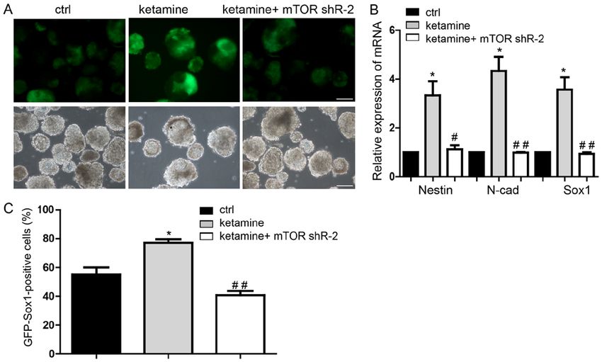
2444 ZHOU et al: LOW KETAMINE LEVELS UPREGULATE p-mTOR PROMOTING NEURAL DIFFERENTIATION
critical genes regulating neural differentiation, for example, was dissociated into single cells by trypsin and counted.
zing finger homeobox (Zfhx)1b has been reported to promote Subsequently, 2x10 4 cells/ml mESCs were washed with
neural stem cell (NSC) colony formation by inducing Sex Glasgow's minimum essential medium (GMEM; Gibco;
determining region Y‑box (Sox)1 expression (19). Sirtuin1 Thermo Fisher Scientific, Inc.) in a 6 cm dish and re‑suspended
could mediate alterations in DNA methylation to modulate in GMEM with 8% knockout serum replacement (Gibco;
embryonic stem cell differentiation (20). The microRNA‑134/ Thermo Fisher Scientific, Inc.), 1% sodium pyruvate, 1%
methyl‑CpG binding domain protein 3 axis could regulate L‑glutamine (Thermo Fisher Scientific, Inc.), 0.1 mM β‑Me.
the reprogramming and pluripotency of induced pluripotent Cells were cultured in a 6 cm ultra‑low attachment petri dish
stem cells, a type of ESC‑like cells, from neural progenitor and passaged every 2 days at 37˚C in a 5% CO2 atmosphere.
cells (NPCs) (21); however, the neuroprotective function of The culture medium was changed every day. Clones exhibiting
ketamine in mESCs on NSC differentiation and its down- GFP fluorescence at the stage of NSC derived from 46C mESCs
stream mechanism remains elusive. were detected with an IX73 + DP80 inverted fluorescence
Mammalian target of rapamycin (mTOR) is a critical regu- microscope (Olympus Corporation, Tokyo, Japan; magni-
lator of growth and homeostasis (22‑24). A growing number fication, x20). During the neural differentiation, ketamine
of studies have demonstrated that the mTOR‑related signaling (final concentration 50 nM) was added into the medium. The
pathway is associated with the differentiation of NPCs and control group consisted of cells treated only with physiological
NSCs (25,26), and is important to regulate oligodendrocyte saline (0.9% NaCl). For the treatment with MK‑801, 10 µg/ml
differentiation and remyelination (27). mTOR also serves MK‑801 was used to treat cells during neural differentiation.
an important role in regulating cortical interneuron number Rapamycin (final concentration 50 µM) was added to the
and autophagy during brain development (28). Rapamycin, medium during the neural differentiation. The control group
the mechanistic target of mTOR, has been associated with was treated with dimethyl sulfoxide, which was additionally
improvements in neurological deficits and increased brain used as the solvent for rapamycin. All the treating or control
water content (29). However, whether mTOR could regulate culture media was changed every day during the 7 days of
the neural differentiation of ESCs has been rarely evaluated. neural differentiation from mESCs.
Besides, whether mTOR participates in ketamine regulatory
signaling pathway or not, is also unclear. Reverse‑transcription quantitative polymerase chain
In the present study, it was determined whether ketamine reaction (RT‑qPCR). Total neural stem cell RNA was isolated
was able to influence the neural differentiation from the by RNaiso plus (Takara Biotechnology Co., Ltd., Dalian,
mESCs and the marker expression of sex‑determining region China), mRNA was reverse transcribed to cDNA at 37˚C for
Y‑box (Sox)1 (30), N‑cadherin (N‑cad) (31) and Nestin (32). 15 min using a RT reagent kit (Perfect Real Time; Takara
The present study suggested a safe dose of ketamine for Biotechnology Co., Ltd.). qPCR was performed using SYBR
clinical application and demonstrated that mTOR may be a Green qPCR Mix (Takara Biotechnology Co., Ltd.). The
potential target of better and safer therapeutics in the future. primers are as follows: Nestin forward, 5'‑CCCTGAAGT
CGAGGAGCTG‑3' and reverse, 5'‑CTGCTGCACCTCTAA
Materials and methods GCGA‑3'; N‑cadherin forward, 5'AGCGCAGTCTTACCGAA
GG‑3' and reverse, 5'‑TCGCTGCTTTCATACTGAACTTT‑3';
mESC culture. The mESC line 46C, containing the Sox1 Sox1 forward, 5'‑AAGGAACACCCGGATTACAAGT‑3' and
promoter and expressing green fluorescence protein (GFP), reverse, 5'‑GTTAGCCCAGCCGTT GAC‑3'; and GAPDH
was employed to indicate the endogenous Sox1 expression forward, 5'‑AGGTCGGTGTGAACGGATTTG‑3' and reverse
during the neural differentiation at NPCs stage and gifted by 5'‑TGTAGACCATGTAGTTGAGGTCA‑3'. The PCR ther-
Dr Xiaoqing Zhang (Tongji University, Shanghai, China) (33). mocycling conditions were as follows: Initial denaturation at
Cells were cultured on feeder cells that are the irradiated 95˚C for 5 min, followed by 40 cycles of denaturation at 95˚C
mouse embryonic fibroblasts in KnockOut™ Dulbecco's for 5 sec, primer annealing at 60˚C for 20 sec, elongation at
modified Eagle's medium (Gibco; Thermo Fisher Scientific, 70˚C for 10 sec. In total, three independent experiments were
Inc., Waltham, MA, USA; cat. no. 10829018) with 15% fetal performed. The relative gene expression was presented as
bovine serum (Gibco; Thermo Fisher Scientific, Inc.), leukemia 2‑∆∆Cq using the relative quantification method and normalized
inhibitory factor (Merck KGaA, Darmstadt, Germany; to the expression of GAPDH (35).
cat. no. LIF2050) and β ‑mercaptoethanol (β ‑Me; 1:10,000,
Sigma‑Aldrich; Merck KGaA) at 37˚C, under a 5% CO2 Western blotting. Cells were lysed by radioimmunoprecipita-
atmosphere. After 48 h, mESCs were digested into single cells tion assay lysis buffer (Beyotime Institute of Biotechnology,
using 0.05% trypsin (Gibco; Thermo Fisher Scientific, Inc.; cat. Haimen, China; cat. no. P0013B) and quantified by a
no. 2520056) and seeded on new feeder cells for passaging. Bicinchoninic Protein Assay Kit (Beyotime Institute of
The feeder cells that were able to secrete leukemia inhibitory Biotechnology; cat. no. P0009). A total of 40 µg protein was
factor to support the growth of the ESCs were made in our lab. loaded for electrophoresis on 10% SDS‑PAGE gels. Proteins
Feeder cells were made from x‑irradiated day 13.5 embryonic were transferred onto polyvinylidene fluoride membranes
fibroblasts. Day 13.5 embryonic fibroblasts were granted from (Merck KGaA; cat. no. MH0323) and blocked with TBS and
Dr Liu lab in Tongji University. Tween 20 with 3% bovine serum albumin (Amresco, Inc.,
Framingham, MA, USA) for 1 h at room temperature and
Neural differentiation of mESCs to NSCs. The protocol was incubated with primary antibodies at 4˚C overnight. The anti-
adapted from a previous study (34). The mESC line, 46C, bodies were as follows: mTOR (cat. no. 2972, Cell Signaling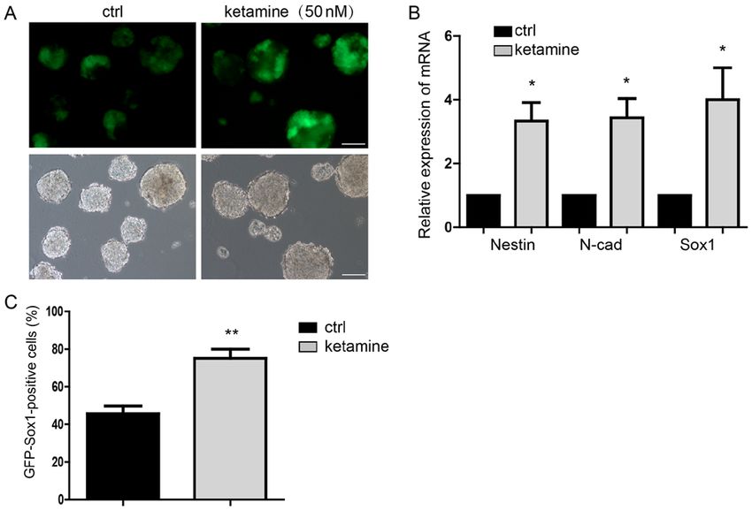
Molecular Medicine REPORTS 21: 2443-2451, 2020                                                           2445

Figure 1. Ketamine promotes neural differentiation. (A) GFP indicated the Sox1 expression of NPCs, suggesting the differentiation potential. Scale bar, 100 µm.
(B) Detection of the NPCs markers expression by RT‑qPCR. (C) Flow cytometry analysis indicated more GFP‑Sox1‑positive cells in the ketamine‑treatment
group. Data are presented as the mean ± standard deviation (n=3). *P
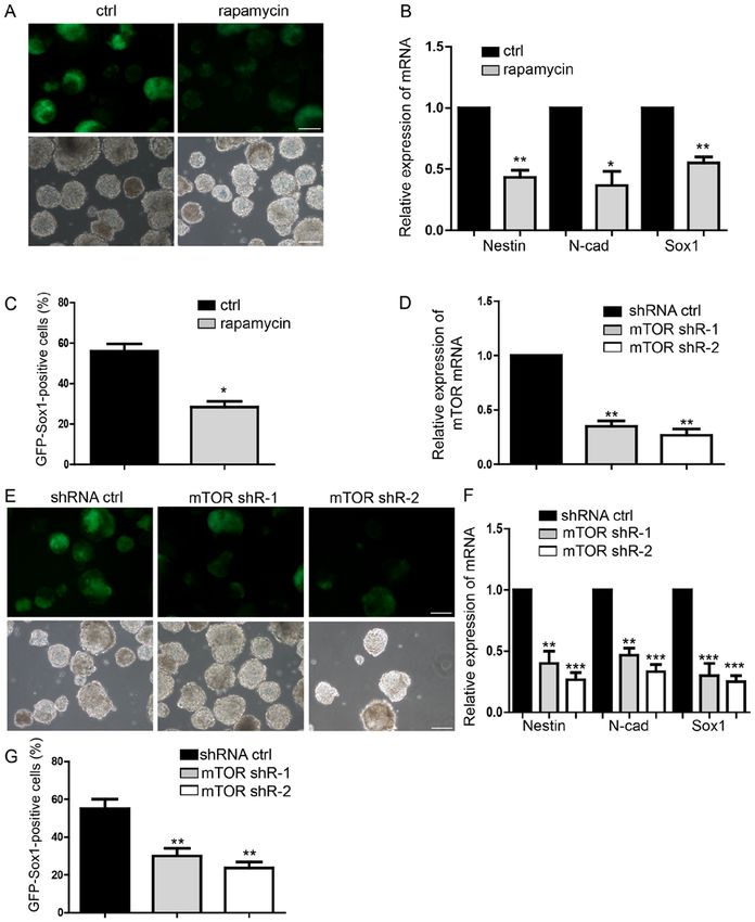
2446             ZHOU et al: LOW KETAMINE LEVELS UPREGULATE p-mTOR PROMOTING NEURAL DIFFERENTIATION

Figure 2. Ketamine activates the mTOR signaling pathway. (A) Representative images of the expression of p‑mTOR in neural progenitor cells derived
from ESCs as detected by western blotting; the ratio of p‑mTOR normalized to GAPDH/total mTOR is presented. (B) Ketamine‑treatment group
demonstrated the upregulation of p‑70SK6. (C) Inhibtion of the NMDA signaling pathway by the NMDA receptor antagonist MK‑801 decreased the
expression levels of p‑mTOR; the ratio of p‑mTOR normalized to GAPDH/total mTOR is presented. (D) 50 µM rapamycin markedly reduced the activa-
tion of the mTOR signaling, which attenuated the function of ketamine on regulating the level of p‑mTOR. Data are presented as the mean ± standard
deviation (n=3). *P
Molecular Medicine REPORTS 21: 2443-2451, 2020                                                          2447

Figure 3. Inhibition of mTOR suppresses neural differentiation. (A) Representative images of neural differentiation in the rapamycin‑treatment and control
groups. (B) Expression of NPCs markers of Nestin, N‑cad and Sox1 by RT‑qPCR. (C) Flow cytometry analysis of rapamycin‑treatment and control group.
(D and E) Detection of the mTOR knockdown effect by shRNA, which suppressed neural differentiation. (F) Expression levels of NPCs markers, as measured
by RT‑qPCR. (G) Flow cytometry analysis indicating less NPCs following transfection with shRNA. Scale bar, 100 µm. Data are presented as the mean ± stan-
dard deviation (n=4). *P
2448               ZHOU et al: LOW KETAMINE LEVELS UPREGULATE p-mTOR PROMOTING NEURAL DIFFERENTIATION

Figure 4. mTOR mediates the function of ketamine‑regulated neural differentiation. (A) Representative images of neural differentiation in rescue experiments.
(B) Downregulation of mTOR restored neural progenitor cell marker expression levels, in the ketamine‑treatment group. (C) Flow cytometry assay also
indicated that mTOR knockdown inhibited the neural differentiation promoted by ketamine. Scale bar, 100 µm. Data are presented as the mean ± standard
deviation (n=3). *P
Molecular Medicine REPORTS 21: 2443-2451, 2020                                            2449

application is an important research goal in clinical practice.   Acknowledgements
The effects of ketamine are not only dependent on its dose,
but also on the frequency of exposure (40‑43). Ketamine           The authors would like to thank Dr Xiaoqing Zhang
has a relative neuroprotective function by relieving pain and     (Tongji University, Shanghai, China) for providing the
inhibiting inflammation (44). Ketamine serves an important        Sox1‑promoter‑GFP 46C mESCs.
role in regulating nerve development (16). In the present
study, ketamine at 50 nM promoted neural differentiation          Funding
and upregulated NSC marker expression levels. The process
of neural differentiation occurs during early develop-            The present study was supported by the National Natural
ment (45,46). The present results additionally demonstrated       Science Foundation of China (grant no. 81571028). Research
the positive effects of ketamine at a low dose, suggesting        funds were from the Shanghai Municipal Science and
the safe clinical use in surgery for pregnant patients and        Technology Commission (grant no. 16XD1401800) and
children in the future.                                           the Natural Science Foundation of Shanghai (grant nos.
    ESCs have been extensively used for studying develop-         17ZR1416400 and 17DZ1205403).
ment, particularly neural development (47-51). Numerous
genes serve an important role in the differentiation into         Availability of data and materials
neural stem cells (52,53). A recent study demonstrated that
fibronectin type III domain‑containing 5 facilitated neural       The datasets used and/or analyzed during the current study are
differentiation by increasing the expression of brain derived     available from the corresponding author on reasonable request.
neurotrophic factor (54). Zfhx1b gene expression has been
confirmed to be notably upregulated via the fibroblast            Authors' contributions
growth factor signaling pathway in mESCs cultured in a
permissive neural‑inducing environment (19). Ketamine             XZ performed the experiments. XL performed the reverse
was proposed to regulate mTOR activity by upregulating            transcription‑quantitative polymerase chain reaction assays
the expression levels of p‑mTOR in the present study. This        and wrote parts of the manuscript. LZ performed the western
was reversed by adding the mTOR inhibitor rapamycin               blotting. JY conducted the statistical analysis. RH performed
or by downregulating mTOR. This suggested a potential             microscopy. YS cultured and prepared the cells. SX analyzed
molecular mechanism of ketamine regulation; however,              the expression level of western blot. HJ provided guidance and
further investigation is required.                                analyzed some of the data. All authors read and approved the
    In neural progenitors, insulin has been demonstrated          final manuscript.
to induce neurogenesis of NPCs by activating mTOR (26).
mTOR is also needed for the of dendritic arbors develop-          Ethics approval and consent to participate
ment and stabilization in the newly born olfactory bulb
neurons (55). The mTOR signaling pathway was reported             Not applicable.
to mediate valproic acid‑induced neural differentiation of
NSCs (56). Inhibition of the mTOR signaling pathway by            Patient consent for publication
rapamycin was observed to suppress neural differentiation
in the present study. The promotion of neural differentiation     Not applicable.
caused by ketamine was also inhibited by silencing mTOR.
The expression of Nestin, Sox1 and N‑cad was also restored        Competing interests
by downregulating mTOR. The NMDA signaling pathway,
was inhibited during the neural differentiation and the levels    The authors declare that they have no competing interests.
of p‑mTOR were also suppressed. This result indicated the
regulatory function of ketamine via a non‑NMDA signaling          References
pathway during neural differentiation. However, a limitation
of the present study is that whether the NMDA receptor             1. Sinner B and Graf BM: Ketamine. Handb Exp Pharmacol:
may influence neural differentiation remains unknown.                 313‑333, 2008.
mTOR complex 1 (mTORC1) was closely associated with the            2. Rofa el H Z , Tu rk a l l R M a nd Ab d el‑R a h m a n MS:
                                                                      Immunomodulation by cocaine and ketamine in postnatal rats.
neuron‑associated biological process downstream target (57).          Toxicology 188: 101‑114, 2003.
The activity of p70S6K, the downstream target of mTORC1,           3. Reitman E and Flood P: Anaesthetic considerations for
was increased by ketamine, indicating that it may participate         non‑obstetric surgery during pregnancy. Br J Anaesth 107
                                                                      (Suppl 1): i72‑i78, 2011.
in the regulation of ketamine. These results determined            4. Wilder RT, Flick RP, Sprung J, Katusic SK, Barbaresi WJ,
that the ketamine/mTOR signaling pathway regulated the                Mickelson C, Gleich SJ, Schroeder DR, Weaver AL and Warner DO:
neural differentiation process of NSCs derived from mESCs;            Early exposure to anesthesia and learning disabilities in a popu-
                                                                      lation‑based birth cohort. Anesthesiology 110: 796‑804, 2009.
however, further investigation is required.                        5. Huang H, Liu CM, Sun J, Hao T, Xu CM, Wang D and Wu YQ:
    In summary, the present study revealed the ketamine/              Ketamine affects the neurogenesis of the hippocampal dentate
mTOR signaling pathway on regulating the neural differentia-          gyrus in 7‑day‑old rats. Neurotox Res 30: 185‑198, 2016.
tion and suggested a potential dose of ketamine. The ketamine/     6. Wang J, Zhou M, Wang X, Yang X, Wang M, Zhang C, Zhou S
                                                                      and Tang N: Impact of ketamine on learning and memory
mTOR signaling pathway needs to be further investigated for           function, neuronal apoptosis and its potential association with
its potential use in clinic.                                          mir‑214 and pten in adolescent rats. PLoS One 9: e99855, 2014.
2450 ZHOU et al: LOW KETAMINE LEVELS UPREGULATE p-mTOR PROMOTING NEURAL DIFFERENTIATION
7. Yan J, Huang Y, Lu Y, Chen J and Jiang H: Repeated adminis- 28. Ka M, Smith AL and Kim WY: MTOR controls genesis and
tration of ketamine can induce hippocampal neurodegeneration autophagy of GABAergic interneurons during brain devel-
and long‑term cognitive impairment via the ROS/HIF‑1α opment. Autophagy 13: 1348‑1363, 2017.
pathway in developing rats. Cell Physiol Biochem 33: 1715‑1732, 29. Xing J and Lu J: Effects of mTOR on neurological deficits after
2014. transient global ischemia. Transl Neurosci 8: 21‑26, 2017.
8. Hudetz JA, Patterson KM, Iqbal Z, Gandhi SD, Byrne AJ, 30. Barraud P, Thompson L, Kirik D, Bjorklund A and Parmar M:
Hudetz AG, Warltier DC and Pagel PS: Ketamine attenuates Isolation and characterization of neural precursor cells from the
delirium after cardiac surgery with cardiopulmonary bypass. Sox1‑GFP reporter mouse. Eur J Neurosci 22: 1555‑1569, 2005.
J Cardiothorac Vasc Anesth 23: 651‑657, 2009. 31. Reinés A, Bernier LP, McAdam R, Belkaid W, Shan W, Koch AW,
9. Hovaguimian F, Tschopp C, Beck‑Schimmer B and Puhan M: Séguéla P, Colman DR and Dhaunchak AS: N‑cadherin
Intraoperative ketamine administration to prevent delirium or prodomain processing regulates synaptogenesis. J Neurosci 32:
postoperative cognitive dysfunction: A systematic review and 6323‑6334, 2012.
meta‑analysis. Acta Anaesthesiol Scand 62: 1182‑1193, 2018. 32. Zhang J and Jiao J: Molecular biomarkers for embryonic and
10. Nikiforuk A and Popik P: Ketamine prevents stress‑induced adult neural stem cell and neurogenesis. Biomed Res Int 2015:
cognitive inflexibility in rats. Psychoneuroendocrinology 40: 727542, 2015.
119‑122, 2014. 33. Ying QL, Stavridis M, Griffiths D, Li M and Smith A: Conversion
11. Wang CQ, Ye Y, Chen F, Han WC, Sun JM, Lu X, Guo R, of embryonic stem cells into neuroectodermal precursors in
Cao K, Zheng MJ and Liao LC: Posttraumatic administration adherent monoculture. Nat Biotechnol 21: 183‑186, 2003.
of a sub‑anesthetic dose of ketamine exerts neuroprotection via 34. Watanabe K, Kamiya D, Nishiyama A, Katayama T, Nozaki S,
attenuating inflammation and autophagy. Neuroscience 343: Kawasaki H, Watanabe Y, Mizuseki K and Sasai Y: Directed
30‑38, 2017. differentiation of telencephalic precursors from embryonic stem
12. Hertle DN, Dreier JP, Woitzik J, Hartings JA, Bullock R, cells. Nat Neurosci 8: 288‑296, 2005.
Okonkwo DO, Shutter LA, Vidgeon S, Strong AJ, Kowoll C, et al: 35. Livak KJ and Schmittgen TD: Analysis of relative gene
Effect of analgesics and sedatives on the occurrence of spreading expression data using real‑time quantitative pcr and the 2(‑delta
depolarizations accompanying acute brain injury. Brain 135: delta c(t)) method. Methods 25: 402‑408, 2001.
2390‑2398, 2012. 36. Gass N, Schwarz AJ, Sartorius A, Schenker E, Risterucci C,
13. Koerner IP and Brambrink AM: Brain protection by anesthetic Spedding M, Zheng L, Meyer‑Lindenberg A and Weber‑Fahr W:
agents. Curr Opin Anaesthesiol 19: 481‑486, 2006. Sub‑anesthetic ketamine modulates intrinsic BOLD connec-
14. Sanders RD, Ma D, Brooks P and Maze M: Balancing paediatric tivity within the hippocampal‑prefrontal circuit in the rat.
anaesthesia: preclinical insights into analgesia, hypnosis, neuro- Neuropsychopharmacology 39: 895‑906, 2014.
protection, and neurotoxicity. Br J Anaesth 101: 597‑609, 2008. 37. Yan J and Jiang H: Dual effects of ketamine: Neurotoxicity
15. Murrough JW: Ketamine for depression: An update. Biol versus neuroprotection in anesthesia for the developing brain.
Psychiatry 80: 416‑418, 2016. J Neurosurg Anesthesiol 26: 155‑160, 2014.
16. Dong C, Rovnaghi CR and Anand KJ: Ketamine alters the 38. Theurillat R, Larenza MP, Feige K, Bettschart‑Wolfensberger R
neurogenesis of rat cortical neural stem progenitor cells. Crit and Thormann W: Development of a method for analysis of
Care Med 40: 2407‑2416, 2012. ketamine and norketamine enantiomers in equine brain and cere-
17. Bosnjak ZJ, Yan Y, Canfield S, Muravyeva MY, Kikuchi C, brospinal fluid by capillary electrophoresis. Electrophoresis 35:
Wells CW, Corbett JA and Bai X: Ketamine induces toxicity 2863‑2869, 2014.
in human neurons differentiated from embryonic stem cells via 39. Li J, Wang B, Wu H, Yu Y, Xue G and Hou Y: 17β‑estradiol attenuates
mitochondrial apoptosis pathway. Curr Drug Saf 7: 106‑119, ketamine‑induced neuroapoptosis and persistent cognitive deficits
2012. in the developing brain. Brain Res 1593: 30‑39, 2014.
18. Czechanski A, Byers C, Greenstein I, Schrode N, Donahue LR, 40. Permoda‑Osip A, Kisielewski J, Bartkowska‑Sniatkowska A and
Hadjantonakis AK and Reinholdt LG: Derivation and charac- Rybakowski JK: Single ketamine infusion and neurocognitive
terization of mouse embryonic stem cells from permissive and performance in bipolar depression. Pharmacopsychiatry 48:
nonpermissive strains. Nat Protoc 9: 559‑574, 2014. 78‑79, 2015.
19. Dang LT, Wong L and Tropepe V: Zfhx1b induces a definitive 41. Diamond PR, Farmery AD, Atkinson S, Haldar J, Williams N,
neural stem cell fate in mouse embryonic stem cells. Stem Cells Cowen PJ, Geddes JR and McShane R: Ketamine infusions for
Dev 21: 2838‑2851, 2012. treatment resistant depression: A series of 28 patients treated
20. Tang S, Huang G, Fan W, Chen Y, Ward JM, Xu X, Xu Q, Kang A, weekly or twice weekly in an ECT clinic. J Psychopharmacol 28:
McBurney MW, Fargo DC, et al: SIRT1‑mediated deacetylation 536‑544, 2014.
of CRABPII regulates cellular retinoic acid signaling and 42. Neri CM, Pestieau SR and Darbari DS: Low‑dose ketamine as
modulates embryonic stem cell differentiation. Mol Cell 55: a potential adjuvant therapy for painful vaso‑occlusive crises in
843‑855, 2014. sickle cell disease. Paediatr Anaesth 23: 684‑689, 2013.
21. Zhang L, Zheng Y, Sun Y, Zhang Y, Yan J, Chen Z and Jiang H: 43. Shibuta S, Morita T, Kosaka J, Kamibayashi T and Fujino Y:
MiR‑134‑Mbd3 axis regulates the induction of pluripotency. Only extra‑high dose of ketamine affects l‑glutamate‑induced
J Cell Mol Med 20: 1150‑1158, 2016. intracellular Ca(2+) elevation and neurotoxicity. Neurosci
22. Meng SS, Guo FM, Zhang XW, Chang W, Peng F, Qiu HB Res 98: 9‑16, 2015.
and Yang Y: mTOR/STAT‑3 pathway mediates mesenchymal 44. Bhutta AT, Sch m itz M L, Swea r ingen C, James LP,
stem cell‑secreted hepatocyte growth factor protective effects Wardbegnoche WL, Lindquist DM, Glasier CM, Tuzcu V,
against lipopolysaccharide‑induced vascular endothelial barrier Prodhan P, Dyamenahalli U, et al: Ketamine as a neuroprotective
dysfunction and apoptosis. J Cell Biochem 120: 3637‑3650, 2018. and anti‑inflammatory agent in children undergoing surgery on
23. Nguyen K, Yan Y, Yuan B, Dasgupta A, Sun JC, Mu H, cardiopulmonary bypass: A pilot randomized, double‑blind,
Do KA, Ueno NT, Andreeff M and Battula VL: ST8SIA1 placebo‑controlled trial. Pediatr Crit Care Med 13: 328‑337,
regulates tumor growth and metastasis in TNBC by activating 2012.
the FAK‑AKT‑mTOR signaling pathway. Mol Cancer Ther 17: 45. Sheridan MA and McLaughlin KA: Dimensions of early expe-
2689‑2701, 2018. rience and neural development: Deprivation and threat. Trends
24. Wang Y, Ma J, Qiu W, Zhang J, Feng S, Zhou X, Wang X, Jin L, Cogn Sci 18: 580‑585, 2014.
Long K, Liu L, et al: Guanidinoacetic acid regulates myogenic 46. Kolb B, Mychasiuk R and Gibb R: Brain development, expe-
differentiation and muscle growth through miR‑133a‑3p and rience, and behavior. Pediatr Blood Cancer 61: 1720‑1723, 2014.
miR‑1a‑3p Co‑mediated Akt/mTOR/S6K signaling pathway. Int 47. Lu AQ, Popova EY and Barnstable CJ: Activin signals through
J Mol Sci 19: pii: E2837, 2018. SMAD2/3 to increase photoreceptor precursor yield during
25. Magri L and Galli R: Mtor signaling in neural stem cells: From embryonic stem cell differentiation. Stem Cell Reports 9:
basic biology to disease. Cell Mol Life Sci 70: 2887‑2898, 2013. 838‑852, 2017.
26. Han J, Wang B, Xiao Z, Gao Y, Zhao Y, Zhang J, Chen B, Wang X 48. Liu Q, Wang G, Chen Y, Li G, Yang D and Kang J: A miR‑590/
and Dai J: Mammalian target of rapamycin (mTOR) is involved Acvr2a/Rad51b axis regulates DNA damage repair during mESC
in the neuronal differentiation of neural progenitors induced by proliferation. Stem Cell Reports 3: 1103‑1117, 2014.
insulin. Mol Cell Neurosci 39: 118‑124, 2008. 49. Xu N, Papagiannakopoulos T, Pan G, Thomson JA and Kosik KS:
27. Dai J, Bercury KK and Macklin WB: Interaction of mTOR and MicroRNA‑145 regulates OCT4, SOX2, and KLF4 and represses
Erk1/2 signaling to regulate oligodendrocyte differentiation. pluripotency in human embryonic stem cells. Cell 137: 647‑658,
Glia 62: 2096‑2109, 2014. 2009.Molecular Medicine REPORTS 21: 2443-2451, 2020 2451
50. Lei J, Yuan Y, Lyu Z, Wang M, Liu Q, Wang H, Yuan L and Chen H: 55. Skalecka A, Liszewska E, Bilinski R, Gkogkas C, Khoutorsky A,
Deciphering the role of sulfonated unit in heparin‑mimicking Malik AR, Sonenberg N and Jaworski J: mTOR kinase is needed
polymer to promote neural differentiation of embryonic stem for the development and stabilization of dendritic arbors in newly
cells. ACS Appl Mater Interfaces 9: 28209‑28221, 2017. born olfactory bulb neurons. Dev Neurobiol 76: 1308‑1327, 2016.
51. Gazina EV, Morrisroe E, Mendis GDC, Michalska AE, Chen J, 56. Zhang X, He X, Li Q, Kong X, Ou Z, Zhang L, Gong Z, Long D,
Nefzger CM, Rollo BN, Reid CA, Pera MF and Petrou S: Method Li J, Zhang M, et al: PI3K/AKT/mTOR signaling mediates
of derivation and differentiation of mouse embryonic stem valproic acid‑induced neuronal differentiation of neural stem
cells generating synchronous neuronal networks. J Neurosci cells through epigenetic modifications. Stem Cell Reports 8:
Methods 293: 53‑58, 2018. 1256‑1269, 2017.
52. Fischer U, Keller A, Voss M, Backes C, Welter C and Meese E: 57. Polchi A, Magini A, Meo DD, Tancini B and Emiliani C: mTOR
Genome‑wide gene amplification during differentiation of neural signaling and neural stem cells: The tuberous sclerosis complex
progenitor cells in vitro. PLoS One 7: e37422, 2012. model. Int J Mol Sci 19: pii: E1474, 2018.
53. Mateo JL, van den Berg DL, Haeussler M, Drechsel D, Gaber ZB,
Castro DS, Robson P, Lu QR, Crawford GE, Flicek P, et al: This work is licensed under a Creative Commons
Characterization of the neural stem cell gene regulatory network Attribution-NonCommercial-NoDerivatives 4.0
identifies OLIG2 as a multifunctional regulator of self‑renewal. International (CC BY-NC-ND 4.0) License.
Genome Res 25: 41‑56, 2015.
54. Forouzanfar M, Rabiee F, Ghaedi K, Beheshti S, Tanhaei S,
Shoaraye Nejati A, Jodeiri Farshbaf M, Baharvand H and
Nasr‑Esfahani MH: Fndc5 overexpression facilitated neural
differentiation of mouse embryonic stem cells. Cell Biol Int 39:
629‑637, 2015.You can also read


