Epigenetic Modifiers in Myeloid Malignancies: The Role of Histone Deacetylase Inhibitors - MDPI
←
→
Page content transcription
If your browser does not render page correctly, please read the page content below
International Journal of
           Molecular Sciences

Review
Epigenetic Modifiers in Myeloid Malignancies:
The Role of Histone Deacetylase Inhibitors
Johanna S. Ungerstedt
 Department of Medicine, Huddinge, Karolinska Institutet, and Hematology Center, and Karolinska University
 Hospital, S-141 86 Stockholm, Sweden; johanna.ungerstedt@ki.se; Tel.: +46-8-5858-0000, Fax: +46-8-5858-2525

 Received: 18 August 2018; Accepted: 5 October 2018; Published: 9 October 2018

 Abstract: Myeloid hematological malignancies are clonal bone marrow neoplasms, comprising of
 acute myeloid leukemia (AML), the myelodysplastic syndromes (MDS), chronic myelomonocytic
 leukemia (CMML), the myeloproliferative neoplasms (MPN) and systemic mastocytosis (SM).
 The field of epigenetic regulation of normal and malignant hematopoiesis is rapidly growing. In recent
 years, heterozygous somatic mutations in genes encoding epigenetic regulators have been found in all
 subtypes of myeloid malignancies, supporting the rationale for treatment with epigenetic modifiers.
 Histone deacetylase inhibitors (HDACi) are epigenetic modifiers that, in vitro, have been shown to
 induce growth arrest, apoptotic or autophagic cell death, and terminal differentiation of myeloid
 tumor cells. These effects were observed both at the bulk tumor level and in the most immature
 CD34+ 38− cell compartments containing the leukemic stem cells. Thus, there is a strong rationale
 supporting HDACi therapy in myeloid malignancies. However, despite initial promising results
 in phase I trials, HDACi in monotherapy as well as in combination with other drugs, have failed
 to improve responses or survival. This review provides an overview of the rationale for HDACi in
 myeloid malignancies, clinical results and speculations on why clinical trials have thus far not met
 the expectations, and how this may be improved in the future.

 Keywords: myelodysplastic syndromes; acute myeloid leukemia; chronic myelomonocytic leukemia;
 systemic mastocytosis; treatment; myeloid mutations

1. Introduction to Myeloid Hematological Diseases and Their Treatment
     The myelodysplastic syndromes (MDS) are a heterogenous group of clonal myeloid hematological
diseases, with a median age of onset of 70 years and a survival of 0.5–8 years, and a 30% risk of
transformation to acute myeloid leukemia (AML) [1]. MDS is curable only by allogeneic stem cell
transplant, for which only a minority of patients are eligible. The only approved therapy for higher
risk MDS is treatment with hypomethylating agents, where two drugs are available, azacitidine and
decitabine. Approximately 50–60% of patients respond to therapy [2]. Azacitidine and decitabine are
considered to be DNA demethylating agents, as preclinical studies in cell lines have shown a quick
and profound global DNA demethylation, as well as site specific promoter demethylation of e.g.,
P15/INK4B [3–5]. However, in vivo and ex vivo studies of MDS patient CD34+ progenitor cells do not
show a clear DNA demethylation in response to azacitidine, and thus the in vivo mechanism of action
of the drugs remains unclear [6,7].
     The Philadelphia chromosome negative myeloproliferative neoplasms (MPN) comprises of
polycythemia vera (PV), essential thrombocytemia (ET), and myelofibrosis (MF) [1]. The rare
diseases chronic neutrophil leukemia and chronic eosinophil leukemia will not be discussed further
in this review, nor will the Philadelphia chromosome positive chronic myeloid leukemia (CML).
The, Janus kinase 2 (JAK2) V617F mutation occurs in around 95% of PV, and 50% of ET and MF, causing
auto-phosphorylation of cytokine receptors and increased JAK-STAT pathway activation [8]. Although

Int. J. Mol. Sci. 2018, 19, 3091; doi:10.3390/ijms19103091                       www.mdpi.com/journal/ijms
Int. J. Mol. Sci. 2018, 19, 3091                                                                    2 of 18

the MPNs share a common driver mutation, there are major clinical differences as ET has a largely
normal life expectancy, PV has a long life expectancy, whereas MF has a significantly shortened life
expectancy [9]. In higher risk disease, oral cytoreductive treatment hydroxycarbamide is used, and for
MF, and now also for PV, the JAK2 inhibitor Ruxolitinib may be applied. Allogeneic stem cell transplant
may be an option for younger, high risk MF patients [10].
      Chronic myelomonocytic leukemia (CMML) is the largest group of MDS/MPN overlap
syndromes [1], where proliferative CMML disease is treated like MPN and dysplastic CMML is
treated like MDS [11], however responses to azacitidine or decitabine are never long lasting, and there
is an imminent need for new treatment options in CMML.
      Systemic mastocytosis (SM) is a rare myeloid malignancy [1]. Over 90% of patients carry the
D816V activating point mutation in the KIT gene. There are two clinical phenotypes, indolent SM with
a normal life expectancy and aggressive SM with a poor prognosis [12]. In the aggressive SM group,
several therapies have been tested, most recently the pan tyrosine kinase inhibitor Midostaurin [12,13],
however to date, no therapy except allogeneic stem cell transplantation has been shown to improve
survival, and as the median age at disease onset is around age 70, only a handful of patients are eligible
for transplant [14].
      Overall, 30% of MDS and CMML patients progress to acute myeloid leukemia (AML). AML is
defined by over 20% myeloblasts in the bone marrow, and may be primary (around 75% of all
AML cases), secondary (to e.g., MDS, MPN, around 15% of cases) or treatment related e.g., after
chemotherapy of hematological or other tumors (10% of cases). Subtypes of AML are well defined
according to cell of origin, specific cytogenetic or other aberrations outlined in the WHO diagnostic
criteria [1]. In general, patients that are not elderly and unfit, are given intensive chemotherapy
(including cytarabine and anthracyclines) to achieve complete remission, and thereafter chemotherapy
consolidation or consolidation with allogeneic stem cell transplant in case of high risk disease [15].
However, a large portion of patients are elderly and fragile, and for these patients lower intensity
treatment, often similar to regimens given to MDS-patients, can be considered if the AML is not highly
proliferative [16]. The 5-year overall survival in adult AML is around 20%.

2. Epigenetic Regulation of Normal and Malignant Hematopoiesis
     Our understanding of how epigenetic regulation of hematopoiesis is orchestrated is rapidly
growing. Epigenetics include DNA methylation (Figure 1) as well as covalent, reversible histone
modifications (Figure 2) [17,18]. DNA methylation is associated with transcriptional repression via
formation of heterochromatin. This is achieved by methylation of the 5-cytosine by DNA methyl
transferase (DNMT) enzymes, where maintenance methylation is exerted by DNMT1, and de novo
methylation is exerted by DNMT3A and 3B (Figure 1). The gene encoding the DNMT3A enzyme is
commonly mutated in AML, leading to loss of function (Table 1). DNA demethylation is a multistep
process exerted by the TET enzymes that oxidize 5-methyl cytosine to cytosine (Figure 1). This oxidation
process requires α-keto-glutarate, which is produced from isocitrate by isocitrate dehydrogenases
1 and 2 (IDH1 and 2) (Figure 1). Mutations in IDH1 and 2 are common in AML, and TET2
mutations are common in all myeloid malignancies (Table 1). Table 1 summarizes the currently
known mutations in epigenetic regulators found in myeloid malignancies, and is compiled from the
pivotal studies of Papaemmanuil [19] and Haferlach [20] for MDS, and Ley for AML [21], as well as
reviews for CMML [22], MPN [23], SM [24], and references [17,18,25,26] for comparison of mutation
frequencies reported.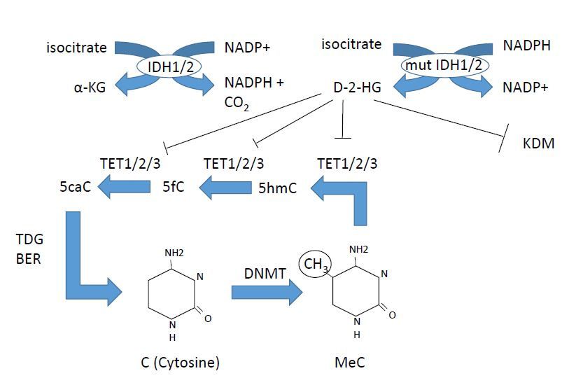
Int. J. Mol. Sci. 2018, 19, 3091                                                                                                                                      3 of 18

      Table 1. In all types of myeloid malignancies, genetic alterations in epigenetic modifiers are found, however the mutation frequency varies between diseases. For
      references please see text.

                Function                     Gene                   Loss/Gain of Function                    Activity                Frequency in Myeloid Malignancies
                                                                                                                                                AML 12–22%
                                                                                                                                                MDS 5–10%
          DNA methylation                  DNMT3A                            loss                   De novo DNA methylation                      CMML 5%
                                                                                                                                                MPN 7–15%
                                                                                                                                                  ASM 1%
                                                                                                                                                 AML 7–23%
                                                                                                                                                 MDS 20–25%
                                                                                                     5-methyl-C to 5-hydroxy
          DNA methylation                    TET2                            loss                                                                CMML 60%
                                                                                                           methyl-C
                                                                                                                                                 MPN 4–13%
                                                                                                                                                  ASM 40%
                                                                                                                                                AML 10–30%
                                                                                                                                                  MDS 3%
          DNA methylation                    IDH1/2                          gain                       Cofactor for TET2
                                                                                                                                                CMML 1–10%
                                                                                                                                                MPN 2.5–5%
                                                                                                                                                  AML rare
                                                                                                                                                  MDS 6%
                                                                                                  Trimethylation of H3K27, part
        Histone methylation                  EZH2                            Loss                                                                CMML 5%
                                                                                                        of PRC2 complex
                                                                                                                                                 MPN 3–13%
                                                                                                                                                  ASM 3%
                                                                                                                                                 AML 5%
                                                                                                                                                MDS 15–20%
        Histone methylation                  ASXL1                           loss                Associates with PRC1 and PRC2                 CMML 40–45%
                                                                                                                                                MPN 2–23%
                                                                                                                                                 ASM 14%
        Histone methylation                  SUZ12                           loss                        Member of PRC2                         MDS rare,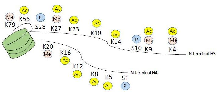
Int. J. Mol. Sci. 2018, 19, 3091                                                                                                           4 of 18

                                                             Table 1. Cont.

                Function               Gene        Loss/Gain of Function                 Activity            Frequency in Myeloid Malignancies
                                                                                H3K9(me1) lysine methyl
        Histone methylation          PRDM16                gain                                                       MDS/AML rare
                                                                                     transferase
                                                                                  H3K36 lysine methyl
        Histone methylation           SETD2                loss                                                          AML 5%
                                                                                     transferase
                                                                                                                sAML(from MDS, MPN) 6.5%
        Histone methylation           JARID2                                     Recruits PRC2 to target
                                                                                                                    MDS, MPN 0.2%
                                                                                                                        AML 3%
                                                                              Counteracts PRC2 by removing             MDS 2.5%
        Histone methylation        UTX (=KDM6A)            loss                di and trimethylated H3K27              CMML 8%
                                                                                                                     MDS/MPN 4.8%
         Histone acetylation       CREBBP (CBP)            gain                  Lysine acetyl transferase               AML rare
         Histone acetylation        P300 (EP300)           gain                  Lysine acetyl transferase               AML rare
        Histone deacetylation         HDAC2                loss                    Lysine deacetylase                    AML rare
        Histone deacetylation         HDAC3                loss                    Lysine deacetylase                    AML rare
Int. J. Mol. Sci. 2018, 19, x FOR PEER REVIEW 5 of 17
Int. J. Mol. Sci. 2018, 19, 3091 5 of 18
Figure 1. DNA methylation and demethylation. DNMT3A is commonly mutated in acute myeloid
Figure (AML),
leukemia 1. DNAIDH1,
methylation and demethylation.
2 mutations DNMT3A
are found in AML, is commonly
and TET2 mutated
is frequently in acute
mutated in allmyeloid
myeloid
leukemia (AML), IDH1, 2 mutations are found in AML, and TET2 is frequently mutated
malignancies. Azacitidine and decitabine are DNA demethylating agents, inhibiting DNA in all myeloid
methyl
malignancies.
transferases Azacitidine and decitabine are DNA demethylating agents, inhibiting DNA methyl
(DNMTs).
transferases (DNMTs).
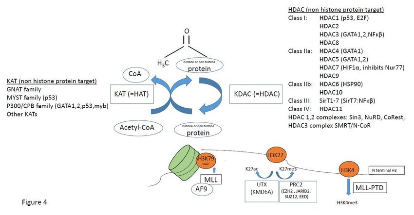
Histone modifications occur on the n-terminal protruding tail of predominantly histone H3
and H4,Histone modifications occur on the n-terminal protruding tail of predominantly histone H3 and
and consist of lysine residues being acetylated or methylated, and serine residues can
H4, and consist of lysine residues being acetylated or methylated, and serine residues can be
be phosphorylated (Figure 2). In addition, arginine may be methylated however this will not be
phosphorylated (Figure 2). In addition, arginine may be methylated however this will not be further
further discussed as it is outside the scope of this paper. Histone acetylation occurs via a family of
discussed as it is outside the scope of this paper. Histone acetylation occurs via a family of lysine
lysine acetyl transferases (KAT), formerly called histone acetyl transferases (HAT), and histone lysine
acetyl transferases (KAT), formerly called histone acetyl transferases (HAT), and histone lysine
methyl transferases (KMT) methylate histones. KAT and KMT enzymes along with protein arginine
methyl transferases (KMT) methylate histones. KAT and KMT enzymes along with protein arginine
methyl
methyl transferases
transferases are arecalled
calledepigenetic
epigeneticwriters,
writers, and
and HDAC, histone lysine
HDAC, histone lysinedemethylases
demethylases(KDM) (KDM)
andand phosphatases are called epigenetic erasers (Figure 3), reviewed in reference
phosphatases are called epigenetic erasers (Figure 3), reviewed in reference [27]. In addition, [27]. In addition,
there areare
there readers
readers that read
that readthe epigenetic
the epigeneticcode.
code.These
These are
are bromodomain containingproteins
bromodomain containing proteinslike
like the
the
BET BETfamily,
family, chromodomain,
chromodomain,PHD PHDfinger
fingerand
andWDWD 40 40 repeats
repeats (Figure 3). Inhibitors
(Figure 3). Inhibitorstotoboth
bothwriters,
writers,
e.g.,e.g.,
DOT1L [28,29] and readers e.g., BET inhibitor JQ1 [30] are in development
DOT1L [28,29] and readers e.g., BET inhibitor JQ1 [30] are in development and early6 of and early clinical trials
clinical
Int. J. Mol. Sci. 2018, 19, x FOR PEER REVIEW 17
for trials
myeloid malignancies
for myeloid malignancies
Figure 2. Histone modifications on the N terminal tail of histone H3 and H4. For simplicity, only
Figure 2. acetylation
methylation, Histone modifications on the N terminal
and phosphorylation are tail of histone
depicted, H3 and modifications
however H4. For simplicity,
alsoonly
include
methylation,
arginine acetylation
methylation and phosphorylation
and ubiquitination marks. are depicted, however modifications also include
arginine methylation and ubiquitination marks.Figure 2. Histone modifications on the N terminal tail of histone H3 and H4. For simplicity, only
methylation, acetylation and phosphorylation are depicted, however modifications also include
Int. J. Mol. Sci. 2018, 19, 3091 6 of 18
arginine methylation and ubiquitination marks.
Figure 3. Epigenetic writers are histone acetyl transferases HAT (KAT), histone lysine methyl
transferase
Figure (KMT) and
3. Epigenetic PRMTs
writers are(protein
histone arginine methyl transferases),
acetyl transferases HAT (KAT), readers are lysine
histone bromodomain
methyl
proteins like
transferase BETand
(KMT) family
PRMTsproteins,
(protein and erasersmethyl
arginine are histone deacetylase
transferases), readersinhibitors (HDACi),
are bromodomain
KDM (lysine/histone
proteins like BET familydemethylases) anderasers
proteins, and phosphatases. Inhibitors
are histone or writers,
deacetylase readers
inhibitors and erasers
(HDACi), KDMare
being developed
(lysine/histone and are in clinical
demethylases) trials for myeloid
and phosphatases. malignancies,
Inhibitors for example
or writers, readers HDACi, bromodomain
and erasers are being
BET inhibitor
developed andQ1
areand DOT1Ltrials
in clinical inhibitors, of which
for myeloid the latter are
malignancies, for in clinicalHDACi,
example phase I trials.
bromodomain BET
inhibitor Q1 and DOT1L inhibitors, of which the latter are in clinical phase I trials.
3. Dysregulation of Histone Acetylation and Methylation in Myeloid Malignancies
3. Dysregulation
KATs modulateof Histone Acetylation
the process and Methylation
of hematopoiesis in Myeloid
both via altering Malignancies
the epigenetic status of chromatin
via histone lysine acetylation [18], and via regulation of non-histone protein acetylation [31]. Mutations
KATs modulate the process of hematopoiesis both via altering the epigenetic status of chromatin
in KATs, e.g., CBP and p300 have been described in myeloid malignancies, although they are rare
via histone lysine acetylation [18], and via regulation of non-histone protein acetylation [31].
events (Table 1, Figure 4). KAT6A (MOZ/myst3, part of the MYST family of KATs) is important in
Mutations in KATs, e.g., CBP and p300 have been described in myeloid malignancies, although they
regulating hematopoietic stem cells, and is a target of translocations causing AML. Recently, Baell et al.
are rare events (Table 1, Figure 4). KAT6A (MOZ/myst3, part of the MYST family of KATs) is
elegantly showed that KAT6A/B (MOZ/myst 3 and myst 4) inhibitors arrest tumor growth and induce
important in regulating hematopoietic stem cells, and is a target of translocations causing AML.
senescence in AML cells, in vitro and in vivo [32]. UTX (KDM6A) acetylates H3K27ac thus mediating
Recently, Baell et al. elegantly showed that KAT6A/B (MOZ/myst 3 and myst 4) inhibitors arrest
active chromatin, and UTX mutations are found in AML (Table 1) [33].
tumor growth and induce senescence in AML cells, in vitro and in vivo [32]. UTX (KDM6A) acetylates
In AML subtype MLL-PTD (MLL mixed lineage leukemia), a mutation causes a partial tandem
H3K27ac thus mediating active chromatin, and UTX mutations are found in AML (Table 1) [33].
duplication (PTD) that confers excessive tri-methylation of H3K4 (Figure 4). MLL, which is a KMT,
may also have over 50 different translocation partners, one of which is MLL-AF9 that recruits DOT1L
to methylate H3K79me2 (Figure 4). The myb oncogene requires myb–p300 interaction for leukemic
transformation of AML oncogenes AML-ETO and MLL-AF9 [34], thus there are many implications of
histone methylation in leukemia.
Mutations in the TP53 gene encoding the P53 tumor suppressor occurs in approximately 10% of
AML and MDS patients, and is associated with a dismal prognosis. The TP53 gene requires coactivator
CBP/p300 acetylation for full transcriptional activation, where the KAT p300 acetylates p53. P53 is
normally de-acetylated by HDAC1. In addition, in AML with inv(16) or t(16;16), p53 activity is inhibited
via interactions between the inv(16) fusion protein CBFβ-SMMHC with HDAC8, where HDAC8
aberrantly deacetylates p53, which promotes leukemia. Inhibition of HDAC8 restores p53 and induces
apoptosis selectively in leukemic inv(16)+ CD34+ cells but spares normal CD34+ cells [35].In AML subtype MLL-PTD (MLL mixed lineage leukemia), a mutation causes a partial tandem
duplication (PTD) that confers excessive tri-methylation of H3K4 (Figure 4). MLL, which is a KMT,
may also have over 50 different translocation partners, one of which is MLL-AF9 that recruits DOT1L
to methylate H3K79me2 (Figure 4). The myb oncogene requires myb–p300 interaction for leukemic
transformation of AML oncogenes AML-ETO and MLL-AF9 [34], thus there are many implications
Int. J. Mol. Sci. 2018, 19, 3091 7 of 18
of histone methylation in leukemia.
Figure 4. Factors associated with histone or non-histone protein lysine acetylation/methylation,
Figure 4.
affected in Factors associated
hematological with histone
malignancies. or non-histone
In brackets are listedprotein lysinetargets
non-histone acetylation/methylation,
of HAT (KAT) and
affected in hematological malignancies. In brackets are listed non-histone targets
histone deacetylases (HDACs). See text for details on how these epigenetic factors areof HAT (KAT)with
associated and
histone deacetylases
myeloid malignancies.(HDACs). See text for details on how these epigenetic factors are associated with
myeloid malignancies.
The HDAC3 containing NCoR complex can be recruited by the oncogenic fusion proteins
Mutations
AML-ETO and in the TP53 gene
PML-RARα, andencoding
HDAC1 the P53 tumorincreases
knockdown suppressor occursininPML-RARα
survival approximately 10% of
mediated
AML and MDS patients, and is associated with a dismal prognosis.
APL. To add to the complexity of HDAC function, a specific HDAC may have different roles over time The TP53 gene requires
coactivator
in leukemia CBP/p300
development. acetylation
The most for well
full transcriptional
studied example activation, where the KATleukemia
is acute promyelocytic p300 acetylates
(APL),
p53. P53
where is normally t(15;17)
a translocation de-acetylated
generatesby the
HDAC1.
PML-RARαIn addition,
fusion in AML Normally,
protein. with inv(16)
RAR or(retinoic
t(16;16),acid
p53
activity isisinhibited
receptor) via interactions
a transcription factor and between the inv(16)
when retinoic acidfusion protein
is absent, RAR CBFβ-SMMHC
associates withwith HDAC8,
HDAC1/2
where HDAC8
containing complexaberrantly deacetylates
SMRT/N-CoR andp53, which transcription.
represses promotes leukemia. RetinoicInhibition of HDAC8
acid causes releaserestores
of the
p53 and induces
corepressor complexapoptosis selectively
and leads in leukemic inv(16)
to transcription. In APL+ CD34 cells butacid
cells, + retinoic spares normal
does CD34+cells
not release the
[35].
corepressor complex, resulting in a differentiation block. However, pharmacological doses of retinoic
acid, The HDAC3
as used containing
in the NCoR complex
clinic, degrade the fusion can be recruited
protein and by thethe oncogenic
cells fusion proteins
differentiate. Thus, HDAC AML-
play an important role in APL, however their role varies over time as HDAC1/2 knockdown To
ETO and PML-RARα, and HDAC1 knockdown increases survival in PML-RARα mediated APL. in
add to
early the complexityexpands
leukemogenesis of HDAC thefunction,
leukemia, a specific
whereasHDAC may have
in leukemic phase,different roles over
knockdown of thetime
same in
leukemia development.
HDAC1/2 The most
cause differentiation andwell studied
apoptosis example
of APL cells,isand acute promyelocytic
increased leukemia
survival of APL mice (APL),
[36].
where
The a translocation
polycomb repressivet(15;17) generates
complex 2 (PRC2) thesilences
PML-RARα H3K27 fusion protein. Normally,
to H3K27me3, RAR
and several (retinoic
genes acid
encoding
receptor) is aoftranscription
components the PRC2; factor
EZH2,and whenJARID2
SUZ12, retinoicand
acidalso is absent, RAR in
mutations associates with HDAC1/2
ASXL1 coding for the
containing
PRC2 complex
associated SMRT/N-CoR
protein ASXL1, have andbeenrepresses
found transcription.
to be mutatedRetinoic in myeloidacidmalignancies
causes release of the
(Table 1,
corepressor
Figure complex and leads to transcription. In APL cells, retinoic acid does not release the
4) [17–23,25,26].
corepressor
HDAC complex,
enzymes resulting
(KDACs)inare a differentiation
commonly mutated block. However,
in solid tumors,pharmacological
with 30% doses of retinoic
of endometrial
acid, as used in the clinic, degrade the fusion protein and the cells
tumors having HDAC mutations, however only 2% of patients with AML have HDAC mutations differentiate. Thus, HDAC play an
[36].
important
A thoroughrole in APL, however
investigation of HDACtheir generole varies over
expression in MDS time andas HDAC1/2
AML showed knockdown in early
that in myeloid
leukemogenesis
malignancies, HDACexpands the leukemia,
expression whereas
is heterogenous, withinnoleukemic
clear pattern phase, knockdown
of over- of the same
or under-expression of
HDAC1/2
any HDACcause [37].differentiation
However, in and the CD34 +
apoptosis of APL cells,
progenitor and increased
compartment survivalwith
of patients of APL
MF,micethere[36].
is
Theincrease
an polycomb repressive
in HDAC levelscomplex 2 (PRC2) silences
[38]. Interestingly, HDACs H3K27actuallyto H3K27me3,
preceded theand several
histone genes
proteins
encoding components
phylogenetically, clearlyofindicating
the PRC2;that EZH2,HDACsSUZ12, JARID2target
primarily and also mutations
non-histone in ASXL1
protein coding[39],
substrates for
and many of these non-histone proteins are the products of tumor suppressor genes, oncogenes or
transcription factors important for hematopoiesis (Figure 4) [31], and as they are deacetylated by
HDACs, they are targets of HDACi treatment.Int. J. Mol. Sci. 2018, 19, 3091 8 of 18
4. Preclinical Experience of HDACi in Myeloid Malignancies
In general, HDACi treatment alters the expression of 5–10% of transcribed genes, depending on
cell type. The specific antitumor activity of HDACi varies between tumor types, and the antitumor
activity on a specific tumor type may vary between different HDACi. The general mechanisms for
HDACi induced cell death include apoptosis or autophagy, increasing ROS production and decreasing
scavengers, increasing DNA damage and decreasing DNA repair, decreasing oncoprotein expression
and stability and stimulating immunogenic cell death [27,40,41]. In myeloid malignancies, HDACi
treatment induces cell death, growth arrest or differentiation, activating chromosome degradation,
altering angiogenesis, inactivating chaperone complexes, and inducing expression of cell cycle
inhibitors e.g., p21, and of pro-apoptotic genes [42–47]. The DNA damage induced by HDACi
can be repaired by normal cells but not by transformed cells [48].
Induction of apoptosis is the major route for HDACi induced cell death, and it may be via either the
intrinsic (mitochondria) or extrinsic (death receptor) pathway. The specific changes in gene expression
leading to apoptosis in myeloid malignancies varies between the different HDACi, but frequently,
extrinsic pathway via TRAIL is used. In AML, HDACi MS275 treatment induces the expression of
TRAIL by activating the TNFS10 gene that encodes TRAIL, triggering death signal via the extrinsic
pathway, and additionally RNA interference against TRAIL blocked downstream caspase activation
and inhibited MS275 mediated apoptosis, suggesting that at least in AML cells, MS275 mechanism of
action is via TRAIL [44]. In K562 leukemic cells, VPA reduces the expression of c-FLP and Bcl.2/Bcl-xL
anti-apoptotic factors, as well as sensitized cells to TRAIL/Apo2L mediated apoptosis, thus acting
on both the intrinsic and extrinsic apoptotic pathways [49]. In APL and AML1-ETO mouse models
in vivo and human cell lines in vitro, HDACi valproic acid upregulates TRAIL, DR5, FasL and Fas in
leukemic cells but not normal progenitors, thus for the sensitivity of HDACi to leukemia a transformed
phenotype is required [42].
In a recent study of several AML cell lines as well as CD33+ progenitor cells from AML and MDS
patients, vorinostat induced gene expressions of COX2, p15, cFOS, genes that are downregulated in
MDS and AML, and suppressed overexpressed genes cyclin D1 and c-MYC [50]. This led to cell cycle
arrest, terminal differentiation and or apoptosis, via mechanisms including modulation of SP1 [50].
Recently, HDACi entinostat has been shown to restore the decreased orphan nuclear receptor Nur77
expression in AML cell lines and in AML patient leukemia cells, especially in the leukemic stem/very
early CD34+ /38− progenitors, and induce apoptosis, presenting a novel mechanism of action of
HDACi and suggesting that Nur77 may be a biomarker for HDACi apoptotic effect [51]. Thus, HDACi
have multiple and broad effects, and likely the mechanism of HDACi induced tumor cell death may
be depending on the molecular defects of the target cell, as well as of the specific HDACi used [43].
In AML/ETO, single agent valproic acid inhibits not only the mature leukemic cells but
also immature progenitors by targeting the AML1/ETO-HDAC complex SMRT/N-CoR, inducing
differentiation [52]. In MPN, preclinical data strongly supports the effect of HDACi inhibiting
proliferation and inducing apoptosis in JAK2 mutated cells, normalizing splenomegaly and blood
counts in JAK2 mutant knock-in mice [53] and promoting proteasome mediated JAK2 degradation
by disrupting HSP90 chaperone function. Treatment of JAK2 mutated CD34+ progenitor cells with
panobinostat induces apoptosis and inhibits JAK2 expression and activity, subsequently reducing
pSTAT3, pSTAT5, pAKT and pGATA1, and partially inhibiting the binding between HSP90 and JAK2,
suggesting that acetylation of HSP90 could mediate JAK2 degradation [54]. The same study showed
a synergistic effect of addition of JAK2 inhibitor to panobinostat.
HDACi therapy in SM was first assessed in a canine model of SM [55]. Our group has shown that
several first and second generation HDACi dose dependently inhibit growth and induce apoptosis in
KIT D816V mutated SM cell lines, and that vorinostat selectively kills KIT D816V mutated primary
patient mast cells whereas normal mast cells are unaffected [56]. To support epigenetics in the
pathogenesis of SM, a recent study shows a deficiency of lysine methylation in aggressive SM [57].
However, to date there have been no clinical trials of HDACi in SM.Int. J. Mol. Sci. 2018, 19, 3091 9 of 18
5. Preclinical Rationale for Combination Therapy Including HDACi
There is a large body of preclinical evidence showing synergistic effects of various first and second
generation HDACi in combination with azacitidine or decitabine in MDS and AML cell lines or ex vivo
cultured patient cells. These include enhanced growth arrest, inhibition of DNA synthesis and loss of
clonogenic potential, and synergistic effects in re-expressing silenced genes [58–60]. When combining
panobinostat and decitabine in vitro, there were synergistic effects in attenuating DNMT1 and EZH2,
de-repression of JunB and enhanced leukemic cell death [61]. Primary AML patient CD34+ cells were
more sensitive than normal CD34+ cells to the treatment, indicating a specific anti-leukemic effect and
that normal progenitors are spared. In a molecular study of clinical samples from the clinical trial
of Tan et al., using azacitidine and panobinostat for MDS and AML [62], Liu et al. analyzed mRNA
of Nur77, p15 and p21 in the clinical patient samples, and found that restored levels of Nur77 and
p21 correlated with clinical responses to the combination therapy [59], in concordance with other
studies suggesting Nur77 as a biomarker of HDACi mediated apoptosis also in the leukemic stem cell
compartment [51]. In MPN, HDACi have been shown to synergize with JAK2 inhibitors in inducing
apoptosis in JAK2 mutated cells [54].
6. Results from Clinical Studies of HDACi Monotherapy and Combination Therapy
for Myeloid Malignancies
Single agent first and second generation HDACi have been tested in several small phase I
and II studies for MDS and AML, showing low overall response rates and 0–10% partial or complete
remissions, reviewed by Morabito et al. and by Stahl et al. [63,64], with the conclusion that combination
treatment is needed to achieve a clinical effect. However, despite preclinical support for synergistic
effects of combination therapy since the pivotal study of Cameron et al. in 1999, showing synergy of
demethylaton and HDAC inhibition in re-expressing silenced genes in cancer [58], and several studies
since references [59–62], and early phase I studies showed promising results, both for vorinostat in
combination with decitabine [65] and panobinostat in combination with azacitidine [62,66], thus far,
the randomized phase II clinical trials of various doses and various HDACi drugs in combination with
azacitidine or decitabine for MDS, CMML and AML have not been able to show an improved clinical
outcome. The recent phase II studies in MDS, CMML and AML are summarized in Table 2. In addition,
a meta-analysis of these trials has been recently published [67]. Of note, there are currently 156 clinical
trials of HDACi mono- or combination therapy registered at clinicaltrials.gov, using a plethora of
HDACi agents (Table 3).Int. J. Mol. Sci. 2018, 19, 3091 10 of 18
Table 2. Phase I/II trials with combination treatment of HDACi and hypomethylating agents, in AML and MDS, sometimes including CMML. OS = overall survival,
ORR = overall response rate, CR = complete remission. In the studies by Uy et al., and Tan et al., there was no control arm thus a comparison of efficacy to monotherapy
could not be made. Out of five evaluable studies, none showed an advantage of combination therapy. 1 Azacitidine 75 mg/m2 Day 1–5/28, 2 panobinostat 3 days/w 7
doses/28 days, phase II 30 mg oral daily Day 1–7/28, 3 decitabine 20 mg/m2 iv Day 1–5, 4 valproic acid 50 mg/kg oral Day 1–7/28, 5 azacitidine 75 mg/m2 Day
1–7/28, 6 vorinostat 300 mg twice daily Day 3–9/28, 7 panobinostat 20–40 mg Day 3, 5, 8, 10, 12, 15, in phase IIb 40 mg, 8 pracinostat 60 mg or placebo oral every 2 days
Day 1–21/28, 9 azacitidine 50 mg/m2 10 days, 10 entinostt 4 mg/m2 Day 3, 10/28, 11 panobinostat three times/week during two weeks/4, phase I dose escalation to
50 mg, phase II 40 mg.
Study, Trial Number Additive Clinical Effect
Disease, Phase Drugs Clinical Response Molecular Markers Analyzed
and Reference of HDACi
Total PBMC histone H3 and H4
Tan [62], ORR 31% in AML, 50% in MDS.
Higher risk MDS, AML. Azacitidine 1 , acetylation higher in responders.
ACTRN12610000924055, NA Median OS 8 months in AML,
n = 39 Panobinostat 2 NUR77 and p21 markers of
Open label, phase Ib/II 16 months in MDS.
treatment efficacy [59]
Issa [68], NCT00414310, Higher risk MDS, AML. Decitabine 3 , No improvement in CR or OS
NO NO
Randomized, Phase II n = 149 valproic acid 4 with adding valproic acid.
NGS. ORR was higher in
ORR 38% monotherapy, 27%
Sekeres [69], 5, DNMT3A mutated patients. ORR
Higher risk MDS, CMML. Azacitidine combination (p = 0.16).
NCT01522976, NO lower for SRSF2 and ASXL1.
n = 184 Vorinostat 6 Study not powered for
Randomized, Phase II Response duration low in TET2
calculating OS.
and TP53 mutated patients.
CR 27.5% in the combination arm, NGS data on 24 myeloid
Garcia-Manero [70], MDS, CMML AML with
Panobinostat 7 , 14.3% in monotherapy. No mutations, no clear correlation
NCT00946647, 20–30% blasts. NO
Azacitidine 5 difference in OS or time between mutation pattern
Randomized phase Ib/II n = 113
to progression. and response.
CR 18% in the combination
Garcia-Manero [71],
5, group, 33% in monotherapy
NCT01873703, MDS (up to 30% blasts). Azacitidine
NO group (p = 0.07). NO
Randomized phase II, n = 102 Pracinostat 8
No difference in OS
double blinded
(16 vs. 19 months).
No correlation between overall
Prebet [72], NCT00313586, 9, methylation decrease and clinical
MDS, CMML, MDS/AML. Azacitidine OS 18 months for monotherapy,
Prebet [73], Open label NO response, or with treatment arm.
n = 149 entinostat 10 13 for combination.
phase II Possible correlation of SOCS1
methylation and response.
Uy [74], NCT00691938, ORR 11/37 AML and 7/14 MDS, Extensive sequencing, complex
AML, MDS. Decitabine 3 ,
Open label observational NA total 36% ORR. Median OS patterns. Mutations persist
n = 52 panobinostat 11
phase I/II 6.4 months. during complete remission.Int. J. Mol. Sci. 2018, 19, 3091 11 of 18
Table 3. HCACi that are listed at clinicaltrials.gov, with at least one listed phase I clinical trial.
Drug Type Compound Name Selectivity Clinical Status Used in Myeloid Disease
Phase II/III. Yes.
Hydroxamates MK0653 (SAHA) Vorinostat Pan HDACi
Approved. Single and combination
Phase II/III. Yes.
LBH589 Panobinostat Pan HDACi
Approved. Single and combination
Phase I/ II/III. Yes.
PXD101 Belinostat Pan HDACi
Approved. Combination therapy
MDS and AML.
JNJ-26481585 Quisinostat HDAC1,3,5,8 Phase I/II
Single therapy
MPN.
ITF2357 Givinostat Class I and II Phase I/II
Single and combination
Yes.
SB939 Pracinostat Class I, II, IV Phase II
Single and combination
SHP141 Remetinostat Phase II/III No
4SC201 Resminostat Pan HDACi Phase I/II No
Phase I/II
Yes.
4SC202 Domatinostat HDAC1,2,3 Approved in melanoma
Single therapy
(combination)
ACY1215 Ricolinostat HDAC6 Phase I/II No
Phase I/II/III. Yes.
Cyclic tetrapeptides FK228 Romidepsin Class I
Approved. Single and combination
Yes
Benzamides MS275 Entinostat HDAC1,2,3 Phase I/II
Combination therapy
Yes
MGCD0103 Mocetinostat Class I Phase I/II
Single and combination
Yes
Fatty acids Valproic acid Valproate Class I and IIa Phase I/ II
Combination therapy
Sodium Butyrate Butyrate Class I and IIa Phase I/II Mostly non cancer diseasesInt. J. Mol. Sci. 2018, 19, 3091 12 of 18
A review of valproic acid effects on AML cells conclude that single agent valproic acid may
stabilize disease in the many old and fragile AML patients that are unfit for more intensive therapy,
however as of yet, no randomized studies have been conducted [75]. However, there is an ongoing
prospective randomized multicenter phase II trial of low dose decitabine alone or in combination with
valproic acid and all-trans-retinoic acid in patients with AML, ineligible for induction chemotherapy,
is also ongoing and an interim report has been published [76]. Thus, there may be therapeutic options
of epigenetic drugs also for the elderly, fragile patients that are not eligible for more intense therapy.
A number of HDACi have been investigated in clinical trials in MPN, recently reviewed by Bose
and Verstovsek [77]. Overall, HDACi monotherapy is effective in MPN however not well tolerated
in ET and PV patients, where published studies show significant drop out due to toxicity, even if
HDACi monotherapy clearly is active and also decreases JAK2 mutation burden in PV and ET [78–80].
Some studies have reported toxicity and high dropout rate also in MF [81], however a recent follow
up study on panobinostat monotherapy in primary MF and post PV/ET MF showed a response
rate of 36% according to IWG-MRT criteria, with a median spleen volume reduction of 34% in eight
evaluable patients, of which one obtained a complete molecular response and six patients remained on
therapy for a median of 18 months [82]. Bose and Verstovsek conclude that that the combination of
HDACi with JAK2 inhibitor in MF is the most promising approach, however toxicity and long-term
tolerability may be future concerns [77]. Currently, three clinical phase I/II trials using HDACi in
combination with JAK2 inhibitor ruxolitinib are ongoing, NCT01693601 (the Prime study, panobinostat
and ruxolitinib, likely to end Feb 2019), NCT01433445 with panobinostat and ruxolitinib, currently in
expansion phase, and NCT02267278 with pracinostat and ruxolitinib). Overall, preclinical data for
combination therapy in MF is solid and there are great expectations on the ongoing combination trials
of HDACi and ruxolitinib in MF. For SM, there have been no clinical trials including HDACi therapy
until now.
7. Why Have the Clinical Studies Failed?
Four recent reviews on the combination trials of azacitidine or decitabine with HDACi conclude
that there may be still a future for the drug combination, despite the lack of beneficial results in phase II
trials [64,83–85]. As preclinical data on how to best combine the drugs is lacking, it may well be that the
clinical trials have administered the drugs with suboptimal timing. Simultaneous administration with
varying dose intervals has been used, perhaps inducing pharmacological antagonism as azacitidine
requires cell division and DNA replication to exert its effects, and HDACi inhibit cell division and
proliferation, thus potentially antagonizing the effect of azacitidine. Another issue that must be met
is how to choose the optimal HDACi for the specific target patient population, regarding selectivity
of inhibition of target proteins, and regarding the pattern of somatic mutations and chromosomal
abnormalities of each patient. Here, novel more selective HDACi are being developed, with focused
targets. In addition, the mechanism of action of HDACi in MDS and AML is unclear. In fact, despite
azacitidine and decitabine being widely used for over 10 years, the mechanism of action of these
drugs in vivo is still unknown, and we, as well as others, have failed to demonstrate demethylation of
MDS progenitor cells upon azacitidine treatment [6,7]. In addition, there are to date no established
biomarkers to assess azacitidine or decitabine effects, nor are there any established biomarkers for
monitoring HDACi effects. Thus, we have no readout for either of the drugs and thus no means of
elucidating which drug is failing, when we combine them in the clinical setting. Currently our only
readout is remission and survival, and possibly decrease of a mutated clone size, however we cannot
measure if the drug effects are counteracting each other as we have no biomarkers of treatment effect.
Thus, before attempting new clinical trials, we need to solve the issue of how to combine the drugs to
optimize synergy and decrease the risk of antagonism or inhibition, and in addition we imperatively
need to establish reliable biomarkers of drug effects.Int. J. Mol. Sci. 2018, 19, 3091 13 of 18
8. Summary
Despite a theoretical rationale and profound preclinical proof of HDACi efficacy in myeloid
malignancies, all phase II randomized clinical trials have failed, except for the combination of
HDACi with JAK2 inhibitor ruxolitinib in MF. However, optimizing combination treatment strategy,
e.g., sequential treatment and not simultaneous, will be a key issue to avoid pharmacological
antagonism, and requires further basic in vitro studies of optimizing drug scheduling and doses,
as well as biomarkers to follow in vivo drug effects. In addition, novel, more selective HDACi should
be preferred, avoiding off target effects. In conclusion, there may still be a role for HDACi in myeloid
malignancies, beyond the promising combination therapy of HDACi and ruxolitinib for MF.
I apologize to all authors that have made significant contributions to the field but was not cited in
the current review, due to practical space limitations.
Conflicts of Interest: The author declares that there is no conflict of interest that could be perceived as prejudicing
the impartiality of the research reported.
References
1. Arber, D.A.; Orazi, A.; Hasserjian, R.; Thiele, J.; Borowitz, M.J.; le Beau, M.M.; Bloomfield, C.D.; Cazzola, M.;
Vardiman, J.W. The 2016 revision to the World Health Organization classification of myeloid neoplasms and
acute leukemia. Blood 2016, 127, 2391–2405. [CrossRef] [PubMed]
2. Madanat, Y.; Sekeres, M.A. Optimizing the use of hypomethylating agents in myelodysplastic syndromes:
Selecting the candidate, predicting the response, and enhancing the activity. Semin. Hematol. 2017, 54, 147–153.
[CrossRef] [PubMed]
3. Hagemann, S.; Heil, O.; Lyko, F.; Brueckner, B. Azacytidine and decitabine induce gene-specific and non-random
DNA demethylation in human cancer cell lines. PLoS ONE 2011, 6, e17388. [CrossRef] [PubMed]
4. Daskalakis, M.; Nguyen, T.T.; Nguyen, C.; Guldberg, P.; Kohler, G.; Wijermans, P.; Jones, P.A.; Lubbert, M.
Demethylation of a hypermethylated P15/INK4B gene in patients with myelodysplastic syndrome by
5-Aza-20 -deoxycytidine (decitabine) treatment. Blood 2002, 100, 2957–2964. [CrossRef] [PubMed]
5. Khan, R.; Schmidt-Mende, J.; Karimi, M.; Gogvadze, V.; Hassan, M.; Ekstrom, T.J.; Zhivotovsky, B.;
Hellstrom-Lindberg, E. Hypomethylation and apoptosis in 5-azacytidine-treated myeloid cells. Exp. Hematol.
2008, 36, 149–157. [CrossRef] [PubMed]
6. Tobiasson, M.; Abdulkadir, H.; Lennartsson, A.; Katayama, S.; Marabita, F.; de Paepe, A.; Karimi, M.;
Krjutskov, K.; Einarsdottir, E.; Grovdal, M.; et al. Comprehensive mapping of the effects of azacitidine on
DNA methylation, repressive/permissive histone marks and gene expression in primary cells from patients
with MDS and MDS-related disease. Oncotarget 2017, 8, 28812–28825. [CrossRef] [PubMed]
7. Wong, Y.F.; Micklem, C.N.; Taguchi, M.; Itonaga, H.; Sawayama, Y.; Imanishi, D.; Nishikawa, S.;
Miyazaki, Y.; Jakt, L.M. Longitudinal Analysis of DNA Methylation in CD34+ Hematopoietic Progenitors in
Myelodysplastic Syndrome. Stem Cells Transl. Med. 2014, 3, 1188–1198. [CrossRef] [PubMed]
8. Rampal, R.; Al-Shahrour, F.; Abdel-Wahab, O.; Patel, J.P.; Brunel, J.P.; Mermel, C.H.; Bass, A.J.; Pretz, J.;
Ahn, J.; Hricik, T.; et al. Integrated genomic analysis illustrates the central role of JAK-STAT pathway
activation in myeloproliferative neoplasm pathogenesis. Blood 2014, 123, e123–e133. [CrossRef] [PubMed]
9. Passamonti, F.; Cervantes, F.; Vannucchi, A.M.; Morra, E.; Rumi, E.; Pereira, A.; Guglielmelli, P.; Pungolino, E.;
Caramella, M.; Maffioli, M.; et al. A dynamic prognostic model to predict survival in primary myelofibrosis:
A study by the IWG-MRT (International Working Group for Myeloproliferative Neoplasms Research and
Treatment). Blood 2010, 115, 1703–1708. [CrossRef] [PubMed]
10. Cervantes, F. How I treat myelofibrosis. Blood 2014, 124, 2635–2642. [CrossRef] [PubMed]
11. Solary, E.; Itzykson, R. How I treat chronic myelomonocytic leukemia. Blood 2017, 130, 126–136. [CrossRef]
[PubMed]
12. Chandesris, M.O.; Damaj, G.; Canioni, D.; Brouzes, C.; Lhermitte, L.; Hanssens, K.; Frenzel, L.; Cherquaoui, Z.;
Durieu, I.; Durupt, S.; et al. Midostaurin in Advanced Systemic Mastocytosis. N. Engl. J. Med. 2016,
374, 2605–2607. [CrossRef] [PubMed]Int. J. Mol. Sci. 2018, 19, 3091 14 of 18
13. Gotlib, J.; Kluin-Nelemans, H.C.; George, T.I.; Akin, C.; Sotlar, K.; Hermine, O.; Awan, F.T.; Hexner, E.;
Mauro, M.J.; Sternberg, D.W.; et al. Efficacy and Safety of Midostaurin in Advanced Systemic Mastocytosis.
N. Engl. J. Med. 2016, 374, 2530–2541. [CrossRef] [PubMed]
14. Ustun, C.; Reiter, A.; Scott, B.L.; Nakamura, R.; Damaj, G.; Kreil, S.; Shanley, R.; Hogan, W.J.; Perales, M.A.;
Shore, T.; et al. Hematopoietic stem-cell transplantation for advanced systemic mastocytosis. J. Clin. Oncol.
2014, 32, 3264–3274. [CrossRef] [PubMed]
15. De Kouchkovsky, I.; Abdul-Hay, M. Acute myeloid leukemia: A comprehensive review and 2016 update.
Blood Cancer J. 2016, 6, e441. [CrossRef] [PubMed]
16. Tenti, E.; Papayannidis, C.; Marconi, G.; Parisi, S.; Simonetti, G.; Paolini, S.; Sartor, C.; Ottaviani, E.;
Testoni, N.; Martinelli, G. Efficacy of Azacitidine in the treatment of adult patients aged 65 years or older
with AML. Expert Opin. Pharmacother. 2016, 17, 2479–2486. [CrossRef] [PubMed]
17. Goyama, S.; Kitamura, T. Epigenetics in normal and malignant hematopoiesis: An overview and update
2017. Cancer Sci. 2017, 108, 553–562. [CrossRef] [PubMed]
18. Sun, X.J.; Man, N.; Tan, Y.; Nimer, S.D.; Wang, L. The Role of Histone Acetyltransferases in Normal and
Malignant Hematopoiesis. Front Oncol. 2015, 5, 108. [CrossRef] [PubMed]
19. Papaemmanuil, E.; Gerstung, M.; Malcovati, L.; Tauro, S.; Gundem, G.; van Loo, P.; Yoon, C.J.; Ellis, P.;
Wedge, D.C.; Pellagatti, A.; et al. Chronic Myeloid Disorders Working Group of the International Cancer
Genome, C. Clinical and biological implications of driver mutations in myelodysplastic syndromes. Blood
2013, 122, 3616–3627. [CrossRef] [PubMed]
20. Haferlach, T.; Nagata, Y.; Grossmann, V.; Okuno, Y.; Bacher, U.; Nagae, G.; Schnittger, S.; Sanada, M.; Kon, A.;
Alpermann, T.; et al. Landscape of genetic lesions in 944 patients with myelodysplastic syndromes. Leukemia
2014, 28, 241–247. [CrossRef] [PubMed]
21. Cancer Genome Atlas Research Network. Genomic and epigenomic landscapes of adult de novo acute
myeloid leukemia. N. Engl. J. Med. 2013, 368, 2059–2074. [CrossRef] [PubMed]
22. Patnaik, M.M.; Tefferi, A. Chronic myelomonocytic leukemia: 2018 update on diagnosis, risk stratification
and management. Am. J. Hematol. 2018, 93, 824–840. [CrossRef] [PubMed]
23. Schischlik, F.; Kralovics, R. Mutations in myeloproliferative neoplasms-their significance and clinical use.
Expert Rev. Hematol. 2017, 10, 961–973. [CrossRef] [PubMed]
24. Schwaab, J.; Schnittger, S.; Sotlar, K.; Walz, C.; Fabarius, A.; Pfirrmann, M.; Kohlmann, A.; Grossmann, V.;
Meggendorfer, M.; Horny, H.P.; et al. Comprehensive mutational profiling in advanced systemic mastocytosis.
Blood 2013, 122, 2460–2466. [CrossRef] [PubMed]
25. Shih, A.H.; Abdel-Wahab, O.; Patel, J.P.; Levine, R.L. The role of mutations in epigenetic regulators in
myeloid malignancies. Nat. Rev. Cancer 2012, 12, 599–612. [CrossRef] [PubMed]
26. Itzykson, R.; Fenaux, P. Epigenetics of myelodysplastic syndromes. Leukemia 2014, 28, 497–506. [CrossRef]
[PubMed]
27. Falkenberg, K.J.; Johnstone, R.W. Histone deacetylases and their inhibitors in cancer, neurological diseases
and immune disorders. Nat. Rev. Drug. Discov. 2014, 13, 673–691. [CrossRef] [PubMed]
28. Steinhilber, D.; Marschalek, R. How to effectively treat acute leukemia patients bearing MLL-rearrangements?
Biochem. Pharmacol. 2018, 147, 183–190. [CrossRef] [PubMed]
29. Waters, N.J. Preclinical Pharmacokinetics and Pharmacodynamics of Pinometostat (EPZ-5676),
a First-in-Class, Small Molecule S-Adenosyl Methionine Competitive Inhibitor of DOT1L. Eur. J. Drug
Metab. Pharmacokinet. 2017, 42, 891–901. [CrossRef] [PubMed]
30. Chaidos, A.; Caputo, V.; Karadimitris, A. Inhibition of bromodomain and extra-terminal proteins (BET) as
a potential therapeutic approach in haematological malignancies: Emerging preclinical and clinical evidence.
Ther. Adv. Hematol. 2015, 6, 128–141. [CrossRef] [PubMed]
31. Singh, B.N.; Zhang, G.; Hwa, Y.L.; Li, J.; Dowdy, S.C.; Jiang, S.W. Nonhistone protein acetylation as cancer
therapy targets. Expert Rev. Anticancer Ther. 2010, 10, 935–954. [CrossRef] [PubMed]
32. Baell, J.B.; Leaver, D.J.; Hermans, S.J.; Kelly, G.L.; Brennan, M.S.; Downer, N.L.; Nguyen, N.; Wichmann, J.;
McRae, H.M.; Yang, Y.; et al. Inhibitors of histone acetyltransferases KAT6A/B induce senescence and arrest
tumour growth. Nature 2018, 560, 253–257. [CrossRef] [PubMed]
33. Puda, A.; Milosevic, J.D.; Berg, T.; Klampfl, T.; Harutyunyan, A.S.; Gisslinger, B.; Rumi, E.; Pietra, D.;
Malcovati, L.; Elena, C.; et al. Frequent deletions of JARID2 in leukemic transformation of chronic myeloid
malignancies. Am. J. Hematol. 2012, 87, 245–250. [CrossRef] [PubMed]Int. J. Mol. Sci. 2018, 19, 3091 15 of 18
34. Pattabiraman, D.R.; McGirr, C.; Shakhbazov, K.; Barbier, V.; Krishnan, K.; Mukhopadhyay, P.; Hawthorne, P.;
Trezise, A.; Ding, J.; Grimmond, S.M.; et al. Interaction of c-Myb with p300 is required for the induction of
acute myeloid leukemia (AML) by human AML oncogenes. Blood 2014, 123, 2682–2690. [CrossRef] [PubMed]
35. Qi, J.; Singh, S.; Hua, W.K.; Cai, Q.; Chao, S.W.; Li, L.; Liu, H.; Ho, Y.; McDonald, T.; Lin, A.; et al. HDAC8
Inhibition Specifically Targets Inv (16) Acute Myeloid Leukemic Stem Cells by Restoring p53 Acetylation.
Cell Stem Cell 2015, 17, 597–610. [CrossRef] [PubMed]
36. Ceccacci, E.; Minucci, S. Inhibition of histone deacetylases in cancer therapy: Lessons from leukaemia.
Br. J. Cancer 2016, 114, 605–611. [CrossRef] [PubMed]
37. Yang, H.; Maddipoti, S.; Quesada, A.; Bohannan, Z.; Cabrero Calvo, M.; Colla, S.; Wei, Y.; Estecio, M.;
Wierda, W.; Bueso-Ramos, C.; et al. Analysis of class I and II histone deacetylase gene expression in human
leukemia. Leuk. Lymphoma 2015, 56, 3426–3433. [CrossRef] [PubMed]
38. Wang, J.C.; Chen, C.; Dumlao, T.; Naik, S.; Chang, T.; Xiao, Y.Y.; Sominsky, I.; Burton, J. Enhanced histone
deacetylase enzyme activity in primary myelofibrosis. Leuk. Lymphoma 2008, 49, 2321–2327. [CrossRef] [PubMed]
39. Gregoretti, I.V.; Lee, Y.M.; Goodson, H.V. Molecular evolution of the histone deacetylase family: Functional
implications of phylogenetic analysis. J. Mol. Biol. 2004, 338, 17–31. [CrossRef] [PubMed]
40. Newbold, A.; Falkenberg, K.J.; Prince, H.M.; Johnstone, R.W. How do tumor cells respond to HDAC
inhibition? FEBS J. 2016, 283, 4032–4046. [CrossRef] [PubMed]
41. Zhang, J.; Zhong, Q. Histone deacetylase inhibitors and cell death. Cell Mol. Life Sci. 2014, 71, 3885–3901.
[CrossRef] [PubMed]
42. Insinga, A.; Monestiroli, S.; Ronzoni, S.; Gelmetti, V.; Marchesi, F.; Viale, A.; Altucci, L.; Nervi, C.; Minucci, S.;
Pelicci, P.G. Inhibitors of histone deacetylases induce tumor-selective apoptosis through activation of the
death receptor pathway. Nat. Med. 2005, 11, 71–76. [CrossRef] [PubMed]
43. Lee, J.H.; Marks, P.A. Histone deacetylase inhibitors in the therapy of cancer: Much to learn. Epigenomics
2010, 2, 723–725. [CrossRef] [PubMed]
44. Nebbioso, A.; Clarke, N.; Voltz, E.; Germain, E.; Ambrosino, C.; Bontempo, P.; Alvarez, R.; Schiavone, E.M.;
Ferrara, F.; Bresciani, F.; et al. Tumor-selective action of HDAC inhibitors involves TRAIL induction in acute
myeloid leukemia cells. Nat. Med. 2005, 11, 77–84. [CrossRef] [PubMed]
45. West, A.C.; Johnstone, R.W. New and emerging HDAC inhibitors for cancer treatment. J. Clin. Invest. 2014,
124, 30–39. [CrossRef] [PubMed]
46. Kosugi, H.; Towatari, M.; Hatano, S.; Kitamura, K.; Kiyoi, H.; Kinoshita, T.; Tanimoto, M.; Murate, T.;
Kawashima, K.; Saito, H.; et al. Histone deacetylase inhibitors are the potent inducer/enhancer of
differentiation in acute myeloid leukemia: A new approach to anti-leukemia therapy. Leukemia 1999,
13, 1316–1324. [CrossRef] [PubMed]
47. Wang, J.; Saunthararajah, Y.; Redner, R.L.; Liu, J.M. Inhibitors of histone deacetylase relieve ETO-mediated
repression and induce differentiation of AML1-ETO leukemia cells. Cancer Res. 1999, 59, 2766–2769. [PubMed]
48. Lee, J.H.; Choy, M.L.; Ngo, L.; Foster, S.S.; Marks, P.A. Histone deacetylase inhibitor induces DNA damage,
which normal but not transformed cells can repair. Proc. Natl. Acad. Sci. USA 2010, 107, 14639–14644. [CrossRef]
[PubMed]
49. Iacomino, G.; Medici, M.C.; Russo, G.L. Valproic acid sensitizes K562 erythroleukemia cells to
TRAIL/Apo2L-induced apoptosis. Anticancer Res. 2008, 28, 855–864. [PubMed]
50. Silva, G.; Cardoso, B.A.; Belo, H.; Almeida, A.M. Vorinostat induces apoptosis and differentiation in myeloid
malignancies: Genetic and molecular mechanisms. PLoS ONE 2013, 8, e53766. [CrossRef] [PubMed]
51. Zhou, L.; Ruvolo, V.R.; McQueen, T.; Chen, W.; Samudio, I.J.; Conneely, O.; Konopleva, M.; Andreeff, M.
HDAC inhibition by SNDX-275 (Entinostat) restores expression of silenced leukemia-associated transcription
factors Nur77 and Nor1 and of key pro-apoptotic proteins in AML. Leukemia 2013, 27, 1358–1368. [CrossRef]
[PubMed]
52. Liu, S.; Klisovic, R.B.; Vukosavljevic, T.; Yu, J.; Paschka, P.; Huynh, L.; Pang, J.; Neviani, P.; Liu, Z.; Blum, W.;
et al. Targeting AML1/ETO-histone deacetylase repressor complex: A novel mechanism for valproic
acid-mediated gene expression and cellular differentiation in AML1/ETO-positive acute myeloid leukemia
cells. J. Pharmacol. Exp. Ther. 2007, 321, 953–960. [CrossRef] [PubMed]
53. Akada, H.; Akada, S.; Gajra, A.; Bair, A.; Graziano, S.; Hutchison, R.E.; Mohi, G. Efficacy of vorinostat in
a murine model of polycythemia vera. Blood 2012, 119, 3779–3789. [CrossRef] [PubMed]Int. J. Mol. Sci. 2018, 19, 3091 16 of 18
54. Wang, Y.; Fiskus, W.; Chong, D.G.; Buckley, K.M.; Natarajan, K.; Rao, R.; Joshi, A.; Balusu, R.; Koul, S.;
Chen, J.; et al. Cotreatment with panobinostat and JAK2 inhibitor TG101209 attenuates JAK2V617F levels
and signaling and exerts synergistic cytotoxic effects against human myeloproliferative neoplastic cells.
Blood 2009, 114, 5024–5033. [CrossRef] [PubMed]
55. Lin, T.Y.; Fenger, J.; Murahari, S.; Bear, M.D.; Kulp, S.K.; Wang, D.; Chen, C.S.; Kisseberth, W.C.;
London, C.A. AR-42, a novel HDAC inhibitor, exhibits biologic activity against malignant mast cell lines via
down-regulation of constitutively activated Kit. Blood 2010, 115, 4217–4225. [CrossRef] [PubMed]
56. Lyberg, K.; Ali, H.A.; Grootens, J.; Kjellander, M.; Tirfing, M.; Arock, M.; Hagglund, H.; Nilsson, G.;
Ungerstedt, J. Histone deacetylase inhibitor SAHA mediates mast cell death and epigenetic silencing
of constitutively active D816V KIT in systemic mastocytosis. Oncotarget 2017, 8, 9647–9659. [CrossRef]
[PubMed]
57. Martinelli, G.; Mancini, M.; De Benedittis, C.; Rondoni, M.; Papayannidis, C.; Manfrini, M.; Meggendorfer, M.;
Calogero, R.; Guadagnuolo, V.; Fontana, M.C.; et al. SETD2 and histone H3 lysine 36 methylation deficiency
in advanced systemic mastocytosis. Leukemia 2018, 32, 139–148. [CrossRef] [PubMed]
58. Cameron, E.E.; Bachman, K.E.; Myohanen, S.; Herman, J.G.; Baylin, S.B. Synergy of demethylation and
histone deacetylase inhibition in the re-expression of genes silenced in cancer. Nat. Genet. 1999, 21, 103–107.
[CrossRef] [PubMed]
59. Liu, H.B.; Urbanavicius, D.; Tan, P.; Spencer, A.; Dear, A.E. Mechanisms and potential molecular markers of
early response to combination epigenetic therapy in patients with myeloid malignancies. Int. J. Oncol. 2014,
45, 1742–1748. [CrossRef] [PubMed]
60. Shaker, S.; Bernstein, M.; Momparler, L.F.; Momparler, R.L. Preclinical evaluation of antineoplastic activity
of inhibitors of DNA methylation (5-aza-20 -deoxycytidine) and histone deacetylation (trichostatin A,
depsipeptide) in combination against myeloid leukemic cells. Leuk. Res. 2003, 27, 437–444. [CrossRef]
61. Fiskus, W.; Buckley, K.; Rao, R.; Mandawat, A.; Yang, Y.; Joshi, R.; Wang, Y.; Balusu, R.; Chen, J.; Koul, S.; et al.
Panobinostat treatment depletes EZH2 and DNMT1 levels and enhances decitabine mediated de-repression
of JunB and loss of survival of human acute leukemia cells. Cancer Biol. Ther. 2009, 8, 939–950. [CrossRef]
[PubMed]
62. Tan, P.; Wei, A.; Mithraprabhu, S.; Cummings, N.; Liu, H.B.; Perugini, M.; Reed, K.; Avery, S.; Patil, S.;
Walker, P.; et al. Dual epigenetic targeting with panobinostat and azacitidine in acute myeloid leukemia and
high-risk myelodysplastic syndrome. Blood Cancer J. 2014, 4, e170. [CrossRef] [PubMed]
63. Morabito, F.; Voso, M.T.; Hohaus, S.; Gentile, M.; Vigna, E.; Recchia, A.G.; Iovino, L.; Benedetti, E.; Lo-Coco, F.;
Galimberti, S. Panobinostat for the treatment of acute myelogenous leukemia. Expert Opin. Investig. Drugs
2016, 25, 1117–1131. [CrossRef] [PubMed]
64. Stahl, M.; Gore, S.D.; Vey, N.; Prebet, T. Lost in translation? Ten years of development of histone deacetylase
inhibitors in acute myeloid leukemia and myelodysplastic syndromes. Expert Opin. Investig. Drugs 2016,
25, 307–317. [CrossRef] [PubMed]
65. Kirschbaum, M.; Gojo, I.; Goldberg, S.L.; Bredeson, C.; Kujawski, L.A.; Yang, A.; Marks, P.; Frankel, P.;
Sun, X.; Tosolini, A.; et al. A phase 1 clinical trial of vorinostat in combination with decitabine in patients
with acute myeloid leukaemia or myelodysplastic syndrome. Br. J. Haematol. 2014, 167, 185–193. [CrossRef]
[PubMed]
66. Kobayashi, Y.; Munakata, W.; Ogura, M.; Uchida, T.; Taniwaki, M.; Kobayashi, T.; Shimada, F.; Yonemura, M.;
Matsuoka, F.; Tajima, T.; et al. Phase I study of panobinostat and 5-azacitidine in Japanese patients with
myelodysplastic syndrome or chronic myelomonocytic leukemia. Int. J. Hematol. 2018, 107, 83–91. [CrossRef]
[PubMed]
67. Pan, T.; Qi, J.; You, T.; Yang, L.; Wu, D.; Han, Y.; Zhu, L. Addition of histone deacetylase inhibitors does not
improve prognosis in patients with myelodysplastic syndrome and acute myeloid leukemia compared with
hypomethylating agents alone: A systematic review and meta-analysis of seven prospective cohort studies.
Leuk. Res. 2018, 71, 13–24. [CrossRef] [PubMed]
68. Issa, J.P.; Garcia-Manero, G.; Huang, X.; Cortes, J.; Ravandi, F.; Jabbour, E.; Borthakur, G.; Brandt, M.;
Pierce, S.; Kantarjian, H.M. Results of phase 2 randomized study of low-dose decitabine with or without
valproic acid in patients with myelodysplastic syndrome and acute myelogenous leukemia. Cancer 2015,
121, 556–561. [CrossRef] [PubMed]You can also read