Diagnosis and Management of Ascites - Hepatitis C Online
←
→
Page content transcription
If your browser does not render page correctly, please read the page content below
© Hepatitis C Online
                                                                                PDF created May 11, 2019, 12:44 am

Diagnosis and Management of Ascites

This is a PDF version of the following document:
Section 3:                 Management of Cirrhosis-Related Complications
Topic 1:                   Diagnosis and Management of Ascites

You can always find the most up to date version of this document at
https://www.hepatitisc.uw.edu/go/management-cirrhosis-related-complications/ascites-diagnosis-
management/core-concept/all.

Evaluation of Ascites
Ascites is defined as an abnormal accumulation of fluid in the abdominal cavity. Ascites is the most
common complication of cirrhosis, with approximately 50% of patients with compensated cirrhosis
developing ascites over the course of 10 years. After developing ascites that necessitates
hospitalization, the risk of mortality increases to 15% at 1 year and nearly 50% at 5 years.
Complications following the development of ascites include spontaneous bacterial peritonitis,
dilutional hyponatremia, refractory ascites, and hepatorenal syndrome. Development of these
complications markedly decreases the likelihood of survival (Figure 1).[1,2] Once ascites develops,
patients should be referred for consideration of liver transplantation.

History and Physical Examination

In the United States, in approximately 85% of patients with ascites, cirrhosis is the cause, but 15%
have a non-hepatic cause of fluid accumulation (Figure 2).[2] Approximately 5% of patients have
“mixed” ascites or have two or more causes for the ascites, typically cirrhosis plus another reason. In
addition to assessing for risk factors for liver disease, history or risk factors for malignancy, heart
failure, nephrotic syndrome, thyroid myxedema, recent abdominal surgery, and tuberculosis should
be elicited. The presence of bulging flanks suggests the presence of ascites (Figure 3).[3] In order for
the flank dullness to be appreciated on physical exam, at least 1500 mL of ascites needs to be
present. The shifting dullness test improves the diagnostic sensitivity of physical examination for
detecting the presence of ascites (Figure 4); this test has 83% sensitivity and 56% specificity in
detecting ascites.[3] An abdominal ultrasound can be done to confirm the presence of ascites when
suspected on history and physical examination.

Diagnostic and Therapeutic Paracentesis

The evaluation for the etiology of clinically apparent ascites should begin with an abdominal
paracentesis with appropriate ascitic fluid analysis. In addition, at time of any hospital admission, a
diagnostic paracentesis should be done to assess for infection. Patients do not need to be fasting for
this procedure. Prophylactic blood products, including fresh frozen plasma and platelets, do not
routinely need to be given prior to a paracentesis in patients with cirrhosis with associated
thrombocytopenia and coagulopathy. The tests for coagulation do not reflect the true bleeding risk in
these patients, as there is diminished production of both procoagulants and anticoagulants. There
are no threshold criteria for coagulation parameters or platelet count for a paracentesis. This
procedure, however, should be avoided in the setting of clinically evident hyperfibrinolysis or
disseminated intravascular coagulation. Epsilon aminocaproic acid can be given to treat
hyperfibrinolysis.[4] Desmopressin may be used in patients with renal failure. The following
summarizes the key steps in performing an abdominal paracentesis.

                                             Page 1/32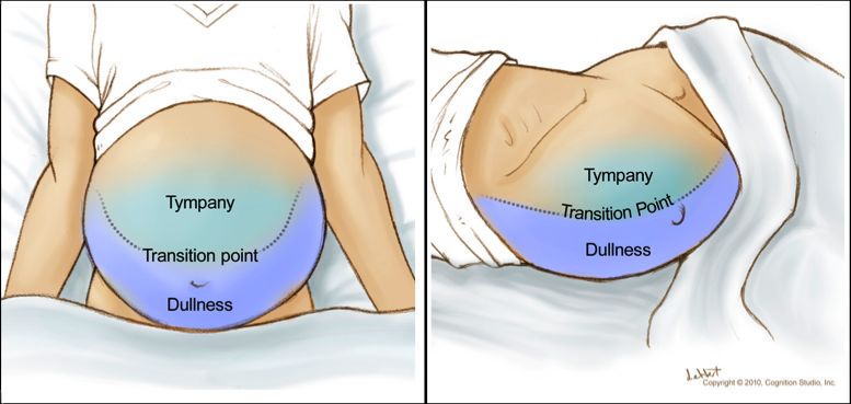
Patient Position and Site for Paracentesis: The procedure is usually performed with the
patient lying supine. As described in the most recent practice guidelines from the American
Association for the Study of Liver Diseases, the left lower quadrant of the abdomen is the
preferred site for the paracentesis and the exact insertion site should be located 2
fingerbreadths (3 cm) cephalad and 2 fingerbreadths (3 cm) medial to the anterior superior
iliac spine (Figure 5).[5] Some experts choose the midline of the abdomen midway between
the pubis and umbilicus, but this site is considered less preferable in obese patients (due to
the increase in midline wall thickness) and in patients with lower volume-ascites (a smaller
pool of fluid in the midline than in the lateral quadrant). The right lower quadrant may be
complicated by a dilated cecum or appendectomy scar. Extreme care should be taken to
avoid the inferior epigastric arteries (Figure 6), which are located halfway between the pubis
and anterior superior iliac spines and run cephalad in the rectus sheath, as well as visible
collaterals in the abdominal wall. In addition, caution is needed in patients who have a
palpable spleen, as it could be ruptured with the left lower quadrant approach. If the ascitic
fluid is difficult to find on physical examination or if there is significant bowel dilatation,
ultrasonography can be used to help locate the fluid pocket and visualize the spleen and
other structures to guide this procedure. Paracentesis sites should be chosen distant from
abdominal surgical scars or under image guidance.
Choosing Needle for Insertion: A 1.0 or 1.5 inch 21 or 22 gauge single hole needle (or 3.5
inch 22 gauge needle for obese patients) can be used for a diagnostic paracentesis, whereas
a 15 or 16 gauge multihole two-piece needle set can be used for therapeutic paracentesis,
involving the removal of more than 5 L of ascites for symptomatic relief from abdominal pain,
early satiety, and/or dyspnea.
Preparation and Insertion Technique: The site should be cleansed with iodine or
chlorhexidine solution and the skin should be anesthetized using 1% lidocaine solution via a
25 or 27 gauge needle. Sterile gloves should be worn to avoid contamination of samples.
After raising a wheal in the superficial skin, 3 to 5 mL of lidocaine is used to anesthetize the
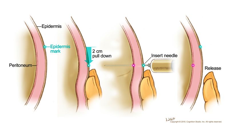
soft tissue tract using the Z-track technique (the skin is pulled downward with the non-
dominant hand, while inserting the needle with the other hand (Figure 7), to decrease the
risk of ascitic fluid leak. The skin is not released until the needle enters the peritoneal cavity,
indicated by the aspiration of ascitic fluid. The paracentesis needle is inserted along the
same line using the Z-track technique. A scalpel can be used to create a skin nick to facilitate
the entry of the larger gauge needle if therapeutic paracentesis is needed. After entry into
the peritoneum, the angle and depth of the paracentesis needle should be stabilized. The
suction applied should be intermittent rather than continuous to avoid pulling in omentum or
bowel into the needle tip and obstructing flow. If the flow of liquid stops, the patient can be
slowly repositioned in an effort to pool more fluid near the needle tip.
Fluid Collection and Samples: For a diagnostic tap, a minimum of 25 mL of fluid should be
collected. One to two mL of ascitic fluid should be injected into a purple top (EDTA) tube for
the cell count and differential tests. Three to four mL of fluid should be directed into a red top
tube for chemical analysis. Fluid should be directly inoculated into blood culture bottles at
the bedside, typically 10 mL into each bottle. If needed, an additional 50 mL of fluid can be
sent in a sterile syringe or cup for cytology or other tests. Vacuum bottles are used to assist
the speed of fluid removal in a therapeutic paracentesis.
Paracentesis Complications: The paracentesis procedure is generally very safe, with only
a 1% risk of abdominal wall hematoma and a less than 0.5% risk of mortality, even in
patients with coagulopathy related to liver disease.[6] Post-paracentesis ascitic fluid leak can
occur in 5% of patients, especially when larger needles are used. More serious complications
such as hemoperitoneum and bowel perforation are extremely rare, reported in less than 1 in
1000 cases.[7] Infections due to this procedure are rare, most often occurring in cases of
bowel injury.[8]
Page 2/32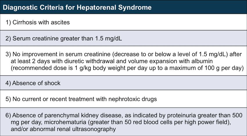
Page 3/32
Analysis of Ascitic Fluid
The following includes a summary of major laboratory tests to consider performing with diagnostic
paracentesis. Other tests not discussed can be ordered if there is suspicion for alternative or
additional causes of ascites. For any initial diagnostic paracentesis to evaluate ascites, it is important
to determine whether portal hypertension is present and whether the ascitic fluid is infected.

        Albumin and Protein: Routinely, an ascitic fluid sample should be sent for albumin and
        total protein. The serum-ascites albumin gradient (SAAG) is calculated by subtracting the
        ascitic fluid albumin value from the serum albumin value obtained on the same day. A SAAG
        value greater than or equal to 1.1 g/dL is indicative of portal hypertension, but does not
        exclude additional causes of ascites in a patient with portal hypertension.[9] An ascitic fluid
        total protein value less than 2.5 g/dL is consistent with ascites from cirrhosis or nephrotic
        syndrome, whereas a high ascitic fluid protein value greater than 2.5 g/dL is seen in patients
        with a cardiac cause of ascites.
        Cell Count and Cultures: Routinely, a cell count and differential should be performed on
        ascitic fluid. With any concern for infection, the fluid should be directly inoculated into
        aerobic and anaerobic blood culture bottles at the bedside prior to the administration of
        antibiotics, as it increases the yield of bacterial growth in culture from 50% to around 80%
        when the polymorphonuclear leukocyte (PMN) count is greater than or equal to 250
        cells/mm3.[10,11] The yield on Gram's staining of ascitic fluid is very low, except in the
        setting of bowel perforation into the ascites. Fungal cultures should be obtained if indicated.
        Mycobacterial Smear and Culture: Ascitic fluid smear and culture for mycobacteria should
        be reserved for patients at high risk for tuberculous peritonitis as the sensitivity of the smear
        is poor and the sensitivity of the fluid culture for mycobacteria is only approximately 50%.
        The 4 to 6 weeks needed before culture results are available delays diagnosis. Ascitic fluid
        polymerase chain reaction (PCR) assays can be done but the utility of these tests has not
        been well established. The gold standard for the diagnosis of tuberculous peritonitis remains
        directed peritoneal biopsy via laparoscopy or mini laparotomy and mycobacterial culture.
        Carcinoembryonic Antigen and Alkaline Phosphatase: Ascitic fluid carcinoembryonic
        antigen greater than 5 ng/mL or ascitic fluid alkaline phosphatase greater than 240 units/L is
        consistent with secondary bacterial peritonitis due to gut perforation.[12] Additional labs that
        support secondary bacterial peritonitis from gastrointestinal perforation include ascitic fluid
        total protein greater than 1 g/dL, glucose less than 50 mg/dL, and lactate dehydrogenase
        (LDH) greater than 225 mU/mL.[13]
        Cytology: Ascitic fluid cytology is expensive and is only revealing in the setting of peritoneal
        carcinomatosis, typically in patients with a history of breast, colon, gastric or pancreatic
        carcinoma. At least 50 mL of fresh warm ascitic fluid needs to be immediately processed for
        optimal yield, with a sensitivity of 82.8% with one sample sent, improving to 96.7% when 3
        samples are sent from different paracenteses.[14]
        Cancer Antigen 125: Serum cancer antigen 125 (CA125) can be elevated in any patient
        with ascites or pleural effusion of any cause, as the level rises when mesothelial cells are
        under pressure in the presence of fluid, so it does not necessarily indicate ovarian
        malignancy in this setting. Thus, CA125 is not routinely ordered as a diagnostic test when
        evaluating ascitic fluid.

Persistent Ascites due to Cirrhosis

Patients undergoing serial outpatient therapeutic paracenteses only need to have the fluid routinely
sent for cell count and differential. At time of any hospital admission, before initiation of antibiotics,
patients should undergo diagnostic paracentesis for cell count and differential and bacterial culture
to assess for spontaneous bacterial peritonitis (SBP). The diagnosis of SBP requires an elevated
ascitic fluid absolute PMN count of greater than or equal to 250 cells/mm3 without an obvious
treatable intraabdominal source of infection, which should prompt empiric antibiotic therapy.[15,16]

                                              Page 4/32
Page 5/32
Basic Management of Ascites
Sodium restriction and diuretics are the mainstays of treatment for patients with ascites due to
portal hypertension, but patients with low SAAG (less than 1.1 g/dL) ascites do not respond well to
these measures, with the exception of those with nephrotic syndrome (Figure 8).[2]

Treatment of the Underlying Disorder

Cessation of alcohol use is vital to the management of ascites due to alcoholic liver disease. In one
study of hospitalized patients with Child-Turcotte-Pugh class C cirrhosis due to severe alcoholic liver
disease, 75% of those who remained abstinent were still alive at 3 years whereas most who
continued to drink alcohol were not.[17] Treatment of autoimmune hepatitis and chronic hepatitis B
can also lead to significant clinical improvement and resolution of ascites in some cases. Similar to
the management of liver-related ascites, treatment of ascites in non-hepatic cases should focus on
treatment of the underlying disorder (e.g. treatment of tuberculosis, treatment of secondary
bacterial peritonitis, or surgical resection of benign ovarian tumor).

Dietary Sodium Restriction

Patients with portal hypertension-associated ascites should restrict their daily dietary sodium intake
to less than 2000 mg (88 mmol).[2] Any further restriction risks malnutrition due to poor palatability
of foods. Twenty-four hour urinary sodium excretion can be measured to assess the adequacy of
fluid loss and dietary sodium restriction. Completeness of the 24-hour collection is estimated by
measurement of 24-hour urinary creatinine; accounting for some anticipated loss of body mass in
the setting of cirrhosis, daily excretion of creatinine should exceed 15 mg/kg body weight in cirrhotic
men and 10 mg/kg body weight in cirrhotic women. The goal of treatment is to increase the daily
urinary excretion of sodium to a value above 78 mmol per day, so that in conjunction with daily
nonurinary sodium excretion, the daily sodium excretion should exceed the allowed daily dietary
intake of sodium.[2] Random urinary sodium concentration is not useful because of the variable
sodium excretion and total urine volume throughout the day, but a random “spot” urine
sodium/potassium ratio correlates with 24-hour urinary sodium excretion, with higher ratios
indicating greater urinary excretion. Thus, a ratio of greater than one is desired. Patients who are
excreting a sufficient amount of urinary sodium (24-hour urinary sodium greater than 78 mmol per
day or spot urine sodium/potassium ratio greater than one) and are not losing weight are likely
consuming more than 2000 mg of sodium daily and need further education and adherence
counseling. On the other hand, the diuretic dose should be increased in patients not excreting a
sufficient amount of urinary sodium, unless they are diuretic resistant.

Fluid Restriction

Dietary sodium restriction is more important than fluid restriction in the management of cirrhosis.
Fluid restriction is not necessary unless the serum sodium concentration is less than 120 mmol/L or
mental status changes attributed to hyponatremia develop. Rapid correction of chronic
hyponatremia (with hypertonic saline or other means) should be avoided due to risk of osmotic
demyelination syndrome.

Diuretics

In patients with portal hypertension, the combination of spironolactone and furosemide, starting at
doses of 100 mg daily and 40 mg daily, respectively, is recommended.[2] Single agent
spironolactone can be used and is superior to single agent furosemide,[18] but combination therapy
leads to more rapid fluid loss in patients with moderate ascites and decreases the risk of
hyperkalemia. If weight loss is insufficient, maintaining the 100 mg:40 mg ratio, the doses of the
diuretics may be increased simultaneously every 3 to 5 days to maximum daily doses of 400 mg of

                                             Page 6/32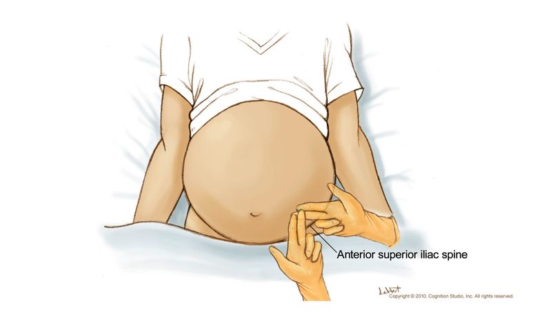
spironolactone and 160 mg of furosemide. The combined single morning dosing improves
compliance, optimizes diuresis, and avoids nocturia. A ratio less than 100 mg:40 mg of
spironolactone and furosemide may be used for patients with parenchymal renal disease with
concern for hyperkalemia. Furosemide can be temporarily held or reduced for those with
hypokalemia.

Option if Intolerant to Spironolactone

For patients unable to tolerate spironolactone due to painful gynecomastia, amiloride (10 to 40 mg
daily) can be substituted, although it has a lower natriuretic effect than spironolactone.[19]
Eplerenone is a newer aldosterone antagonist used to treat heart failure and is not associated with
gynecomastia but has not been extensively studied yet for the management of ascites.[20]
Hydrochlorothiazide in combination with furosemide is not recommended due to combined
hypokalemia. Torsemide and bumetanide have also been used in combination with spironolactone in
the management of ascites, but they have not demonstrated superiority over furosemide.

Daily Limit for Weight Loss

In patients with significant peripheral edema, there is no limit for daily weight loss, but in those
without peripheral edema, daily weight loss should be restricted to 0.5 kg maximum. Diuretics may
need to be held in the setting of significant volume loss such as active gastrointestinal hemorrhage
or diarrhea, uncontrolled or recurrent hepatic encephalopathy, significant hyponatremia (serum
sodium less than 120 mmol/L) despite fluid restriction, or renal dysfunction (e.g. serum creatinine
greater than 2.0 mg/dL).

Medications to Avoid

The use of angiotensin converting enzyme inhibitors and angiotensin receptor blockers should be
avoided in patients with cirrhosis, due to concerns of renal failure and increased mortality for those
who develop hypotension. Hypotension (mean arterial pressure less than or equal to 82 mmHg)
independently predicts increased one-year mortality in patients with cirrhosis. In patients with
refractory ascites, propranolol is associated with decreased survival perhaps due to the increased
risk of paracentesis-induced circulatory dysfunction, so, the risks and benefits of its use should be
considered individually for each patient.[21] Nonsteroidal anti-inflammatory drugs (NSAIDS),
including aspirin, should also be avoided due to the risk of reduced urinary sodium excretion and
renal failure. Although vaptans can improve hyponatremia, there are significant risks associated with
use of these types of agents in patients with cirrhosis. For example, tolvaptan, a selective oral
vasopressin V2-receptor antagonist used to treat hypervolemic and euvolemic hyponatremia, has
been shown to be effective in patients with refractory ascites, but is contraindicated for use in
persons with underlying liver disease, including those with cirrhosis, due to risk of causing severe
hepatotoxicity.[22,23] Moreover, hyponatremia recurs upon discontinuation of the medication.[24]
Satavaptan was evaluated for the management of ascites in patients with cirrhosis and was
potentially associated with a higher risk of mortality.[25]

Management of Tense Ascites

A single large volume paracentesis followed by dietary sodium restriction and initiation of diuretics is
appropriate as initial therapy for new onset large volume ascites.[26] Up to 5 liters can be removed
without significant disturbances in systemic and renal hemodynamics,[27] but if more than 5 liters of
ascitic fluid is removed, then intravenous albumin (8 g/L of fluid removed) should be given.[28]

                                             Page 7/32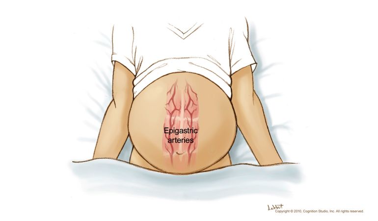
Management of Refractory Ascites
Among patients with cirrhosis and ascites, fewer than 10% will develop refractory ascites, which is
defined as ascites that is unresponsive to dietary sodium restriction and maximal diuretic dosing
(typically, spironolactone 400 mg daily and furosemide 160 mg daily), or that which recurs rapidly
after therapeutic paracentesis.[29] There are two different subtypes: diuretic-resistant ascites (lack
of response to dietary sodium restriction and intensive diuretic treatment) and diuretic-intractable
ascites (diuretic-induced complications such as hepatic encephalopathy, renal insufficiency,
hyponatremia, or hyperkalemia that prevent optimization of diuretic dosing). Once refractory ascites
develops, one-year mortality is approximately 50%. Options for treatment include optimization of
medical management, serial large volume paracenteses, transjugular intrahepatic portosystemic
shunt (TIPS), peritoneovenous shunt, and liver transplantation.

Medical Treatment

As mentioned previously, propranolol has been shown to be associated with decreased survival in
the setting of refractory ascites and discontinuation should be considered. Angiotensin-converting
enzyme inhibitors and angiotensin receptor blockers should be avoided. Oral midodrine, an agent
used to treat hypotension, was shown to increase mean arterial pressure and improve survival in a
pilot study in cirrhotic patients with refractory or recurrent ascites.[30] The dose is typically initiated
at 5 mg orally three times daily and titrated upward in 2.5 mg increments for each dose daily, with a
maximum dose of 17.5 mg three times daily to achieve an increase in systolic blood pressure of 10
to 15 mmHg. A favorable clinical response to midodrine may allow for reinitiation of diuretic therapy.

Serial Large Volume Therapeutic Paracenteses

Once a patient is deemed diuretic resistant, diuretics should be discontinued, and management may
rely upon serial large volume therapeutic paracenteses alone. Typically, a large volume paracentesis
(up to 10 L removed) performed every 2 weeks should control ascites in a patient who is compliant
with dietary sodium restriction.[31] Need for more frequent paracenteses suggests dietary non-
compliance. The use of indwelling intraabdominal catheters is reserved for patients with malignancy-
associated ascites and is not recommended in this situation. Long-term serial paracenteses can lead
to significant loss of protein and worsen malnutrition, but placement of a percutaneous endoscopic
gastrostomy (PEG) tube in an effort to provide nutrition should be avoided in these patients due to
the high risk of mortality associated with performing the procedure.[32]

Albumin Infusions with Therapeutic Paracentesis

In one randomized study, the use of intravenous albumin (10 grams administered per liter of fluid
removed) in the setting of therapeutic paracentesis decreased the risk of negative changes in
plasma renin and serum creatinine levels.[33] A meta-analysis of 17 trials demonstrated a reduction
in risk of post-paracentesis circulatory dysfunction, hyponatremia, and mortality in the albumin
group (odds ratio of death 0.64, 95% CI, 0.41-0.98); study protocols typically used a 20% or higher
concentration of albumin solution, and administered 5 to 10 g of albumin per liter of fluid
removed.[28] With a large volume paracentesis (5 liters or more removed), some experts
recommend giving 6 to 8 g of intravenous albumin for every liter of ascitic fluid removed, with the
albumin infused during or immediately following the paracentesis. In the United States, both 5% and
25% concentrations of intravenous albumin are available but the 25% solution is preferred since the
5% solution contains 5 times the amount of sodium.

Transjugular Intrahepatic Portosystemic Shunt (TIPS)

TIPS, which is a side-to-side portocaval shunt placed by Interventional Radiology, has been shown in
multiple multicenter randomized controlled trials to be superior to serial large volume paracenteses

                                              Page 8/32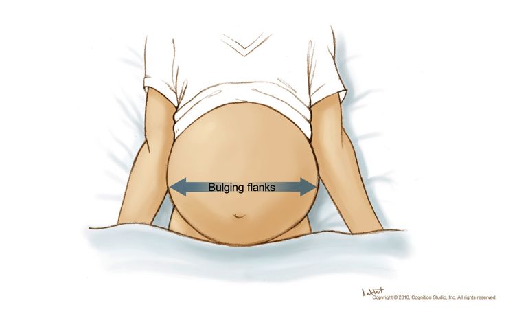
in the control of ascites, but with varying results on the impact on overall transplant-free survival
and the potential risk of inducing or worsening hepatic encephalopathy.[34,35,36,37,38,39]
Polytetrafluoroethylene-covered stents are preferred over uncovered stents due to decreased rates
of TIPS occlusion.
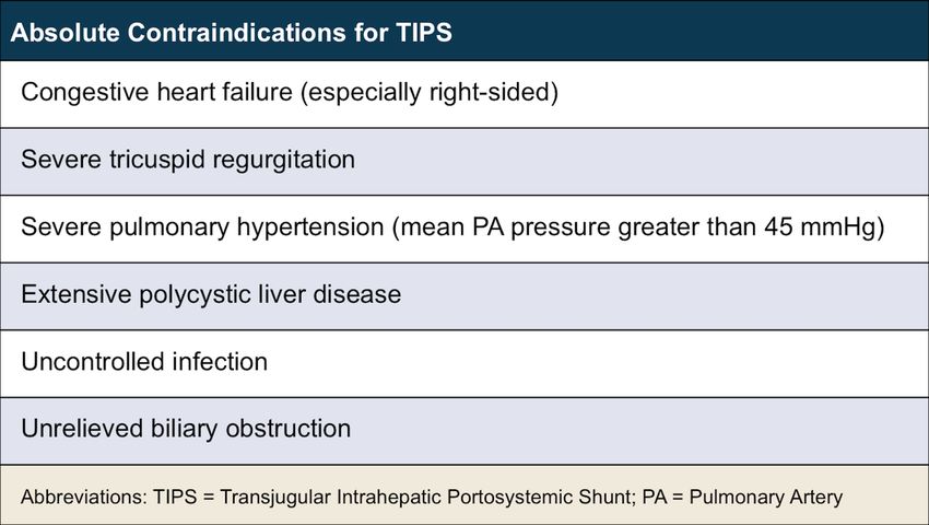
Absolute Contraindications: The absolute contraindications to placement of TIPS include
congestive heart failure (particularly right-sided heart failure), severe tricuspid regurgitation,
severe pulmonary hypertension (mean pulmonary pressure greater than 45 mmHg),
extensive polycystic liver disease, and uncontrolled infection or biliary obstruction (Figure 9
).[34]
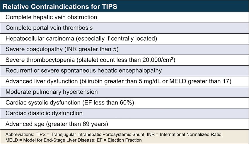
Relative Contraindications: The relative contraindications for performing TIPS include
obstruction of all hepatic veins, complete portal vein thrombosis, hepatocellular carcinoma
(especially if centrally located), severe coagulopathy (international normalized ratio [INR]
greater than 5) or thrombocytopenia (platelet count less than 20,000/cm3), moderate
pulmonary hypertension, recurrent or persistent severe spontaneous hepatic
encephalopathy, advanced liver failure (bilirubin greater than 5 mg/dL or Model for End-stage
Liver Disease [MELD] score greater than 17), cardiac dysfunction (ejection fraction less than
60%), cardiac diastolic dysfunction, and advanced age (e.g. greater than 69 years) (Figure 10
).[34]
Outcome after TIPS: Short- and long-term mortality rates following TIPS can be estimated
using MELD and Child-Turcotte-Pugh scoring systems. Clinical improvement in ascites
following TIPS is seen in 74% of patients.[40] Diuretics may need to be continued even after
placement of TIPS. Approximately 30% of patients develop hepatic encephalopathy after
TIPS, though most can be managed medically (e.g. lactulose). Risk factors for the
development of hepatic encephalopathy after TIPS include older age and history of pre-TIPS
hepatic encephalopathy.[41] Narrowing or occluding the TIPS can treat severe debilitating
hepatic encephalopathy resistant to medical therapy, which, fortunately, is rare. Those with
renal dysfunction, especially those on dialysis, may have a reduced response to TIPS.
Peritoneovenous Shunts
The use of peritoneovenous shunts for management of ascites has fallen out of favor due to limited
long-term patency (less than 20% at 2 years), risk of complications, and no improvement in survival
compared to medical therapy.[42,43,44] It is reserved as palliative treatment in select patients who
are not candidates for transplantation, TIPS, or serial therapeutic paracenteses.[2]
Page 9/32
Complications Associated with Ascites
Spontaneous Bacterial Peritonitis (SBP)

Diagnosis of SBP requires an ascitic fluid absolute polymorphonuclear count greater than or equal to
250 cells/mm3 without an obvious intraabdominal, surgically-treatable source and should prompt
empiric antibiotic treatment with an intravenous third-generation cephalosporin, preferably
cefotaxime 2 g every 8 hours, for 5 days.[45,46] Patients with serum creatinine greater than 1
mg/dL, blood urea nitrogen greater than 30 mg/dL, or total bilirubin greater than 4 mg/dL should
receive intravenous albumin 1.5 g per kg body weight upon diagnosis and 1.0 g per kg body weight
on day 3 after diagnosis.[47,48] After an episode of SBP, patients should receive long-term
prophylaxis with daily norfloxacin or trimethoprim-sulfamethoxazole.[49] More detailed information
regarding diagnosis, treatment, and prevention of SBP is provided in Lesson 2 of this Module.

Dilutional Hyponatremia

Vasodilatation in cirrhosis triggers activation of the renin-angiotensin system and sympathetic
nervous system, leading to avid sodium and water retention with increased antidiuretic hormone
release, resulting in dilutional hyponatremia. Up to 50% of patients with cirrhosis and ascites have a
serum sodium concentration less than 135 mmol/L. Hyponatremia is an independent risk factor for
morbidity and mortality in patients with cirrhosis and has been proposed as an addition to the MELD
score for liver transplant prioritization.[1,19,50]

       Indication for the Treatment of Hyponatremia: Treatment specifically for hyponatremia
       is not necessary unless the serum sodium concentration drops below 120 mmol/L, which
       occurs in only 1% of patients, or if there are neurologic symptoms attributed to
       hyponatremia. If treated, the rate of correction should not exceed an increase of more than 9
       mmol/L per day, with a goal of increasing only 4 to 6 mmol/L per day, in order to avoid the
       risk of osmotic demyelination syndrome.[51]
       Approach to Treatment of Hyponatremia: Relative fluid restriction (1000 to 1500 mL free
       water per day) and discontinuation of diuretics should be the first line of treatment; true fluid
       restriction (total fluid intake less than urine volume) is difficult to achieve. Treatment of
       hypokalemia may also raise serum sodium concentration. Vasopressin receptor antagonists
       (vaptans) cause selective water diuresis and raise serum sodium concentrations, but are not
       routinely used in patients with cirrhosis. Conivaptan is a V1a receptor blocker that requires
       intravenous administration and is not recommended in patients with cirrhosis because of the
       concern that it can increase the risk of hypotension and renal compromise. Tolvaptan, an oral
       V2 receptor blocker, should also be avoided in patients with cirrhosis due to concerns for liver
       injury; this side effect was observed in a clinical study of tolvaptan in patients with polycystic
       kidney disease. The utility of demeclocycline, a tetracycline derivative, in patients with
       cirrhosis is limited due to the risk of nephrotoxicity.[52] Hypertonic saline is generally
       avoided in patients with cirrhosis except to partially correct severe hyponatremia
       immediately prior to liver transplantation. Over-rapid correction before, during, and after liver
       transplantation should be avoided.

Hepatorenal Syndrome

Approximately 20% of hospitalized patients with cirrhosis and ascites will develop some type of renal
dysfunction. In one study, over a mean follow-up of 41 months, 7.6% of hospitalized patients with
ascites and cirrhosis developed HRS.[53]

       Diagnostic Criteria for Hepatorenal Syndrome: The diagnostic criteria for hepatorenal
       syndrome requires all of the following: (1) cirrhosis with ascites, (2) serum creatinine greater
       than 1.5 mg/dL, (3) no improvement in serum creatinine (decrease to or below a level of 1.5

                                             Page 10/32
mg/dL) after at least 2 days with diuretic withdrawal and volume expansion with albumin
(recommended dose is 1 g/kg body weight per day up to a maximum of 100 g per day), (4)
absence of shock, (5) no current or recent treatment with nephrotoxic drugs, and (6) absence
of parenchymal kidney disease, as indicated by proteinuria greater than 500 mg per day,
microhematuria (greater than 50 red blood cells per high power field), and/or abnormal renal
ultrasonography (Figure 11).[37]
Classification of Hepatorenal Syndrome: There are two types of hepatorenal syndrome:
type 1 and type 2.[54] Type 1 hepatorenal syndrome is characterized by rapidly progressive
renal failure with a doubling in the initial serum creatinine to a level greater than 2.5 mg/dL
(or 50% reduction in the initial 24-hour creatinine clearance to a level lower than 20 mL/min)
in less than two weeks; it is frequently triggered by a precipitating event, such as SBP,
urinary tract infection, or intravascular volume contraction, and is associated with acute rapid
deterioration of circulatory function with hypotension and activation of endogenous
vasoconstrictor systems, and leads to a very poor prognosis, with a median survival of
around 2 weeks in untreated patients. Type 2 hepatorenal syndrome is typically associated
with refractory ascites and is characterized by a slower, progressive decline in renal function,
typically with a serum creatinine that ranges from 1.5 to 2.5 mg/dL, and a median survival of
4 to 6 months.
Management of Type 1 Hepatorenal Syndrome: Management is focused on the
treatment of the precipitating event, the renal failure, and the systemic inflammatory
response syndrome. Measures to prevent Type 1 HRS include the use intravenous albumin in
patients with SBP at high risk for developing HRS and the use of prophylactic antibiotics in
cirrhotic patients with gastrointestinal bleeding.[55] Once Type 1 HRS is established,
diuretics should be discontinued and vasoconstrictors used to decrease systemic
vasodilatation and improve renal perfusion. The combination of terlipressin and albumin has
been shown to be superior to albumin alone and placebo for the treatment of Type 1
hepatorenal syndrome and may be effective in more than 30% of cases.[56,57] Terlipressin
is not available in the United States, so midodrine, an alpha-agonist, is used instead (starting
at a dose of 5 to 7.5 mg orally three times daily, titrated up to 15 mg three times daily), in
combination with octreotide, starting with 100 mcg subcutaneously three times daily, titrated
up to 200 mcg three times daily and albumin (up to 40 g daily in divided doses), with a goal
of increasing mean arterial pressure by 15 mmHg. This combination achieves a response rate
of around 30% as shown in case series.[58] For patients in the intensive care unit, albumin
infusion with norepinephrine can be considered as well for Type 1 hepatorenal syndrome.[59]
In addition, TIPS can be used to improve renal function, but should be avoided in patients
with advanced liver dysfunction. Ultimately, liver transplantation is the definitive treatment
for this condition, and some even require renal replacement therapy as a bridge to
transplantation.[60]
Management of Type 2 Hepatorenal Syndrome: Treatment of type 2 hepatorenal
syndrome is typically centered on management of the refractory ascites, with measures such
as TIPS. If eligible, these patients should be referred for consideration of liver transplantation.
Umbilical Hernia
Up to 20% of patients with cirrhosis and ascites can develop umbilical hernias. Complications related
to these hernias include omental or bowel strangulation, typically after paracentesis or shunt
procedure, and hernia perforation.[61] Patients should wear an abdominal binder to minimize strain
and enlargement of the hernia and should be educated on the warning symptoms of an incarcerated
hernia. Pre-emptive TIPS should be considered to prevent rupture of thin-walled umbilical
hernias.[62,63] The risks and benefits of elective surgical repair need to be assessed individually. In
patients who are medical candidates for surgery (e.g. Child-Turcotte-Pugh class A cirrhosis), the
ascites needs to be controlled first with optimal medical management or TIPS; otherwise, the hernia
will recur in over 70% of patients.[64] Emergent surgical repair due to incarceration or rupture
should be performed by surgeons experienced with patients with cirrhosis. If feasible, TIPS should be
considered before or after the surgery, along with dietary sodium and fluid restriction.
Page 11/32Hepatic Hydrothorax
Approximately 5 to 10% of patients with cirrhosis and ascites develop hepatic hydrothorax, which is
typically a right-sided pleural effusion.[65] It is a result of fluid being drawn up from the peritoneal
cavity into the pleural space through small defects in the diaphragm. Sometimes, minimal to almost
no fluid remains in the abdomen. Injection of technetium-radiolabeled sulfur colloid into the
abdomen followed by transdiaphragmatic flow of the isotope into the thoracic space can confirm
ascites as the origin of the pleural effusion, if needed.[66] Thoracentesis does not require platelet or
fresh frozen plasma transfusions, and, there is no limit to the amount of fluid that can be
removed.[67] Due to differences in hydrostatic pressure, the protein concentration is higher in
pleural fluid than ascites. Spontaneous bacterial empyema can occur in the absence of spontaneous
bacterial peritonitis and can be treated with appropriate antibiotic therapy without placement of a
chest tube.[68] Chest tube placement in patients with hepatic hydrothorax is associated with
massive fluid losses, high morbidity (greater than 90%) and high mortality (greater than 30% in the
absence of TIPS), so it should be avoided.[69,70] Treatment should start with dietary sodium
restriction and diuretics. Therapeutic thoracentesis can be done for dyspnea. TIPS can be performed
as treatment for refractory hepatic hydrothorax. Most patients with hepatic hydrothorax will not be
candidates for pleurodesis due to the rapid rate of fluid reaccumulation.
Page 12/32Summary Points
The development of ascites indicates decompensation of cirrhosis, and patients should be
referred for liver transplantation evaluation.
Prophylactic blood products do not need to be administered prior to paracentesis, even in the
setting of coagulopathy or thrombocytopenia, but paracentesis should be avoided in patients
with disseminated intravascular coagulation or untreated hyperfibrinolysis.
A SAAG of greater than or equal to 1.1 g/dL indicates portal hypertension as the cause of
ascites, with cirrhosis or heart failure common causes of the portal hypertension. Additional
diagnostic tests can be ordered based on clinical suspicion.
Treatment of ascites in patients with cirrhosis should be focused on dietary sodium restriction
of less than 2000 mg daily and the use of diuretics, specifically, spironolactone and
furosemide, titrated using a respective ratio of 100 mg:40 mg. Fluid restriction is reserved
only for those with a serum sodium concentration of less than 120 mmol/L or symptomatic
hyponatremia.
Treatment options for the management of refractory ascites include optimization of medical
therapy, serial large volume therapeutic paracenteses with the use of intravenous albumin,
TIPS in selected candidates, and liver transplantation. Peritoneovenous shunt is a palliative
measure reserved only for patients who are not candidates for the other therapies.
An ascitic fluid absolute polymorphonuclear count greater than or equal to 250 cells/mm3
should prompt empiric antibiotic treatment for spontaneous bacterial peritonitis with
intravenous cefotaxime (2 g every 8 hours) for five days.
Patients with untreated Type 1 hepatorenal syndrome have very poor short-term survival and
should be referred for urgent liver transplantation evaluation.
In most circumstances, placement of a chest tube is contraindicated in patients with hepatic
hydrothorax due to risk of massive fluid loss and high morbidity and mortality.
Page 13/32Citations
1. Planas R, Montoliu S, Ballesté B, et al. Natural history of patients hospitalized for
management of cirrhotic ascites. Clin Gastroenterol Hepatol. 2006;4:1385-94.
[PubMed Abstract] -
2. Runyon BA. American Association for the Study of Liver Diseases (AASLD) Practice Guidelines
Committee. Management of Adult Patients with Ascites Due to Cirrhosis: Update 2012.
[AASLD Practice Guidelines] -
3. Cattau EL Jr, Benjamin SB, Knuff TE, Castell DO. The accuracy of the physical examination in
the diagnosis of suspected ascites. JAMA. 1982;247:1164-6.
[PubMed Abstract] -
4. Gunawan B, Runyon B. The efficacy and safety of epsilon-aminocaproic acid treatment in
patients with cirrhosis and hyperfibrinolysis. Aliment Pharmacol Ther. 2006;23:115-20.
[PubMed Abstract] -
5. Sakai H, Sheer TA, Mendler MH, Runyon BA. Choosing the location for non-image guided
abdominal paracentesis. Liver Int. 2005;25:984-6.
[PubMed Abstract] -
6. De Gottardi A, Thévenot T, Spahr L, Morard I, Bresson-Hadni S, Torres F, Giostra E, Hadengue
A. Risk of complications after abdominal paracentesis in cirrhotic patients: a prospective
study. Clin Gastroenterol Hepatol. 2009;7:206-9.
[PubMed Abstract] -
7. Pache I, Bilodeau M. Severe haemorrhage following abdominal paracentesis or ascites in
patients with liver failure. Aliment Pharmacol Ther. 2005;21:525-9.
[PubMed Abstract] -
8. Runyon BA. Paracentesis of ascitic fluid: a safe procedure. Arch Intern Med.
1986;146:2259-61.
[PubMed Abstract] -
9. Runyon BA, Montano AA, Akriviadis EA, Antillon MR, Irving MA, McHutchison JG. The serum-
ascites albumin gradient is superior to the exudate-transudate concept in the differential
diagnosis of ascites. Ann Intern Med. 1992;117:215-20.
[PubMed Abstract] -
10. Runyon BA, Antillon MR, Akriviadis EA, McHutchinson JG. Bedside inoculation of blood culture
bottles is superior to delayed inoculation in the detection of spontaneous bacterial peritonitis.
J Clin Microbiol. 1990;28:2811-2.
[PubMed Abstract] -
11. Runyon BA, Canawati HN, Akriviadis EA. Optimization of ascitic fluid culture technique.
Gastroenterology. 1988;95:1351-5.
[PubMed Abstract] -
12. Wu SS, Lin OS, Chen Y-Y, Hwang KL, Soon MS, Keeffe EB. Ascitic fluid carcinoembryonic
antigen and alkaline phosphatase levels for the differentiation of primary from secondary
bacterial peritonitis with intestinal perforation. J Hepatol. 2001;34:215-21.
[PubMed Abstract] -
13. Runyon BA, Hoefs JC. Ascitic fluid analysis in the differentiation of spontaneous bacterial
Page 14/32peritonitis from gastrointestinal tract perforation into ascitic fluid. Hepatology.
1984;4:447-50.
[PubMed Abstract] -
14. Runyon BA. Malignancy-related ascites and ascitic fluid “humoral tests of malignancy.” J Clin
Gastroenterol. 1994;18:94-8.
[PubMed Abstract] -
15. Runyon BA. Management of adult patients with ascites due to cirrhosis: an update.
Hepatology. 2009;49:2087-107.
[PubMed Abstract] -
16. Such J, Runyon BA. Spontaneous bacterial peritonitis. Clin Infect Dis. 1998;27:669-74.
[PubMed Abstract] -
17. Veldt BJ, Lainé F, Guillygomarc’h A, et al. Indication of liver transplantation in severe alcoholic
liver cirrhosis: quantitative evaluation and optimal timing. J Hepatol. 2002;36:93-8.
[PubMed Abstract] -
18. Pérez-Ayuso RM, Arroyo V, Planas R, et al. Randomized comparative study of efficacy of
furosemide versus spironolactone in nonazotemic cirrhosis with ascites. Relationship between
the diuretic response and the activity of the renin-aldosterone system. Gastroenterology.
1983;84:961-8.
[PubMed Abstract] -
19. Angeli P, Dalla Pria M, De Bei E, Albino G, Caregaro L, Merkel C, Ceolotto G, Gatta A.
Randomized clinical study of the efficacy of amiloride and potassium canrenoate in
nonazotemic cirrhotic patients with ascites. Hepatology. 1994;19:72-9.
[PubMed Abstract] -
20. Dimitriadis G, Papadopoulos V, Mimidis K. Eplerenone reverses spironolactone-induced
painful gynaecomastia in cirrhotics. Hepatol Int. 2011;5:738-9.
[PubMed Abstract] -
21. Sersté T, Melot C, Francoz C, et al. Deleterious effects of beta-blockers on survival in patients
with cirrhosis and refractory ascites. Hepatology. 2010;52:1017-22.
[PubMed Abstract] -
22. Zhang X, Wang SZ, Zheng JF, et al. Clinical efficacy of tolvaptan for treatment of refractory
ascites in liver cirrhosis patients. World J Gastroenterol. 2014;20:11400-5.
[PubMed Abstract] -
23. Ohki T, Sato K, Yamada T, et al. Efficacy of tolvaptan in patients with refractory ascites in a
clinical setting. World J Hepatol. 2015;7:1685-93.
[PubMed Abstract] -
24. Cárdenas A, Ginès P, Marotta P, et al. Tolvaptan, an oral vasopressin antagonist, in the
treatment of hyponatremia in cirrhosis. J Hepatol. 2012;56:571-8.
[PubMed Abstract] -
25. Wong F, Watson H, Gerbes A, et al. Satavaptan for the management of ascites in cirrhosis:
efficacy and safety across the spectrum of ascites severity. Gut. 2012;61:108-16.
[PubMed Abstract] -
26. Ginès P, Arroyo V, Quintero E, et al. Comparison of paracentesis and diuretics in the
Page 15/32treatment of cirrhotics with tense ascites. Results of a randomized study. Gastroenterology.
1987;93:234-41.
[PubMed Abstract] -
27. Peltekian KM, Wong F, Liu PP, Logan AG, Sherman M, Blendis LM. Cardiovascular, renal and
neurohumoral responses to single large-volume paracentesis patients with cirrhosis and
diuretic-resistant ascites. Am J Gastroenterol. 1997;92:394-9.
[PubMed Abstract] -
28. Bernardi M, Caraceni P, Navickis RJ, Wilkes MM. Albumin infusion in patients undergoing large-
volume paracentesis: a meta-analysis of randomized trials. Hepatology. 2012;55:1172-81.
[PubMed Abstract] -
29. Arroyo V, Ginès P, Gerbes AL, et al. Definition and diagnostic criteria of refractory ascites and
hepatorenal syndrome in cirrhosis. International Ascites Club. Hepatology. 1996;23:164-76.
[PubMed Abstract] -
30. Singh V, Dhungana SP, Singh B, et al. Midodrine in patients with cirrhosis and refractory or
recurrent ascites: a randomized pilot study. J Hepatol. 2012;56:348-54.
[PubMed Abstract] -
31. Titó L, Ginès P, Arroyo V, et al. Total paracentesis associated with intravenous albumin
management of patients with cirrhosis and ascites. Gastroenterology. 1990;98:146-51.
[PubMed Abstract] -
32. Baltz JG, Argo CK, Al-Osaimi AM, Northup PG. Mortality after percutaneous endoscopic
gastrostomy in patients with cirrhosis: a case series. Gastrointest Endscop. 2010;72:1072-5.
[PubMed Abstract] -
33. Ginès P, Titó L, Arroyo V, et al. Randomized comparative study of therapeutic paracentesis
with and without intravenous albumin in cirrhosis. Gastroenterology. 1988;94;1493-502.
[PubMed Abstract] -
34. Boyer TD, Haskal ZJ; American Association for the Study of Liver Diseases. The role of
transjugular intrahepatic portosystemic shunt (TIPS) in the management of portal
hypertension: update 2009. Hepatology. 2010;51:1-16.
[AASLD Practice Guidelines] -
35. Ginès P, Uriz J, Calahorra B, et al. Transjugular intrahepatic portosystemic shunting versus
paracentesis plus albumin for refractory ascites in cirrhosis. Gastroenterology.
2002;123:1839-47.
[PubMed Abstract] -
36. Rössle M, Ochs A, Gülberg V, et al. A comparison of paracentesis and transjugular
intrahepatic portosystemic shunting in patients with ascites. N Engl J Med. 2000;342:1701-7.
[PubMed Abstract] -
37. Salerno F, Cammà C, Enea M, Rössle M, Wong F. Transjugular intrahepatic portosystemic
shunt for refractory ascites: a meta-analysis of individual patient data. Gastroenterology.
2007;133:825-34.
[PubMed Abstract] -
38. Salerno F, Merli M, Riggio O, et al. Randomized controlled study of TIPS versus paracentesis
plus albumin in cirrhosis with severe ascites. Hepatology. 2004;40:629-35.
[PubMed Abstract] -
Page 16/3239. Sanyal AJ, Genning C, Reddy KR, et al. The North American Study for the Treatment of
Refractory Ascites. Gastroenterology. 2003;124:634-41.
[PubMed Abstract] -
40. Somberg KA, Lake JR, Tomlanovich SJ, LaBerge JM, Feldstein V, Bass NM. Transjugular
intrahepatic portosystemic shunts for refractory ascites: assessment of clinical and hormonal
response and renal function. Hepatology. 1995;21:709-16.
[PubMed Abstract] -
41. Sanyal AJ, Freedman AM, Shiffman ML, Purdum PP 3rd, Luketic VA, Cheatham AK.
Portosystemic encephalopathy after transjugular intrahepatic portosystemic shunt: results of
a prospective controlled study. Hepatology. 1994;20:46-55.
[PubMed Abstract] -
42. Ginès P, Arroyo V, Vargas V, et al. Paracentesis with intravenous infusion of albumin as
compared with peritoneovenous shunting in cirrhosis with refractory ascites. N Engl J Med.
1991;325:829-35.
[PubMed Abstract] -
43. Guardiola J, Xiol X, Escribá JM, et al. Prognosis assessment of cirrhotic patients with refractory
ascites treated with a peritoneovenous shunt. Am J Gastroenterol. 1995;90:2097-102.
[PubMed Abstract] -
44. Stanley MM, Ochi S, Lee KK, et al. Peritoneovenous shunting as compared with medical
treatment in patients with alcoholic cirrhosis and massive ascites. Veterans Administration
Cooperative Study on Treatment of Alcoholic Cirrhosis with Ascites. N Engl J Med.
1989;321:1632-8.
[PubMed Abstract] -
45. Hoefs JC, Canawati HN, Sapico FL, Hopkins RR, Weiner J, Montgomerie JZ. Spontaneous
bacterial peritonitis. Hepatology. 1982;2:399-407.
[PubMed Abstract] -
46. Runyon BA, McHutchinson JG, Antillon MR, Akriviadis EA, Montano A. Short-course versus long-
course antibiotic treatment of spontaneous bacterial peritonitis: a randomized controlled trial
of 100 patients. Gastroenterology. 1991;100:1737-42.
[PubMed Abstract] -
47. Sigal SH, Stanca CM, Fernandez J, Arroyo V, Navasa M. Restricted use of albumin for
spontaneous bacterial peritonitis. Gut. 2007;56:597-9.
[Gut] -
48. Sort P, Navasa M, Arroyo V, et al. Effect of intravenous albumin on renal impairment and
mortality in patients with cirrhosis and spontaneous bacterial peritonitis. N Engl J Med.
1999;341:403-9.
[PubMed Abstract] -
49. Ginès P, Rimola A, Plana R, et al. Norfloxacin prevents spontaneous bacterial peritonitis
recurrence in cirrhosis: results of a double-blind, placebo-controlled trial. Hepatology.
1990;12:716-24.
[PubMed Abstract] -
50. Heuman DM, Abou-Assi SG, Habib A, et al. Persistent ascites and low serum sodium to
identify patients with cirrhosis and low MELD scores who are high risk for early death.
Page 17/32Hepatology. 2004;40:802-10.
[PubMed Abstract] -
51. Gankam Kengne F, Decaux G. Hyponatremia and the Brain. Kidney Int Rep. 2018;3:24-35.
[PubMed Abstract] -
52. Miller PD, Linas SL, Schrier RW. Plasma demeclocycline levels and nephrotoxicity. Correlation
in hyponatremic cirrhotic patients. JAMA. 1980;243;2513-5.
[PubMed Abstract] -
53. Montoliu S, Ballesté B, Planas R, et al. Incidence and prognosis of different types of functional
renal failure in cirrhotic patients with ascites. Clin Gastroenterol Hepatol. 2010;8:616-22.
[PubMed Abstract] -
54. Egerod Israelsen M, Gluud LL, Krag A. Acute kidney injury and hepatorenal syndrome in
cirrhosis. J Gastroenterol Hepatol. 2015;30:236-43.
[PubMed Abstract] -
55. Ginès P, Guevera M, Arroyo V, Rodés J. Hepatorenal syndrome. Lancet. 2003;362:1819-27.
[PubMed Abstract] -
56. Martín-Llahí M, Pépin MN, Guevara M, et al. Terlipressin and albumin vs albumin in patients
with cirrhosis and hepatorenal syndrome: a randomized study. Gastroenterology.
2008;134:1352-9.
[PubMed Abstract] -
57. Sanyal AJ, Boyer T, Garcia-Tsao G, et al. A randomized, prospective, double-blind, placebo-
controlled trial of terlipressin for type 1 hepatorenal syndrome. Gastroenterology.
2008;134:1360-8.
[PubMed Abstract] -
58. Esrailian E, Pantangco ER, Kyulo NL, Hu KQ, Runyon BA. Octreotide/Midodrine therapy
significantly improves renal function and 30-day survival in patients with type 1 hepatorenal
syndrome. Dig Dis Sci. 2007;52:742-8.
[PubMed Abstract] -
59. Gluud LL, Christensen K, Christensen E, Krag A. Systematic review of randomized trials on
vasoconstrictor drugs for hepatorenal syndrome. Hepatology. 2010;51:576-84.
[PubMed Abstract] -
60. Wong F. Recent advances in our understanding of hepatorenal syndrome. Nat Rev
Gastroenterol Hepatol. 2012;9:382-91.
[PubMed Abstract] -
61. Belghiti J, Durand F. Abdominal wall hernias in the setting of cirrhosis. Semin Liver Dis.
1997;17:219-26.
[PubMed Abstract] -
62. Telem DA, Schiano T, Divino CM. Complicated hernia presentation in patients with advanced
cirrhosis and refractory ascites: management and outcome. Surgery. 2010;148:538-43.
[PubMed Abstract] -
63. Triantos CK, Kehagias I, Nikolopoulou V, Burrhoughs AK. Surgical repair of umbilical hernias in
cirrhosis with ascites. Am J Med Sci. 2011;341:222-6.
[PubMed Abstract] -
Page 18/3264. Runyon BA, Juler GL. Natural history of repaired umbilical hernias in patients with and without
cirrhosis. Am J Gastroenterol. 1985;80:38-9.
[PubMed Abstract] -
65. Strauss RM, Boyer TD. Hepatic hydrothorax. Semin Liver Dis. 1997;17:227-32.
[PubMed Abstract] -
66. Rubinstein D, McInnes IE, Dudley FJ. Hepatic hydrothorax in the absence of clinical ascites:
diagnosis and management. Gastroenterology. 1985;88:188-91.
[PubMed Abstract] -
67. Xiol X, Castellote J, Cortes-Beut R, Delgado M, Guardiola J, Sesé E. Usefulness and
complications of thoracentesis in cirrhotic patients. Am J Med. 2001;111:67-9.
[Elsevier] -
68. Xiol X, Castellví JM, Guardiola J, Sesé E, Castellote J, Perelló A, Cervantes X, Iborra MJ.
Spontaneous bacterial empyema in cirrhotic patients: a prospective study. Hepatology.
1996;23: 719-23.
[PubMed Abstract] -
69. Orman ES, Lok AS. Outcomes of patients with chest tube insertion for hepatic hydrothorax.
Hepatol Int. 2009;3:582-6.
[PubMed Abstract] -
70. Runyon BA, Greenblatt M, Ming RH. Hepatic hydrothorax is a relative contraindication to
chest tube insertion. Am J Gastroenterol. 1986;81:566-7.
[PubMed Abstract] -
References
Angeli P, Fasolato S, Mazza E, et al. Combined versus sequential diuretic treatment of ascites
in non-azotaemic patients with cirrhosis: results of an open randomised clinical trial. Gut.
2010;59:98-104.
[PubMed Abstract] -
Angeli P, Wong F, Watson H, Ginès P; CAPPS Investigators. Hyponatremia in cirrhosis: Results
of a patient population survey. Hepatology. 2006;44:1535-42.
[PubMed Abstract] -
Ginès P, Arroyo V, Quintero E, et al. Comparison of paracentesis and diuretics in the
treatment of cirrhotics with tense ascites. Results of a randomized study. Gastroenterology.
1987;93:234-41.
[PubMed Abstract] -
Heuman DM, Abou-Assi SG, Habib A, et al. Persistent ascites and low serum sodium to
identify patients with cirrhosis and low MELD scores who are high risk for early death.
Hepatology. 2004;40:802-10.
[PubMed Abstract] -
Hoefs JC, Canawati HN, Sapico FL, Hopkins RR, Weiner J, Montgomerie JZ. Spontaneous
bacterial peritonitis. Hepatology. 1982;2:399-407.
[PubMed Abstract] -
Llach J, Ginès P, Arroyo V, et al. Prognostic value of arterial pressure, endogenous vasoactive
Page 19/32systems, and renal function in cirrhotic patients admitted to the hospital for the treatment of
ascites. Gastroenterology. 1988;94:482-7.
[PubMed Abstract] -
Pockros PJ, Reynolds TB. Rapid diuresis in patients with ascites from chronic liver disease: the
importance of peripheral edema. Gastroenterology. 1986;90:1827-33.
[PubMed Abstract] -
Runyon BA, Hoefs JC, Canawati HN. Polymicrobial bacterascites. A unique entity in the
spectrum of infected ascitic fluid. Arch Intern Med. 1986;146:2173-5.
[PubMed Abstract] -
Runyon BA, Montano AA, Akriviadis EA, Antillon MR, Irving MA, McHutchison JG. The serum-
ascites albumin gradient is superior to the exudate-transudate concept in the differential
diagnosis of ascites. Ann Intern Med. 1992;117:215-20.
[PubMed Abstract] -
Runyon BA. American Association for the Study of Liver Diseases (AASLD) Practice Guidelines
Committee. Management of Adult Patients with Ascites Due to Cirrhosis: Update 2012.
[AASLD Practice Guidelines] -
Salerno F, Gerbes A, Ginès P, Wong F, Arroyo V. Diagnosis, prevention and treatment of
hepatorenal syndrome in cirrhosis. Gut. 2007;56:1310-8.
[PubMed Abstract] -
Santos J, Planas R, Pardo A, et al. Spironolactone alone or in combination with furosemide in
the treatment of moderate ascites in nonazotemic cirrhosis. A randomized comparative study
of efficacy and safety. J Hepatol. 2003;39:187-92.
[PubMed Abstract] -
Schrier RW, Gross P, Gheorghiade M, et al. Tolvaptan, a selective oral vasopressin
V2-receptor antagonist, for hyponatremia. N Engl J Med. 2006;355:2099-112.
[PubMed Abstract] -
Sersté T, Melot C, Francoz C, et al. Deleterious effects of beta-blockers on survival in patients
with cirrhosis and refractory ascites. Hepatology. 2010;52:1017-22.
[PubMed Abstract] -
Singh V, Dheerendra PC, Singh B, et al. Midodrine versus albumin in the prevention of
paracentesis-induced circulatory dysfunction in cirrhotics: a randomized pilot study. Am J
Gastroenterol. 2008;103:1399-405.
[PubMed Abstract] -
Wong F, Blei AT, Blendis LM, Thuluvath PJ. A vasopressin receptor antagonist (VPA-985)
improves serum sodium concentration in patients with hyponatremia: a multicenter,
randomized, placebo-controlled trial. Hepatology. 2003;37:182-91.
[PubMed Abstract] -
Page 20/32Figures
Figure 1 Natural History and Survival of Patients with Ascites
This figure shows the 1- and 5-year survival of patients with ascites. Patients who do not develop
complications have markedly better survival than those who develop dilutional hyponatremia,
refractory ascites, or hepatorenal syndrome.
Source: Planas R, Montoliu S, Ballesté B, Rivera M, Miquel M, Masnou H, Galeras JA, Giménez MD,
Santos J, Cirera I, Morillas RM, Coll S, Solà R. Natural history of patients hospitalized for
management of cirrhotic ascites. Clin Gastroenterol Hepatol. 2006;4:1385-94.
Page 21/32Figure 2 Differential Diagnosis of Ascites
Abbreviations: SAAG = serum-ascites albumin gradient; SBP = spontaneous bacterial peritonitis;
CHF = congestive heart failure; LDH = lactate dehydrogenase; CEA = carcinoembryonic antigen.
Source: Runyon BA, Montano AA, Akriviadis EA, Antillon MR, Irving MA, McHutchinson JG. The serum-
ascites albumin gradient is superior to the exudate-transudate concept in the differential diagnosis
of ascites. Ann Intern Med. 1992; 117:215-20.
Page 22/32Figure 3 Bulging Flanks
This illustration shows a patient with bulging flanks.
Page 23/32Figure 4 Shifting Dullness
To perform the shifting dullness test, place the patient in the supine position, percuss the entire
abdominal region, and mark the dullness-tympany transition point (left figure). Then place the
patient in the right lateral decubitus position, wait 30 to 60 seconds, repeat the percussion, and
again mark the dullness-tympany transition point (right figure). A positive shifting dullness test is
indicated by a shifting of the transition point.
Page 24/32You can also read