Light Chain Diversity among the Botulinum Neurotoxins - MDPI
←
→
Page content transcription
If your browser does not render page correctly, please read the page content below
toxins
Review
Light Chain Diversity among the
Botulinum Neurotoxins
Alexander P. Gardner and Joseph T. Barbieri *
 Microbiology and Immunology, Medical College of Wisconsin, 8701 Watertown Plank Rd.,
 Microbiology and Immunology, BSB-255, Milwaukee, WI 53226, USA; agardner@mcw.edu
 * Correspondence: jtb01@mcw.edu; Tel.: +1-414-955-8412

 Received: 1 June 2018; Accepted: 21 June 2018; Published: 2 July 2018

 Abstract: Botulinum neurotoxins (BoNT) are produced by several species of clostridium. There are
 seven immunologically unique BoNT serotypes (A–G). The Centers for Disease Control classifies
 BoNTs as ‘Category A’ select agents and are the most lethal protein toxins for humans. Recently,
 BoNT-like proteins have also been identified in several non-clostridia. BoNTs are di-chain proteins
 comprised of an N-terminal zinc metalloprotease Light Chain (LC) and a C-terminal Heavy Chain
 (HC) which includes the translocation and receptor binding domains. The two chains are held
 together by a disulfide bond. The LC cleaves Soluble N-ethylmaleimide-sensitive factor attachment
 protein receptors (SNAREs). The cleavage of SNAREs inhibits the fusion of synaptic vesicles to
 the cell membrane and the subsequent release of acetylcholine, which results in flaccid paralysis.
 The LC controls the catalytic properties and the duration of BoNT action. This review discusses
 the mechanism for LC catalysis, LC translocation, and the basis for the duration of LC action.
 Understanding these properties of the LC may expand the applications of BoNT as human therapies.

 Keywords: botulinum neurotoxins; SNARE proteins; zinc metalloproteases; neurons; toxins;
 synaptic vesicles

 Key Contribution: The botulinum neurotoxins are zinc metalloproteases and are the most toxic
 proteins for human. This review discusses the mechanism for Light Chain catalysis and role of
 the Light Chain in the potency and duration of Botulinum neurotoxin action. We also compare
 the properties of the Botulinum neurotoxin to several recently described non-clostridial botulinum
 toxin-like proteins.

1. Background on Botulinum Neurotoxin
     Botulinum neurotoxins (BoNT), the causative agents of botulism, are produced by several species of
Clostridium, including botulinum, baratii, and butyricum. Clostridia are Gram-positive, spore-forming,
anaerobic bacteria [1–3]. There are seven immunologically different serotypes of BoNT (A–G),
categorized based upon immunity cross protection [4]. Using bioinformatic tools, several new BoNT
derivatives have been identified, including BoNT/FA, BoNT/en (eBoNT/J) [5,6], BoNT/Wo [7], and
BoNT/X [8], which represent potentially new BoNT serotypes. Other references that discuss and/or
analyze sequence variation in the BoNT light chain include Ref. [9,10].
     Select agents are “agents and toxins that have been determined to have the potential to pose a
severe threat to public health and safety, to animal and plant health, or to animal or plant product” [11].
The CDC has catalogued select agents and toxins into three Categories A, B, and C, where Category A
agents and toxins pose the greatest risk to national security [12]. There are six ‘Category A’ agents:
Anthrax, Botulism, Plague, Smallpox, Tularemia, and viral hemorrhagic fevers such as Ebola, Marbury,
and Dengue [11,13,14]. In contrast, tetanus toxin and diphtheria toxin are not cataloged as a risk in the

Toxins 2018, 10, 268; doi:10.3390/toxins10070268                               www.mdpi.com/journal/toxins
Toxins 2018, 10, 268                                                                               2 of 14

United States and other developed countries due to routine vaccination with tetanus and diphtheria
toxoids [15].
     BoNTs are physically organized as a N-terminal catalytic domain termed Light Chain (LC) and
a C-terminal translocation/receptor binding domain termed Heavy Chain (HC). This review will
describe our current understanding of the biological and biochemical action of the LC.

2. BoNT Structure and Function
     BoNT are synthesized as single chain proteins which are typically isolated as di-chain forms [16],
except BoNT/E which is isolated as a single chain toxin and cleaved by host proteases into the di-chain
form [16–18]. Di-chain BoNT/E is ~100-fold more toxic than in the single chain form [18], showing that
di-chain cleavage is a step in the BoNT activation process. BoNTs are tri-domain proteins that are 1300
amino acids in length. In the di-chain form, BoNTs are classified as AB toxins. The A subunit (50 kDa)
is the catalytically active LC. The B subunit (100 kDa) is the HC, which contains the translocation
domain (HCN) and the receptor binding domain (HCC). The A and B subunit are covalently linked by
a disulfide bond [19–21].
     The HCC is 50 kDa and binds polysialoganglioside (gangliosides containing sialic acids) enriched on
the presynaptic membrane exposed to the extracellular environment. Upon membrane depolarization,
the HCC will bind to the luminal domain of the Synaptic Vesicle protein 2 (SV2), or synaptotagmin and
is endocytosed into a synaptic vesicle [22,23]. As the synaptic vesicle matures, a Na+ -K+ ATPase reloads
synaptic vesicles with neurotransmitter molecules into the acidified vesicle lumen. As the vesicle
acidifies, at ~pH 4.5 a conformational change occurs in the HCN and embeds into the membrane
of the synaptic vesicle forming a pore of ~15 Å in diameter for disulfide bond reduction and LC
translocation into the cytosol [24,25]. The translocation mechanism is not fully understood, but if
similar to diphtheria toxin, translocation initiates at the C terminus of the LC [26]. The HCN then aids
in the refolding of the cytosolic LC [27]. Once refolded in the cytosol, LC then anterograde traffics to
the membrane to cleave the appropriate substrate.
     A subset of zinc metalloproteases, including BoNT, contain a consensus sequence His-Glu-X-X-His
(Figure 1) [28,29]. BoNT belongs to the thermolysin family of zinc metalloproteases [30]. BoNTs cleave
Soluble N-ethylmaleimide-sensitive factor Attachment protein REceptors (SNARE) where each BoNT
serotype cleaves a unique site within a SNARE protein or multiple SNARE proteins [31]. Light
Chain A (LC/A) and LC/E cleave (Figure 2) synaptosomal-associated protein (SNAP-25) [32–34].
LC/C (Figure 2) also cleaves SNAP-25 and two isoforms of Syntaxin (Syntaxin-1A and Syntaxin-1B),
a T-SNARE [33]. Light Chain of Tetanus toxin (LC/T), LC/B, LC/D, LC/F, LC/FA and LC/G (Figure 2)
cleave vesicle-associated membrane protein (VAMP)/Synaptobrevin, a V-SNARE [32,35–38]. BoNT/FA,
previously named as a new serotype, BoNT/H, is a chimera between BoNT/F and BoNT/A [39].
LC/FA shares 81% homology with LC/F5, while the HCC/FA has 93% identity to BoNT/A1 [40].
Among the non-clostridia BoNT-like proteins, BoNT/X cleaves VAMP1-3 at a unique site, and cleaves
VAMP 4 and 5, as well as Ykt6 (a VAMP family protein). LC/X is also ~10 fold more efficient at
cleavage of VAMP1 with a turnover rate of 271 min−1 compared to LC/B, which has a turnover rate of
47 min−1 [41]. Another non-clostridia LC/en cleaved VAMP 1-3, Syntaxin (Syx 1B and Syx 4), as well
as SNAP-23 and SNAP-25, albeit with a low efficiency for SNAP-25 and Syx 1B and Syx 4 was not
cleaved. At high concentrations, LC/en cleaved Syx 4 [5]. LC/Wo cleaves VAMP-2 [7].
     In neurons, SNARE proteins on synaptic vesicles and on the cytosolic face of the plasma membrane
constitute the mechanism machinery that initially engages synaptic vesicles with the plasma membrane
and then coil to facilitate the fusion of the synaptic vesicle into the plasma membrane to allow the
release of neurotransmitters at the neuromuscular junction [42]. LCs recognize SNARE protein lengths
of 16 to >50 amino acids, while other zinc proteases cleave peptides as short as two amino acids [30].
SNARE protein cleavage inhibits neurotransmitter, acetylcholine, release, which inhibits muscle
contraction at the neuromuscular junction [16]. This leads to the flaccid paralysis characteristic of
botulism [43]. This extended region of sequence recognition helps define the neuronal specificity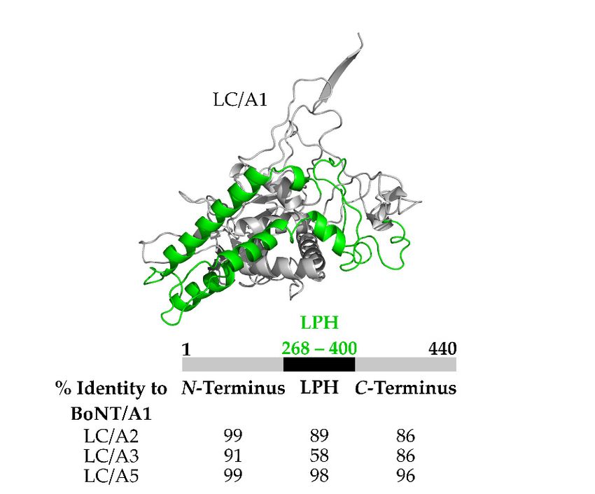
Toxins 2018, 10, 268 3 of 14
of BoNTs as neurotrophic toxins, by reducing the possibility of off-target cleavage of non-neuronal
isotype SNARE
Toxins 2018, proteins.
10, x FOR This adds to the utility of BoNTs as therapeutic agents.
PEER REVIEW 3 of 14
Toxins 2018, 10, x FOR PEER REVIEW 3 of 14
Figure 1.
1. Clostridial
Clostridial BoNT and
BoNTand non-clostridial
andnon-clostridial
non-clostridial BoNT
BoNT derivatives.
derivatives.(Left) Structure
(Left) and alignment
Structure (Right) of
Figure
Figure 1. Clostridial BoNT BoNT derivatives. (Left) Structure andand alignment
alignment (Right)
(Right) of
the
of theconserved
conserved zinc
zincbinding
binding motif
motifH-E-X-X-H
H-E-X-X-H in red lettering. Residues in highlighted in green are
the conserved zinc binding motif H-E-X-X-H in inred
redlettering.
lettering.Residues
Residuesininhighlighted
highlightediningreen
green are
are
conserved. Accession
conserved. Accession numbers
numbers BoNT
BoNTA-GA-Gfrom
fromUniProt
UniProtBoNT/A:
BoNT/A:P10845,
P10845,BoNT/B:
BoNT/B:P10844,
P10844,BoNT/C1:
BoNT/C1:
conserved. Accession numbers BoNT A-G from UniProt BoNT/A: P10845, BoNT/B: P10844, BoNT/C1:
P18640, BoNT/D:
P18640, BoNT/D:P19321,
P19321,BoNT/E:
BoNT/E:Q00496, BoNT/F:
Q00496, BoNT/F: P30996 and BoNT/G: Q60393. BoNT/FA: GenBank
P18640, BoNT/D: P19321, BoNT/E: Q00496, BoNT/F: P30996P30996 and BoNT/G:
and BoNT/G: Q60393. Q60393.
BoNT/FA:BoNT/FA:
GenBank
(KGO15617.1), BoNT/X: UniParc (UPI0005822796), BoNT/Wo:
GenBank (KGO15617.1), BoNT/X: UniParc (UPI0005822796), BoNT/Wo: UniProtK (A0A069CUU9 UniProtK (A0A069CUU9
(KGO15617.1), BoNT/X: UniParc (UPI0005822796), BoNT/Wo: UniProtK (A0A069CUU9
(A0A069CUU9_WEIOS)) and
(A0A069CUU9_WEIOS)) and BoNT/en:
BoNT/en:GenBank
GenBank (OTO22244.1).
(OTO22244.1).
(A0A069CUU9_WEIOS)) and BoNT/en: GenBank (OTO22244.1).
Figure 2. BoNT serotype (A–G) SNARE cleavage. BoNT cleavage prevents interaction of the T-SNARE
Figure 2. BoNT serotype (A–G) SNARE cleavage. BoNT cleavage prevents interaction of the T-SNARE
and V-SNARE,
Figure preventing
2. BoNT serotype acetylcholine
(A–G) release.BoNT
SNARE cleavage. BoNT/A and /E
cleavage cleave interaction
prevents SNAP-25, ofBoNT/C cleaves
the T-SNARE
and V-SNARE, preventing acetylcholine release. BoNT/A and /E cleave SNAP-25, BoNT/C cleaves
SNAP-25 and Syntaxin which are located on the cytosolic face of the cell membrane.
and V-SNARE, preventing acetylcholine release. BoNT/A and /E cleave SNAP-25, BoNT/C cleaves BoNT/B, /D, /F,
SNAP-25 and Syntaxin which are located on the cytosolic face of the cell membrane. BoNT/B, /D, /F,
/G, and tetanus
SNAP-25 toxin (TeNT)
and Syntaxin whichcleave VAMP/
are located on Synaptobrevin which
the cytosolic face is located
of the on the cytosolic
cell membrane. BoNT/B, face of
/D,
/G, and tetanus toxin (TeNT) cleave VAMP/ Synaptobrevin which is located on the cytosolic face of
synaptic
/F, vesicles.
/G, and tetanusIntoxin
red are the amino
(TeNT) cleave acid
VAMP/# of Synaptobrevin
the P1′ scissile bond
whichcleavage byon
is located thethe
indicated
cytosolicBoNT
face
synaptic vesicles. In red are the amino acid # of the P1′ scissile bond cleavage by the indicated BoNT
synaptic vesicles. In red are the amino acid # of the P10 scissile bond cleavage by the indicated
serotype.
of
serotype.
BoNT serotype.
3. Cleavage of BoNT Substrates
3. Cleavage of BoNT Substrates
3. Cleavage of BoNT Substrates
Several groups have studied the sites recognized by each of the seven BoNT serotypes, as well
Several groups have studied the sites recognized by each of the seven BoNT serotypes, as well
as the non-clostridial
Several groups haveBoNT derivatives
studied the sites for cleavagebyofeach
recognized various
of theSNARE proteins.
seven BoNT Binz etasal.
serotypes, used
well as
as the non-clostridial BoNT derivatives for cleavage of various SNARE proteins. Binz et al. used
several
the techniques,
non-clostridial suchderivatives
BoNT as radiolabeled SNAP-25,
for cleavage HPLC,
of various and microsequencing,
SNARE to used
proteins. Binz et al. identify the
several
several techniques, such as radiolabeled SNAP-25, HPLC, and microsequencing, to identify the
cleavage site
techniques, of LC/A
such and LC/E.SNAP-25,
as radiolabeled Incubation of radiolabeled
HPLC, SNAP-25 with
and microsequencing, the LC/A
to identify the yielded
cleavagea
cleavage site of LC/A and LC/E. Incubation of radiolabeled SNAP-25 with197the LC/A yielded a
nanopeptide
site of LC/A andcorresponding to the C
LC/E. Incubation of terminus SNAP-25
radiolabeled SNAP-25between residues
with the LC/AEyielded
and aRnanopeptide
198 . Using the
nanopeptide corresponding to the C terminus SNAP-25 between residues E197 and R198. Using the
same techniques, LC/E cleaved between residues R180 and I181 [44]. More recent experiments show in
same techniques, LC/E cleaved between residues R180 and I181 [44]. More recent experiments show in
a competition experiment between LC/A and LC/E, LC/E outcompeted LC/A for cleavage of SNAP-
a competition experiment between LC/A and LC/E, LC/E outcompeted LC/A for cleavage of SNAP-
25, due to LC/E cleavage site being upstream of LC/A [45].
25, due to LC/E cleavage site being upstream of LC/A [45].Toxins 2018, 10, 268 4 of 14
corresponding to the C terminus SNAP-25 between residues E197 and R198 . Using the same techniques,
LC/E cleaved between residues R180 and I181 [44]. More recent experiments show in a competition
experiment between LC/A and LC/E, LC/E outcompeted LC/A for cleavage of SNAP-25, due to
LC/E cleavage site being upstream of LC/A [45].
BoNT/C is the only BoNT serotype to cleave Syntaxin-1A between K253 and A254 and Syntaxin-1B
between K252 and A253 [46]. Another group showed LC/C also cleaved SNAP-25 adjacent to the bond
that BoNT/A cleaves, between residues R198 and A199 [47]. LC/B, LC/D, LC/F, and LC/G cleave
the vesicle-bound SNARE protein VAMP/Synaptobrevin. Schiavo et al. showed BoNT/B inhibited
acetylcholine release by proteolytic cleavage of synaptobrevin between E76 and F77 and showed
tetanus toxin, a related clostridial neurotoxin, cleaved synaptobrevin between the same residues, but
in inhibitory neurons which led to spastic paralysis [35]. Yamasaki et al. used radiolabeled Aplysia
synaptobrevin to show LC/D and LC/F cleaved synaptobrevin between K49 and I50 and E48 and K49 ,
respectively [36]. BoNT/FA cleaved the same substrate as BoNT/F5 between residues L54 and E55
on synaptobrevin. Other BoNT/F subtypes cleaved synaptobrevin between E58 and K59 [37,48–51].
BoNT/G cleaved synaptobrevin-1 between A82 and A83 and synaptobrevin-2 between A81 and A82 [38].
BoNT/X cleaves VAMP 1-5 and Ykt6 between R66 and A67 . Cleavage of VAMP-4, VAMP-5 and
Ykt6 is unique to BoNT/X [41]. BoNT/en cleaved VAMP-2 between A67 and D68 , Syx 1B between M182
and D183 , and Syx 4 between K191 and D192 . Zhang et al. were not able to determine the cleavage site
of SNAP-23/25 due to multiple smeared bands of substrate arising upon incubation with high LC/en
concentrations [5]. BoNT/Wo cleaved close to the C terminus of VAMP-2, within the juxtamembrane
domain between residues W89 and W90 [7]. Overall, the specificity for substrate is greater for the BoNTs,
relative to the non-clostridial BoNT-like proteins, suggesting that the evolution of the neurotrophic
nature of the BoNTs included an enhanced specificity for substrate recognition. Future studies may
reveal the molecular basis for the differential substrate specificity that may provide insight into the
steps required for the modulation of LCs to customize substrate cleavage.
4. Exosite-Pocket Model for LC-Mediated SNARE Cleavage
Unexpectedly, SNAP-25 binds to the grove within LC/A, which is also the binding region of the
HCN belt to the LC. The HCN belt comprises residues 458–547 of the HC (Figure 3) [52–54]. The HCN
belt region and SNAP-25 align along a stretch of 38-amino acids which aligned within the LC/A
crystal structure (Figure 3). Initial studies proposed that SNAP-25 aligned via an exosite orientation
that was then refined to a pocket model to describe the interactions between LC/A and SNAP-25.
Detailed reciprocal mutagenesis of LC and SNAP-25 defined a series of dis-contiguous interactions
between SNAP-25 and LC/A. Optimal scissile bond cleavage utilized five discreet pockets of amino
acids within LC/A that contribute to either efficient binding or cleavage of SNAP-25 between Q197
and R198 [44,53].
The mechanism of scissile bond cleavage for SNAP-25 by LC/A has been studied in detail and
proceeds as follows. Upon SNAP-25 binding the zinc ion the coordination of zinc binding changes
position which allows the carbonyl oxygen of E197 in SNAP-25 to displace a catalytic water molecule,
allowing the water molecule to form two hydrogen bonds with E224 , while staying weakly coordinated
to the zinc ion. This intermediate state allows E224 which acts as a proton shuttle to perform a
nucleophilic attack on the carbonyl carbon of the scissile peptide bond. This intermediate is stabilized
by Y366 which provides the hydroxide for interaction. The last step in the cleavage of the scissile
bond occurs when the intermediate state collapses and E224 mediates the transfer of two protons
onto the scissile amide, which generates a stable amino group that then leaves the active site [55].
Understanding how the mechanism of cleavage works allows the field to optimize the efficiency of
LC-based therapeutics.belt region and SNAP-25 align along a stretch of 38-amino acids which aligned within the LC/A
crystal structure (Figure 3). Initial studies proposed that SNAP-25 aligned via an exosite orientation that
was then refined to a pocket model to describe the interactions between LC/A and SNAP-25. Detailed
reciprocal mutagenesis of LC and SNAP-25 defined a series of dis-contiguous interactions between SNAP-
25 and
Toxins LC/A.
2018, Optimal scissile bond cleavage utilized five discreet pockets of amino acids within LC/A
10, 268 5 of 14
that contribute to either efficient binding or cleavage of SNAP-25 between Q and R [44,53].
197 198
Figure 3. Structural alignment of BoNT/A heavy chain loop with SNAP-25. (A) Structural alignment
Figure 3. Structural alignment of BoNT/A heavy chain loop with SNAP-25. (A) Structural alignment
of the HC loop of BoNT/A and SNAP-25 with LC/A. Left panel shows the alignment from the C
of the HC loop of BoNT/A and SNAP-25 with LC/A. Left panel shows the alignment from the C
terminus of the HC loop of BoNT/A and SNAP-25 on the face of LC/A that continues in the right panel
terminus of the HC loop of BoNT/A and SNAP-25 on the face of LC/A that continues in the right
along the back of LC/A toward the N termini. The blue ribbon represents the HC loop; the red ribbon
panel along the back of LC/A toward the N termini. The blue ribbon represents the HC loop; the red
represents SNAP-25. Asp193 of SNAP-25 and Glu528 of the HC loop interactions with LC/A S5 pocket
ribbon represents SNAP-25. Asp193 of SNAP-25 and Glu528 of the HC loop interactions with LC/A S5
ends the similarity between SNAP-25 and the HC loop of BoNT/A; (B) Sequence and spatial similarity
pocket ends the similarity between SNAP-25 and the HC loop of BoNT/A; (B) Sequence and spatial
of HC loop of BoNT/A and SNAP-25 interacting with LC/A. The spatial overlap of the HC loop of
similarity of HC loop of BoNT/A and SNAP-25 interacting with LC/A. The spatial overlap of the HC
BoNT/A and residues of SNAP-25 that interact with LC/A is indicated by (:) or partial overlap (.),
loop of BoNT/A and residues of SNAP-25 that interact with LC/A is indicated by (:) or partial overlap
which was determined by manual alignment of the two structures. Note the structural overlap
(.), which was determined by manual alignment of the two structures. Note the structural overlap
between the HC loop of BoNT/A and SNAP-25 ends at Asp193. Reproduced from [53]. Copyright
2007, American Society for Biochemistry and Molecular Biology, Rockville, MD, USA.
5. BoNT Duration and Potency
Several pathways are responsible for protein turnover in eukaryotic cells, including lysosome- and
ubiquitin-mediated degradation. Lysosome-mediated proteolysis often involves the initial trafficking
of cell surface proteins into endocytic vesicles that mature into endosomes and eventually fuse with
lysosomes, which contain numerous degradative enzymes, including proteases [56]. An alternative
to the lysosomal mediated-proteolysis is the ubiquitin-proteasome pathway, the major pathway
for intracellular protein degradation in eukaryotic cells. Ubiquitin is a 76-amino acid polypeptide
that is attached to proteins destined for degradation via covalent binding to K48 by the targeted
protein, through a process using E1, E2, or two different E3s (RING or HECT) [56]. There are
also chain elongation factors known as E4, which are a subclass of E3 which only catalyze chain
extension of ubiquitin onto the protein [57]. Binding to E1 activates ubiquitin, which is transferred
to E2. The final transfer of ubiquitin to the target protein is performed by E3. E3 (HECT) takes
ubiquitin from E2 and transfers ubiquitin to the protein targeted for degradation. A second pathway
is performed by E3 (RING) which mediates transfer of ubiquitin from E2 to the protein targeted for
degradation without directly contacting ubiquitin [56,57]. Many proteins are polyubiquitinated, and
once ubiquitinated, proteins are degraded by the proteasome into amino acids [56]. Proteins are also
degraded during apoptosis, where caspases are used for mediated degradation. Caspases contain a
conserved Gln-Ala-Cys-X-Gly and cleave proteins at aspartic acid. During apoptosis, caspases cleave
cellular substrates such as poly (ADP-ribose) polymerase and lamins [58].
The average protein in a eukaryote cell has a typical half-life of ~1.5 to 48 h [59–61]. While a
plethora of long-lived proteins (LLPs) are found in the nucleus, such as histones and nuclear
pore complexes, LLPs have also been identified within other intracellular locations. The current
understanding for the slow turnover of LLPs is based upon slow turnover rates when found in protein
complexes or cellular structures that cannot be replaced without disrupting the complex [62]. Recently,
Heo et al. characterized the half-life for proteins in the synapse, using stable isotope labeling where
heavy-isotope-labeled amino acids (Lys-13-C6 ) were introduced into the diet of mice for 7 weeks and, toToxins 2018, 10, 268 6 of 14
measure turnover, followed with a normal diet, where 164 LLPs were found in synaptosomes. In these
experiments, proteins present in both the cytosol and the synaptosomes had higher turnover rates
when present in the cytosol. The authors proposed that proteins in the synapse formed more stable
complexes than in the cytosol which were then more accessible to protein degradation machinery [63].
BoNT duration of action is unique to each serotype and appears to be a LC function [64], ranging
from weeks to several months, [65]. In general, LC duration of action in mice and human cells for
BoNT/A and BoNT/C are longer than LC duration of BoNT/B, /D, /F, and /G which are longer
thanToxins
the duration thanPEER
2018, 10, x FOR LC REVIEW
of BoNT/E (Table 1) [66]. BoNT/A has eight subtypes (A1–A8) in the6 of 14
current PubMed database that differ in amino acid homology up to 9.5%, have different durations of
action
rat[67,68],
spinal but
cordeachcellsLC subtype cleaves
intoxicated SNAP-25/A2,
with BoNT/A1, at theandsame
/A5location.
had 50% Pellett et al. reported
of uncleaved SNAP-25 that
after 9
primary rat spinal
months, whilecord cells intoxicated
primary withcells
rat spinal cord BoNT/A1, /A2, with
intoxicated and /A5 had 50%
BoNT/A4 hadof25%
uncleaved SNAP-25
uncleaved SNAP-25
afterafter
9 months,
9 months while primary
[45]. rat spinal
In addition, BoNT/A4cord cells
had 10intoxicated
3-fold reduced with activity
BoNT/A4 had 25%
compared touncleaved
BoNT/A1 [69].
SNAP-25 after 9 months [45]. In addition, BoNT/A4 had 10 3 -fold reduced activity compared to
Unexpectedly, BoNT/A3 elicited a shorter half-life of duration than other BoNT subtypes where at 5
BoNT/A1
months[69].postUnexpectedly,
primary rat spinalBoNT/A3 cordelicited a shorter half-life
cell intoxication cleaved of duration
SNAP-25 wasthan
notother BoNT [45].
observed
subtypes where at 5 months post primary rat spinal cord cell intoxication cleaved
Recently, utilizing BoNT/A subtype chimeras, the lower duration of BoNT/A3 action was localized SNAP-25 was not
observed
to LC[45]. Recently,
action. utilizing BoNT/A
A structure-based subtype
alignment chimeras,
showed the lower
that LC/A3 had duration of BoNT/A3amino
a region comprising actionacids
was localized
268–396 withto LC action.
only ~58% A amino
structure-based alignment
acid homology showed
relative that LC/A3
to LC/A1, LC/A2,hadandaLC/A5
region(Figure
comprising
4) which
amino acids 268–396 with only ~58% amino acid homology relative to LC/A1,
was termed the Low Primary amino acid Homology region (LPH). Utilizing GFP-LC fusion proteins LC/A2, and LC/A5
(Figure
as a4)reporter,
which was termed the
GFP-LC/A1 was Low Primary
found amino to
to localized acidtheHomology regionmembrane,
host cell plasma (LPH). Utilizing
whileGFP-LC
GFP-LC/A3
fusion proteins as a reporter, GFP-LC/A1 was found to localized to the host cell
was present in the cytosol [64,70]. Thus, intracellular localization may contribute to the duration ofplasma membrane,
whileBoNT/LC
GFP-LC/A3 was present
duration. Earlier in the cytosol
studies showed [64,70]. Thus,
that the intracellular
N-terminal localization
17-amino acids ofmay
LC/A1 contribute
contributed
to the
toduration of BoNT/LC
LCA1 membrane duration.[71].
localization Earlier
Thus,studies
two orshowed that themay
more regions N-terminal 17-amino
contribute to the acids of of
targeting
LC/A1 contributed to LCA1
LC/A1 to the cell membrane. membrane localization [71]. Thus, two or more regions may contribute to
the targeting of LC/A1 to the cell membrane.
Figure 4. Structure
Figure and alignment
4. Structure of theofLow
and alignment Primary
the Low amino
Primary acid Homology
amino domain
acid Homology (LPH)(LPH)
domain of LC/A3.
of LC/A3.
(Top)(Top)
Crystal structure
Crystal of BoNT
structure LC/A1
of BoNT with with
LC/A1 LPH LPH
highlighted in green
highlighted PDB:3BTA.
in green (Bottom)
PDB:3BTA. Percent
(Bottom) Percent
(%) homology
(%) homologyfor domains found
for domains in LC/A2,
found LC/A3,
in LC/A2, LC/A3,andand
LC/A5
LC/A5 compared
compared toto LC/A1.N-terminal
LC/A1. N-terminal and
and C-terminal
C-terminalregions
regionsofofhigh
high homology.
homology. LowLow primary
primary amino
amino acid
acid homology (LPH) spans residues 268–
268–400
400 for
forLC/A1,
LC/A1, /A2,/A2,and
and/A5
/A5while
whileresidues
residues268–396
268–396for
forLC/A3.
LC/A3.For For sequencealignment
sequence alignmentused
usedblastp
blastpsuite,
suite,with
with accession
accession numbers
numbersfromfromUniProt BoNT/A1:
UniProt BoNT/A1: A2I2U2, BoNT/A2:
A2I2U2, BoNT/A2:Q84GH1, BoNT/A3:
Q84GH1, BoNT/A3:
Q3LRX, BoNT/A5:
Q3LRX, BoNT/A5: C7BEA8.
C7BEA8.
BoNT/B includes seven subtypes that share 93% primary amino acid homology [72]. The
duration of BoNT/B action ranged from 2–4 months [73–76]. BoNT/C (also known as C1) has a
duration of action that approaches BoNT/A, ranging from 4 to 6 months [73]. Native chimeras of
BoNT/C and BoNT/D, termed BoNT/CD and BoNT/DC may help decipher the basis for the varied
range of BoNT action [77]. BoNT/D duration of action is short, ranging in weeks and BoNT/D had a
relatively short duration of action in human neurons, lasting ~3 weeks post-intoxication. BoNT/D alsoToxins 2018, 10, 268 7 of 14
BoNT/B includes seven subtypes that share 93% primary amino acid homology [72]. The duration
of BoNT/B action ranged from 2–4 months [73–76]. BoNT/C (also known as C1) has a duration of
action that approaches BoNT/A, ranging from 4 to 6 months [73]. Native chimeras of BoNT/C and
BoNT/D, termed BoNT/CD and BoNT/DC may help decipher the basis for the varied range of BoNT
action [77]. BoNT/D duration of action is short, ranging in weeks and BoNT/D had a relatively
short duration of action in human neurons, lasting ~3 weeks post-intoxication. BoNT/D also has a
lower toxicity relative to BoNT/A [78]. Each of the 14 subtypes of BoNT/E have a short duration of
action in human neurons, lasting from 2–3 weeks [45,79]. Like LC/A3, LC/E is present in the cytosol
when expressed in eukaryotic cells [70]. Based upon these correlates, we hypothesize intracellular
localization may contribute to the duration of LC action. BoNT/F has 8 subtypes (1–8) that differ in
amino acid homology by up to 30.9% [79]. In rats, BoNT/F had a shorter duration than BoNT/A [80].
The last of the novel serotypes is BoNT/G of which there are not any known subtypes and has a similar
duration of action as BoNT/A and BoNT/B [81].
Depending on the serotype, BoNTs have different specific toxicities. Most studies determine
specific toxicities in the lab, using animal models of intoxication, typically using oral or injection in
mice. By intraperitoneal (injection into the gastrocnemius muscle) or intravenous data, the specific
toxicity for the BoNT serotypes ranges from 1.1–1.2 ng for BoNT/A, BoNT/B, BoNT/C1, and BoNT/E
to 0.4 ng for BoNT/D (Table 1) [82–86]. While BoNT/F, which is the least toxic of the serotypes tested,
with a lethal dose of 2.4 ng (Table 1) intravenous intoxication [87]. BoNT/G toxicity is not known at
this time.
BoNT/A is the most widely studied serotype and several subtypes have been characterized for
their specific toxicity. The specific toxicity of BoNT/A subtypes are similar to BoNT/A1 which is
between 1 × 10⁸ –2 × 10⁸ LD50 /mg in mouse models of botulism. However, [88] BoNT/A4 has a
specific toxicity of 1.0–1.25 × 10⁵ LD50 /mg [69]. Continued analysis should determine the specific
toxicity for the other subtypes of BoNT/A as well as other BoNT serotypes. Understanding the basis
for LC localization may help define the duration of LC, while toxicity does not seem to be correlated to
LC localization.
Table 1. Toxicity and duration of all seven BoNT serotypes (A–G).
Serotype 1 Toxicity Duration
/A 1.2 ng [82]Toxins 2018, 10, 268 8 of 14
factor 2 a RING E3 ligase was responsible for LC ubiquitination [92]. Shi et al. showed that when
ubiquitinated, BoNT/B has decreased biological activity compared to non-ubiquitinated BoNT/B [93].
Shi also showed that in the presence of expoxomicin, a proteasome inhibitor, ubiquitinated LC/B
increased compared to the phorbol 12-myristate 13 acetate (PMA) treated SH-SY5Y cells [93]; PMA
is a ubiquitination enhancer [94]. Thus, ubiquitination appears to contribute to the LC longevity.
Understanding how the LC avoids degradation will allow the modulation of LC duration for
therapeutic application.
7. BoNT Engineering
A subfield of BoNT engineering has emerged to enhance the beneficial therapeutic potential
of BoNT [64]. Pellett et al. recombined LC and HC domains of different subtypes of BoNT/A to
create hybrid toxins that comprised reciprocal LC-HC components with potentially new properties
but retain therapeutic and vaccine sensitivities of the parental proteins. Based upon the differential
duration of toxin action, Pellett et al., characterized BoNTA1/A3 and BoNTA3/A1 chimeras [64].
While BoNTA1/A3 had greater potency than BoNTA3, showing a role for HC in potency, BoNTA3/A1
had a slight reduction in potency relative to BoNT/A1, but retained the short duration of action of
BoNT/A3. Thus, this experiment showed the dominance of the HC as rate-limiting for the specific
activity of botulism and that LC contributed to the duration of action in this mouse model [64]. These
experiments show the utility of BoNT chimeras to define the molecular and cellular aspects of BoNT
action, as tools for next generation BoNT therapies.
BoNT engineering has also changed the substrate specificity based upon understanding LC
substrate binding. LC/E substrate specificity was extended to cleave SNAP-23, by changing residue
224 of LC/E D→K (D224 K). SNAP-23 is a nonneuronal isoform of SNAP-25, which is involved with
several cellular processes, such as membrane repair, cytokinesis, and synaptic transmission [95–97].
The kinetics of SNAP-23 cleavage by LC/E (D224 K) were similar to LC/B cleavage of VAMP-2, ~17 S−1
which is 5-fold lower than wild-type LC/E cleavage of SNAP-25. LC/E (D224 K) cleavage of SNAP-23
in Hela cells inhibited TNF-α-mediated mucin and IL-8 secretion. Inhibiting mucin and IL-8 by LC/E
(D224 K) shows potential therapy for several human diseases that involve hypersecretion such as asthma
and inflammatory disease [98], therapeutics outside the scope of neurological conditions. Recently, Binz
et al. mapped the region that is responsible for making human SNAP-23 resistant to cleavage by LC/A
at physiological concentrations. Mutating 10 amino acids in SNAP-23 (between residues 172–206)
yielded a substrate that was cleaved by LC/A with similar properties as SNAP-25. Re-engineering
LC/A was also performed, using a yeast-based screening system to identify residues involved in
the P20 pocket recognition. The LC/A(A308 V) mutation was 2.5-fold more efficient for SNAP-23
cleavage than wild-type LC/A. Although more experiments are needed to further understand the rate
limiting steps, both in catalysis and binding, for LC/A cleavage of SNAP-23, these mutations are a
significant advancement towards the development of pharmaceuticals for use to treat non-neuronal
based diseases of hypersecretion [99].
Due to BoNTs neuronal specificity for entry into the presynaptic membrane of neurons within
synaptic vesicles as well as the trafficking of LC to the inner leaflet of the plasma membrane or
synaptic vesicles, an emerging field of study is the use of BoNT as a vehicle for therapeutic delivery
inside a cell. Dolly and coworkers have engineered various BoNT-derivatives with substitute host
and bacterial factors that have provided insight into BoNT action and the use of BoNT as conjugate
carriers [100,101]. Vazquez-Citron et al. designed an atoxic derivative of BoNT/C1, which possessed
the same key entry characteristics of wild-type BoNT/C1, while being 5 × 10⁶ -fold less toxic than
wild-type BoNT/C1 [102]. While the use of BoNT as a vehicle is promising, a caveat to this technique
is the assurance that the vehicle has a large enough therapeutic range that toxicity will not be a factor.
Naturally occurring BoNTs are encased by progenitor toxin complexes, which are high molecular
weight multi-protein complexes composed of BoNT and several non-toxic neurotoxin-associated
proteins (NAPs) [103]. One NAP is a non-toxic non-hemagglutinin (NTNHA) protein [104,105].Toxins 2018, 10, 268 9 of 14
To study this progenitor toxin complex, BoNT/A1 was engineered with a three-point mutation in
the LC (E224 Q/R363 A/Y366 F). Gu et al. added a BoNT/A-specific nanobody which improved crystal
packing and diffraction quality. Progenitor toxin complex crystal structure provided orientation and
identified interactions between the two complexes. A better understanding of the progenitor toxin
complex could render the complex as a drug delivery system [106].
Use of the current chemically inactivated BoNT vaccine has been discontinued based upon the
loss of vaccine potency [107]. Recently, Webb et al. engineered full-length BoNT/A (H223 A, E224 A,
H227 A) as a vaccine candidate that was more potent than the HCC based vaccine. Note, the H223 A,
E224 A, H227 A mutations lie within the Zn binding motif of the LC (Figure 1) [108]. A second vaccine
strategy, to further reduce residual toxicity and potential reversion, engineered recombinant, full length
BoNT/A with three-point mutations within the LC (E224 A, R363 A, Y366 F), along with a point mutation
in the ganglioside binding pocket W1226 A [109], which also proved to be a potent vaccine candidate.
Recombinant, full length BoNT vaccines, inactivated via mutations in LC function, may provide a
platform towards the development of an FDA-approved vaccine.
8. Conclusions
This review addresses the molecular, cellular, and structural properties of LCs with respect to
toxicity and duration of BoNT action, which provide the basis for BoNT engineering and BoNT vaccine
production. Advances in understanding LC action also provides the basis for BoNT as a therapeutic
and as a delivery platform.
Conflicts of Interest: The authors declare no conflict of interest.
References
1. Schiavo, G.; Matteoli, M.; Montecucco, C. Neurotoxins affecting neuroexocytosis. Physiol. Rev. 2000, 80,
717–766. [CrossRef] [PubMed]
2. Johnson, E.A.; Bradshaw, M. Clostridium botulinum and its neurotoxins: A metabolic and cellular perspective.
Toxicon 2001, 39, 1703–1722. [CrossRef]
3. Scott, A.B. Botulinum toxin injection of eye muscles to correct strabismus. Trans. Am. Ophthalmol. Soc. 1981,
79, 734–770. [PubMed]
4. Niemann, H.; Blasi, J.; Jahn, R. Clostridial neurotoxins: New tools for dissecting exocytosis. Trends Cell Biol.
1994, 4, 179–185. [CrossRef]
5. Zhang, S.; Lebreton, F.; Mansfield, M.J.; Miyashita, S.I.; Zhang, J.; Schwartzman, J.A.; Tao, L.; Masuyer, G.;
Martinez-Carranza, M.; Stenmark, P.; et al. Identification of a botulinum neurotoxin-like toxin in a commensal
strain of Enterococcus faecium. Cell Host Microbe 2018, 23, 169.e6–176.e6. [CrossRef] [PubMed]
6. Brunt, J.; Carter, A.T.; Stringer, S.C.; Peck, M.W. Identification of a novel botulinum neurotoxin gene cluster
in Enterococcus. FEBS Lett. 2018, 592, 310–317. [CrossRef] [PubMed]
7. Zornetta, I.; Azarnia Tehran, D.; Arrigoni, G.; Anniballi, F.; Bano, L.; Leka, O.; Zanotti, G.; Binz, T.;
Montecucco, C. The first non clostridial botulinum-like toxin cleaves VAMP within the juxtamembrane
domain. Sci. Rep. 2016, 6, 30257. [CrossRef] [PubMed]
8. Zhang, S.; Masuyer, G.; Zhang, J.; Shen, Y.; Lundin, D.; Henriksson, L.; Miyashita, S.I.; Martinez-Carranza, M.;
Dong, M.; Stenmark, P. Identification and characterization of a novel botulinum neurotoxin. Nat. Commun.
2017, 8, 14130. [CrossRef] [PubMed]
9. Montecucco, C.; Berica Rasotto, M.B. On Botulinum Neurotoxin Variability. mBio 2015, 6, e02131-14.
[CrossRef] [PubMed]
10. Mansfield, M.J.; Doxey, A.C. Genomic insights into the evolution and ecology of botulinum neurotoxins.
Pathog. Dis. 2018, 76, fty040. [CrossRef] [PubMed]
11. Division of Select Agents and Toxins: What Is a Select Agent? Available online: https://www.cdc.gov/
phpr/dsat/what-is-select-agents.htm (accessed on 10 April 2018).
12. Bioterrorism Agents/Diseases. Available online: https://emergency.cdc.gov/agent/agentlist-category.asp
(accessed on 10 April 2018).Toxins 2018, 10, 268 10 of 14
13. Potential Bioterrorism Agents. Available online: https://www.bcm.edu/departments/molecular-virology-
and-microbiology/emerging-infections-and-biodefense/potential-bioterrorism-agents (accessed on 10
April 2018).
14. Select Agents and Toxins List. Available online: https://www.selectagents.gov/selectagentsandtoxinslist.
html (accessed on 10 April 2018).
15. Hambrosky, J.; Kroeger, A.; Wolf, S. Epidemiology and prevention of vaccine-preventable diseases. In Centers
for Disease Control and Prevention; Public Health Foundation: Washington, DC, USA, 2015; Volume 13.
16. Aoki, K.R.; Guyer, B. Botulinum toxin type A and other botulinum toxin serotypes: A comparative review of
biochemical and pharmacological actions. Eur. J. Neurol. 2001, 8 (Suppl. 5), 21–29. [CrossRef] [PubMed]
17. Singh, B.R. Scientific and Therapeutic Aspects of Botulinum Toxin; Brin, M.F., Jankovic, J., Hallet, M., Eds.;
Lippincott Williams & Wilkins: Philadelphia, PA, USA, 2001; pp. 75–88.
18. Singh, B.R.; DasGupta, B.R. Conformational changes associated with the nicking and activation of botulinum
neurotoxin type E. Biophys. Chem. 1990, 38, 123–130. [CrossRef]
19. Oguma, K.; Fujinaga, Y.; Inoue, K. Structure and function of Clostridium botulinum toxins. Microbiol. Immunol.
1995, 39, 161–168. [CrossRef] [PubMed]
20. Li, L.; Binz, T.; Niemann, H.; Singh, B.R. Probing the mechanistic role of glutamate residue in the zinc-binding
motif of type a botulinum neurotoxin light chain. Biochemistry 2000, 39, 2399–2405. [CrossRef] [PubMed]
21. Sathyamoorthy, V.; DasGupta, B.R. Separation, purification, partial characterization and comparison of the
heavy and light chains of botulinum neurotoxin types A, B, and E. J. Biol. Chem. 1985, 260, 10461–10466.
[PubMed]
22. Binz, T.; Rummel, A. Cell entry strategy of clostridial neurotoxins. J. Neurochem. 2009, 109, 1584–1595.
[CrossRef] [PubMed]
23. Rummel, A. Double receptor anchorage of botulinum neurotoxins accounts for their exquisite neurospecificity.
Curr. Top. Microbiol. Immunol. 2013, 364, 61–90. [PubMed]
24. Hoch, D.H.; Romero-Mira, M.; Ehrlich, B.E.; Finkelstein, A.; DasGupta, B.R.; Simpson, L.L. Channels formed
by botulinum, tetanus, and diphtheria toxins in planar lipid bilayers: Relevance to translocation of proteins
across membranes. Proc. Natl. Acad. Sci. USA 1985, 82, 1692–1696. [CrossRef] [PubMed]
25. Smart, O.S.; Breed, J.; Smith, G.R.; Sansom, M.S. A novel method for structure-based prediction of ion
channel conductance properties. Biophys. J. 1997, 72, 1109–1126. [CrossRef]
26. Falnes, P.O.; Olsnes, S. Cell-mediated reduction and incomplete membrane translocation of diphtheria
toxin mutants with internal disulfides in the A fragment. J. Biol. Chem. 1995, 270, 20787–20793. [CrossRef]
[PubMed]
27. Koriazova, L.K.; Montal, M. Translocation of botulinum neurotoxin light chain protease through the heavy
chain channel. Nat. Struct. Biol. 2003, 10, 13–18. [CrossRef] [PubMed]
28. Schiavo, G.; Rossetto, O.; Santucci, A.; DasGupta, B.R.; Montecucco, C. Botulinum neurotoxins are zinc
proteins. J. Biol. Chem. 1992, 267, 23479–23483. [PubMed]
29. Rawlings, N.; Barret, A. Introduction: Metallopeptidases and their clans. In Handbook of Proteolytic Enzyme;
Barret, A., Rawlings, N., Woessner, J., Eds.; Academic Press: San Diego, CA, USA, 2004; Volume 2,
pp. 231–268.
30. Segelke, B.; Knapp, M.; Kadkhodayan, S.; Balhorn, R.; Rupp, B. Crystal structure of Clostridium botulinum
neurotoxin protease in a product-bound state: Evidence for noncanonical zinc protease activity. Proc. Natl.
Acad. Sci. USA 2004, 101, 6888–6893. [CrossRef] [PubMed]
31. Sudhof, T.C.; Rothman, J.E. Membrane fusion: Grappling with SNARE and SM proteins. Science 2009, 323,
474–477. [CrossRef] [PubMed]
32. Schiavo, G.; Rossetto, O.; Catsicas, S.; Polverino de Laureto, P.; DasGupta, B.R.; Benfenati, F.; Montecucco, C.
Identification of the nerve terminal targets of botulinum neurotoxin serotypes A, D, and E. J. Biol. Chem.
1993, 268, 23784–23787. [PubMed]
33. Schiavo, G.; Santucci, A.; Dasgupta, B.R.; Mehta, P.P.; Jontes, J.; Benfenati, F.; Wilson, M.C.; Montecucco, C.
Botulinum neurotoxins serotypes A and E cleave SNAP-25 at distinct COOH-terminal peptide bonds.
FEBS Lett. 1993, 335, 99–103. [CrossRef]
34. Blasi, J.; Chapman, E.R.; Yamasaki, S.; Binz, T.; Niemann, H.; Jahn, R. Botulinum neurotoxin C1 blocks
neurotransmitter release by means of cleaving HPC-1/syntaxin. EMBO J 1993, 12, 4821–4828. [PubMed]Toxins 2018, 10, 268 11 of 14
35. Schiavo, G.; Benfenati, F.; Poulain, B.; Rossetto, O.; Polverino de Laureto, P.; DasGupta, B.R.;
Montecucco, C. Tetanus and botulinum-B neurotoxins block neurotransmitter release by proteolytic cleavage
of synaptobrevin. Nature 1992, 359, 832–835. [CrossRef] [PubMed]
36. Yamasaki, S.; Hu, Y.; Binz, T.; Kalkuhl, A.; Kurazono, H.; Tamura, T.; Jahn, R.; Kandel, E.; Niemann, H.
Synaptobrevin/vesicle-associated membrane protein (VAMP) of Aplysia californica: Structure and proteolysis
by tetanus toxin and botulinal neurotoxins type D and F. Proc. Natl. Acad. Sci. USA 1994, 91, 4688–4692.
[CrossRef] [PubMed]
37. Schiavo, G.; Shone, C.C.; Rossetto, O.; Alexander, F.C.; Montecucco, C. Botulinum neurotoxin serotype F is a
zinc endopeptidase specific for VAMP/synaptobrevin. J. Biol. Chem. 1993, 268, 11516–11519. [PubMed]
38. Schiavo, G.; Malizio, C.; Trimble, W.S.; Polverino de Laureto, P.; Milan, G.; Sugiyama, H.; Johnson, E.A.;
Montecucco, C. Botulinum G neurotoxin cleaves VAMP/synaptobrevin at a single Ala-Ala peptide bond.
J. Biol. Chem. 1994, 269, 20213–20216. [PubMed]
39. Hackett, G.; Moore, K.; Burgin, D.; Hornby, F.; Gray, B.; Elliott, M.; Mir, I.; Beard, M. Purification and
characterization of recombinant botulinum neurotoxin serotype FA, also known as serotype H. Toxins 2018,
10, 195. [CrossRef] [PubMed]
40. Dover, N.; Barash, J.R.; Hill, K.K.; Xie, G.; Arnon, S.S. Molecular characterization of a novel botulinum
neurotoxin type H gene. J. Infect. Dis. 2014, 209, 192–202. [CrossRef] [PubMed]
41. Masuyer, G.; Zhang, S.; Barkho, S.; Shen, Y.; Henriksson, L.; Kosenina, S.; Dong, M.; Stenmark, P. Structural
characterisation of the catalytic domain of botulinum neurotoxin X—High activity and unique substrate
specificity. Sci. Rep. 2018, 8, 4518. [CrossRef] [PubMed]
42. Kiessling, V.; Ahmed, S.; Domanska, M.K.; Holt, M.G.; Jahn, R.; Tamm, L.K. Rapid fusion of synaptic vesicles
with reconstituted target SNARE membranes. Biophys. J. 2013, 104, 1950–1958. [CrossRef] [PubMed]
43. Mayorov, A.V.; Willis, B.; Di Mola, A.; Adler, D.; Borgia, J.; Jackson, O.; Wang, J.; Luo, Y.; Tang, L.;
Knapp, R.J.; et al. Symptomatic relief of botulinum neurotoxin/a intoxication with aminopyridines: A new
twist on an old molecule. ACS Chem. Biol. 2010, 5, 1183–1191. [CrossRef] [PubMed]
44. Binz, T.; Blasi, J.; Yamasaki, S.; Baumeister, A.; Link, E.; Sudhof, T.C.; Jahn, R.; Niemann, H. Proteolysis of
SNAP-25 by types E and A botulinal neurotoxins. J. Biol. Chem. 1994, 269, 1617–1620. [PubMed]
45. Whitemarsh, R.C.; Tepp, W.H.; Johnson, E.A.; Pellett, S. Persistence of botulinum neurotoxin a subtypes 1–5
in primary rat spinal cord cells. PLoS ONE 2014, 9, e90252. [CrossRef] [PubMed]
46. Schiavo, G.; Shone, C.C.; Bennett, M.K.; Scheller, R.H.; Montecucco, C. Botulinum neurotoxin type C cleaves
a single Lys-Ala bond within the carboxyl-terminal region of syntaxins. J. Biol. Chem. 1995, 270, 10566–10570.
[CrossRef] [PubMed]
47. Foran, P.; Lawrence, G.W.; Shone, C.C.; Foster, K.A.; Dolly, J.O. Botulinum neurotoxin C1 cleaves both
syntaxin and SNAP-25 in intact and permeabilized chromaffin cells: Correlation with its blockade of
catecholamine release. Biochemistry 1996, 35, 2630–2636. [CrossRef] [PubMed]
48. Kalb, S.R.; Baudys, J.; Egan, C.; Smith, T.J.; Smith, L.A.; Pirkle, J.L.; Barr, J.R. Different substrate recognition
requirements for cleavage of synaptobrevin-2 by Clostridium baratii and Clostridium botulinum type F
neurotoxins. Appl. Environ. Microbiol. 2011, 77, 1301–1308. [CrossRef] [PubMed]
49. Kalb, S.R.; Baudys, J.; Raphael, B.H.; Dykes, J.K.; Luquez, C.; Maslanka, S.E.; Barr, J.R. Functional
characterization of botulinum neurotoxin serotype H as a hybrid of known serotypes F and A (BoNT
F/A). Anal. Chem. 2015, 87, 3911–3917. [CrossRef] [PubMed]
50. Kalb, S.R.; Baudys, J.; Webb, R.P.; Wright, P.; Smith, T.J.; Smith, L.A.; Fernandez, R.; Raphael, B.H.;
Maslanka, S.E.; Pirkle, J.L.; et al. Discovery of a novel enzymatic cleavage site for botulinum neurotoxin F5.
FEBS Lett. 2012, 586, 109–115. [CrossRef] [PubMed]
51. Pellett, S.; Tepp, W.H.; Lin, G.; Johnson, E.A. Substrate cleavage and duration of action of botulinum
neurotoxin type FA (“H, HA”). Toxicon 2018, 147, 38–46. [CrossRef] [PubMed]
52. Lacy, D.B.; Tepp, W.; Cohen, A.C.; DasGupta, B.R.; Stevens, R.C. Crystal structure of botulinum neurotoxin
type A and implications for toxicity. Nat. Struct. Biol. 1998, 5, 898–902. [CrossRef] [PubMed]
53. Chen, S.; Kim, J.J.; Barbieri, J.T. Mechanism of substrate recognition by botulinum neurotoxin serotype A.
J. Biol. Chem. 2007, 282, 9621–9627. [CrossRef] [PubMed]
54. Masuyer, G.; Davies, J.R.; Moore, K.; Chaddock, J.A.; Ravi Acharya, K. Structural analysis of Clostridium
botulinum neurotoxin type D as a platform for the development of targeted secretion inhibitors. Sci. Rep.
2015, 5, 13397. [CrossRef] [PubMed]Toxins 2018, 10, 268 12 of 14
55. Brunger, A.T.; Rummel, A. Receptor and substrate interactions of clostridial neurotoxins. Toxicon 2009, 54,
550–560. [CrossRef] [PubMed]
56. Cooper, G. A Molecular Approach, 2nd ed.; ASM Press: Washington, DC, USA; Sinauer Associates: Sunderland,
MA, USA; Boston University: Boston, MA, USA, 2000.
57. Kipreos, E.T. Ubiquitin-mediated pathways in c. elegans. WormBook 2005, 1–24. [CrossRef] [PubMed]
58. Cohen, G.M. Caspases: The executioners of apoptosis. Biochem. J. 1997, 326 Pt 1, 1–16. [CrossRef] [PubMed]
59. Belle, A.; Tanay, A.; Bitincka, L.; Shamir, R.; O’Shea, E.K. Quantification of protein half-lives in the budding
yeast proteome. Proc. Natl. Acad. Sci. USA 2006, 103, 13004–13009. [CrossRef] [PubMed]
60. Cambridge, S.B.; Gnad, F.; Nguyen, C.; Bermejo, J.L.; Kruger, M.; Mann, M. Systems-wide proteomic analysis
in mammalian cells reveals conserved, functional protein turnover. J. Proteome Res. 2011, 10, 5275–5284.
[CrossRef] [PubMed]
61. Price, J.C.; Guan, S.; Burlingame, A.; Prusiner, S.B.; Ghaemmaghami, S. Analysis of proteome dynamics in
the mouse brain. Proc. Natl. Acad. Sci. USA 2010, 107, 14508–14513. [CrossRef] [PubMed]
62. Toyama, B.H.; Savas, J.N.; Park, S.K.; Harris, M.S.; Ingolia, N.T.; Yates, J.R., 3rd; Hetzer, M.W. Identification
of long-lived proteins reveals exceptional stability of essential cellular structures. Cell 2013, 154, 971–982.
[CrossRef] [PubMed]
63. Heo, S.; Diering, G.H.; Na, C.H.; Nirujogi, R.S.; Bachman, J.L.; Pandey, A.; Huganir, R.L. Identification
of long-lived synaptic proteins by proteomic analysis of synaptosome protein turnover. Proc. Natl. Acad.
Sci. USA 2018, 115, E3827–E3836. [CrossRef] [PubMed]
64. Pellett, S.; Bradshaw, M.; Tepp, W.H.; Pier, C.L.; Whitemarsh, R.C.M.; Chen, C.; Barbieri, J.T.; Johnson, E.A.
The light chain defines the duration of action of botulinum toxin serotype A subtypes. MBio 2018, 9,
e00089-18. [CrossRef] [PubMed]
65. Pellett, S.; Yaksh, T.L.; Ramachandran, R. Current status and future directions of botulinum neurotoxins for
targeting pain processing. Toxins 2015, 7, 4519–4563. [CrossRef] [PubMed]
66. Foran, P.G.; Mohammed, N.; Lisk, G.O.; Nagwaney, S.; Lawrence, G.W.; Johnson, E.; Smith, L.; Aoki, K.R.;
Dolly, J.O. Evaluation of the therapeutic usefulness of botulinum neurotoxin B, C1, E, and F compared with
the long lasting type A. Basis for distinct durations of inhibition of exocytosis in central neurons. J. Biol. Chem.
2003, 278, 1363–1371. [CrossRef] [PubMed]
67. Kull, S.; Schulz, K.M.; Weisemann, J.; Kirchner, S.; Schreiber, T.; Bollenbach, A.; Dabrowski, P.W.; Nitsche, A.;
Kalb, S.R.; Dorner, M.B.; et al. Isolation and functional characterization of the novel Clostridium botulinum
neurotoxin A8 subtype. PLoS ONE 2015, 10, e0116381. [CrossRef] [PubMed]
68. Pellett, S.; Tepp, W.H.; Whitemarsh, R.C.; Bradshaw, M.; Johnson, E.A. In vivo onset and duration of action
varies for botulinum neurotoxin A subtypes 1–5. Toxicon 2015, 107, 37–42. [CrossRef] [PubMed]
69. Whitemarsh, R.C.; Tepp, W.H.; Bradshaw, M.; Lin, G.; Pier, C.L.; Scherf, J.M.; Johnson, E.A.; Pellett, S.
Characterization of botulinum neurotoxin a subtypes 1 through 5 by investigation of activities in mice,
in neuronal cell cultures, and in vitro. Infect. Immun. 2013, 81, 3894–3902. [CrossRef] [PubMed]
70. Fernandez-Salas, E.; Steward, L.E.; Ho, H.; Garay, P.E.; Sun, S.W.; Gilmore, M.A.; Ordas, J.V.; Wang, J.;
Francis, J.; Aoki, K.R. Plasma membrane localization signals in the light chain of botulinum neurotoxin.
Proc. Natl. Acad. Sci. USA 2004, 101, 3208–3213. [CrossRef] [PubMed]
71. Chen, S.; Barbieri, J.T. Association of botulinum neurotoxin serotype A light chain with plasma
membrane-bound SNAP-25. J. Biol. Chem. 2011, 286, 15067–15072. [CrossRef] [PubMed]
72. Kalb, S.R.; Baudys, J.; Rees, J.C.; Smith, T.J.; Smith, L.A.; Helma, C.H.; Hill, K.; Kull, S.; Kirchner, S.;
Dorner, M.B.; et al. De novo subtype and strain identification of botulinum neurotoxin type B through toxin
proteomics. Anal. Bioanal. Chem. 2012, 403, 215–226. [CrossRef] [PubMed]
73. Eleopra, R.; Tugnoli, V.; Rossetto, O.; Montecucco, C.; De Grandis, D. Botulinum neurotoxin serotype C: A
novel effective botulinum toxin therapy in human. Neurosci. Lett. 1997, 224, 91–94. [CrossRef]
74. Sloop, R.R.; Cole, B.A.; Escutin, R.O. Human response to botulinum toxin injection: Type B compared with
type A. Neurology 1997, 49, 189–194. [CrossRef] [PubMed]
75. Eleopra, R.; Tugnoli, V.; Rossetto, O.; De Grandis, D.; Montecucco, C. Different time courses of recovery
after poisoning with botulinum neurotoxin serotypes A and E in humans. Neurosci. Lett. 1998, 256, 135–138.
[CrossRef]Toxins 2018, 10, 268 13 of 14
76. Brin, M.F.; Lew, M.F.; Adler, C.H.; Comella, C.L.; Factor, S.A.; Jankovic, J.; O’Brien, C.; Murray, J.J.;
Wallace, J.D.; Willmer-Hulme, A.; et al. Safety and efficacy of neurobloc (botulinum toxin type B) in
type A-resistant cervical dystonia. Neurology 1999, 53, 1431–1438. [CrossRef] [PubMed]
77. Nakamura, K.; Kohda, T.; Shibata, Y.; Tsukamoto, K.; Arimitsu, H.; Hayashi, M.; Mukamoto, M.; Sasakawa, N.;
Kozaki, S. Unique biological activity of botulinum D/C mosaic neurotoxin in murine species. Infect. Immun.
2012, 80, 2886–2893. [CrossRef] [PubMed]
78. Pellett, S.; Tepp, W.H.; Scherf, J.M.; Pier, C.L.; Johnson, E.A. Activity of botulinum neurotoxin type D (strain
1873) in human neurons. Toxicon 2015, 101, 63–69. [CrossRef] [PubMed]
79. Peck, M.W.; Smith, T.J.; Anniballi, F.; Austin, J.W.; Bano, L.; Bradshaw, M.; Cuervo, P.; Cheng, L.W.;
Derman, Y.; Dorner, B.G.; et al. Historical perspectives and guidelines for botulinum neurotoxin subtype
nomenclature. Toxins 2017, 9, 38. [CrossRef] [PubMed]
80. Kauffman, J.A.; Way, J.F., Jr.; Siegel, L.S.; Sellin, L.C. Comparison of the action of types a and f botulinum
toxin at the rat neuromuscular junction. Toxicol. Appl. Pharmacol. 1985, 79, 211–217. [CrossRef]
81. Rossetto, O.; Pirazzini, M.; Montecucco, C. Botulinum neurotoxins: Genetic, structural and mechanistic
insights. Nat. Rev. Microbiol. 2014, 12, 535–549. [CrossRef] [PubMed]
82. Lamanna, C.; Mc, E.O.; Eklund, H.W. The purification and crystallization of Clostridium botulinum type A
toxin. Science 1946, 103, 613–614. [CrossRef] [PubMed]
83. Duff, J.T.; Klerer, J.; Bibler, R.H.; Moore, D.E.; Gottfried, C.; Wright, G.G. Studies on immunity to toxins of
Clostridium botulinum. II. Production and purification of type B toxin for toxoid. J. Bacteriol. 1957, 73, 597–601.
[PubMed]
84. Ohishi, I.; Iwasaki, M.; Sakaguchi, G. Purification and characterization of two components of botulinum C2
toxin. Infect. Immun. 1980, 30, 668–673. [PubMed]
85. Cardella, M.A.; Duff, J.T.; Wingfield, B.H.; Gottfried, C. Studies on immunity to toxins of Clostridium
botulinum. VI. Purification and detoxification of type D toxin and the immunological response to toxoid.
J. Bacteriol. 1960, 79, 372–378. [PubMed]
86. Gerwing, J.; Dolman, C.E.; Ko, A. Mechanism of tryptic activation of clostridium botulinum type E toxin.
J. Bacteriol. 1965, 89, 1176–1179. [PubMed]
87. Oishi, I.; Sakaguchi, G. Purification of Clostridium botuliunum type F progenitor toxin. Appl. Microbiol. 1974,
28, 923–928. [PubMed]
88. Tepp, W.H.; Lin, G.; Johnson, E.A. Purification and characterization of a novel subtype A3 botulinum
neurotoxin. Appl. Environ. Microbiol. 2012, 78, 3108–3113. [CrossRef] [PubMed]
89. Syuto, B.; Kubo, S. Isolation and molecular size of clostridium botulinum type C toxin. Appl. Environ. Microbiol.
1977, 33, 400–405. [PubMed]
90. Ciccarelli, A.S.; Whaley, D.N.; McCroskey, L.M.; Gimenez, D.F.; Dowell, V.R., Jr.; Hatheway, C.L. Cultural
and physiological characteristics of Clostridium botulinum type G and the susceptibility of certain animals to
its toxin. Appl. Environ. Microbiol. 1977, 34, 843–848. [PubMed]
91. Tsai, Y.C.; Kotiya, A.; Kiris, E.; Yang, M.; Bavari, S.; Tessarollo, L.; Oyler, G.A.; Weissman, A.M.
Deubiquitinating enzyme VCIP135 dictates the duration of botulinum neurotoxin type A intoxication.
Proc. Natl. Acad. Sci. USA 2017, 114, E5158–E5166. [CrossRef] [PubMed]
92. Tsai, Y.C.; Maditz, R.; Kuo, C.L.; Fishman, P.S.; Shoemaker, C.B.; Oyler, G.A.; Weissman, A.M. Targeting
botulinum neurotoxin persistence by the ubiquitin-proteasome system. Proc. Natl. Acad. Sci. USA 2010, 107,
16554–16559. [CrossRef] [PubMed]
93. Shi, X.; Curran, J.E.; Liao, Z.; Gordon, R.K. The biological activity of ubiquitinated BoNT/B light chain
in vitro and in human SHSY-5Y neuronal cells. J. Cell. Biochem. 2009, 108, 660–667. [CrossRef] [PubMed]
94. Zhang, Y.; Wang, Z.; Liu, D.X.; Pagano, M.; Ravid, K. Ubiquitin-dependent degradation of cyclin B is
accelerated in polyploid megakaryocytes. J. Biol. Chem. 1998, 273, 1387–1392. [CrossRef] [PubMed]
95. Sadoul, K.; Berger, A.; Niemann, H.; Weller, U.; Roche, P.A.; Klip, A.; Trimble, W.S.; Regazzi, R.; Catsicas, S.;
Halban, P.A. SNAP-23 is not cleaved by botulinum neurotoxin E and can replace SNAP-25 in the process of
insulin secretion. J. Biol. Chem. 1997, 272, 33023–33027. [CrossRef] [PubMed]
96. Vaidyanathan, V.V.; Yoshino, K.; Jahnz, M.; Dorries, C.; Bade, S.; Nauenburg, S.; Niemann, H.; Binz, T.
Proteolysis of SNAP-25 isoforms by botulinum neurotoxin types A, C, and E: Domains and amino acid
residues controlling the formation of enzyme-substrate complexes and cleavage. J. Neurochem. 1999, 72,
327–337. [CrossRef] [PubMed]You can also read