Sorting Mechanisms for MicroRNAs into Extracellular Vesicles and Their Associated Diseases - MDPI
←
→
Page content transcription
If your browser does not render page correctly, please read the page content below
cells
Review
Sorting Mechanisms for MicroRNAs into
Extracellular Vesicles and Their Associated Diseases
Michael Groot 1          and Heedoo Lee 2, *
 1    Department of Medicine, Boston University Medical Campus, Boston, MA 02118, USA; mgroot@bu.edu
 2    Department of Biology and Chemistry, Changwon National University, Changwon 51140, Korea
 *    Correspondence: leehd@changwon.ac.kr; Tel.: +82-55-213-3452

 Received: 7 April 2020; Accepted: 21 April 2020; Published: 22 April 2020

 Abstract: Extracellular vesicles (EV) are secretory membranous elements used by cells to transport
 proteins, lipids, mRNAs, and microRNAs (miRNAs). While their existence has been known for many
 years, only recently has research begun to identify their function in intercellular communication
 and gene regulation. Importantly, cells have the ability to selectively sort miRNA into EVs for
 secretion to nearby or distant targets. These mechanisms broadly include RNA-binding proteins
 such as hnRNPA2B1 and Argonaute-2, but also membranous proteins involved in EV biogenesis
 such as Caveolin-1 and Neural Sphingomyelinase 2. Moreover, certain disease states have also
 identified dysregulated EV-miRNA content, shedding light on the potential role of selective sorting in
 pathogenesis. These pathologies include chronic lung disease, immune response, neuroinflammation,
 diabetes mellitus, cancer, and heart disease. In this review, we will overview the mechanisms
 whereby cells selectively sort miRNA into EVs and also outline disease states where EV-miRNAs
 become dysregulated.

 Keywords: extracellular vesicle; exosome; microvesicle; microRNA; RNA-binding protein

1. Introduction
     EVs are a broad group of membranous vesicles classified based on size, function, RNA profiles, or
method of biogenesis. Based on classifications from the International Society of Extracellular Vesicles,
EVs can be subdivided into exosomes, microvesicles (MVs), and apoptotic bodies (ABs) [1]. Regardless
of the subtype, EVs are gaining increased attention for their function in transporting both proteins and
RNA extracellularly for wide-ranging effects [2,3]. In particular, this exosome-mediated transfer of
mRNA and microRNA (miRNA) has been shown to induce effects on recipient cells, such as regulate
protein expression, suggesting an in vivo functional role of exosome-derived mRNA and miRNA [4].
To achieve these regulatory functions, the RNA and miRNA content of EVs is markedly different from
the RNA content of the parent cell, meaning that cells are capable of increasing or decreasing the
concentration of EV RNAs [5–7]. Furthermore, certain populations of miRNA-rich EVs have been
identified that represent 6% of the total EVs but approximately 39% of the total EV-derived RNA [8].
Collectively, this evidence supports the concept that miRNAs can be selectively sorted into EVs through
a purposeful rather than passive process. Thus far, research has identified several miRNA sorting
mechanisms, but the specific details remain incompletely understood.
     In this review, we will attempt to overview the current understanding of the miRNA sorting
processes into EVs while demonstrating the areas still in need of further research. Specifically, we will
identify the miRNAs involved in each sorting process and the underlying proteins driving each
mechanism. This will be divided into a description of RNA-binding protein (RBP) mechanisms as
well as membranous proteins involved in EV biogenesis. Finally, we will discuss the effect of certain
disease states such as heart disease and diabetes mellitus (DM) on miRNA packaging.

Cells 2020, 9, 1044; doi:10.3390/cells9041044                                   www.mdpi.com/journal/cells
Cells 2020, 9, 1044                                                                            2 of 16

2. RNA-Binding Proteins

2.1. Heterogeneous Nuclear Ribonucleoproteins
     RBPs bind specific RNA molecules to assist with the sorting process into exosomes. Heterogeneous
nuclear ribonucleoprotein A2B1 (hnRNPA2B1) is one such RNA binding protein capable of
targeting miRNAs to control the loading into exosomes [9]. The hnRNPA2B1 protein was found
to undergo sumoylation and bind miRNA-198 [9]. This binding interaction then localizes the
hnRNPA2B1-miRNA-198 complex into exosomes for extracellular transport (Figure 1). Interestingly,
Villarroya-Beltri et al. identified GGAG/UGCA motifs for the interaction between hnRNPA2B1 and
miR-198 and -601 [9]. Another report showed that AGG/UAG motifs are specifically recognized by
hnRNPA2B1 and Lee et al. demonstrated that hnRNPA2B1 strongly interacts with MV-associated
miRNA-17 and -93, which both have AGG/UAG motifs [10,11]. These binding motifs could provide a
potential mechanism whereby hnRNPA2B1 exerts regulatory control over miRNA sorting. Another
RBP, heterogeneous nuclear ribonucleoprotein A1 (hnRNPA1), also appears to demonstrate binding
affinity to miRNA-198. It becomes sumoylated in a similar fashion to hnRNPA2B1, suggesting a
potential role of sorting miRNA into EVs [12]. While these collective findings have demonstrated a
positive interaction between hnRNPs and miRNA loading into exosomes, other reports have specifically
shown a negative interaction between hnRNPA2B1 and miR-503 [13]. Knockdown of hnRNPA2B1
correlated with increased miR-503 levels within exosomes. Importantly, miR-503 does not contain any
motifs known to bind hnRNPA2B1. This suggests that hnRNPA2B1 could be involved in shuttling
certain miRNAs into exosomes via specific RNA motifs while selectively preventing the addition of
other miRNAs via another unidentified mechanism.
     Synaptotagmin-binding cytoplasmic RNA-interaction protein (SYNCRIP) is another member
of the hnRNP protein family and is also known as hnRNP-Q. SYNCRIP was found to associate
and precipitate with miR-3470a and miR-194-2-3p, which are both miRNAs highly enriched in
exosomes [14]. SYNCRIP was not found to associate with cytoplasmic miRNAs or other random
sequences, suggesting a high level of specificity for the exosome-derived miRNAs. Further, shRNAs
against SYNCRIP caused a reduction in the miRNA levels within exosomes and an increase in the
cytoplasmic miRNA [14]. These findings suggest that SYNCRIP is involved in loading specific miRNAs
into exosomes, and deficiency in SYNCRIP causes accumulation of miRNA in the cytosol. As a potential
mechanism for this selective miRNA sorting, SYNCRIP was capable of binding a GGCU sequence
found in certain exosome-enriched miRNAs, such as miR-3470a and miR-194-2-3p [14]. Further
work has identified that SYNCRIP contains an N-terminal unit for RNA recognition (NURR) domain
capable of binding to the GGCU sequence contained within miR-3470 and other exosome-associated
miRNAs [15]. Removal of the NURR domain from SYNCRIP impairs the SYNCRIP-miR-3470 binding,
providing further evidence that these domains are critical in exosome loading.
Cells 2020, 9, 1044 3 of 16
Cells 2020, 9, x 3 of 16
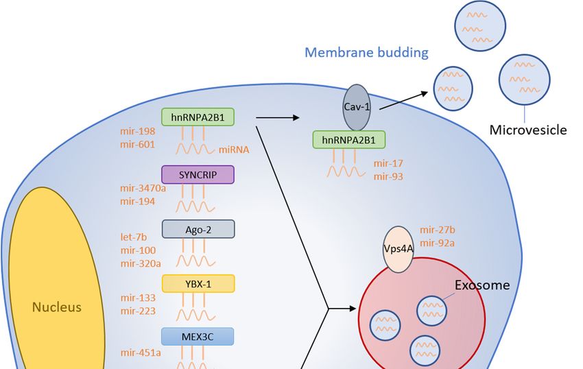
Figure 1. Summary of miRNA sorting into EVs. RNA binding proteins bind specific miRNAs
and selectively shuttle them into EVs. Membranous proteins are also involved in the miRNA
Figure 1. Summary of miRNA sorting into EVs. RNA binding proteins bind specific miRNAs and
sorting mechanism.
selectively shuttle them into EVs. Membranous proteins are also involved in the miRNA sorting
mechanism.
2.2. Argonaute 2
2.2. Cells exert 2control over endogenous mRNA through RNA interference (RNAi) using proteins
Argonaute
such as Drosha, Dicer, and RISC. The argonaute2 (Ago2) protein is a component of the RISC complex
Cells exert control over endogenous mRNA through RNA interference (RNAi) using proteins
that binds miRNAs and facilitates mRNA degradation through endonuclease activity. Recent findings
such as Drosha, Dicer, and RISC. The argonaute2 (Ago2) protein is a component of the RISC complex
have shown the presence of circulating Ago2-miRNA complexes in human plasma, which suggests
that binds miRNAs and facilitates mRNA degradation through endonuclease activity. Recent
that Ago2 might have an important role in the stability of secreted miRNA [16]. Additionally, Ago2
findings have shown the presence of circulating Ago2-miRNA complexes in human plasma, which
has been identified within exosomes and has separately been shown to protect miRNA contained
suggests that Ago2 might have an important role in the stability of secreted miRNA [16].
within MVs from RNase degradation [17]. Expectedly, Ago2 has also been implicated in binding
Additionally, Ago2 has been identified within exosomes and has separately been shown to protect
and sorting miRNA into EVs through the KRAS-MEK-ERK signaling pathway [18]. Specifically,
miRNA contained within MVs from RNase degradation [17]. Expectedly, Ago2 has also been
phosphorylated Ago2 inhibits its co-localization with MVs and decreases secretion into endosomes.
implicated in binding and sorting miRNA into EVs through the KRAS-MEK-ERK signaling pathway
Conversely, inhibition of MEK and ERK was shown to decrease Ago2 phosphorylation and increase
[18]. Specifically, phosphorylated Ago2 inhibits its co-localization with MVs and decreases secretion
Ago2 accumulation inside exosomes [18]. Thus, the miRNA sorting exerted by Ago2 appears to be
into endosomes. Conversely, inhibition of MEK and ERK was shown to decrease Ago2
controlled upstream by the KRAS-MEK-ERK pathway. Further, KRAS-MEK-ERK pathway-dependent
phosphorylation and increase Ago2 accumulation inside exosomes [18]. Thus, the miRNA sorting
phosphorylation of Ago2 has been demonstrated to exert some specific control over the sorting of
exerted by Ago2 appears to be controlled upstream by the KRAS-MEK-ERK pathway. Further,
let-7a, miR-100, and miR-320a into exosomes [18]. Strengthening these findings, miR-100 levels within
KRAS-MEK-ERK pathway-dependent phosphorylation of Ago2 has been demonstrated to exert
exosomes are elevated in oncogenic KRAS mutants with overactive phosphorylation [19]. Collectively,
some specific control over the sorting of let-7a, miR-100, and miR-320a into exosomes [18].
KRAS-MEK-ERK has general regulatory ability over MV and exosomal levels of Ago2, in addition to
Strengthening these findings, miR-100 levels within exosomes are elevated in oncogenic KRAS
specific control over select miRNAs.
mutants with overactive phosphorylation [19]. Collectively, KRAS-MEK-ERK has general regulatory
ability over MV and exosomal levels of Ago2, in addition to specific control over select miRNAs.
2.3. Y-Box Binding Protein 1Cells 2020, 9, 1044 4 of 16
2.3. Y-Box Binding Protein 1
Y-Box Binding Protein 1 (YBX-1) is another protein with RNA-binding domains and exerts a
range of functions including mRNA splicing and transport [20,21]. More recently, YBX-1 has been
found to regulate miR-133 packaging into exosomes after hyperoxia/reperfusion (H/R) treatment of
endothelial progenitor cells (EPC) [22]. Silencing YBX-1 through siRNA causes decreased miR-133
localization within H/R EPC-derived exosomes with no change in expression in the EPC cytosol.
Conversely, overexpression of YBX-1 with miR-133 mimics increased miR-133 quantity within H/R
EPC-derived exosomes.
Additionally, YBX-1 was also identified through liquid chromatography and mass spectrometry
as a candidate for binding miR-223 [23]. From this identification, it was found that YBX-1 was required
to selectively package miR-223 into exosomes derived from HEK293T cells. To confirm that the effect
of YBX-1 is selective to miR-223, they also evaluated the influence of YBX-1 over miR-190 packaging.
miR-190 is not normally known to be present in exosomes, and YBX-1 indeed produced no change in
miR-190 presence within exosomes, suggesting that YBX-1 does have a selective effect on miR-223.
As mentioned, YBX-1 contains RNA-binding domains to achieve its endogenous functions within the
cell, so it likely interacts with miR-133 and miR-223 through specific RNA-binding domains.
2.4. MEX3C
MEX3C functions as an RNA-binding E3 ubiquitin ligase to assist with mRNA degradation.
However, recent evidence has suggested that MEX3C could also play a role in miRNA sorting
into EVs [24]. Immunoprecipitation experiments demonstrate that MEX3C associates with Ago2,
which has been shown in this review to be involved in miRNA sorting [24]. Recently, MEX3C has
also been shown to co-localize with adaptor-related protein complex 2 (AP-2), which is an adaptor
protein implicated in clathrin-mediated endocytosis [25]. As endosomes are processed by the cell,
they form into multivesicular bodies (MVB). Exosomes are then formed when MVBs fuse with the
plasma membranes and release their cargo proteins and RNAs. Given the MEX3C-endosome and
MEX3C-Ago2 relationships, it is reasonable to predict that MEX3C exerts some influence over exosome
biogenesis and potential miRNA sorting. In an additional experiment, siRNA molecules targeting
MEX3C cause decreased exosomal miR-451a [25]. This study found no complementary sequences
between MEX3C and miR-451a, so the authors suggested that Ago2 might serve as the intermediary
between MEX3C and miR-451a. Collectively, this MEX3C-Ago2 complex could shuttle miR-451a
into exosomes.
2.5. Major Vault Protein
Major vault protein (MVP) is a ribonucleoprotein involved in transporting RNA from the nucleus
to the cytoplasm [26]. Recently, MVP has also been proposed as a regulator of miRNAs sorting
into exosomes [27]. MVP knockout in CT26 colon cancer cells caused increased cellular miR-193a,
but decreased levels within CT26-derived exosomes [27]. This MVP knockout was miR-193a selective
because there were no observed expression changes in another tested miRNA, miR-126a. Expectedly,
MVP overexpression in CT26 cells caused reduced miR-193a levels within the cytosol and increased
exosomal concentration [27]. To further strengthen these findings, MVP was identified as a potential
binding partner of miR-193a through mass spectrometry [27]. Collectively, these findings indicate
that miR-193a is sorted into exosomes via an MVP-dependent process, but the specific mechanism is
still unknown.
2.6. La Protein
The La protein is an RNA-binding protein that functions as a transcription factor for RNA
polymerase III that shuttles between the nucleus and cytoplasm [28]. La-depleted cytosol was associated
with a 4-fold reduction of miR-122 sequestration into EVs [29]. However, miR-122 levels returned toCells 2020, 9, 1044 5 of 16
normal with the addition of La to the cytosol [29]. Importantly, the addition of La exclusively without
cytosol produced no effects, indicating the necessity of other unknown cytoplasmic proteins in the
mechanism. Furthermore, electrophoretic mobility shift assays demonstrated a higher binding affinity
between La and miR-122 (Kd = 4.8 nM) than another RNA-binding protein, Ago2 (Kd = 10 nM) [29].
An overview of this mechanism and all other RNA-binding proteins is presented in Figure 1 and
Table 1.
Table 1. List of miRNA sorting mechanisms regulated by RNA-binding proteins.
RNA-Binding Protein Mechanism Reference
Binds miR-198 and miR-601 via potential
[9]
Heterogeneous nuclear GGAG/UGCA motifs to load into exosomes
ribonucleoprotein A2B1 Binds miR-17 and miR-93 via potential AGG/UAG
[10]
motifs to load into MVs
Synaptotagmin-binding
Binds miR-3470a and miR-194-2-3p via potential
cytoplasmic RNA-interaction [14]
GGCU motif to load into exosomes
protein
Loads let-7a, miR-100, and miR-320a into EVs through
Argonaute 2 [18]
KRAS-MEK-ERK signaling pathway
Y-Box Binding Protein 1 Binds miR-133 and miR-223 to load into exosomes [22,23]
MEX3C combines with AP-2 and is involved in
MEX3C [25]
exosome biogenesis and sorting of miR-451a
Major Vault Protein MVP shuttles miR-193a into exosomes [27]
La protein La protein shuttles miR-122 into EVs [29]
3. Membrane Proteins
3.1. Caveolin-1
Caveolae are small 50 to 100nm invaginations of the plasma membrane that assist with
receptor-independent endocytosis and exocytosis [30]. Caveolin-1 (Cav-1) proteins are localized
within these caveolae and demonstrate a critical role in the regulation of membrane trafficking. Under
periods of cell stress, such as during hyperoxia and ROS generation, Cav-1 becomes upregulated in MV
membranes [11]. Furthermore, Cav-1 overexpression caused increased release of hnRNPA2B1 into MVs
as well as elevated levels of hnRNPA2B1-associated miRNAs in MVs of hyperoxia-treated cells [11].
Cav-1 deletion caused decreased expression of hnRNPA2B1 in MVs in response to hyperoxia [11].
Collectively, these results suggest that Cav-1 is integral in the trafficking process of hnRNPA2B1 and
hnRNPA2B1-associated miRNAs into MVs. Additionally, Cav-1 has been identified in populations of
miRNA-rich EVs derived from lung epithelial cells [8]. This further strengthens the concept that Cav-1
plays an important role in selective transport of miRNAs into EVs.
Low molecular weight hyaluronan (LMW-HA) is another plasma membrane component and is
known to cause ROS production inside cells. Treating cells with LMW-HA causes time-dependent
induction of exosome production [31]. However, this effect disappears when caveolin-enriched
microdomains (CEM) are inhibited from forming. CEM are plasma membrane regions enriched with
Cav-1 [32]. Thus, without the presence of Cav-1, exosome biogenesis appears to be impaired. These
findings indicate that Cav-1 is a necessary component of EV biogenesis during ROS-induced cell stress.
3.2. Neural Sphingomyelinase 2
Neural Sphingomyelinase 2 (nSMase2) is a hydrolase involved in the metabolism of sphingolipids,
which are a critical component of the plasma membrane. Specifically, it functions as the rate-limiting
enzyme in ceramide biosynthesis [33]. Ceramide has been implicated in exosome biogenesis, making
the nSMase2 enzyme a likely regulator of exosome synthesis by extension [34]. Inhibition of nSMase2Cells 2020, 9, 1044 6 of 16
greatly reduced secretion of miR-16 and miR-146a within exosomes without any change in cellular
miRNA levels [33]. Likewise, overexpression of nSMase2 increased expression of miR-16 and miR-146a
in exosomes with no effect on cellular miRNA levels [33].
Human tumors have been shown to employ exosomes as a mode of signaling to nearby cells [35].
The nSMase2-dependent method of miRNA sorting also has potential implications in tumor signaling
and progression. Exosome levels of miR-16 decreased significantly in nSMase2-knockdown mouse
cancer cells but increased in nSMase2-overexpressing cancer cells [36]. nSMase2-knockdown cancer
cells also demonstrated decreased lung metastatic colonization, with the nSMase2-overexpressing
cells predictably increasing metastatic progression. In a separate study, inhibition of miR-16 has been
shown to stimulate cell proliferation, suggesting that miR-16 has an important role in preventing
proliferation and tumorigenesis [37]. Furthermore, apoptosis increased in tumor cells when transfected
with pre-miR-16 when compared to controls [37]. Thus, the nSMase2-induced shuttling of miR-16 into
exosomes could provide evidence for the theory that exosomes can be used as disposal mechanisms
for unnecessary or unwanted mRNA or miRNA. Tumor cells could shuttle tumor suppressors such as
miR-16 into exosomes for disposal via a nSMase2-dependent mechanism to prevent apoptosis and
promote proliferation.
3.3. Vacuolar Protein Sorting-Associated Protein 4
The vacuolar protein sorting-associated protein 4 (Vps4A) is required for normal endosomal
trafficking and MVB sorting, but has also recently been implicated in cancer [38,39]. Hepatocellular
carcinoma cells (HCC) overexpressing Vps4A caused elevated exosomal levels of miR-27b-3p and
miR-92a-3p, which are both known oncomiRs [40]. On the other hand, inhibition of Vps4A in HEK293
cells caused reduced levels of EV-derived miR-92a and miR-150 [41]. Interestingly, miR-27b-3p
promotes migration and invasion of colorectal cancer cells while miR-92a-3p promotes proliferation,
migration, and invasion of esophageal squamous cell cancer [42,43]. HCC cells overexpressing Vps4A
also resulted in elevated levels of miR-193a-3p, miR-320a, and miR-132-3p, which are all known tumor
suppressor miRNAs [40]. As evident by these results, Vps4A produces a tumor suppressor effect
within the same cell but appears to secrete oncomiRs via exosomes that could potentially promote
tumor migration in recipient other cells. More research needs to be conducted to determine the exact
mechanism that Vps4A uses to sort miRNAs into EVs as well as determine its regulation over tumor
growth. The membranous proteins that are involved in the miRNA sorting are graphically summarized
in Figure 1.
4. Disease States
4.1. Chronic Lung Disease
The miRNAs contained within EVs originally come from the cytosol of the parent cell, meaning
that there is a relationship between cytosolic and EV levels of miRNAs. As such, any condition or
disease state that manipulates miRNA expression within individual cells can modulate the expression
within EVs [44]. Thus, the miRNA content of EVs is thought to reflect the cellular response to stressors.
Endogenously, certain cell stressors such as hyperoxia or hypoxia are widely known to vary the gene
expression within a particular cell. Hyperoxia-treated lung epithelial cells caused an upregulation
of miR-320a and miR-221 in MVs [45]. In another recent in vivo experiment by Go and colleagues,
2020, hyperoxia-treated mice developed increased levels of miR-21 in serum EVs [46]. These authors
furthered their findings by measuring EV miRNA in serum derived from premature human infants as
a model of chronic lung disease (CLD). Predictably based on the mouse experiments, they identified
elevated serum EV levels of miR-21 in human infants [46]. These findings suggest that miR-21 is likely
involved in the lung injury process associated with CLD. MicroRNA-21 has been previously shown to
target and suppress the tumor suppressor protein Programmed Cell Death 4 (PDCD4) [47]. Go andCells 2020, 9, 1044 7 of 16
colleagues, 2020, also identified that miR-21 suppressed PDCD4, and thus, suggested that the elevated
serum EV miR-21 could be a potential biomarker of CLD [46].
4.2. Immune Response
Interleukin-4 (IL-4) is a cytokine known to activate IgE production and to aid the proliferation
of helper T cells into Th-2 cells. Additionally, IL-4 has been shown to act on bone marrow-derived
macrophages (BMDM) to produce differential expression of 40 miRNAs from BMDM-derived
exosomes [48]. Of the differentially expressed miRNAs, miR-138-5p and miR-149-5p were among the
most highly upregulated. While IL-4 caused an upregulation of these transcripts inside exosomes,
artificial overexpression of miRNA target sequences caused an accumulation in P-bodies and a
reduction from exosomes [48]. Again, this could provide further evidence of exosomes serving as a
disposal mechanism for overly abundant miRNA levels.
4.3. Neuroinflammation
Various miRNAs have been implicated in neuroinflammation and neurological diseases such as
Parkinson’s disease, Alzheimer’s disease, amyotrophic lateral sclerosis, and depression [49]. Astrocytes
stimulated with the pro-inflammatory IL-1β both increased exosomal secretion as well as altering
the miRNA content within the exosomes [50]. While certain exosomal miRNAs were increased from
baseline, a subpopulation of miRNA, including let-7d, miR-126, miR-130b, miR-139-5p, and miR-141-3p,
was only present in the IL-1β stimulated group [50]. This means that this population of five miRNAs
was exclusively shuttled into exosomes due to the pro-inflammatory conditions. Through target scan
predictions, the authors identified several potential locations where these upregulated miRNAs could
target mRNAs involved in apoptosis and cell death [50].
While these miRNAs are implicated in the progression towards cell death, other groups of miRNAs
have demonstrated the ability to protect against neuroinflammation. Subarachnoid hemorrhage (SAH)
cause neuroinflammation leading to early brain injury [51]. As a proposed treatment, exosomes
experimentally loaded with miR-193-3p and delivered to a mouse model of SAH caused a reduction in
HDAC3 and a subsequent increase in acetylated-p65 levels [52]. Acetylation causes p65 to remain in the
inactive form and prevents it from activating NF-κB [53]. Thus, miR-193-3p has the ability to prevent
NF-κB activation subsequently reduce NF-κB-mediated inflammation. Additionally, miR-193-3p
delivery also caused a reduction of pro-inflammatory mediators Caspase-3, IL-1β, IL-6, and TNF-α,
further attenuating the inflammatory response [52]. Ultimately, this miR-193-3p exosomal treatment
reduced brain edema and improved neurological scores in the mouse models of SAH [52].
4.4. Diabetes Mellitus
DM is characterized by high levels of circulating glucose and is associated with endothelial
damage and dysfunction [54,55]. Furthermore, DM has been shown to increase total levels of MV
released from various cell types, raising the question of whether endothelial cells might also be involved
in this MV production [56,57]. Within this population of elevated MVs, hyperglycemic conditions
in vitro caused a significant reduction in MV-derived miR-126 and miR-26a [58]. These results have
been strengthened by other reports confirming a reduction in MV-derived and AB-derived miR-126
under hyperglycemic conditions when compared to normal controls [59]. However, alternative reports
have identified an increase in miR-126 within plasma-derived EVs from human diabetic neuropathy
patients [60]. The reduction of MV-derived miR-126 and elevation of EV-derived miR-126 hint at the
possible mechanistic differences between sorting into the various types of extracellular bodies.
MVs have been shown to deliver miRNAs to endothelial progenitor cells (EPCs) and induce
regulation over the recipient cell [61]. Specifically, delivery of miR-126 to damaged EPCs aids in
the recovery process and reverses any impairment [61]. Further, EV-mediated delivery of miR-126
improved endothelial barrier function in an in vitro model [60]. Functionally, miR-126 has been
shown to protect endothelial cells against H/R mediated damage, while miR-26a protects endothelialCells 2020, 9, 1044 8 of 16
cells against ischemia-reperfusion injury [62,63]. Finally, miR-126 inhibits expression of vascular cell
adhesion molecule 1 (VCAM-1), which functions to bind leukocytes and promotes inflammation [64].
Thus, the hyperglycemic-mediated downregulation of miR-126 and miR-26a inside MVs might
reflect a concomitant decrease in cytoplasmic miR-126 expression, which could be partly driving
the pathogenesis. This downregulation could eliminate the protective functions as well as stimulate
VCAM-1. Higher VCAM-1 would promote the endothelial inflammation associated with DM.
Collectively, these results point towards a regulatory mechanism whereby hyperglycemia selectively
alters the packaging of miR-126 and miR-26a into MVs and contributes to the pathogenic process.
4.5. Cancer
A growing hypothesis regarding cancer suppression theorizes that a select population of miRNAs
are secreted from healthy cells that provide growth-suppressing signals to nearby cancerous cells [65].
Syndecan-1 is a cell surface heparan sulfate proteoglycan and is known to have functions in cancer cell
signaling, such as in multiple myeloma and breast cancer [66–68]. Analyzing exosomes from A549
cancer cells expressing syndecan-1 showed an upregulation of 43 miRNAs and a downregulation of 91
miRNAs when compared to exosomes from syndecan-1 deleted cells [69]. Of the upregulated miRNAs,
exosomal expression of has-miR-485-3p was upregulated by 184-fold. In a separate report, miR-485-3p
has been shown to suppress cancer growth and was found to be downregulated in breast cancer [70].
Further, A549 cells cultured with exosomes isolated from syndecan-1 expressing cells demonstrated
decreased rates of proliferation compared to exosomes from syndecan-1 deficient cells [69]. Collectively,
this data suggests that syndecan-1 plays a role in selectively packaging has-miR-485-3p into exosomes.
These has-miR-485-3p-enriched exosomes then exert anti-tumorigenic effects over nearby cells.
As mentioned earlier in this review, several mechanisms of miRNA packaging have instead
been implicated in cancer progression including MVP, nSMase2, and Vps4A [27,36,40]. However,
other mechanisms have also been implicated in selective miRNA packaging into exosomes to cause
tumorigenesis. In particular, tumor-derived exosomes contain the miRNA processing enzyme Dicer,
which appears to actively convert pre-miRNAs to mature miRNAs within the exosomes [71]. Dicer
was not found in exosomes derived from normal cells, and thus, pre-miRNA conversion to miRNA
was also absent [71]. This Dicer-dependent mechanism potentially contributes to cancer progression.
Healthy MCF-10A cells from human mammary glands exposed to exosomes produced by breast cancer
MDA-MB-231 cells caused increased survival and proliferation [71]. However, deletion of Dicer in
tumor-derived exosomes caused a reduction in the growth of MCF-10A cells [71]. This suggests that
tumors have the ability to convey pro-tumorigenic signals to nearby cells through exosomes via a
potential Dicer-dependent mechanism.
Cholangiocarcinoma (CCA) is a rare form of bile duct cancer also implicated in miRNA sorting [72].
Proteomic analysis of EVs derived from CCAs revealed differential expression of 95 proteins relative to
control while EVs from hepatocellular carcinomas (HCC) revealed 98 dysregulated proteins relative to
control [73]. Furthermore, analysis of bile-derived EVs from CCA patients revealed an upregulation
of miR-191, miR-486-3p, and miR-1274b [74]. In addition to the dysregulated EVs, other reports
have demonstrated a downregulation of miR-195 within the CCA cells [75]. Treating CCA cells with
miR-195-loaded EVs restored expression and reduced growth and invasiveness [75]. This suggests
that CCA cells are able to selectively remove miR-195 from the cytosol, but sheds light on potential
EV-based therapy.
Significant work has also been conducted on examining circulating exosomal miRNAs in
ovarian cancer (OC). Initial studies into OC identified a correlation between tumor miRNAs and
exosomal-derived miRNAs [76]. Specifically, miR-21, miR-141, miR-200a, miR-200c, miR-200b,
miR-203, miR-205, and miR-214 were upregulated in both OC tumor cells and OC-derived exosomes.
In additional studies of plasma-derived exosomes from human OC patients, miR-205-5p, miR-145-5p,
miR-10a-5p, miR-346, and miR-328-3p were all found to be upregulated [77]. Interestingly, in a
meta-analysis study of exosomal miRNAs from solid tumors, poor prognosis was associated withCells 2020, 9, 1044 9 of 16
upregulated miR-21, miR-200a, miR-200b, miR-200c, and miR-203 [78]. While this meta-analysis was
not specific to OC, it indicates that some of the upregulated exosome-derived miRNAs from OC
patients could be used as biomarkers of disease progression.
4.6. Heart Disease and Atherosclerosis
In response to cardiovascular stressors such as hypertension, the heart can undergo hypertrophic
remodeling [79]. While the mechanism behind cardiac remodeling involves many factors,
fibroblasts are essential to cardiac hypertrophy [80]. Artificially treating cardiomyocytes with media
depleted of fibroblast-derived exosomes produced no cardiomyocyte hypertrophy, suggesting that
fibroblast-derived exosomes play a critical role in cell growth [81]. The authors further found that certain
miRNAs could mediate this hypertrophy. Transfecting fibroblasts with pre-miR-21 caused a resultant
increase in miR-21 expression in cardiomyocytes after 72h [81]. Finally, transfection of fibroblast-derived
exosomes enriched in miR-21 produced a resultant hypertrophy of the cardiomyocytes [81].
Atherosclerosis is widely known to be the driving force behind coronary artery disease and high
levels of oxidized LDL are a significant risk factor for atherosclerosis development and progression [82].
Recently, it has been demonstrated that oxidized LDL (oxLDL) has the ability to selectively shuttle
miRNAs into MVs [83]. Specifically, miR-92a-3p, miR-222-3p, and miR-26a-5p were selectively sorted
into MVs through an oxLDL-dependent mechanism. In addition to oxLDL, IL-6 is a cytokine known to
drive endothelial dysfunction and atherosclerosis [84]. Treating endothelial cells with IL-6 also caused
increased miR-92a-3p sequestration into MVs [83]. The authors further demonstrated that oxLDL
increased STAT3 phosphorylation and potentiated miR-92a-3p shuttling into MVs [83]. In mouse
models, miR-92a-3p inhibition caused a reduction of aortic atherosclerotic lesions size, suggesting that
miR-92a-3p is indeed involved in the propagation of atherosclerosis [85]. Thus, the oxLDL-STAT3
mechanism serves to shuttle miR-9sa-3p into MVs and promotes atherosclerosis.
Other miRNA sorting mechanisms have been found to promote atheroprotection. In particular,
miR-143/145 are downregulated in atherosclerosis and have been shown to regulate smooth muscle
cell (SMC) differentiation [86]. Additionally, aorta SMC differentiation is partially controlled by a
transcription factor known as Krüppel-like Factor 2 (KLF2) [87]. Thus, it is reasonable to hypothesize
a connection between KLF2 and miR-143/145 expression. KLF2 overexpression has recently been
shown to selectively transfer miR-143/145 into endothelial EVs and that these miR-143/145-enriched
EVs can be transferred to SMCs [88]. Interestingly, KLF2 overexpression in vivo caused a reduction in
fatty lesions within the aortas of mice [88]. Overall, this suggests that KLF2-induced packaging of
miR-143/145 into EVs exerts regulatory control over the atherosclerotic process in SMCs and helps
prevent atherosclerosis propagation. An overview of this mechanism and other diseases involving
miRNA-derived EV mechanisms is listed in Table 2.
Table 2. List of diseases and the associated effect on EV miRNA.
Disease miRNA Involved Reference
Hyperoxia-treated lung epithelial cells caused an upregulation
[45]
of miR-320a and miR-221 in MVs
Chronic Lung Disease
CLD causes elevated serum EV levels of miR-21 in human
[46]
infants
IL-4 causes upregulated miR-138-5p and miR-149-5p in
Immune Response [48]
BMDM-derived exosomes
Astrocytes stimulated with IL-1β increased exosomal levels of
[50]
let-7d, miR-126, miR-130b, miR-139-5p, and miR-141-3p
Neuroinflammation
Exosomal miR-193-3p delivered to a SAH mouse caused a
[52]
reduction in HDAC3 reduced inflammationCells 2020, 9, 1044 10 of 16
Table 2. Cont.
Disease miRNA Involved Reference
Hyperglycemia causes a reduction in MV and AB-derived
[58,59]
miR-126 and MV-derived miR-26a
Diabetes Mellitus Diabetic neuropathy patients demonstrate increased miR-126
[60]
within plasma-derived EVs
EV-mediated delivery of miR-126 improved endothelial barrier
[60,61]
function and aids in EPC recovery
In A549 cells, Syndecan-1 expression caused a 184-fold
upregulation of exosome-derived has-miR-485-3p, which has [69,70]
been shown to suppress cancer growth
Tumor-derived exosomes Dicer, which converts pre-miRNAs to
[71]
mature miRNAs to promote proliferation
Cancer Bile-derived EVs from CCA patients have upregulated miR-191,
miR-486-3p, and miR-1274b and downregulated miR-195 within [74,75]
the CCA cells
miR-21, miR-141, miR-200a, miR-200c, miR-200b, miR-203,
miR-205, and miR-214 are upregulated in both OC tumor cells
and OC-derived exosomes, while miR-205-5p, miR-145-5p, [76,77]
miR-10a-5p, miR-346, and miR-328-3p were all found to be
upregulated exclusively in OC-derived exosomes
Fibroblast-derived exosomes mediate cardiac hypertrophy via
[81]
potential miR-21 dependent mechanism
Heart Disease and miR-92a-3p, miR-222-3p, and miR-26a-5p are selectively sorted
Atherosclerosis [83]
into MVs through an oxLDL-dependent mechanism
KLF2 assists with packaging miR-143/145 into EVs for
[88]
atheroprotective effect of SMCs
5. Discussion
Exosomes, MVs, and ABs are all generated via independent mechanisms, but all are known
to contain miRNAs. Research into EVs has expanded rapidly in recent years, largely due to newly
found regulatory mechanisms whereby cells can selectively control their miRNA cargo. In order to
selectively sort miRNA, RBPs such as hnRNPA2B1, Ago2, YBX-1, MEX3C, MVP, and the La protein all
appear to bind miRNAs and facilitate their transfer into EVs. For many of these RBPs, the literature
has recently begun to identify specific miRNA-binding motifs capable of exerting selectivity over the
miRNAs shuttled into EVs. However, the specifics of these mechanisms are still largely unknown.
Alternatively, certain membrane proteins such as Cav-1 nSMase2 and Vps4A have been implicated in
EV biogenesis, and in the process have been shown to selectively shuttle miRNAs. Finally, certain
disease states such as chronic lung disease, immune response, neuroinflammation, diabetes mellitus,
cancer, and heart disease can cause dysregulation over EV-miRNA content. In some cases, selective
shuttling of miRNAs into EVs has been directly implicated in the pathological process of the disease,
as is the case with miR-92a-3p and atherosclerosis. However, other research has simply demonstrated
differential expression of EV-derived miRNAs without a clear mechanism of pathogenic control over
the disease, as is the case with miR-320a and miR-221 over hyperoxia.
Despite the knowledge that cells can selectively sort miRNA into EVs, the purpose of this sorting
is still under debate. One overarching idea theorizes that EVs are secreted from parent cells to have
a regulatory influence over daughter cells. Evidence has already shown that secreted EVs can be
endocytosed by daughter cells and alter the mRNA or miRNA content of those cells. However, another
main theory indicates that cells might secrete EVs to eliminate unnecessary RNAs, including miRNAs.
While cells still could be selectively sorting miRNAs into EVs for export, the purpose would simply beCells 2020, 9, 1044 11 of 16
for garbage disposal rather than complex signaling. In reality, cells likely use these miRNA sorting
mechanisms for both signaling and disposal, but more work needs to be conducted to determine
the ultimate function. Regardless of the reason that cells employ miRNA packaging, evidence has
demonstrated an ability to harness this clinically. In this review, we have shown that artificially
delivering EV-mediated miRNAs is beneficial in reducing neuroinflammation after SAH. This potential
treatment opens the door for future artificial EV-mediated therapy.
It is important to note that there are some potential limitations to research into miRNA sorting
mechanisms. Some mechanisms have been quite heavily researched, such as is the case with hnRNPA2B1
and Ago2. However, there are still relatively few studies on other miRNA sorting mechanisms, such as
the MVP or La protein mechanisms. As miRNA sorting is still a relatively novel field, it is possible that
the current body of literature only represents studies that have reported positive findings, especially
on the less well researched mechanisms. Additionally, miRNA sorting is a complicated phenomenon
likely requiring a cascade of protein-mediated events. Thus far, research has identified certain proteins
associated with upregulated or downregulated miRNA levels within EVs, but specific step-by-step
mechanisms are largely unreported. Therefore, it is difficult to determine whether the sorting proteins
reported in this review are part of independent sorting mechanisms, or whether they all play a role in
a unified larger picture sorting mechanism.
To best propel this field forward, future research should be directed towards strengthening and
verifying the current findings. As mentioned, certain sorting mechanisms have little research and need
additional findings to be able to make stronger conclusions. Once these sorting mechanisms have been
thoroughly established, more in vivo experiments must be conducted. Most work until this point has
focused on in vitro studies, but something as complicated as EV-mediated miRNA signaling must be
conducted in vivo to allow for more definitive conclusions. Finally, research can aim to manipulate the
miRNA sorting mechanisms listed in this review to alter the miRNA expression of naturally secreted
EVs with the ultimate goal of providing therapy.
Author Contributions: H.L. conceptualized the topic and developed the outline. M.G. conducted the original
draft preparation. H.L. reviewed the article and provided editing. All authors have read and agreed to the
published version of the manuscript.
Funding: This paper was supported by Changwon National University in 2019–2020.
Acknowledgments: We thank Janet Yan, Frankie Yang, and the Special Issue editors in Cells for helping us to
write this review article.
Conflicts of Interest: The authors declare no conflict of interest.
References
1. Crescitelli, R.; Lässer, C.; Szabó, T.G.; Kittel, A.; Eldh, M.; Dianzani, I.; Buzás, E.I.; Lötvall, J. Distinct RNA
profiles in subpopulations of extracellular vesicles: Apoptotic bodies, microvesicles and exosomes. J. Extracell.
Vesicles 2013, 2, 20677. [CrossRef] [PubMed]
2. Frühbeis, C.; Fröhlich, D.; Krämer-Albers, E.M. Emerging roles of exosomes in neuron-glia communication.
Front. Physiol. 2012, 3, 119. [CrossRef]
3. Luga, V.; Zhang, L.; Viloria-Petit, A.M.; Ogunjimi, A.A.; Inanlou, M.R.; Chiu, E.; Buchanan, M.; Hosein, A.N.;
Basik, M.; Wrana, J.L. Exosomes mediate stromal mobilization of autocrine Wnt-PCP signaling in breast
cancer cell migration. Cell 2012, 151, 1542–1556. [CrossRef]
4. Valadi, H.; Ekström, K.; Bossios, A.; Sjöstrand, M.; Lee, J.J.; Lötvall, J.O. Exosome-mediated transfer of
mRNAs and microRNAs is a novel mechanism of genetic exchange between cells. Nat. Cell Biol. 2007, 9,
654–659. [CrossRef] [PubMed]
5. Jenjaroenpun, P.; Kremenska, Y.; Nair, V.M.; Kremenskoy, M.; Joseph, B.; Kurochkin, I.V. Characterization of
RNA in exosomes secreted by human breast cancer cell lines using next-generation sequencing. PeerJ 2013, 1,
e201. [CrossRef] [PubMed]Cells 2020, 9, 1044 12 of 16
6. Skog, J.; Würdinger, T.; van Rijn, S.; Meijer, D.H.; Gainche, L.; Curry, W.T.; Carter, B.S.; Krichevsky, A.M.;
Breakefield, X.O. Glioblastoma microvesicles transport RNA and proteins that promote tumour growth and
provide diagnostic biomarkers. Nat. Cell Biol. 2008, 10, 1470–1476. [CrossRef]
7. Nolte’T Hoen, E.N.M.; Buermans, H.P.J.; Waasdorp, M.; Stoorvogel, W.; Wauben, M.H.M.; ’T Hoen, P.A.C. Deep
sequencing of RNA from immune cell-derived vesicles uncovers the selective incorporation of small non-coding
RNA biotypes with potential regulatory functions. Nucleic Acids Res. 2012, 40, 9272–9285. [CrossRef]
8. Lee, H.; Groot, M.; Pinilla-Vera, M.; Fredenburgh, L.E.; Jin, Y. Identification of miRNA-rich vesicles in
bronchoalveolar lavage fluid: Insights into the function and heterogeneity of extracellular vesicles. J. Control.
Release 2019, 294, 43–52. [CrossRef]
9. Villarroya-Beltri, C.; Gutiérrez-Vázquez, C.; Sánchez-Cabo, F.; Pérez-Hernández, D.; Vázquez, J.;
Martin-Cofreces, N.; Martinez-Herrera, D.J.; Pascual-Montano, A.; Mittelbrunn, M.; Sánchez-Madrid, F.
Sumoylated hnRNPA2B1 controls the sorting of miRNAs into exosomes through binding to specific motifs.
Nat. Commun. 2013, 4, 2980. [CrossRef]
10. Wu, B.; Su, S.; Patil, D.P.; Liu, H.; Gan, J.; Jaffrey, S.R.; Ma, J. Molecular basis for the specific and multivariant
recognitions of RNA substrates by human hnRNP A2/B1. Nat. Commun. 2018, 9, 1–12. [CrossRef]
11. Lee, H.; Li, C.; Zhang, Y.; Zhang, D.; Otterbein, L.E.; Jin, Y. Caveolin-1 selectively regulates microRNA sorting
into microvesicles after noxious stimuli. J. Exp. Med. 2019, 216, 2202–2220. [CrossRef] [PubMed]
12. Li, T.; Evdokimov, E.; Shen, R.F.; Chao, C.C.; Tekle, E.; Wang, T.; Stadtman, E.R.; Yang, D.C.H.; Chock, P.B.
Sumoylation of heterogeneous nuclear ribonucleoproteins, zinc finger proteins, and nuclear pore complex
proteins: A proteomic analysis. Proc. Natl. Acad. Sci. USA 2004, 101, 8551–8556. [CrossRef] [PubMed]
13. Pérez-Boza, J.; Boeckx, A.; Lion, M.; Dequiedt, F.; Struman, I. hnRNPA2B1 inhibits the exosomal export of
miR-503 in endothelial cells. Cell. Mol. Life Sci. 2020. [CrossRef]
14. Santangelo, L.; Giurato, G.; Cicchini, C.; Montaldo, C.; Mancone, C.; Tarallo, R.; Battistelli, C.; Alonzi, T.;
Weisz, A.; Tripodi, M. The RNA-Binding Protein SYNCRIP Is a Component of the Hepatocyte Exosomal
Machinery Controlling MicroRNA Sorting. Cell Rep. 2016, 17, 799–808. [CrossRef] [PubMed]
15. Hobor, F.; Dallmann, A.; Ball, N.J.; Cicchini, C.; Battistelli, C.; Ogrodowicz, R.W.; Christodoulou, E.;
Martin, S.R.; Castello, A.; Tripodi, M.; et al. A cryptic RNA-binding domain mediates Syncrip recognition
and exosomal partitioning of miRNA targets. Nat. Commun. 2018, 9, 831. [CrossRef] [PubMed]
16. Arroyo, J.D.; Chevillet, J.R.; Kroh, E.M.; Ruf, I.K.; Pritchard, C.C.; Gibson, D.F.; Mitchell, P.S.; Bennett, C.F.;
Pogosova-Agadjanyan, E.L.; Stirewalt, D.L.; et al. Argonaute2 complexes carry a population of circulating
microRNAs independent of vesicles in human plasma. Proc. Natl. Acad. Sci. USA 2011, 108, 5003–5008.
[CrossRef]
17. Li, L.; Zhu, D.; Huang, L.; Zhang, J.; Bian, Z.; Chen, X.; Liu, Y.; Zhang, C.Y.; Zen, K. Argonaute 2 Complexes
Selectively Protect the Circulating MicroRNAs in Cell-Secreted Microvesicles. PLoS ONE 2012, 7, e0046957.
[CrossRef]
18. McKenzie, A.J.; Hoshino, D.; Hong, N.H.; Cha, D.J.; Franklin, J.L.; Coffey, R.J.; Patton, J.G.; Weaver, A.M.
KRAS-MEK Signaling Controls Ago2 Sorting into Exosomes. Cell Rep. 2016, 15, 978–987. [CrossRef]
19. Cha, D.J.; Franklin, J.L.; Dou, Y.; Liu, Q.; Higginbotham, J.N.; Beckler, M.D.; Weaver, A.M.; Vickers, K.;
Prasad, N.; Levy, S.; et al. KRAS-dependent sorting of miRNA to exosomes. eLife 2015, 4, e07197. [CrossRef]
20. Chen, Y.; Sekine, K.; Nakamura, K.; Yanai, H.; Tanaka, M.; Miyajima, A. Y-Box Binding Protein-1
Down-Regulates Expression of Carbamoyl Phosphate Synthetase-I by Suppressing CCAAT Enhancer-Binding
Protein-Alpha Function in Mice. Gastroenterology 2009, 137, 330–340. [CrossRef]
21. Matsumoto, K.; Wolffe, A.P. Gene regulation by Y-box proteins: Coupling control of transcription and
translation. Trends Cell Biol. 1998, 8, 318–323. [CrossRef]
22. Lin, F.; Zeng, Z.; Song, Y.; Li, L.; Wu, Z.; Zhang, X.; Li, Z.; Ke, X.; Hu, X. YBX-1 mediated sorting of miR-133
into hypoxia/reoxygenation-induced EPC-derived exosomes to increase fibroblast angiogenesis and MEndoT.
Stem Cell Res. Ther. 2019, 10, 263. [CrossRef] [PubMed]
23. Shurtleff, M.J.; Temoche-Diaz, M.M.; Karfilis, K.V.; Ri, S.; Schekman, R. Y-box protein 1 is required to sort
microRNAs into exosomes in cells and in a cell-free reaction. eLife 2016, 5, e19276. [CrossRef] [PubMed]
24. Buchet-Poyau, K.; Courchet, J.; Le Hir, H.; Séraphin, B.; Scoazec, J.Y.; Duret, L.; Domon-Dell, C.; Freund, J.N.;
Billaud, M. Identification and characterization of human Mex-3 proteins, a novel family of evolutionarily
conserved RNA-binding proteins differentially localized to processing bodies. Nucleic Acids Res. 2007, 35,
1289–1300. [CrossRef]Cells 2020, 9, 1044 13 of 16
25. Lu, P.; Li, H.; Li, N.; Singh, R.N.; Bishop, C.E.; Chen, X.; Lu, B. MEX3C interacts with adaptor-related protein
complex 2 and involves in miR-451a exosomal sorting. PLoS ONE 2017, 12, e0185992. [CrossRef] [PubMed]
26. Chung, J.-H.; Eng, C. Nuclear-Cytoplasmic Partitioning of Phosphatase and Tensin Homologue Deleted
on Chromosome 10 (PTEN) Differentially Regulates the Cell Cycle and Apoptosis. Cancer Res. 2005, 65,
8096–8100. [CrossRef]
27. Teng, Y.; Ren, Y.; Hu, X.; Mu, J.; Samykutty, A.; Zhuang, X.; Deng, Z.; Kumar, A.; Zhang, L.; Merchant, M.L.;
et al. MVP-mediated exosomal sorting of miR-193a promotes colon cancer progression. Nat. Commun. 2017,
8, 14448. [CrossRef]
28. Intine, R.V.; Tenenbaum, S.A.; Sakulich, A.L.; Keene, J.D.; Maraia, R.J. Differential phosphorylation and
subcellular localization of La RNPs associated with precursor tRNAs and translation-related mRNAs. Mol.
Cell 2003, 12, 1301–1307. [CrossRef]
29. Temoche-Diaz, M.M.; Shurtleff, M.J.; Nottingham, R.M.; Yao, J.; Fadadu, R.P.; Lambowitz, A.M.; Schekman, R.
Distinct mechanisms of microrna sorting into cancer cell-derived extracellular vesicle subtypes. eLife 2019, 8,
e47544. [CrossRef]
30. Rothberg, K.G.; Heuser, J.E.; Donzell, W.C.; Ying, Y.S.; Glenney, J.R.; Anderson, R.G.W. Caveolin, a protein
component of caveolae membrane coats. Cell 1992, 68, 673–682. [CrossRef]
31. Mirzapoiazova, T.; Lennon, F.E.; Mambetsariev, B.; Allen, M.; Riehm, J.; Poroyko, V.A.; Singleton, P.A.
Extracellular Vesicles from Caveolin-Enriched Microdomains Regulate Hyaluronan-Mediated Sustained
Vascular Integrity. Int. J. Cell Biol. 2015, 2015, 481493. [CrossRef]
32. Drab, M.; Verkade, P.; Elger, M.; Kasper, M.; Lohn, M.; Lauterbach, B.; Menne, J.; Lindschau, C.; Mende, F.;
Luft, F.C.; et al. Loss of caveolae, vascular dysfunction, and pulmonary defects in caveolin-1 gene-disrupted
mice. Science 2001, 293, 2449–2452. [CrossRef]
33. Kosaka, N.; Iguchi, H.; Yoshioka, Y.; Takeshita, F.; Matsuki, Y.; Ochiya, T. Secretory mechanisms and
intercellular transfer of microRNAs in living cells. J. Biol. Chem. 2010, 285, 17442–17452. [CrossRef] [PubMed]
34. Trajkovic, K.; Hsu, C.; Chiantia, S.; Rajendran, L.; Wenzel, D.; Wieland, F.; Schwille, P.; Brugger, B.; Simons, M.
Ceramide triggers budding of exosome vesicles into multivesicular endosomes. Science 2008, 319, 1244–1247.
[CrossRef] [PubMed]
35. Meckes, D.G.; Shair, K.H.Y.; Marquitz, A.R.; Kung, C.P.; Edwards, R.H.; Raab-Traub, N. Human tumor
virus utilizes exosomes for intercellular communication. Proc. Natl. Acad. Sci. USA 2010, 107, 20370–20375.
[CrossRef] [PubMed]
36. Kosaka, N.; Iguchi, H.; Hagiwara, K.; Yoshioka, Y.; Takeshita, F.; Ochiya, T. Neutral sphingomyelinase 2
(nSMase2)-dependent exosomal transfer of angiogenic micrornas regulate cancer cell metastasis. J. Biol.
Chem. 2013, 288, 10849–10859. [CrossRef]
37. Yan, X.; Liang, H.; Deng, T.; Zhu, K.; Zhang, S.; Wang, N.; Jiang, X.; Wang, X.; Liu, R.; Zen, K.; et al. The
identification of novel targets of miR-16 and characterization of their biological functions in cancer cells. Mol.
Cancer 2013, 12, 92. [CrossRef]
38. Bishop, N.; Woodman, P. ATPase-defective mammalian VPS4 localizes to aberrant endosomes and impairs
cholesterol trafficking. Mol. Biol. Cell 2000, 11, 227–239. [CrossRef]
39. Raiborg, C.; Rusten, T.E.; Stenmark, H. Protein sorting into multivesicular endosomes. Curr. Opin. Cell Biol.
2003, 15, 446–455. [CrossRef]
40. Wei, J.-X.; Lv, L.-H.; Wan, Y.-L.; Cao, Y.; Li, G.-L.; Lin, H.-M.; Zhou, R.; Shang, C.-Z.; Cao, J.; He, H.; et al.
Vps4A functions as a tumor suppressor by regulating the secretion and uptake of exosomal microRNAs in
human hepatoma cells. Hepatology 2015, 61, 1284–1294. [CrossRef]
41. Jackson, C.E.; Scruggs, B.S.; Schaffer, J.E.; Hanson, P.I. Effects of Inhibiting VPS4 Support a General Role for
ESCRTs in Extracellular Vesicle Biogenesis. Biophys. J. 2017, 113, 1342–1352. [CrossRef] [PubMed]
42. Yang, X.; Chen, J.; Liao, Y.; Huang, L.; Wen, C.; Lin, M.; Li, W.; Zhu, Y.; Wu, X.; Iwamoto, A.; et al. MiR-27b-3p
promotes migration and invasion in colorectal cancer cells by targeting HOXA10. Biosci. Rep. 2019, 39.
[CrossRef] [PubMed]
43. Li, X.; Guo, S.; Min, L.; Guo, Q.; Zhang, S. MiR-92a-3p promotes the proliferation, migration and invasion
of esophageal squamous cell cancer by regulating PTEN. Int. J. Mol. Med. 2019, 44, 973–981. [CrossRef]
[PubMed]Cells 2020, 9, 1044 14 of 16
44. de Jong, O.G.; Verhaar, M.C.; Chen, Y.; Vader, P.; Gremmels, H.; Posthuma, G.; Schiffelers, R.M.; Gucek, M.;
van Balkom, B.W.M. Cellular stress conditions are reflected in the protein and RNA content of endothelial
cell-derived exosomes. J. Extracell. Vesicles 2012, 1, 18396. [CrossRef]
45. Lee, H.; Zhang, D.; Zhu, Z.; Dela Cruz, C.S.; Jin, Y. Epithelial cell-derived microvesicles activate macrophages
and promote inflammation via microvesicle-containing microRNAs. Sci. Rep. 2016, 6, 35250. [CrossRef]
46. Go, H.; Maeda, H.; Miyazaki, K.; Maeda, R.; Kume, Y.; Namba, F.; Momoi, N.; Hashimoto, K.; Otsuru, S.;
Kawasaki, Y.; et al. Extracellular vesicle miRNA-21 is a potential biomarker for predicting chronic lung
disease in premature infants. Am. J. Physiol. Lung Cell. Mol. Physiol. 2020. [CrossRef]
47. Frankel, L.B.; Christoffersen, N.R.; Jacobsen, A.; Lindow, M.; Krogh, A.; Lund, A.H. Programmed cell death 4
(PDCD4) is an important functional target of the microRNA miR-21 in breast cancer cells. J. Biol. Chem. 2008,
283, 1026–1033. [CrossRef]
48. Squadrito, M.L.; Baer, C.; Burdet, F.; Maderna, C.; Gilfillan, G.D.; Lyle, R.; Ibberson, M.; De Palma, M.
Endogenous RNAs Modulate MicroRNA Sorting to Exosomes and Transfer to Acceptor Cells. Cell Rep. 2014,
8, 1432–1446. [CrossRef]
49. Brites, D.; Fernandes, A. Neuroinflammation and depression: Microglia activation, extracellular microvesicles
and microRNA dysregulation. Front. Cell. Neurosci. 2015, 9, 476. [CrossRef]
50. Gayen, M.; Bhomia, M.; Balakathiresan, N. Exosomal MicroRNAs Released by Activated Astrocytes as
Potential Neuroinflammatory Biomarkers. Int. J. Mol. Sci. 2020, 21, 2312. [CrossRef]
51. Lucke-Wold, B.P.; Logsdon, A.F.; Manoranjan, B.; Turner, R.C.; McConnell, E.; Vates, G.E.; Huber, J.D.;
Rosen, C.L.; Simard, J.M. Aneurysmal subarachnoid hemorrhage and neuroinflammation: A comprehensive
review. Int. J. Mol. Sci. 2016, 17, 497. [CrossRef] [PubMed]
52. Lai, N.; Wu, D.; Liang, T.; Pan, P.; Yuan, G.; Li, X.; Li, H.; Shen, H.; Wang, Z.; Chen, G. Systemic exosomal
miR-193b-3p delivery attenuates neuroinflammation in early brain injury after subarachnoid hemorrhage in
mice. J. Neuroinflamm. 2020, 17, 74. [CrossRef] [PubMed]
53. Chen, S.; Ye, J.; Chen, X.; Shi, J.; Wu, W.; Lin, W.; Lin, W.; Li, Y.; Fu, H.; Li, S. Valproic acid attenuates
traumatic spinal cord injury-induced inflammation via STAT1 and NF-KB pathway dependent of HDAC3.
J. Neuroinflamm. 2018, 15, 150. [CrossRef] [PubMed]
54. Tabit, C.E.; Chung, W.B.; Hamburg, N.M.; Vita, J.A. Endothelial dysfunction in diabetes mellitus: Molecular
mechanisms and clinical implications. Rev. Endocr. Metab. Disord. 2010, 11, 61–74. [CrossRef]
55. Hadi, H.A.R.; Suwaidi, J.A. Endothelial dysfunction in diabetes mellitus. Vasc. Health Risk Manag. 2007, 3,
853–876.
56. Ogata, N.; Nomura, S.; Shouzu, A.; Imaizumi, M.; Arichi, M.; Matsumura, M. Elevation of monocyte-derived
microparticles in patients with diabetic retinopathy. Diabetes Res. Clin. Pract. 2006, 73, 241–248. [CrossRef]
57. Tan, K.T.; Tayebjee, M.H.; Lim, H.S.; Lip, G.Y.H. Clinically apparent atherosclerotic disease in diabetes is
associated with an increase in platelet microparticle levels. Diabet. Med. 2005, 22, 1657–1662. [CrossRef]
58. Jansen, F.; Wang, H.; Przybilla, D.; Franklin, B.S.; Dolf, A.; Pfeifer, P.; Schmitz, T.; Flender, A.; Endl, E.;
Nickenig, G.; et al. Vascular endothelial microparticles-incorporated microRNAs are altered in patients with
diabetes mellitus. Cardiovasc. Diabetol. 2016, 15, 49. [CrossRef]
59. Zampetaki, A.; Kiechl, S.; Drozdov, I.; Willeit, P.; Mayr, U.; Prokopi, M.; Mayr, A.; Weger, S.; Oberhollenzer, F.;
Bonora, E.; et al. Plasma MicroRNA profiling reveals loss of endothelial MiR-126 and other MicroRNAs in
type 2 diabetes. Circ. Res. 2010, 107, 810–817. [CrossRef]
60. Florijn, B.W.; Duijs, J.M.G.J.; Levels, J.H.; Dallinga-Thie, G.M.; Wang, Y.; Boing, A.N.; Yuana, Y.; Stam, W.;
Limpens, R.W.A.L.; Au, Y.W.; et al. Diabetic Nephropathy Alters the Distribution of Circulating Angiogenic
MicroRNAs Among Extracellular Vesicles, HDL, and Ago-2. Diabetes 2019, 68, 2287–2300. [CrossRef]
61. Alexandru, N.; Andrei, E.; Niculescu, L.; Dragan, E.; Ristoiu, V.; Georgescu, A. Microparticles of healthy
origins improve endothelial progenitor cell dysfunction via microRNA transfer in an atherosclerotic hamster
model. Acta Physiol. 2017, 221, 230–249. [CrossRef] [PubMed]
62. Yang, H.-H.; Chen, Y.; Gao, C.-Y.; Cui, Z.-T.; Yao, J.-M. Protective Effects of MicroRNA-126 on Human
Cardiac Microvascular Endothelial Cells Against Hypoxia/Reoxygenation-Induced Injury and Inflammatory
Response by Activating PI3K/Akt/eNOS Signaling Pathway. Cell. Physiol. Biochem. 2017, 42, 506–518.
[CrossRef] [PubMed]Cells 2020, 9, 1044 15 of 16
63. Wu, Y.; Zhang, M.; Xue, Y.; Zhang, T.; Wu, N.; Guo, W.; Du, X.; Xu, Y. Effect of microRNA-26a on vascular
endothelial cell injury caused by lower extremity ischemia–reperfusion injury through the AMPK pathway
by targeting PFKFB3. J. Cell. Physiol. 2019, 234, 2916–2928. [CrossRef]
64. Harris, T.A.; Yamakuchi, M.; Ferlito, M.; Mendell, J.T.; Lowenstein, C.J. MicroRNA-126 regulates endothelial
expression of vascular cell adhesion molecule 1. Proc. Natl. Acad. Sci. USA 2008, 105, 1516–1521. [CrossRef]
65. Kosaka, N.; Yoshioka, Y.; Fujita, Y.; Ochiya, T. Versatile roles of extracellular vesicles in cancer. J. Clin. Investig.
2016, 126, 1163–1172. [CrossRef]
66. Szatmári, T.; Ötvös, R.; Hjerpe, A.; Dobra, K. Syndecan-1 in Cancer: Implications for Cell Signaling,
Differentiation, and Prognostication. Dis. Markers 2015, 2015, 796052. [CrossRef]
67. Ren, Z.; Van Andel, H.; De Lau, W.; Hartholt, R.B.; Maurice, M.M.; Clevers, H.; Kersten, M.J.; Spaargaren, M.;
Pals, S.T. Syndecan-1 promotes Wnt/b-catenin signaling in multiple myeloma by presenting Wnts and
R-spondins. Blood 2018, 131, 982–994. [CrossRef] [PubMed]
68. Ibrahim, S.A.; Gadalla, R.; El-Ghonaimy, E.A.; Samir, O.; Mohamed, H.T.; Hassan, H.; Greve, B.; El-Shinawi, M.;
Mohamed, M.M.; Götte, M. Syndecan-1 is a novel molecular marker for triple negative inflammatory breast
cancer and modulates the cancer stem cell phenotype via the IL-6/STAT3, Notch and EGFR signaling
pathways. Mol. Cancer 2017, 16, 57. [CrossRef] [PubMed]
69. Parimon, T.; Brauer, R.; Schlesinger, S.Y.; Xie, T.; Jiang, D.; Ge, L.; Huang, Y.; Birkland, T.P.; Parks, W.C.;
Habiel, D.M.; et al. Syndecan-1 Controls Lung Tumorigenesis by Regulating miRNAs Packaged in Exosomes.
Am. J. Pathol. 2018, 188, 1094–1103. [CrossRef] [PubMed]
70. Lou, C.; Xiao, M.; Cheng, S.; Lu, X.; Jia, S.; Ren, Y.; Li, Z. MiR-485-3p and mir-485-5p suppress breast cancer
cell metastasis by inhibiting PGC-1α expression. Cell Death Dis. 2016, 7, e2159. [CrossRef]
71. Melo, S.A.; Sugimoto, H.; O’Connell, J.T.; Kato, N.; Villanueva, A.; Vidal, A.; Qiu, L.; Vitkin, E.; Perelman, L.T.;
Melo, C.A.; et al. Cancer Exosomes Perform Cell-Independent MicroRNA Biogenesis and Promote
Tumorigenesis. Cancer Cell 2014, 26, 707–721. [CrossRef] [PubMed]
72. Olaizola, P.; Lee-Law, P.Y.; Arbelaiz, A.; Lapitz, A.; Perugorria, M.J.; Bujanda, L.; Banales, J.M. MicroRNAs
and extracellular vesicles in cholangiopathies. Biochim. Biophys. Acta Mol. Basis Dis. 2018, 1864, 1293–1307.
[CrossRef] [PubMed]
73. Arbelaiz, A.; Azkargorta, M.; Krawczyk, M.; Santos-Laso, A.; Lapitz, A.; Perugorria, M.J.; Erice, O.;
Gonzalez, E.; Jimenez-Agüero, R.; Lacasta, A.; et al. Serum extracellular vesicles contain protein biomarkers
for primary sclerosing cholangitis and cholangiocarcinoma: Arbelaiz et al. Hepatology 2017, 66, 1125–1143.
[CrossRef] [PubMed]
74. Li, L.; Masica, D.; Ishida, M.; Tomuleasa, C.; Umegaki, S.; Kalloo, A.N.; Georgiades, C.; Singh, V.K.;
Khashab, M.; Amateau, S.; et al. Human bile contains MicroRNA-laden extracellular vesicles that can be
used for cholangiocarcinoma diagnosis. Hepatology 2014, 60, 896–907. [CrossRef] [PubMed]
75. Li, L.; Piontek, K.; Ishida, M.; Fausther, M.; Dranoff, J.A.; Fu, R.; Mezey, E.; Gould, S.J.; Fordjour, F.K.;
Meltzer, S.J.; et al. Extracellular vesicles carry microRNA-195 to intrahepatic cholangiocarcinoma and
improve survival in a rat model. Hepatology 2017, 65, 501–514. [CrossRef] [PubMed]
76. Taylor, D.D.; Gercel-Taylor, C. MicroRNA signatures of tumor-derived exosomes as diagnostic biomarkers of
ovarian cancer. Gynecol. Oncol. 2008, 110, 13–21. [CrossRef] [PubMed]
77. Wang, W.; Yin, Y.; Shan, X.; Zhou, X.; Liu, P.; Cao, Q.; Zhu, D.; Zhang, J.; Zhu, W. The Value of Plasma-Based
MicroRNAs as Diagnostic Biomarkers for Ovarian Cancer. Am. J. Med. Sci. 2019, 358, 256–267. [CrossRef]
78. Zhou, J.; Guo, H.; Yang, Y.; Zhang, Y.; Liu, H. A meta-analysis on the prognosis of exosomal miRNAs in all
solid tumor patients. Medicine 2019, 98, e15335. [CrossRef]
79. Nadruz, W. Myocardial remodeling in hypertension. J. Hum. Hypertens. 2015, 29, 1–6. [CrossRef]
80. Takeda, N.; Manabe, I.; Uchino, Y.; Eguchi, K.; Matsumoto, S.; Nishimura, S.; Shindo, T.; Sano, M.; Otsu, K.;
Snider, P.; et al. Cardiac fibroblasts are essential for the adaptive response of the murine heart to pressure
overload. J. Clin. Investig. 2010, 120, 254–265. [CrossRef] [PubMed]
81. Bang, C.; Batkai, S.; Dangwal, S.; Gupta, S.K.; Foinquinos, A.; Holzmann, A.; Just, A.; Remke, J.; Zimmer, K.;
Zeug, A.; et al. Cardiac fibroblast-derived microRNA passenger strand-enriched exosomes mediate
cardiomyocyte hypertrophy. J. Clin. Investig. 2014, 124, 2136–2146. [CrossRef] [PubMed]
82. Chen, M.; Masaki, T.; Sawamura, T. LOX-1, the receptor for oxidized low-density lipoprotein identified from
endothelial cells: Implications in endothelial dysfunction and atherosclerosis. Pharmacol. Ther. 2002, 95,
89–100. [CrossRef]You can also read